Article

Synthesis and Structure of bis(Dibutyldithiocarbamate)zinc(II): Zn2[(n-Bu)2NCSS]4

1
Department of Chemistry, South China Normal University, Guangzhou 510631, P.R. China
2
Department of Chemistry, Lanzhou University,Lanzhou 730000, P.R. China
3
Chengdu Centre of Analysis and Measurement, Chinese Academy of Sciences, Chengdu 610015, P.R. China
*
Authors to whom correspondence should be addressed.
Molecules 2003, 8(5), 411-417; https://doi.org/10.3390/80500411
Submission received: 9 February 2003 / Revised: 3 April 2003 / Accepted: 6 April 2003 / Published: 31 May 2003

Abstract

:
The binuclear zinc (II) complex Zn2[(n-Bu)2NCSS]4 has been prepared, and its crystal and molecular structure have been determined by x-ray diffraction. The crystal is monoclinic, space group C2/c, with a=23.329(3)Å, b=17.090(2)Å, c=16.115(2)Å, α =90°, β=127.560(10)°, γ=90°, z=4, V=5039.1(11)Å3, F(000)=2016, R=0.0450, and Rw=0.1192. The crystal structure shows that two S-N-S atom chains, belonging to the different dibutyldithiocarbamate ligands, bridge two zinc (II) ions. Each zinc (II) ion coordinates to four S atoms. The coordination geometry around the zinc (II) is a tetrahedron, however, the coordination sphere of two zinc ions in the dimer is best described as a distorted octagon. The X-ray photoelectron spectra, IR and UV data have been used to study the structure and spectra properties of the complex.

Introduction

Investigation of the role that zinc thiolate complexes play in biological chemistry has stimulated numerous studies on zinc coordination compounds with sulfur ligation [1,2,3,4]. In particular, zinc complexes with NS2 ligand cores are of increasing interest as structural and spectroscopic models for metal binding sites in a number of metallothioneins and metalloregulatory proteins [5,6,7]. Dithiocarbamates are more frequently used in these model compound studies. As an extension of our previous studies [8,9,10], we synthesized the zinc complex with dibutyldithiocarbamate and analyzed its structure by x-ray diffraction. The X-ray photoelectron spectra, IR and UV data were also used to study the structure and spectral properties of the complex.

Results and Discussion

X-Ray Crystallography [11]
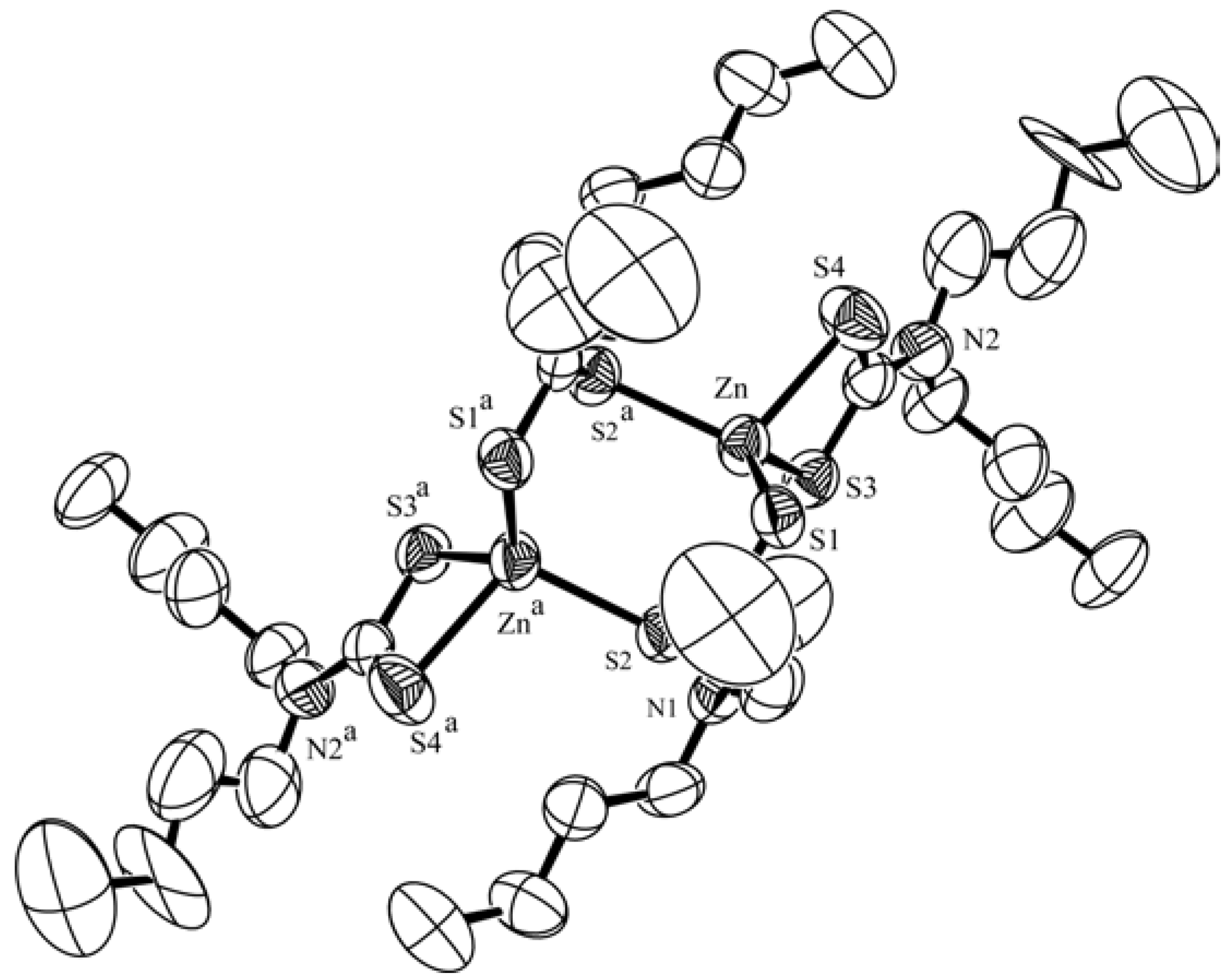
Figure 1 shows the molecular structure of Zn2[(n-Bu)2NCSS]4. Figure 2 shows the molecular packing arrangement in the unit cell. The crystal structure of the zinc (II) complex Zn2[(n-Bu)2NCSS]4 is built up of centro-symmetric dimeric entities. The coordination sphere of two zinc ions is best described as a distorted octagon. Four sulfur atoms belonging to two dibutyldithiocarbamate ligands occupy the basal coordination positions. Therefore, each bridging sulfur simultaneous occupies an apical site of tetrahedron. The topology of the formed dimer is then viewed as an edge-sharing distorted octagon. Four bridging S atoms are strictly planar because the S(2)-C(1) bond distance is equal to S(2)a-C(1)a (0.1725nm) and the S(1)-C(1) is equal to S(1)a-C(1)a (0.1704nm).
Figure 1. The molecular structure of Zn2[(n-Bu)2NCSS]4 with the hydrogen atoms omitted for clarity
Molecules 08 00411 g001
The Zn(1)-C(1)-Zn(1)a-C(1)a is strictly planar too, due to the inversion center. The carbon-sulfur bonds have an average length of 0.171(9) nm and the ligand “ bite” angle S-C-S has a mean value of 117.8(5) deg. The N(1)-C(1) and N(2)-C(10) bond distances [0.131(0)nm, 0.133(2)nm] are shorter than the other N-C bond distances, and are indicative of considerable double-bond character. It is informative to examine the structure of the Zn complex. This is true for bidentate dithiocarbamate adducts:
Molecules 08 00411 i001
but not for monodentate analogues: Molecules 08 00411 i002
One possible reason is that the enhancement of the Zn-S bond, which originates from reduction of a negative residual charge on the central ion, is partly compensated by the fact that one molecule of dithiocarbamate is replaced by two molecules of a monodentate ligand which results in less-stable crystal packing. On the other hand, the chelating mode of coordination of the bidentate ligand dibutyldithiocarbamate allows easy electron delocalization over a large conjugate system and the rigid plane permits a more stable crystal packing [12].
Figure 2. The molecular packing arrangement in the unit cell with the butyl groups omitted for clarity
Molecules 08 00411 g002

X-ray photoelectron spectroscopy (XPS)

The measured binding energies are given in Table 2. The C(1s) spectrum of free ligand Na[S2CN(n-C4H9)2] showed a main peak and a weak peak on the higher energy side. The former has been assigned to the carbon in the alkyl and the latter to the CSS. Its N(1s) spectrum showed a single full-width peak, but the S(2p) spectrum showed a broad peak with a shoulder suggesting two types of sulfur atoms. The two peaks indicated by the broken lines, which were obtained by a graphical resolution, were almost equal in intensity. The peak with the lower binding energy of the two in the S(2p) spectrum of Na[S2CN(n-C4H9)2] can be readily assigned to the double-bond sulfur atom and the other to the single-bond sulfur atom which bonds with a sodium ion.
In general, coordination of a ligand to a metal ion causes an increase in the electron density on the metal, resulting in a decrease in the metal ion’s electron binding energy [12]. The Zn(2p)3/2 chemical shift, which are the differences between the Zn(2p)3/2 binding energies of the complex and ZnCl2 (the former is smaller than the latter), was 1.7eV. From this result, it may be concluded that coordination bonds are formed in the complex. The C(1s), N(1s) and S(2p) spectrum of Zn2[S2CN(n-C4H9)2]4 showed a single peak, respectively. This indicated the bidentate behavior of the dithio group in complex[13]. It is similar to the conclusion obtained by the result of the crystal structure discussed above.
Table 2. Crystal data and structure refinement
CompoundsBinding energies /eV
Zn(2p)3/2C(1s)N(1s)S(2p)
ZnCl2 [12]1023.1
Na[S2CN(n-C4H9)2] 287.0, 284.9398.9164.2, 162.2
Zn2[S2CN(n-C4H9)2]41021.6287.0, 284.6399.7162.0

IR spectra

The ligand shows a strong absorption at 1472 cm-1, which is assigned to the C-N stretching frequency, while the complex showed an absorption at 1495 cm-1, which is assigned to the C=N stretching frequency. In the complex, if Zn-S chelate bonds are formed, then the great contribution of the latter structure will cause the ν(C-N) bands of the complex to shift to higher frequency. Compared with the ligand, the ν(C-N) band of the complex is indeed shifted towards higher frequency by 23 cm-1. Thus, the dibutyldithiocarbamate ligand coordinates to zinc ions through bidentate sulfur atoms. The presence of a single strong band (958 cm-1) due to a ν(CSS) mode in the spectra of the complex is strongly indicative of the bidentate behaviour of the dithio ligand in the complexes, otherwise a doublet (978, 959 cm-1) would be expected in the 1000 ± 70 region, as in the case of monodentate coordination [14].

Electronic Spectra

The complex showed two absorption bands due to the CS2 group (these absorptions are indicated as Band 1 and Bond 2). Band 1 (236 nm) is attributed to the л→л* transition of the S·····C····· S group [15]. This band does not undergo splitting and this fact clearly indicates the equivalence of the C·····S bond in the complexes. Bond 2(267nm) is assigned to a n→π* transition , i.e. the transition of an electron of the lone pair on the sulfur atom to an antibonding л–orbital [15].

Conclusions

We have reported the synthesis and X-ray crystal structure of Zn2[(n-Bu)2NCSS]4. The structure and spectral properties of the complex have been characterized by means of X-ray photoelectron spectroscopic, IR and UV data.

Experimental

General

All reagents were of analytical purity, made in China, and used without further purification. XPS analysis was performed using a PHI-5702 Multi-functional X-ray Photoelectron Spectrometer, MgK(alpha) radiation as the excitation source, pass energy 29.3 eV, contaminated carbon as reference (C(1s): 284.6 eV), resolution: (positive or negative) 0.2 eV. Carbon, nitrogen and hydrogen were determined using a Vario EL elemental analyzer. The IR spectra were recorded in the region of 4000 ~ 400 cm-1 on a Perkin-Elmer 17300 FT-IR spectrometer using KBr pellets. Electronic spectra were recorded in chloroform solutions in the 190-500 nm region with a Shimadzu UV-240 spectrophotometer.

Preparation of bis(dibutyldithiocarbamate) zinc(II)

Dibutylamine (0.01 mol) in water (20mL) was added to carbon disulfide (0.01 mol) at 0°C. The mixture was stirred for 1h at room temperature. Zinc chloride (0.005 mol) in water (20mL) was added to above mixture slowly and was continually stirred for 4h at room temperature. Then ethyl ether (40mL) was added. The organic phase was separated and washed with water three times. The ethyl ether solution was filtered and allowed to evaporate very slowly. Colorless crystals were crystallized from the solution after 15 days. Anal. Calcd. for C36H72N4S8Zn2: C: 45.60; H: 7.65; N: 5.91; Found: C: 45.54; H: 7.20; N: 6.28.

X-Ray Crystallography

A colorless single crystal of Zn2[(n-Bu)2NCSS]4 with the dimensions 0.54×0.50×0.42 mm was mounted on glass fibers and data collections were performed on an R3M/E diffractometer with graphite-monochromated Mo-Κα radiation(λ=0.71073Å) at 296K, using the ω/2θ scan technique. The reflections were corrected for absorption by the Gaussian integration method, for Lorentz polarization and secondary extinction effects. Crystal data are listed in Table 2.
The structure was solved by the Patterson method and subsequent difference Fourier techniques and refined by full-matrix least-squarres methods with anisotropic thermal factors for all non-hydrogen atoms; a number of the hydrogen atoms were placed in calculated positions. All calculations were performed using the SHELXTL system of computer programs. The final values R=0.0450, Rw=0.1192 were obtained.
Table 2. Crystal data and structure refinement
a. FormulaZn2C36H72N4S8
b. Crystal system Monoclinic
c. Space group C2/c
d. Unit cell dimensions a = 23.329(3) Å α = 90 deg.
b = 17.090(2) Å β = 127.560(10) deg.
c = 16.115(2) Å γ = 90 deg.
e. Volume, Z 5093.1(11) Å 3, 8
f. Density (calculated) 1.237 mg/m3
g. Absorption coefficient 1.297 mm-1
h. F(000) 2016
i. Crystal size 0.54 x 0.50 x 0.42 mm
j. θ range for data collection 1.62 to 25.00 deg.
k. Reflections collected 5841
l. Independent reflections 4489 [R(int) = 0.0133]
m. Max. and min. transmission 0.6437 and 0.5521
n. Final R indices [I>2sigma(I)]R1 = 0.0450, wR2 = 0.1192
o. Extinction coefficient 0.00028(10)
p. Largest diff. peak and hole0.472 and -0.351 e/Å 3

Acknowledgments

Project supported by Natural Science Foundation of Guangdong Province (990463) and National Natural Science Foundation of China (29561002).

References and Notes

  1. Dance, I. G.; Garbutt, R. G.; Scudder, M. L. Inorg. Chem. 1990, 29, 1571.
  2. Bochmann, M.; Webb, K. J.; Hursthouse, M. B.; Mazid, M. J. Chem. Soc., Dalton Trans. 1991, 2317.
  3. Santos, R. A.; Gruff, E. S.; Koch, S.A. J. Am. Chem. Soc. 1991, 113, 469.
  4. Jian, F.; Wang, Z.; Bai, Z.; You, X.; Fun, H.K.; Chinnakali, K. J. Chem. Crystallogr. 1999, 29, 227.
  5. Zhang, C.; Chadha, R.; Reddy, H. K.; Schrauzer, G. N. Inorg. Chem. 1991, 30, 763.
  6. Casals, I.; Gonzalez-Duarte, P.; Lopez, C.; Solans, X. Polyhedron 1990, 9, 763.
  7. Mann, S.; Ozin, G. A. Nature 1996, 382, 313.
  8. Zhu, H.; Tang, N.; Gan, X.; Zhang, W.; Tan, M.; Wang, A. Polyhedron 1993, 8, 945.
  9. Su, C.; Tang, N.; Tan, M.; Liu, W.; Gan, X. Polyhedron 1996, 15, 73.
  10. Jiang, X.; Zhang, W.; Zhong, Y.; Wang, S. L. Coll. Czech. Chem. Commun. 2002, 11, 1629.
  11. CCDC 160049 contains the supplementary crystallographic data for this paper. These data can be obtained free of charge via www.ccdc.cam.ac.uk/conts/retrieving.html (or from the CCDC, 12 Union Road, Cambridge CB2 1EZ, UK; fax: +44 1223 336033; e-mail: [email protected])
  12. Shu, C. Y.; Tan, M. N.; Zhang, Z. F.; Tang, N.; Cai, L. P.; Xue, Q. J. Synth. React. Inorg. Met-Org. Chem. 1999, 29, 35.
  13. John, F. M.; William, F. S.; Peter, E. S.; Kenneth, D. B. Handbook of X-ray Photoelectron Spectroscopy; Physical Electronica Inc.: Minnesota, 1995; p. 122. [Google Scholar]
  14. Yoshida, T.; Yamasaki, K.; Sawada, S. Bull. Chem. Soc. Jpn. 1979, 52, 2908.
  15. Nakamoto, K. IR and Raman Spectra of Inorganic and Coordination Compounds; John Wiley & Sons: New York, 1978; p. 339. [Google Scholar]
  16. Janssen, M. Rec. Trav. Chim. 1960, 79, 1066.
  • Sample Availability: Available from the authors.