Article

Zerumbone Protects against Carbon Tetrachloride (CCl4)-Induced Acute Liver Injury in Mice via Inhibiting Oxidative Stress and the Inflammatory Response: Involving the TLR4/NF-κB/COX-2 Pathway

Medical College, Henan University of Science and Technology, Luoyang 471023, China
*
Author to whom correspondence should be addressed.
These two authors contributed equally to this paper.
Molecules 2019, 24(10), 1964; https://doi.org/10.3390/molecules24101964
Submission received: 16 April 2019 / Revised: 10 May 2019 / Accepted: 15 May 2019 / Published: 22 May 2019

Abstract

:
The natural compound Zerumbone (hereinafter referred to as ZER), a monocyclic sesquiterpenoid, has been reported to possess many pharmacological properties, including antioxidant and anti-inflammatory properties. This study aimed to investigate the underlying mechanism of ZER against acute liver injury (ALI) in CCl4-induced mice models. ICR mice were pretreated intraperitoneally with ZER for five days, then received a CCl4 injection two hours after the last ZER administration and were sacrificed 24 h later. Examination of serum aspartate aminotransferase (AST) and alanine aminotransferase (ALT) activities and the histopathological analysis confirmed the hepatoprotective effect of ZER. Biochemical assays revealed that ZER pretreatment recovered the activities of antioxidant enzymes superoxide dismutase (SOD) and glutathione peroxidase (GSH-Px), restored the glutathione (GSH) reservoir, and reduced the production of malondialdehyde (MDA), all in a dose-dependent manner. Furthermore, administration of ZER in vivo reduced the release amounts of pro-inflammatory cytokines interleukin-6 (IL-6) and tumor necrosis factor alpha (TNF-α) and inhibited the increased protein levels of Toll-like receptor 4 (TLR4), nuclear factor-kappaB (NF-κB) p-p65, and cyclooxygenase (COX-2). Further studies in lipopolysaccharide (LPS)-induced Raw264.7 inflammatory cellular models verified that ZER could inhibit inflammation via inactivating the TLR4/NF-κB/COX-2 pathway. Thus, our study indicated that ZER exhibited a hepatoprotective effect against ALI through its antioxidant and anti-inflammatory activities and the possible mechanism might be mediated by the TLR4/NF-κB/COX-2 pathway. Collectively, our studies indicate ZER could be a potential candidate for chemical liver injury treatment.

1. Introduction

The liver, the largest digestive gland in human body, not only carries out the metabolism of carbohydrates, proteins, and lipids, but is also involved in secretion and biotransformation. Meanwhile, as the main executer of detoxification, the liver is highly vulnerable to all kinds of xenobiotics, such as exogenous chemicals, viruses, many drugs, and bacteria metabolites [1]. Using drug-induced liver injury as an example, the annual incidence in the general population was estimated to be 23.80 per 100,000 persons in mainland China [2]. Clearly, hepatic disease has become a major global burden on human health. However, it is still a therapeutic challenge to develop hepatoprotective drugs with high efficacy and low side effects [1,3].
A large amount of evidence indicates that oxidative stress and inflammation are two of the most important pathogenic mechanisms for hepatic disease [4]. CCl4 is a well-known hepatotoxin. Acute administration of CCl4 to rodents, which rapidly leads to oxidative stress and an inflammatory response, is the most widely accepted in vivo experimental model [5,6] and is extensively used to identify new, effective drugs and investigate the underlying mechanisms [7,8]. The toxic effects observed in CCl4 intoxication come from a toxic metabolite of CCl4, trichloromethylperoxy radical, which triggers lipid peroxidation and oxidative stress [4,9]. CCl4 intoxication also causes localized necrosis of liver tissues, followed by the activation of Kupffer cells [10]. Together with the damaged hepatocytes, the activated Kupffer cells release chemokines and inflammatory cytokines, such as TNF-α and IL-6, which lead to an inflammatory response [7,11,12]. Healthy inflammation helps to clear the necrotic cell debris and stimulate regeneration of hepatocytes, however, unregulated inflammatory response can provoke further hepatic damage [13]. Many pharmaceutical interventions that are effective against acute liver injury (ALI) have shown the benefits of inflammation inhibition [14,15].
Toll-like receptors (TLRs) are a well-known family of pattern-recognition receptors, which are expressed in a series of cell types including Kupffer cells and other inflammatory cells [16,17]. During acute liver injury (ALI), necrosis of the hepatocytes gives rise to the release of cellular constituents such as ATP, mitochondrial DNA, and molecules like high mobility group box-1 protein (HMGB1), which are recognized as damage-associated molecular patterns (DAMPs) to trigger the TLRs [18]. TLR triggering leads to the production of pro-inflammatory cytokines and mediators such as TNF-α, IL-6, and COX-2 via the phosphorylation and activation of NF-κB [18,19,20].
Recently, various natural products extracted from traditional Chinese medicine plants have drawn great attention in treatment of liver diseases [7,12]. Zerumbone (ZER) is a monocyclic sesquiterpenoid isolated from Zingiber zerumbet Smith, a perennial medicine herb. It has many biomedical properties, such as antioxidant, anti-inflammatory, anti-allergic, and anti-neoplastic properties [21]. Over the past few years, evidence has accumulated showing that ZER is a multi-targeted phytochemical with potential to treat many diseases, including osteoarthritis [22], diabetic complications [23,24], atherosclerosis [25], neuropathic pain [26], acute lung injury [27], and cancer, through the modulation of various molecular targets and cellular signaling cascades [21,28].
ZER is reported to have pharmacological activity against hepatic disease, including nonalcoholic fatty liver disease, chronic liver fibrosis, and ALI [29,30,31]. However, the underlying mechanism for ALI has not yet been well-studied. Therefore, the present study was undertaken to explore the hepatoprotective mechanism of ZER against ALI in vivo and in vitro. A CCl4-induced ALI mice model was employed. ICR mice were pretreated intraperitoneally with ZER at 1.25, 5, and 20 μmol/kg for five days, then received a CCl4 injection two hours after the last ZER administration. Then, the oxidative stress levels and the extent of the inflammatory response was evaluated. Our in vivo data indicate that ZER exhibited hepatoprotective effects against ALI by alleviating oxidative stress and inhibiting the inflammatory response and the possible mechanism might be mediated by the TLR4/NF-κB/COX-2 pathway. Our in vitro experiments confirmed that ZER could inhibit the inflammatory response through the TLR4/NF-κB/COX-2 pathway.

2. Results

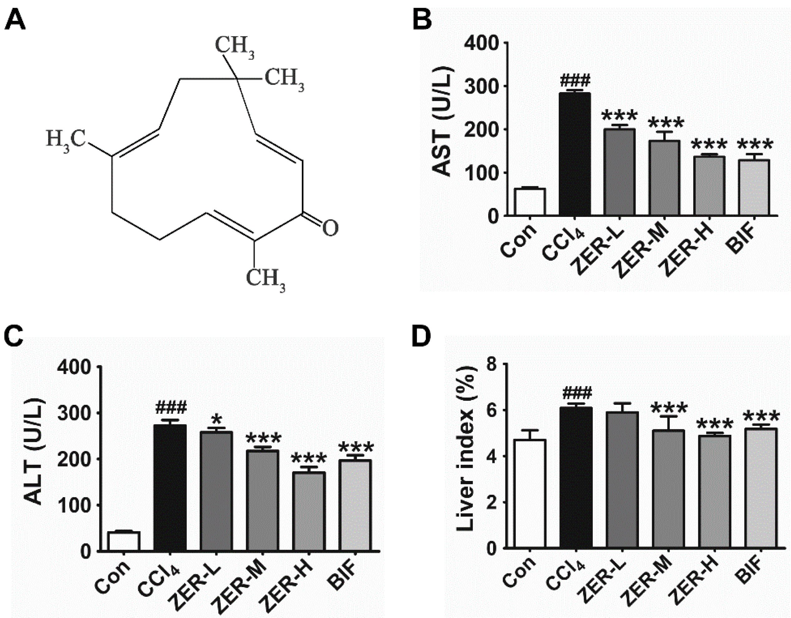
2.1. Effects of ZER on Serum Aspartate Aminotransferase (AST), Alanine Aminotransferase (ALT), and Liver Index

The structure of ZER is shown in Figure 1A. Bifendate (BIF) was used as a positive control, which is widely prescribed to treat hepatic injury. The mice were pretreated with ZER for five consecutive days and then challenged with a one-time intraperitoneal injection. The protective effects of ZER were first assessed by serum biochemical parameters. As shown in Figure 1B and Figure 1C, the activities of serum AST and ALT were markedly elevated in the CCl4 injection group with respect to the control group, indicating that the model of ALI induced by CCl4 was successfully established. ZER pretreatment effectively lowered the increase of serum AST and ALT activities in a dose-dependent manner. The ALT activity in the 20 μmol/kg ZER group was even lower than the BIF-positive control group. These results suggested that the CCl4-induced ALI mice model was successfully established and ZER harbors hepatoprotective properties against CCl4-induced ALI.
Liver disease usually accompanied the rise of liver index. In the CCl4-intoxicated animals, we also observed the increase of the liver index with respect to the normal group, which is shown in Figure 1D. Unsurprisingly, ZER pretreatment markedly decreased the liver index in a dose-dependent fashion, as demonstrated by the gradual decrease of the liver index with respect to that of the CC14 group.

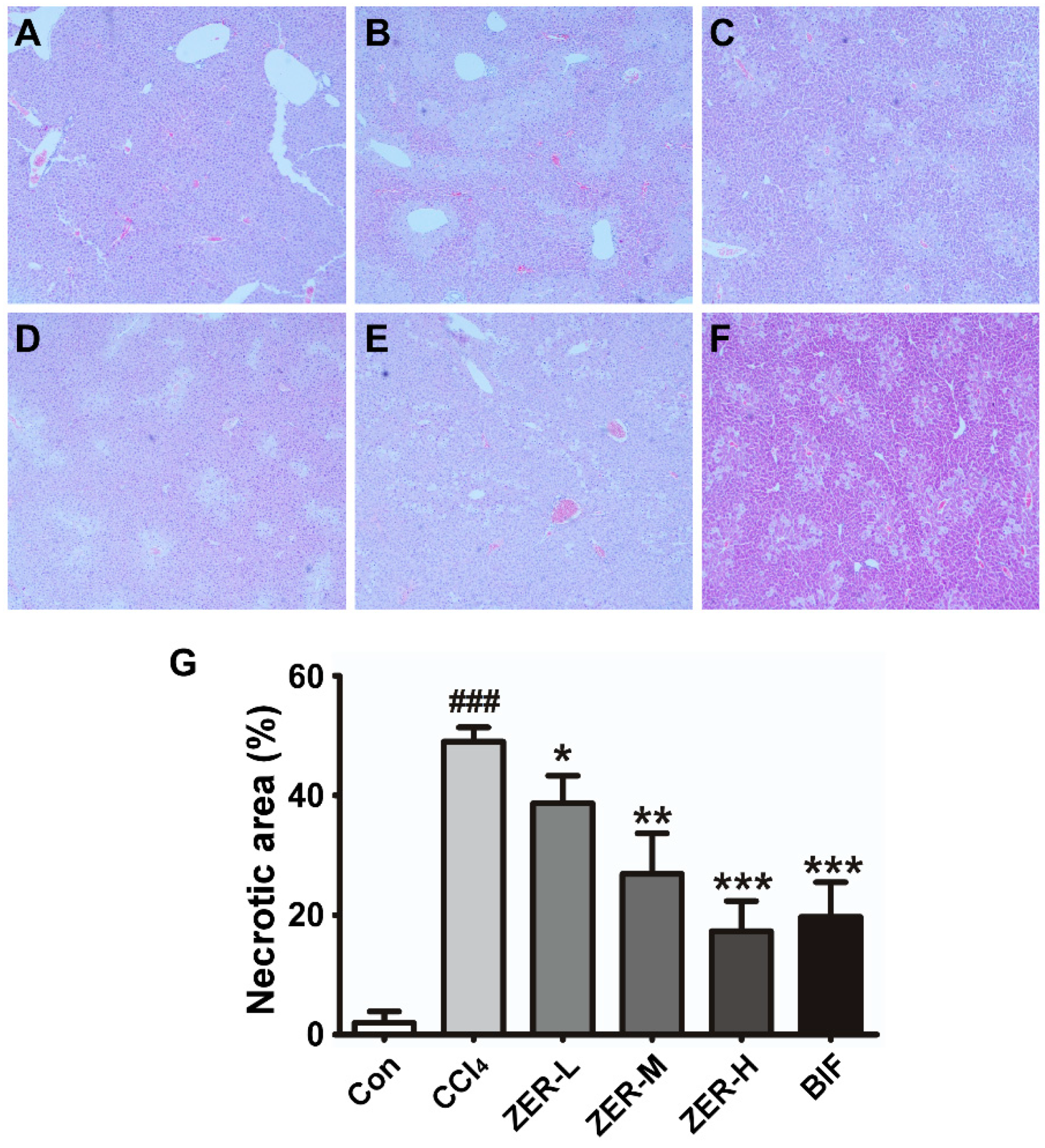
2.2. Histopathological Effect of ZER in Liver

To further assess the protective effects of ZER on CCl4-induced ALI, an examination of the liver histophathology was employed to evaluate the extent of the hepatic damage. After intoxication of the mice with CCl4, a moderate degree of hepatocyte necrosis was evident in the central lobular areas, which accounted for 49% (the model) of the liver section, indicating a successful mouse model of ALI (Figure 2B,G). However, ZER pretreatment significantly decreased the necrosis in a dose-dependent fashion (Figure 2C–G). The necrotic area decreased from 49% to 39.4% (ZER-L), 26.9% (ZER-M), or 19.7% (ZER-H) (Figure 2G). The histopathological results demonstrated that pretreatment with ZER effectively alleviated CCl4-induced centrolobular necrosis.

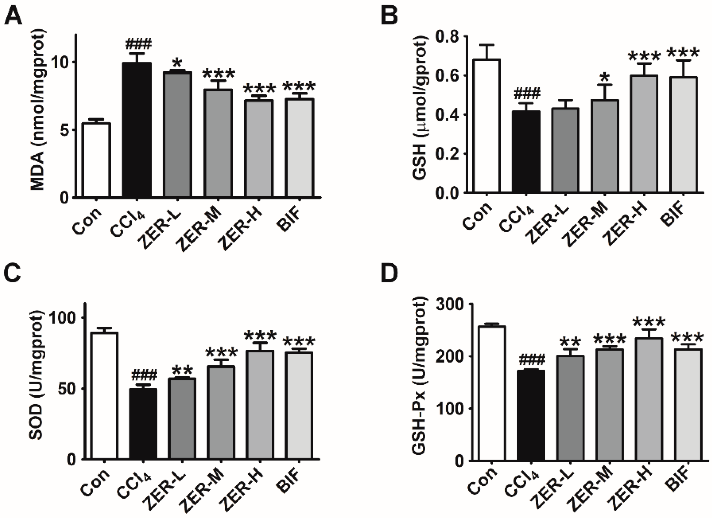
2.3. Effects of ZER on Oxidative Stress Parameters

It is well elucidated that oxidative stress is an important mechanism of action of hepatotoxicity induced by CCl4. CCl4 intoxication causes increasing generation of reactive oxygen species (ROS) in hepatocytes, which triggers GSH depletion and lipid peroxidation. MDA is the end product of lipid peroxidation and can be used as a marker for oxidative stress. ROS also results in a significant decrease in antioxidant enzyme activity, including SOD and GSH-Px. To understand the potential mechanisms of the observed hepatoprotective effects, oxidative stress parameters was evaluated by measuring MDA levels, GSH levels, and the activities of SOD and GSH-Px. As can be seen in Figure 3, MDA contents was remarkably increased, GSH levels were remarkably decreased, and SOD and GSH-Px activities were significantly decreased comparing to those in the control group after intoxication with CCl4. However, with ZER pretreatment, the generation of MDA in liver tissues was reduced, the depleted GSH levels were restored, and the decreased SOD and GSH-Px activities were increased, all in a dose-dependent manner. These results suggest that ZER exerts hepatoprotective effects by alleviating oxidative stress.

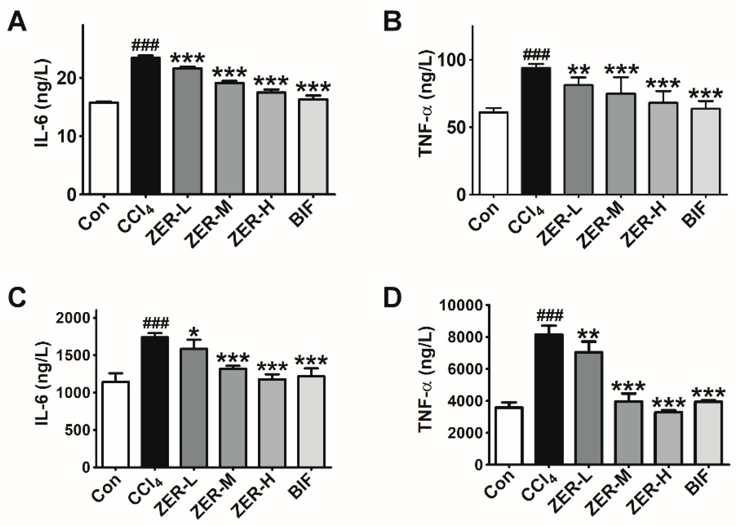
2.4. Effects of ZER on the Levels of TNF-α and IL-6 In Vivo

Besides oxidative stress, CCl4-induced ALI is also characterized by an inflammatory response due to pro-inflammatory cytokines such as TNF-α and IL-6 released by damaged hepatocytes, sinusoidal endothelial cells, and activated Kupffer cells. To investigate if ZER provides protection against CCl4-induced ALI via anti-inflammation, the change in levels of two main pro-inflammatory cytokines, TNF-α and IL-6, was examined. As can be seen in Figure 4, both serum and liver tissue TNF-α and IL-6 levels were elevated in the CCl4-intoxication group than in the control group, as determined by ELISA. However, pretreatment with ZER inhibited TNF-α and IL-6 production with respect to the CCl4-intoxication group in a dose-dependent manner. The results clearly showed that ZER-pretreatment could inhibit the CCl4-intoxication-induced production of inflammatory cytokines TNF-α and IL-6. These results hint that the hepatoprotective effect of ZER is achieved by down-regulating the inflammatory response.

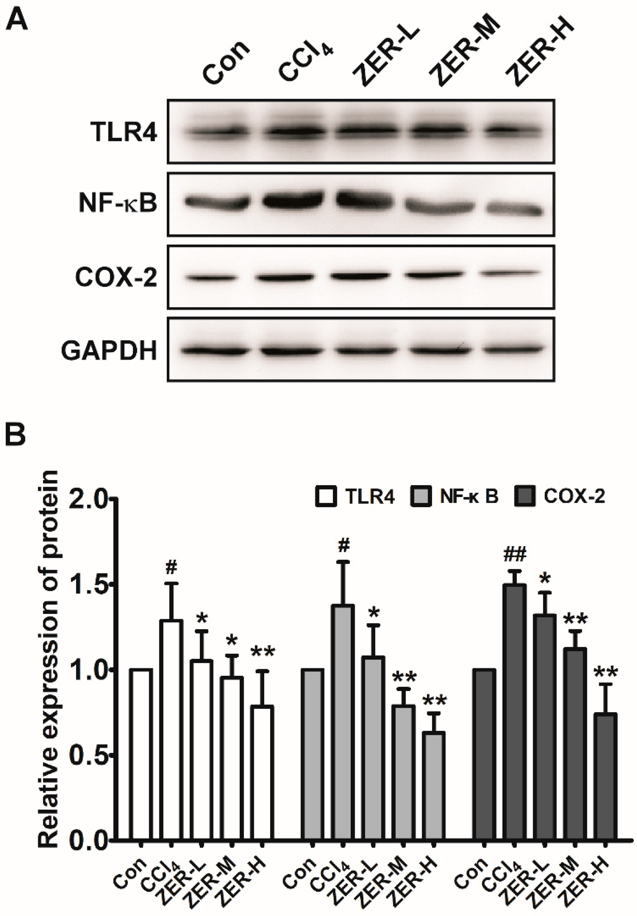
2.5. Effects of ZER on TLR4/NF-κB/COX-2 Signaling In Vivo

We further explored the underlying anti-inflammatory mechanism of ZER against ALI induced by CCl4. As is well-known, the production of pro-inflammatory cytokines TNF-α and IL-6 are induced by activation of NF-κB signaling, so we first detected the change in the level of phosphorylated p65, which forms the most common heterodimer of NF-κB together with p50. As shown in Figure 5, compared with the control group, the protein level of p-p65 was increased in the liver tissue of the CCl4-challenged model group. The levels of p-p65 in the ZER-pretreated groups were decreased compared with that of the model group and the decrement was exhibited in a dose-dependent manner. We then detected the change in the levels of TLR4 and COX-2 proteins, which are the upstream and downstream molecules of the NF-κB pathway, respectively. The change in the levels of TLR4 and COX-2 were in accordance with that of p65, as can be seen in Figure 5. These results suggested that CCl4 intoxication may mediate the inflammatory response via TLR4/NF-κB/COX-2 signaling and ZER could inhibit inflammation by blocking the activation of the TLR4/NF-κB/COX-2 signaling pathway.

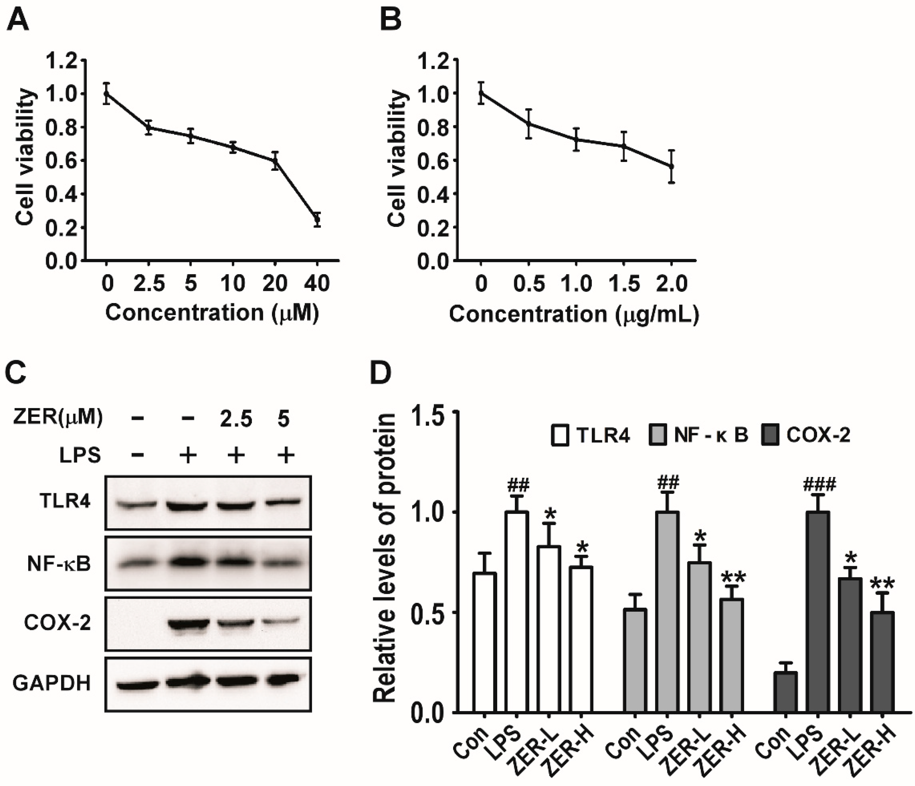
2.6. Effects of ZER on TLR4/NF-κB/COX-2 Signaling In Vitro

The expression analysis of the liver tissue samples suggested that ZER protects CCl4-induced ALI by ameliorating the inflammatory response through TLR4/NF-κB/COX-2 signaling. To further verify the involvement of this pathway in vitro, anLPS-induced Raw264.7 inflammatory cellular model was established and employed. We first assessed the potential inhibitory effects of ZER and LPS on cell viability against Raw264.7 cells at different concentrations. As shown in Figure 6A, the 75% viability of ZER inhibition of cell viability was observed at about 7.5 μmol/L and 50% at about 25 μmol/L. According to the 3-[4,5-dimethylthiazol-2-yl]-2,5 diphenyl tetrazolium bromide (MTT) results, we set two ZER treatment groups: 2.5 μmol/L as the low dose group and 5 μmol/L as the high dose group. As shown in Figure 6B, the cell viability of Raw264.7 cells treated with 0.5 μg/mL LPS for 36 h was about 80%. We further determined the activation situation of TLR4/NF-κB/COX-2 signaling upon ZER pretreatment in LPS-stimulated Raw264.7 cells. The results showed that LPS stimulation significantly activated the TLR4/NF-κB/COX-2 signaling, as evidenced by the up-regulated protein levels of TLR4, p-p65, and COX-2. Unsurprisingly, after treatment with different concentrations of ZER, the protein levels of TLR4, p-p65, and COX-2 were markedly decreased, as shown in Figure 6C,D. These results clearly confirmed that ZER could inhibit inflammation by blocking the activation of the TLR4/NF-κB/COX-2 signaling pathway.

3. Discussion

ALI, which is caused by a number of factors, such as toxic chemicals, drugs, viruses, and bacteria, seriously threatens human health [1,4]. Recently, great attention and effort have been devoted to discovering phytochemicals with preventive or therapeutic effects against ALI [6,7,11,12,16,17,30]. In the current study, we found that ZER exhibits a hepatoprotective activity against CCl4-induced ALI by reducing the level of oxidative stress and inhibiting the inflammatory response. Further results revealed that the anti-inflammation mechanism for ZER against CCl4-induced ALI involved inactivation of the TLR4/NF-κB/COX-2 signaling pathway. Together, these findings explain the pharmacological mechanism of ZER acting on CCl4-induced ALI and provided an experimental and theoretical basis for the application of ZER in the clinical treatment of ALI.
The decreased serum levels of ALT and AST upon ZER pretreatment imply the hepatoprotective effect against CCl4-induced ALI and pathological analysis further confirmed this beneficial effect by effectively decreasing the area of centrolobular necrosis. When trying to elucidate the underlying mechanism, two pathogenic events were taken into consideration, oxidative stress and inflammation, since these two events during ALI normally serve as the mechanisms for liver damage [4,32]. Meanwhile, ZER is reported to possess many pharmacological properties, including antioxidant and anti-inflammatory properties [33]. Therefore, we hypothesized that ZER would exert hepatoprotective activity against ALI by alleviating oxidative stress and the inflammatory response. In light of the assessment of oxidative stress parameters in the liver tissues, including SOD, GSH-Px, GSH, and MDA, we confirmed that ZER exerts hepatoprotective effects against CCl4-induced ALI through reducing oxidative stress by recovering the activities of antioxidant enzymes, restoring the GSH reservoir, and reducing lipid peroxidation specifically. Afterward, the evaluation of the change in serum and hepatic levels of pro-inflammatory cytokines IL-6 and TNF-α upon ZER pretreatment confirmed the involvement of anti-inflammation for ZER against ALI, which is consistent with Kim et al.’s report [30]. Based on this observation, we tried to seek the signaling pathways responsible for the protection of ZER against ALI. Protein expression analysis of liver tissue samples hinted at the TLR4/NF-κB/COX-2 signaling pathway. Further in vitro studies in Raw264.7 cells verified the involvement of this pathway, as evidenced by the reduced release amounts of IL-6 and TNF-α and down-regulated protein levels of TLR4, NF-κB, and COX-2, which were originally elevated upon LPS stimulation. Thus, the anti-inflammation activity for ZER against CCl4-induced ALI may occur through the TLR4/NF-κB/COX-2 signaling pathway.
Antioxidant enzymes, such as SOD and GSH-Px plus GSH, which are scavengers of ROS in the liver, are the first line of defense against oxidative stress-induced tissue injury [34]. Several previous studies have shown that ZER can inhibit the generation of ROS [35,36,37,38]. For instance, ZER was found to provide protection to pancreatic β cells from high glucose-induced cytotoxicity through directly interfering with ROS production [39]. Our data also suggested this inhibition of ROS generation. However, contrary observations have been reported. For example, treatment with ZER has been found to significantly increase reactive oxygen species levels in colorectal cancer cells and melanoma cells [40,41]. These discrepancies may arise from distinct disease contexts and different cellular processes involved in the studies; the observations of ZER in favor of ROS generation were implicated in apoptosis under the cancer context [40,41,42,43,44], whereas the observations of ZER against ROS generation were mostly related to inflammation and inflammatory-associated diseases, such as hepatic disease, osteoarthritis, and diabetes and diabetic complications [22,24,39,45].
An excessive inflammatory response can aggravate injured hepatic tissue and hinder repair processes [13,32]. Previous studies have reported the hepatoprotective activity of ZER against ALI via anti-inflammation [30,31]. However, our results further suggested that ZER might inhibit CCl4-induced inflammation through TLR4/NF-κB/COX-2 signaling. There are studies in the literature that support our observations. An in vitro study conducted in human leukemic cell line THP-1 cell-derived macrophages activated by LPS showed that ZER inhibits the secretion of pro-inflammatory cytokines TNF-α and IL-6, the induction of NF-κB and COX-2, and also decreases the elevated mRNA levels of TLR2 and TLR4 [46]. Lee and Haque reported the inhibition of NF-κB p65 phosphorylation and pro-inflammatory cytokines release by ZER, respectively [27,47]. It is well established that hepatic macrophages, including activated Kupffer cells, hold central functions in initiating, perpetuating, and even restricting inflammation in the liver [10,13]. According to our results both in vivo and in vitro, it is likely that ZER targets hepatic macrophages to inhibit unregulated inflammatory responses, thus preventing further hepatic injury and promoting hepatic tissue repair. However, this hypothesis needs to be tested.
Hepatocellular injury leads to cell death, including both necrosis and apoptosis [48,49,50]. However, uncontrolled necrosis and apoptosis can provoke further hepatic damage [30,51]. Our results show that ZER pretreatment effectively decreases the necrotic area of the injured liver by inhibiting the inflammatory response through the TLR4/NF-κB/COX-2 signaling pathway. Meanwhile, Kim et al. reported that ZER exerts a hepatoprotective activity by inhibiting apoptosis through inhibiting the translocation of activated hepatic JNK to mitochondria, thus ameliorating CCl4-intoxication-induced oxidative stress. Hence, ZER provides protection against CCl4-induced ALI via alleviating both necrosis and apoptosis.
The present study revealed that ZER, which is a monocyclic sesquiterpene [30], provided protection against ALI through ameliorating oxidative stress and inflammation. Meanwhile, several previous studies showed that sesquiterpene or sesquiterpene lactones exhibit hepatoproctive activities toward different types of hepatic disease [8,52,53,54]. Another two studies showed that a mixture with 21 kinds of sesquiterpenoids extracted from the root of Panax Ginseng exhibited strong hepatoproctive effects, which were also mediated by reducing oxidative stress and inhibiting the inflammation response [7]. In fact, more and more sesquiterpenoids, including sesquiterpenes and sesquiterpene lactones, have been reported as having either anti-oxidation or anti-inflammation activities, or both [7,22,55,56]. Given that oxidative stress and inflammation are usually present simultaneously and intertwined to aggravate already-existing liver tissue damage [4,13] and the clinical consideration and therapeutic strategy for liver diseases predominantly focuses on anti-oxidation and anti-inflammation [1,14], we speculate that natural products of sesquiterpenoids extracted from plants may be a promising reservoir to screen effective therapeutic drugs for hepatic disease with high efficacy and low toxicity.
Collectively, our work revealed a new mechanism of ZER against ALI, which is through ameliorating oxidative stress. Furthermore, unlike the previous study, we unraveled the involvement of inactivation of the TLR4/NF-κB/COX-2 signaling pathway in the modulation of the inflammatory response upon ZER pretreatment. We also acknowledge that the validation data about the involvement of TLR4/NF-κB/COX-2 signaling was only conducted in vitro and so may not be convincing. Employment of an NF-κB inhibitor in vivo is a better way to confirm the correlation. Last but not least, a better understanding of the mechanism underlying the hepatoprotective activity of ZER against ALI is very necessary so that successful translation of ZER to clinical applications can be developed rapidly for liver disease.
In conclusion, our study raises the possibility that ZER could be used as a potential therapeutic drug for hepatic disease.

4. Materials and Methods

4.1. Chemicals and Reagents

Zerumbone (ZER) was purchased from Sigma Aldrich (No.Z3902, St. Louis, MO, USA), dissolved in DMSO (Dimethyl sulfoxide). Bifendate (BIF) was purchased from Beijing Yuehe Pharmaceutical Factory (H11020980, Beijing, China). LPS was purchased from Beyotime Biotechnology (No.S1732, Shanghai, China). RPMI 1640 Medium was purchased from Hyclone (Logan, UT, USA). Fetal bovine serum (FBS) was purchased from Gibco (Gaithersburg, MA, USA). The assay kits for ALT, AST, SOD, GSH-Px, GSH, and MDA were purchased from Nanjing Jiancheng Bioengineering Institute (Nanjing, China). Bicinchoninic acid (BCA) assay kit and MTT were purchased from Solarbio (Beijing, China). Mouse IL-6 and TNF-α ELISA kits was purchased from ShangHai Haling Biological Technology Co., Ltd. (ShangHai, China). Rabbit anti-TLR4 antibody (#14358, 1:1000), rabbit anti-COX-2 antibody (#4842, 1:1000), rabbit anti-p-NF-κB p65 (Ser536) antibody (#3033, 1:1000), mouse anti-β-Actin antibody (#3700, 1:2000) were purchased from Cell Signaling Technology (Danvers, MA, USA). All other chemicals and reagents were purchased from local firms.

4.2. Animals

Specific pathogen-free (SPF) male ICR mice (SCXK20140007), weighting 20 ± 2g, 7~8 weeks, were purchased from Jinan Pengyue Experimental Animal Breeding Co., Ltd. (Jinan, Shandong, China). The mice were kept in the Laboratory Animal Centre of the First Affiliated Hospital of Henan University of Science and Technology. The feeding conditions for the mice were provided with a constant temperature of 22 ± 2 °C and a relative humidity of 55 ± 5% with a 12 h/12 h light–dark cycle. During the experiment, the mice were given a standard diet with clean mineral water and pellet feed. All experiments were conducted in accordance with the Guide for the Care and Use of Laboratory Animals and the animal experiment protocols were approved by the Institutional Ethical Committee of Henan University of Science and Technology.

4.3. Experimental Design

After a five-day acclimation based on body weight, sixty ICR mice were equally divided into six groups of 10 animals each, as follows: The control (Con) group, the CCl4 model group, the ZER-L (1.25 μmol/kg of body weight (BW), dissolved in DMSO) group, the ZER-M (5 μmol/Kg) group, the ZER-H (20 μmol/Kg) group, and the positive control group treated with BIF (350 μmol/Kg/day). The final concentration (v/v) of DMSO in the in vivo experiment was 1%. During the experiment, the mice were given a standard diet. All mice were intraperitoneally injected with corresponding drugs for 5 consecutive days: The control group and the CCl4 model group injected with DMSO, three ZER groups with ZER with the corresponding concentrations, and the positive control group with BIF. Two hours after the last administration, all the animals from each group received a one-time intraperitoneal injection of CCl4 (0.01 mL/g body weight, 0.2% in soybean oil) to establish the ALI model, except for the control group, which were received the same volume of soybean oil. Twelve hours fasting followed with the CCl4 challenge.
The mice were sacrificed under anesthesia (6% chloral hydrate, 10 mL/kg) 12 h after the CCl4 challenge. Blood and liver tissue were collected for experiments. The serum was obtained by centrifuging (3000 rpm, 4 °C, 15 min) and then immediately subjected to the following biochemical analysis. For the liver, a whole lobe without any mechanical damage was fixed in 10% formalin for histopathological analysis, while the rest of the liver tissue samples were stored in liquid nitrogen for further experiments.

4.4. Detections of Biochemistry Indexes

The activities of ALT and AST in serum and the activities or levels of SOD, GSH-Px, GSH, and MDA in liver homogenates were determined by commercial kits according to the manufacturer’s instructions. The 10% liver homogenates were prepared as follows: The liver tissues were homogenized in nine-fold (w/v) cold saline on ice and then centrifuged (12,000 rpm, 30 min, 4 °C), supernatants were collected for detection.

4.5. Histopathological Analysis

The liver lobes fixed in 10% formalin were embedded in paraffin and processed into 4 μm thick sections, which were subjected to H&E staining. All sections were examined and pictured under an optical microscope. Areas of necrosis were quantified using ImageJ v1.8.0 software (National Institutes of Health, Bethesda, MD, USA).

4.6. Detections of Inflammatory Cytokines IL-6 and TNF-α

The levels of IL-6 and TNF-α both in serum and in liver tissue were analyzed by ELISA kits according to the manufacturer’s instructions. The serum was prepared as in Section 4.3 and 10% liver homogenates were prepared as in Section 4.4.

4.7. Cell Culture and LPS Treatment

The Raw264.7 cell line was a generous gift from Dr Ruifang Li, who also works in Medical School of Henan University of Science and Technology. The Raw264.7 cell line was cultured in Roswell Park Memorial Institute (RPMI) 1640 medium supplemented with 10% (v/v) FBS and antibiotics (100 U/mL penicillin G and streptomycin sulfate) in an incubator at 37 °C with 5% CO2 and saturated humidity. ZER was dissolved in DMSO to a concentration of 10 mM for the in vitro experiment. The final concentration (v/v) of DMSO in the in vivo experiment was 0.05%. For LPS stimulation, the dosage and timing for activating the Raw264.7 cells was 0.5 μg/mL LPS for 36 h. The cells in the exponential growth phase were harvested and seeded into 6-well plates at 6 × 10⁵ cells per well, then stimulated by LPS (0.5 μg/mL) for 12 h when the cells were of 80% confluence. ZER was then added for another 24 h. The cells were lysed and subjected to Western blotting analysis.

4.8. Cytotoxicity Assessment of ZER and LPS by MTT Assay

The cytotoxicity of ZER and LPS toward Raw264.7 cells were evaluated by MMT assays. The procedure was carried out as previously described with little modification [57]. Raw264.7 cells were seeded into 96-well plates with 1 × 10⁴ cells in 100 μL medium per well overnight. Then, the cells were treated with different concentrations of ZER (0, 2.5, 5, 10, 20, and 40 μmol/L) for 24 h or different concentrations of LPS (0, 0.5, 1.0, 1.5, and 2.0 μg/mL) for 36 h. The control group was treated with DMSO or ddH2O. Subsequently, 15 μL of MTT (5 mg/mL, Sigma, St. Louis, MO, USA) was added to each well and 4 h later, the medium was discarded and the purple formazan crystals were solubilized in 150 μL DMSO per well. Absorbance was detected at 490 nm on Microplate Readers (Bio-Tek, USA). The viability rate was calculated as follows: Viability rate (%) = (A model/A control) × 100%.

4.9. Western Blotting Analysis

This procedure was described in our previous study. The total proteins of liver tissue or Raw264.7 cells were extracted by Radio Immunoprecipitation Assay (RIPA) lysis solution (Solarbio, Beijing, China) with phenylmethanesulfonyl fluoride (PMSF) on ice following the user guide. BCA protein content determination was used for quantification. The same amount of protein (40 μg) was subjected to Western blotting analysis as previously described [53]. The signals were detected by the Western BrightTM electrochemiluminescence (ECL)kit (Advansta, Menlo Park, CA, USA), then acquired and analyzed by Image Lab Software (Bio-RadLaboratories, Hercules, CA, USA).

4.10. Statistical Analysis

All experimental data were expressed as mean ± SD (standard deviation). Statistical significance was determined by SPSS 16.0 software (IBM,.Chicago, IL, USA), using one-way analysis of variance (ANOVA) with LSD Fisher post hoc test. p < 0.05 was considered as significant.

Author Contributions

M.W. and S.L. conceived and designed the research; J.N. and Y.W. performed the experiments; L.O. and B.D. performed the statistical analysis; M.W. and S.L. wrote the main manuscript text; M.W. and J.N. prepared figures. All authors reviewed the manuscript.

Funding

This research was funded by the National Natural Science Foundation of China (No. 31500631).

Acknowledgments

The cell used in this work was generously given by Ruifang Li, who also works in Medical School of Henan University of Science and Technology. The experimental animal room was provided generously by Shegan Gao, who is the dean of Medical School of Henan University of Science and Technology.

Conflicts of Interest

The authors declare no conflict of interest.

Abbreviations

CCl4Carbon tetrachloride
ALIAcute liver injury
ZERZerumbone
ASTAspartate aminotransferase
ALTAlanine aminotransferase
SODSuperoxide dismutase
GSH-PxGlutathione peroxidase
GSHGlutathione
MDAMalondialdehyde
IL-6Interleukin-6
TNF-αTumor necrosis factor alpha
TLR4Toll-like Receptor 4
NF-κBNuclear factor -κB
COX-2Cyclooxygenase-2
TLRsToll-like receptors
HMGB1High mobility group box-1 protein
DAMPsDamage associated molecular patterns
BIFBifendate
LPSLipopolysaccharide
ROSReactive oxygen species
FBSFetal bovine serum
H&EHematoxylin and Eosin

References

  1. Patten, D.A.; Shepherd, E.L.; Weston, C.J.; Shetty, S. Novel Targets in the Immune Microenvironment of the Hepatic Sinusoids for Treating Liver Diseases. Semin. Liver Dis. 2019, 39, 111–123. [Google Scholar]
  2. Shen, T.; Liu, Y.; Shang, J.; Xie, Q.; Li, J.; Yan, M.; Xu, J.M.; Niu, J.Q.; Liu, J.J.; Paul, B.W.; et al. Incidence and Etiology of Drug-Induced Liver Injury in Mainland China. Gastroenterology 2019, 156, 1–12. [Google Scholar] [CrossRef]
  3. Trepotec, Z.; Lichtenegger, E.; Plank, C.; Aneja, M.K.; Rudolph, C. Delivery of mRNA Therapeutics for the Treatment of Hepatic Diseases. Mol. Ther. 2019, 27, 794–802. [Google Scholar] [CrossRef] [PubMed] [Green Version]
  4. Reyes-Gordillo, K.; Shah, R.; Muriel, P. Oxidative Stress and Inflammation in Hepatic Diseases: Current and Future Therapy. Oxid. Med. Cell Longev. 2017, 2017, 3140673. [Google Scholar] [CrossRef] [PubMed]
  5. Dai, C.; Xiao, X.; Li, D.; Tun, S.; Wang, Y.; Velkov, T.; Tang, S.S. Chloroquine ameliorates carbon tetrachloride-induced acute liver injury in mice via the concomitant inhibition of inflammation and induction of apoptosis. Cell Death Dis. 2018, 9, 1164. [Google Scholar] [CrossRef]
  6. Mitazaki, S.; Kotajima, N.; Matsuda, S.; Ida, N.; Iide, M.; Honma, S.; Suto, M.; Kato, N.; Kurode, N.; Hiraiwa, K.; et al. Dimethylthiourea ameliorates carbon tetrachloride-induced acute liver injury in ovariectomized mice. Biomed. Pharmacother. 2018, 104, 427–436. [Google Scholar] [CrossRef]
  7. Wang, W.; Wang, S.; Liu, J.; Cai, E.; Zhu, H.; He, Z.M.; Gao, Y.G.; Li, P.Y.; Zhao, Y. Sesquiterpenoids from the root of Panax Ginseng protect CCl4-induced acute liver injury by anti-inflammatory and anti-oxidative capabilities in mice. Biomed. Pharmacother. 2018, 102, 412–419. [Google Scholar] [CrossRef]
  8. Mahesh, A.; Jeyachandran, R.; Cindrella, L.; Thangadurai, D.; Veerapur, V.P.; Muralidha, P. Hepatocurative potential of sesquiterpene lactones of Taraxacum officinale on carbon tetrachloride induced liver toxicity in mice. Acta Biol. Hung. 2010, 61, 175–190. [Google Scholar] [CrossRef] [PubMed]
  9. Arosio, B.; Gagliano, N.; Fusaro, L.M.; Parmeggiani, L.; Tagliabue, J.; Galetti, P.; Castri, D.D.; Moscheni, C.; Annoni, G. Aloe-Emodin quinone pretreatment reduces acute liver injury induced by carbon tetrachloride. Pharmacol. Toxicol. 2000, 87, 229–233. [Google Scholar] [PubMed]
  10. Li, P.; He, K.; Li, J.; Li, Z.J.; Gong, J.P. The role of Kupffer cells in hepatic diseases. Mol. Immunol. 2017, 85, 222–229. [Google Scholar] [CrossRef] [PubMed]
  11. Yin, Y.; Zhang, Y.; Li, H.; Zhao, Y.; Cai, E.; Zhu, H.Y.; Li, P.Y. Triterpenoids from fruits of Sorbus pohuashanensis inhibit acetaminophen-induced acute liver injury in mice. Biomed. Pharmacother. 2019, 109, 493–502. [Google Scholar] [CrossRef] [PubMed]
  12. Meng, X.; Wang, Z.; Liang, S.; Tang, Z.; Liu, J.; Xin, Y.; Kuang, H.; Wang, Q. Hepatoprotective effect of a polysaccharide from Radix Cyathulae officinalis Kuan against CCl4-induced acute liver injury in rat. Int. J. Biol. Macromol. 2019, 132, 1057–1067. [Google Scholar] [CrossRef] [PubMed]
  13. Tacke, F. Targeting hepatic macrophages to treat liver diseases. J. Hepatol. 2017, 66, 1300–1312. [Google Scholar] [CrossRef] [PubMed]
  14. Woolbright, B.L.; Jaeschke, H. Sterile inflammation in acute liver injury: Myth or mystery? Expert Rev. Gastroenterol. Hepatol. 2015, 9, 1027–1029. [Google Scholar] [CrossRef]
  15. Li, M.; Wang, S.; Li, X.; Kou, R.; Wang, Q.; Wang, X.; Zhao, N.; Zeng, T.; Xie, K. Diallyl sulfide treatment protects against acetaminophen-/carbon tetrachloride-induced acute liver injury by inhibiting oxidative stress, inflammation and apoptosis in mice. Toxicol. Res. 2019, 8, 67–76. [Google Scholar] [CrossRef] [Green Version]
  16. Chen, C.Y.; Kao, C.L.; Liu, C.M. The Cancer Prevention, Anti-Inflammatory and Anti-Oxidation of Bioactive Phytochemicals Targeting the TLR4 Signaling Pathway. Int. J. Mol. Sci. 2018, 19, 2729. [Google Scholar] [CrossRef]
  17. Ding, Y.; Liu, P.; Chen, Z.L.; Zhang, S.J.; Wang, Y.Q.; Cai, X.; Luo, L.; Zhou, X.; Zhao, L. Emodin Attenuates Lipopolysaccharide-Induced Acute Liver Injury via Inhibiting the TLR4 Signaling Pathway in vitro and in vivo. Front. Pharmacol. 2018, 9, 962. [Google Scholar] [CrossRef]
  18. Kuramochi, M.; Izawa, T.; Pervin, M.; Bondoc, A.; Kuwamura, M.; Yamate, J. The kinetics of damage-associated molecular patterns (DAMPs) and toll-like receptors during thioacetamide-induced acute liver injury in rats. Exp. Toxicol. Pathol. 2016, 68, 471–477. [Google Scholar] [CrossRef] [PubMed]
  19. Roy, A.; Srivastava, M.; Saqib, U.; Liu, D.; Faisal, S.M.; Sugathan, S.; Bishnoi, S.; Baij, M.S. Potential therapeutic targets for inflammation in toll-like receptor 4 (TLR4)-mediated signaling pathways. Int. Immunopharmacol. 2016, 40, 79–89. [Google Scholar] [CrossRef] [PubMed]
  20. Ceccarelli, S.; Nobili, V.; Alisi, A. Toll-like receptor-mediated signaling cascade as a regulator of the inflammation network during alcoholic liver disease. World J. Gastroenterol. 2014, 20, 16443–16451. [Google Scholar] [CrossRef] [PubMed]
  21. Girisa, S.; Shabnam, B.; Monisha, J.; Fan, L.; Halim, C.E.; Arfuso, F.; Ahn, K.S.; Sethi, G.; Kunnumakkara, A.B. Potential of Zerumbone as an Anti-Cancer Agent. Molecules 2019, 24, 734. [Google Scholar] [CrossRef]
  22. Chien, T.Y.; Huang, S.K.; Lee, C.J.; Tsai, P.W.; Wang, C.C. Antinociceptive and Anti-Inflammatory Effects of Zerumbone against Mono-Iodoacetate-Induced Arthritis. Int. J. Mol. Sci. 2016, 17, 249. [Google Scholar] [CrossRef]
  23. Liu, W.Y.; Tzeng, T.F.; Liu, I.M. Zerumbone, a Bioactive Sesquiterpene, Ameliorates Diabetes-Induced Retinal Microvascular Damage through Inhibition of Phospho-p38 Mitogen-Activated Protein Kinase and Nuclear Factor-kappaB Pathways. Molecules 2016, 21, 1708. [Google Scholar] [CrossRef]
  24. Tzeng, T.F.; Liou, S.S.; Chang, C.J.; Liu, I.M. Zerumbone, a tropical ginger sesquiterpene, ameliorates streptozotocin-induced diabetic nephropathy in rats by reducing the hyperglycemia-induced inflammatory response. Nutr. Metab. 2013, 10, 64. [Google Scholar] [CrossRef]
  25. Hemn, H.O.; Noordin, M.M.; Rahman, H.S.; Hazilawati, H.; Zuki, A.; Chartrand, M.S. Antihypercholesterolemic and antioxidant efficacies of zerumbone on the formation, development, and establishment of atherosclerosis in cholesterol-fed rabbits. Drug Des. Dev. Ther. 2015, 9, 4173–4208. [Google Scholar]
  26. Zulazmi, N.A.; Gopalsamy, B.; Min, J.C.; Farouk, A.A.; Sulaiman, M.R.; Bharatham, B.H.; Perimal, E.K. Zerumbone Alleviates Neuropathic Pain through the Involvement of l-Arginine-Nitric Oxide-cGMP-K(+) ATP Channel Pathways in Chronic Constriction Injury in Mice Model. Molecules 2017, 22, 555. [Google Scholar] [CrossRef]
  27. Lee, C.Y.; Chen, S.P.; Su, C.H.; Ho, Y.C.; Yang, M.L.; Lee, S.S.; Huang, L.R.; Yang, C.P.; Chan, C.J.; Kuan, Y.H. Zerumbone from Zingiber zerumbet Ameliorates Lipopolysaccharide-Induced ICAM-1 and Cytokines Expression via p38 MAPK/JNK-IkappaB/NF-kappaB Pathway in Mouse Model of Acute Lung Injury. Chin. J. Physiol. 2018, 61, 171–180. [Google Scholar] [CrossRef]
  28. Prasannan, R.; Kalesh, K.A.; Shanmugam, M.K.; Nachiyappan, A.; Ramachandran, L.; Nguyen, A.H.; Kumar, A.P.; Lakshmanan, M.; Sethi, G. Key cell signaling pathways modulated by zerumbone: Role in the prevention and treatment of cancer. Biochem. Pharmacol. 2012, 84, 1268–1276. [Google Scholar] [CrossRef] [Green Version]
  29. Tzeng, T.F.; Liou, S.S.; Chang, C.J.; Liu, M.H. Zerumbone, a Natural Cyclic Sesquiterpene of Zingiber zerumbet Smith, Attenuates Nonalcoholic Fatty Liver Disease in Hamsters Fed on High-Fat Diet. Evid. Based Complement. Alternat. Med. 2013, 2013, 303061. [Google Scholar] [CrossRef]
  30. Kim, J.W.; Yang, D.; Jeong, H.; Park, I.S.; Lee, M.H.; Lim, C.W.; Kim, B. Dietary zerumbone, a sesquiterpene, ameliorates hepatotoxin-mediated acute and chronic liver injury in mice. Phytother. Res. 2019, 33, 1–13. [Google Scholar] [CrossRef]
  31. Hamid, A.; Lee, L.S.; Karim, S.R.; Jufri, N.F. Hepatoprotective Effects of Zerumbone against Paracetamol-Induced Acute Hepatotoxicity in Rats. Malays. J. Med. Sci. 2018, 25, 64–71. [Google Scholar] [CrossRef]
  32. Woolbright, B.L.; Jaeschke, H. The impact of sterile inflammation in acute liver injury. J. Clin. Transl. Res. 2017, 3, 170–188. [Google Scholar] [CrossRef] [Green Version]
  33. Kalantari, K.; Moniri, M.; Boroumand, M.A.; Abdul Rahim, R.; Bin Ariff, A.; Izadiyan, Z.; Mohamad, R.; et al. A Review of the Biomedical Applications of Zerumbone and the Techniques for Its Extraction from Ginger Rhizomes. Molecules 2017, 22, 1645. [Google Scholar] [CrossRef]
  34. Xu, M.; Rui, D.; Yan, Y.; Xu, S.; Niu, Q.; Feng, G.; Wang, Y.; Li, S.; Jing, M. Oxidative Damage Induced by Arsenic in Mice or Rats: A Systematic Review and Meta-Analysis. Biol. Trace Elem. Res. 2017, 176, 154–175. [Google Scholar] [CrossRef]
  35. Ghazalee, N.S.; Jantan, I.; Arshad, L.; Haque, M.A. Immunosuppressive effects of the standardized extract of Zingiber zerumbet on innate immune responses in Wistar rats. Phytother. Res. 2019, 33, 1–10. [Google Scholar] [CrossRef]
  36. Yang, H.L.; Lee, C.L.; Korivi, M.; Liao, J.W.; Rajendran, P.; Wu, J.J.; Hseu, Y.C. Zerumbone protects human skin keratinocytes against UVA-irradiated damages through Nrf2 induction. Biochem. Pharmacol. 2018, 148, 130–146. [Google Scholar] [CrossRef]
  37. Fadhel Abbas Albaayit, S.; Maharjan, R.; Liao, J.W.; Rajendran, P.; Wu, J.J.; Hseu, Y.C. Immunomodulation of Zerumbone via Decreasing the Production of Reactive Oxygen Species from Immune Cells. Pak. J. Biol. Sci. 2018, 21, 475–479. [Google Scholar] [CrossRef] [Green Version]
  38. Al-Suhaimi, E.A.; Al-Riziza, N.A.; Al-Essa, R.A. Physiological and therapeutical roles of ginger and turmeric on endocrine functions. Am. J. Chin. Med. 2011, 39, 215–231. [Google Scholar] [CrossRef]
  39. Wang, C.; Zou, S.; Cui, Z.; Guo, P.; Meng, Q.; Shi, X.; Gao, Y.; Yang, G.; Han, Z. Zerumbone protects INS-1 rat pancreatic beta cells from high glucose-induced apoptosis through generation of reactive oxygen species. Biochem. Biophys. Res. Commun. 2015, 460, 205–209. [Google Scholar] [CrossRef]
  40. Sithara, T.; Dhanya, B.P.; Arun, K.B.; Sini, S.; Dan, M.; Kokkuvayil-Vasu, R.; Nisha, P. Zerumbone, a Cyclic Sesquiterpene from Zingiber zerumbet Induces Apoptosis, Cell Cycle Arrest, and Antimigratory Effects in SW480 Colorectal Cancer Cells. J. Agric. Food Chem. 2018, 66, 602–612. [Google Scholar] [CrossRef]
  41. Yan, H.; Ren, M.Y.; Wang, Z.X.; Feng, S.J.; Li, S.; Cheng, Y.; Hu, C.X.; Gao, S.Q.; Zhang, G.Q. Zerumbone inhibits melanoma cell proliferation and migration by altering mitochondrial functions. Oncol. Lett. 2017, 13, 2397–2402. [Google Scholar] [CrossRef] [Green Version]
  42. Rajan, I.; Jayasree, P.R.; Kumar, P.R. Zerumbone induces mitochondria-mediated apoptosis via increased calcium, generation of reactive oxygen species and upregulation of soluble histone H2AX in K562 chronic myelogenous leukemia cells. Tumour Biol. 2015, 36, 8479–8489. [Google Scholar] [CrossRef] [PubMed]
  43. Hu, Z.; Zeng, Q.; Zhang, B.; Liu, H.; Wang, W. Promotion of p53 expression and reactive oxidative stress production is involved in zerumbone-induced cisplatin sensitization of non-small cell lung cancer cells. Biochimie 2014, 107, 257–262. [Google Scholar] [CrossRef] [PubMed]
  44. Sobhan, P.K.; Seervi, M.; Deb, L.; Varghese, S.; Soman, A.; Joseph, J.; Mathew, K.A.; Raghu, G.; Thomas, G. Calpain and reactive oxygen species targets Bax for mitochondrial permeabilisation and caspase activation in zerumbone induced apoptosis. PLoS ONE 2013, 8, e59350. [Google Scholar] [CrossRef] [PubMed]
  45. Tzeng, T.F.; Liou, S.S.; Tzeng, Y.C.; Liu, I.M. Zerumbone, a Phytochemical of Subtropical Ginger, Protects against Hyperglycemia-Induced Retinal Damage in Experimental Diabetic Rats. Nutrients 2016, 8, 449. [Google Scholar] [CrossRef]
  46. Kim, M.J.; Yun, J.M. Molecular Mechanism of the Protective Effect of Zerumbone on Lipopolysaccharide-Induced Inflammation of THP-1 Cell-Derived Macrophages. J. Med. Food 2019, 22, 62–73. [Google Scholar] [CrossRef]
  47. Haque, M.A.; Jantan, I.; Harikrishnan, H. Zerumbone suppresses the activation of inflammatory mediators in LPS-stimulated U937 macrophages through MyD88-dependent NF-kappaB/MAPK/PI3K-Akt signaling pathways. Int. Immunopharmacol. 2018, 55, 312–322. [Google Scholar] [CrossRef] [PubMed]
  48. Yang, D.W.; Qian, G.B.; Jiang, M.J.; Wang, P.; Wang, K.Z. Inhibition of microRNA-495 suppresses chondrocyte apoptosis through activation of the NF-kappaB signaling pathway by regulating CCL4 in osteoarthritis. Gene Ther. 2019, 26, 1–13. [Google Scholar] [CrossRef]
  49. Hamid, M.; Liu, D.; Abdulrahim, Y.; Khan, A.; Qian, G.; Huang, G. Inactivation of Kupffer cells by selenizing astragalus polysaccharides prevents CCl4-Induced hepatocellular necrosis in the male Wistar rat. Biol. Trace Elem. Res. 2017, 179, 226–236. [Google Scholar] [CrossRef]
  50. Iracheta-Vellve, A.; Petrasek, J.; Gyongyosi, B.; Abhishek, S.; Patrick, L.; Karen, K.; Donna, C.; Charles, D.; Calenda, E.; Kurt, J.; Katherine, A.; Gyongyi, S. Endoplasmic reticulum stress-induced hepatocellular death pathways mediate liver injury and fibrosis via stimulator of interferon genes. J. Biol. Chem. 2016, 291, 26794–26805. [Google Scholar] [CrossRef]
  51. Li, H.; Zhang, T.; Wang, K.; Lu, M.; Guo, Y.H.; Zhang, Y.; Chen, Z.N.; Bian, H.J. MFGE8 protects against CCl4 -induced liver injury by reducing apoptosis and promoting proliferation of hepatocytes. J. Cell Physiol. 2019, 234, 1–12. [Google Scholar] [CrossRef]
  52. Wang, B.; Zhou, T.Y.; Nie, C.H.; Wan, D.L.; Zheng, S.S. Bigelovin, a sesquiterpene lactone, suppresses tumor growth through inducing apoptosis and autophagy via the inhibition of mTOR pathway regulated by ROS generation in liver cancer. Biochem. Biophys. Res. Commun. 2018, 499, 156–163. [Google Scholar] [CrossRef]
  53. Jian, T.; Ao, X.; Wu, Y.; Lv, H.; Ma, L.; Zhao, L.; Tong, B.; Ren, B.; Chen, J.; Li, W. Total sesquiterpene glycosides from Loquat (Eriobotrya japonica) leaf alleviate high-fat diet induced non-alcoholic fatty liver disease through cytochrome P450 2E1 inhibition. Biomed. Pharmacother. 2017, 91, 229–237. [Google Scholar] [CrossRef]
  54. Xin, X.; Yang, W.; Yasen, M.; Zhao, H.; Aisa, H. The mechanism of hepatoprotective effect of sesquiterpene rich fraction from Cichorum glandulosum Boiss. et Huet on immune reaction-induced liver injury in mice. J. Ethnopharmacol. 2014, 155, 1068–1075. [Google Scholar]
  55. Zwicker, P.; Schultze, N.; Niehs, S.; Albrecht, D.; Methling, K.; Wurster, M.; Wachlin, G.; Lalk, M.; Lindequist, U.; Haertel, B. Differential effects of Helenalin, an anti-inflammatory sesquiterpene lactone, on the proteome, metabolome and the oxidative stress response in several immune cell types. Toxicol. In Vitro 2017, 40, 45–54. [Google Scholar] [CrossRef]
  56. Jodynis-Liebert, J.; Murias, M.; Bloszyk, E. Effect of sesquiterpene lactones on antioxidant enzymes and some drug-metabolizing enzymes in rat liver and kidney. Planta Med. 2000, 66, 199–205. [Google Scholar] [CrossRef]
  57. Meilin, W.; Jingling, N.; Lei, G.; Yang, G.; Shegan, G. Zerombone inhibits migration in ESCC via promoting Rac1 ubiquitination. Biomed. Pharmacother. 2019, 109, 2447–2455. [Google Scholar]
Sample Availability: Samples of the compounds are not available from the authors.
Figure 1. The structure of ZER (A) and the inhibitory effects of ZER on serum aspartate aminotransferase AST (B), alanine aminotransferase ALT (C), and liver index (D) on CCl4-induced acute liver injury (ALI) in mice. ZER-L, low dose group; ZER-M, medium dose group; ZER-L, high dose group. Values represent the mean ± SD (n = 10). ### p < 0.001 compared with the control group; * p < 0.05 and *** p < 0.001 compared with the CCl4-treated group.
Molecules 24 01964 g001
Figure 2. Protective effects of ZER on liver histopathology in CCl4-induced ALI mice. Liver tissues were stained with Hematoxylin and Eosin (H&E) staining (40×). (A), control group; (B), CCl4 group; (C), ZER-L group; (D), ZER-M group; (E), ZER-H group; (F), Bifendate (BIF) group. The pale pink areas are the necrotic areas. (G) Quantification of liver necrosis in histological sections (n = 10). Values represent the mean ± SD. ### p < 0.001 compared with the control group; * p < 0.05, ** p < 0.01, and *** p < 0.001 compared with the CCl4-treated group.
Molecules 24 01964 g002
Figure 3. Effects of ZER on oxidative stress parameters (A) MDA, (B) GSH, (C) SOD, and (D) GSH-Px in liver tissue of CCl4-induced ALI mice. Values represent the mean ± SD (n = 10). ### p < 0.001 compared with the control group; * p < 0.05, ** p < 0.01, and *** p < 0.001 compared with the CCl4-treated group.
Molecules 24 01964 g003
Figure 4. Effects of ZER on the levels of IL-6 ((A) serum and (C) liver tissue) and TNF-α ((B) serum and (D) liver tissue) in CCl4-induced ALI mice. Values represent the mean ± SD (n = 10). ### p < 0.001 compared with the control group; * p < 0.05, ** p < 0.01, and *** p < 0.001 compared with the CCl4-treated group.
Molecules 24 01964 g004
Figure 5. Effects of ZER on the protein levels of TLR4, NF-κB (p-p65), and COX-2 in liver tissue of CCl4-induced ALI mice. (A) Western blotting analysis of TLR4, NF-κB (p-p65), and COX-2; Glyceraldehyde-3-phosphate dehydrogenase (GAPDH) was used as the loading control. (B) Quantification of the relative protein levels of TLR4, NF-κB (p-p65), and COX-2. Values represent the mean ± SD (n = 3). # p < 0.05 and ## p < 0.01 compared with the control group; * p < 0.05 and ** p < 0.01 compared with the CCl4-treated group.
Molecules 24 01964 g005
Figure 6. Effects of ZER on TLR4/NF-κB/COX-2 signaling in LPS-induced Raw264.7 cells. (A) The cytotoxicity of ZER was evaluated by MTT assay. (B) The cytotoxicity of LPS was evaluated by MTT assay. (C) Western blotting analysis of TLR4, NF-κB (p-p65), and COX-2; GAPDH was used as the loading control. (D) Quantification of the relative protein levels of TLR4, NF-κB (p-p65), and COX-2. Values represent the mean ± SD (n = 3). ## p < 0.01 and ### p < 0.001 compared with the control group; * p < 0.05 and ** p < 0.01 compared with the LPS treated group.
Molecules 24 01964 g006

