Article

The Phytochemical and Biological Investigation of Jatropha pelargoniifolia Root Native to the Kingdom of Saudi Arabia

1
Department of Pharmacognosy, Faculty of Pharmacy, King Saud University, P.O. Box 2457, Riyadh 11451, Saudi Arabia
2
Department of Pharmacognosy, College of Pharmacy, Mansoura University, El-Mansoura 35516, Egypt
3
Technical Biochemistry, TU Dortmund University, Emil-Figge-Strasse 66, D-44227 Dortmund, Germany
*
Author to whom correspondence should be addressed.
Molecules 2018, 23(8), 1892; https://doi.org/10.3390/molecules23081892
Submission received: 29 May 2018 / Revised: 19 July 2018 / Accepted: 25 July 2018 / Published: 28 July 2018

Abstract

:
Extensive phytochemical analysis of different root fractions of Jatropha pelargoniifolia Courb. (Euphorbiaceae) has resulted in the isolation and identification of 22 secondary metabolites. 6-hydroxy-8-methoxycoumarin-7-O-β-d-glycopyranoside (15) and 2-hydroxymethyl N-methyltryptamine (18) were isolated and identified as new compounds along with the known diterpenoid (1, 3, 4, and 7), triterpenoid (2 and 6), flavonoid (5, 11, 13, 14, and 16), coumarinolignan (810), coumarin (15), pyrimidine (12), indole (17, 18), and tyramine-derived molecules (1922). The anti-inflammatory, analgesic, and antipyretic activities were evaluated for fifteen of the adequately available isolated compounds (16, 811, 13, 14, 16, 21, and 22). Seven (4, 6, 10, 5, 13, 16, and 22) of the tested compounds showed a significant analgesic effect ranging from 40% to 80% at 10 mg/kg in two in vivo models. Compound 1 could also prove its analgesic property (67.21%) when it was evaluated on a third in vivo model at the same dose. The in vitro anti-inflammatory activity was also recorded where all compounds showed the ability to scavenge nitric oxide (NO) radical in a dose-dependent manner. However, eight compounds (1, 4, 5, 6, 10, 13, 16, and 22) out of the fifteen tested compounds exhibited considerable in vivo anti-inflammatory activity which reached 64.91% for compound 10 at a dose of 10 mg/kg. Moreover, the tested compounds exhibited an antipyretic effect in a yeast-induced hyperthermia in mice. The activity was found to be highly pronounced with compounds 1, 5, 6, 10, 13, and 16 which decreased the rectal temperature to about 37 °C after 2 h of the induced hyperthermia (~39 °C) at a dose of 10 mg/kg. This study could provide scientific evidence for the traditional use of J. pelargoniifolia as an anti-inflammatory, analgesic, and antipyretic.

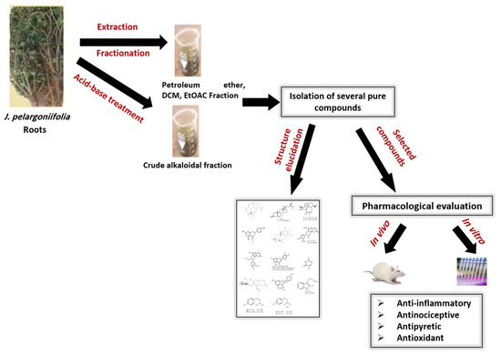
Graphical Abstract

1. Introduction

Euphorbiaceae is considered as one of the largest families of flowering plants which includes approximately 7800 species that are distributed among 300 genera and five subfamilies in tropical and subtropical regions [1,2]. Among the main genera of this family, Jatropha L. is represented by approximately 200 species [2]. Jatropha species are used in folk medicine to treat various diseases, such as skin inflammation, eye infection, chest pain, stomach pain, itching, and as a vermifuge, or as ornamental plants and energy crops in Latin America, Africa, and Asia [3]. J. gossypiifolia, J. elliptica, J. curcas, and J. mollissima, among other species of Jatropha, have been reported for their chemical constituents, biological activities, and medicinal uses [4]. Jatropha glauca, J. curcas, J. spinose, and J. pelargoniifolia are the only four species that are distributed in Saudi Arabia and are employed as traditional herbal medicines, owing to their anti-inflammatory, antioxidant, antiseptic, and analgesic properties [5,6].
J. pelargoniifolia Courb. of the current study is grown as a shrub and is widely known as “Obab” in Arabic. It is widely distributed in East Tropical Africa (Sudan, Eritrea, Ethiopia, Somalia, and Kenya) and the Arabian Peninsula (Yemen, Oman, and Saudi Arabia) [7]. The plant is sometimes collected from the wild for local medicinal use, especially the petiole sap which is applied to treat ulcers, severe skin inflammation, and for wound healing [7].
Previous phytochemical studies on the plants belonging to the genus Jatropha revealed a broad range of isolated secondary metabolites, such as diterpenoids, triterpenoids, non-conventional coumarino-lignans, alkaloids, coumarins, flavonoids, cyclic peptides, and steroids [8,9]. However, accordingly reviewed by Zhang et al. [8], the main compounds isolated from Jatropha genus are the terpenoids. J. gossypiifolia was subjected to extensive phytochemical studies that resulted in the isolation of many secondary metabolites, such as propacin, venkatasin, citlalitrione, ricinine, apigenin, jatropholones A& B, and jatrophone [4]. Moreover, curcusones A–D, taraxerol, nobiletin, curacyclines A & B, as well uracil, have been isolated from J. curcas. [5,6,8]. Additionally, many reported studies showed the isolation of multidione, multifidone, multifolone, and multifidol glucoside from J. multifida, while from J. podagrica, there was japodic acid, erythrinasinate, γ-sitosterol, japodagrin, and podacyclines A & B [3,8,9]. This is indeed a reflection of the versatility of the enzymatic system that is present in Euphorbiaceous plants, however nothing was reported regarding J. pelargoniifolia. Thus, it was of interest to explore the active constituents and their biological activity to provide evidence for the traditional use of J. pelargoniifolia.

2. Result and Discussion

2.1. Isolation of Compounds

The alcoholic extract of J. pelargoniifolia roots powder was successively partitioned with petroleum ether (60 °C), dichloromethane (DCM), ethyl acetate (EtOAc), and then n-butanol (n-BuOH) to give the correspondent organic fractions. Each fraction was subjected to chromatographic separation on normal and reversed phase (RP) silica gel to yield compounds 1, 6, 7, 10, 11, and 16 from petroleum ether, DCM2, and EtOAc fractions, respectively. Furthermore, the organic extract that was obtained after an acid-base treatment of the roots powder was isolated on a normal silica gel column which was followed by purification on RP-HPLC and/or crystallization to afford compounds 1722 (Figure 1).

2.2. Structure Elucidation

The new compound 15 was obtained as white crystals. The NMR and ESIMS (Electronspray Ionization Mass Spectrometry) data established the molecular formula of 15 to be C16H18O10. The IR absorption bands at max 3349, 1719, and 1625 cm−1 suggested the presence of hydroxyl, ester carbonyl, and aromatic functionalities, respectively. Furthermore, the 13C nuclear magnetic resonance (NMR) spectrum of 15, which was measured in deuterated methanol (CD3OD, displayed sixteen signals of nine sp2 and seven sp3 carbons (including that of a methoxyl group). Its 1H NMR spectrum showed a pair of ortho-coupled protons at δH 6.26 and 7.88 (each, 1H, d, J = 9.5 Hz) as was observed by 1H-1H correlated spectroscopy (COSY) that was assignable to H-3 and H-4 of an α-pyrone ring system of a coumarin, respectively [10]. This was further evidenced from the 13C NMR carbon signals of the -pyrone at δC 163.5 (C, C-2), 146.5 (CH, C-4), 144.4 (C, C-8a), 116.2 (CH, C-3), and 112.7 (C, C-4a) (Table 1). Moreover, the single aromatic singlet appearing at δH 7.00 (1H, s) suggested 15 to be a trisubstituted coumarin. Six proton signals at δH 3.30–4.99 ppm, a doublet of an anomeric proton at δH 4.99 (1H, d, J = 7.8 Hz), and six 13C NMR signals at δC 106.2 (CH, C-1′), 75.5 (CH, C-2′), 77.8 (CH, C-3′), 71.0 (CH, C-4′), 78.5 (CH, C-5′), and 62.2 (CH2, C-6′) indicated the presence of a β-D-glucopyranosyl substituent. An aromatic methoxy substituent (δHC 3.91/57.0) was also revealed. The 3JCH correlations that were observed in the heteronuclear multiple bond correlation (HMBC) spectrum linked these two substituents to the coumarin carbons at δC 133.2 and 147.5, respectively (Figure 2). Thus, a hydroxy group should represent the third substituent on the coumarin carbon at δC 145.7. The long-range correlations (HMBC) that were found from H-5 (δH 7.00, 1H, s) to the carbons at δC 146.5 (C-4), 145.7, C-8a (δC 144.4), and δC 133.2 (C-7) indicated that C-8 (δC 147.5) is the position of the methoxyl group. To confirm the locations of the glucosyl and hydroxyl groups, the NMR data of 15 were further compared to those of 5-hydroxy-7-methoxycoumarin-8-O-β-d-glucoside and other closely related coumarin derivatives that were previously isolated from Daphne pseudo-mezereum [11] and Tetraphis pellucida [12], respectively. The structure of compound 15 was thus established as a new natural product and was identified as 6-hydroxy-8-methoxy coumarin-7-O-β-d-glycopyranoside.
Compound 18 was isolated from the organic extract of the acid-base treated root powder as white needle-shaped crystals. It produced a positive Dragendorff’s test, indicating its alkaloid nature. The IR absorption band with a spike at max 3309 cm−1 suggested the presence of hydroxyl and/or secondary amine functionality. The UV absorptions at max 295, 287, 279, 230 nm in MeOH were characteristic to an indole chromophore. The COSY correlations (Figure 2) disclosed the ABCD system of the aromatic protons at δH 7.41/7.00 (Table 2), which is consistent with 2,3-disubstituted indole alkaloids. A side chain of an ethylene and a N-methyl was linked to C-3 of the indole as it was manifested by 2D NMR correlations. However, comparison of 1H NMR data of 18 with those of N-methyltryptamine (17) revealed that the 1H proton singlet at position 2 in 17 was replaced in 18 by a 2H singlet of a hydroxymethyl proton at δH 3.91 ppm. The 13C NMR spectroscopic and ESIMS data of 18 was thus consistent with a molecular formula C12H16N2O of 30 mass units more than that of 17 (C11H14N2). Furthermore, the HMBC correlation that was found from the methylene protons (δH 3.91, 2H, s) to C-2 and C-3 confirmed its C-2 location of the hydroxymethyl group. Finally, a full analysis of th COSY and HMBC spectral correlations (Figure 2) assigned the structure of compound 18 to be 3-(2-(methylamino) ethyl)-1H-indol-2-yl) methanol or 2-hydroxymethyl N-methyltryptamine, a new indole alkaloid.
Compounds 114, 1617 and 1922, which were also isolated from J. pelargoniifolia roots, were found to be identical to the previously reported natural products by comparison of their spectroscopic (IR, MS, and NMR) data and were identified as jatrophadiketone (1) was isolated from the roots of J. curcas [13], β-sitosterol (2) isolated from J. curcas seed kernels and from the methanolic extract of the root bark of Calotropis gigantean (Linn.), [14,15], curcuson D (3) and curcuson C (4) were isolated from J. curcas root extract [16], naringenin (5) was isolated from the root extract of J. gossypifolia [17,18], β-sitosterol glucoside (6) was isolated from the leave and twig extract of J. curcas, [19], spruceanol (7) was isolated from both the aerial extract of J. divaricate and the bark extract of Aleurites moluccana [20,21], propacin (8), cleomiscosin B (9), cleomiscosin A (10) compounds 8 and 10 were isolated from whole plant extracts of J. gossypifolia, while compound 9 was isolated from Mallotus apelta [22,23,24,25,26], apigenin (11) was identified in J. gossypifolia [4,27], uracil (12) was isolated from the leaves of J. curcas [28,29,30], cynaroside (13) was identified in Scabiosa atropurpurea aerial parts extract [31], linarin (14) was isolated from the extracts of aerial parts of both Bupleurum chinense and Valeriana officinalis [32,33], hovetricoside C (16) was separated from Artocarpus tonkinensis [34], N-methyltryptamine (17) was isolated from Zanthoxylum arborescens [35], N-methyltyramine (19) was isolated from a beer [36], and hordenine (20) was separated and identified from Ephedra aphylla that was growing in Egypt [37]. Compounds 21 (hordenine HCl) and 22 (N-methyltyramine HCl) were identical to the authentic samples that were purchased from Sigma-Aldrich (St Louis, MO, USA), and on the basis of their 1H NMR and TLC co-chromtographic data, they were isolated previously from Ariocarpus kotschoubeyanus [38].

2.3. Biological Activity

The alcoholic extract of J. pelargoniifolia was found to possess significant anti-inflammatory, analgesic, and antipyretic activities when it was tested on in vivo models in a dose-dependent manner [39]. This prompted us to extend the study of these activities on the isolated compounds. The anti-inflammatory, analgesic, antipyretic, and antioxidant activities for the compounds which have been isolated in good yields (16, 811, 13, 14, 16, 21, and 22) were thus evaluated for their analgesic, anti-inflammatory, antipyretic, and antioxidant activities using in vivo and in vitro models. The analgesic activities were assessed in mice via acetic acid-induced writhing, hot-plate, and tail-flick methods.
In the acetic acid-induced writhing method, compounds 1, 4, 6, 9, 11, 13, 14, 16, and 22 showed a dose-dependent analgesic activity by the reduction in the number of writhings. However, the diterpenoids (1 and 4), β-sitosterol glucoside (6), flavonoids (5 and 13), and tryptamine HCl (22) exhibited the strongest analgesic activity by inhibiting writhing in mice (49.07–65.74% inhibition) at a dose of 10 mg/kg compared with the standard antinociceptive drug (indomethacin), which showed 72.68% reduction in the number of writhings at a concentration of 4 mg/kg (Table S9). The coumarinolignan (8) and hordenine HCl (21) did not show any inhibition either at 5 or at 10 mg/kg.
In the hot plate method, the thermal responses in the mice that were treated with selected compounds after half, one, and two hours were significantly reduced (p < 0.001). Especially in the mice that were treated with a dose 10 mg/kg of compounds 22, 10, 6, 4, 5, 13, 16, and 14, the antinociceptive effects were reduced by 78.57, 76.19, 74.46, 73.33, 65.95, 59.57, 53.48, and 34.88%, respectively (Table S10). While in the tail-flick method, the tested animals that were treated with 10 mg/kg of compounds 22, 1, 10, 4, 13, 16, 5, 6, 21, 3, and 9 showed a significant (p < 0.001) reduction in antinociceptive activity (67.32, 67.21, 60.45, 57.59, 54.04, 53.59, 45.58, 40.30, 21.75, 20.39, and 10.87%, respectively) compared with indomethacin (95.61%), as depicted from Table S11. The obtained results confirmed that the strong analgesic activity that is exhibited by the roots of J. pelargoniifolia could be due to its bioactive compounds that may exert their analgesic activities through different CNS (Central Nervous System) mechanisms (peripheral and central). Therefore, further studies with purified compounds should be conducted in the future for further pharmacological and toxicological characterization in order to elucidate the mechanisms that are involved in the central analgesic effect of these compounds.
The anti-inflammatory activities of the major isolated compounds were evaluated by using the carrageenan-induced paw edema model in rats. It was found that the size of the edema was significantly reduced (p < 0.05–0.001) in the animals that were treated with the low doses of 5 and 10 mg/kg compared with the standard anti-inflammatory drug (phenylbutazone) at a high dose of 100 mg/kg. The rats that were treated with compounds 10, 16, 1, 5, 6, 22, 4, 13, 3, 8, 9, 14, and 21 exhibited a significant reduction in their hind paw edema in a dose-dependent manner. Therefore, at a dose of 5 mg/kg, the edema size was reduced by 20.44, 47.23, 17.95, 45.85, 33.97, 44.47, 13.53, 26.51, 7.18, 2.20, 5.52, 6.35, and 6.35, respectively, while at 10 mg/kg, the edema size was reduced by 64.91, 55.24, 54.94, 51.38, 51.10, 50.27, 49.17, 48.61, 13.53, 12.98, 10.22, 10.22, and 8.01%, respectively relative to that reduced by phenylbutazone at 100 mg/kg (69.06%) which was almost similar to that produced by a dose of 10 mg/kg of compound 10 (64.91%; Table S12). These anti-inflammatory results were almost compatible with those of the above mentioned antinociceptive activity for the tested compounds.
In addition, the isolated compounds from the J. pelargoniifolia roots were tested for their antipyretic activity against yeast-induced hyperthermia in mice. All tested compounds, which were administered at doses of 5 and 10 mg/kg, showed a considerable reduction in the rectal temperature of the hyper-thermic mice, ranging between 36.73 ± 0.13 °C and 38.56 ± 0.16 °C as compared with the hypothermic effect (36.33 ± 0.11 °C) resulting from indomethacin administration (Table S13). Moreover, compounds 5, 6, 10, 13, and 16 displayed about a 1 °C reduction in temperature less than that of the yeast-induced hyperthermia control (~ 38.8 °C) in the first 30 min. of the experiment.
The percentage inhibition ± SD of the nitric oxide-scavenging activity was determined for the selected compounds at concentrations of 20, 40, 60, 80, and 100 µg/mL, and the obtained results were compared with a standard antioxidant drug (ascorbic acid). Compounds 22, 4, 2, 10, 1, 14, 21, 9, 8, 11, 6, 3, 13, 5, and 16 exhibited significant free radical-scavenging potency when compared with the free radical-scavenging activity of a strong known antioxidant drug (ascorbic acid) 87.23 ± 0.98. The ability of the tested compounds to produce antioxidant effects was found to be concentration-dependent. At a 100 µg/mL dose, the % inhibition ± SD of the tested compounds were 77.60 ± 4.22, 77.36 ± 4.22, 76.83 ± 5.01, 75.26 ± 5.54, 71.66 ± 0.70, 70.36 ± 14.73, 67.67 ± 5.75, 63.67 ± 12.85, 63.67 ± 12.85, 57.00 ± 10.21, 56.54 ± 6.03, 38.30 ± 5.63, 33.06 ± 1.86, 27.00 ± 7.85, and 25.61 ± 5.18, respectively (Table S14). The significant antioxidant activity that was associated with the administration of J. pelargoniifolia roots was perhaps due to its content of several phenolic and polyphenolic compounds which play an important role in free radical-scavenging activity with less cytotoxicity.
It is important to mention here that in our previous study, which was carried out on the crude alcoholic extract of J. pelargoniifolia roots, we observed a significant anti-inflammatory activity and analgesic potency [39], likely resulting from the presence of cleomiscosin A, hovetricoside C, jatrophadiketone, naringenin, β-sitosterol glucoside, N-methyltyramine HCL, curcuson C, cynaroside, curcuson D, propacin, cleomiscosin B, linarin, and hordenine HCL in good yield. Undoubtedly, a synergistic effect between these bioactive constituents produces significant antinociceptive and anti-inflammatory effects. These results justify the use of this plant in folk medicine for the treatment of pain and several inflammatory conditions. Further study will be conducted on the pure isolated compounds to investigate the exact mechanisms underlying their promising biological activities.
Our study proved that J. pelargoniifolia roots can be considered as a source of several biologically- active compounds such as hordenine, which exhibited various biological activities like inhibiting melanogenesis in human melanocytes, increasing the respiratory and heart rates [40], the stimulation of gastrin release, inhibition of monoamine oxidase B, and antibacterial properties [41]. Furthermore, Chrisitine et al. reported that N-methyltyramine increases blood pressure in an anaesthetized rat, relaxes guinea pig ileum, and increases both the force and the rate of contraction of guinea-pig right atrium by inducing the release of noradrenaline [42]. Additionally, naringenin has been reported to have several pharmacological properties, including anti-dyslipidemic, anti-obesity and antidiabetic, and antifibrotic [43]. Moreover, cleomiscosin A showed strong anti-inflammatory activity and has analgesic and antipyretic potencies [44]. Curcuson C has been reported to have antipyretic activity in vivo [45].

3. Materials and Methods

3.1. Chemicals and Analytical Instruments

The high-resolution electron spray ionization-mass spectrometry (HRESI-MS) analyses (Bruker, Bremen, Germany) were carried out on an Agilent Triple Quadrupole 6410 QQQ LC-MS mass spectrometer (Central Lab. College of Pharmacy, King Saud University (KSU)). The infra-red spectra were generally recorded in the potassium bromide pellets, unless otherwise specified, using the FTIR spectrophotometer (FT-IR Microscope Transmission, company, Waltham, MA, USA). The melting points were recorded by using a Mettler FP 80 Central Processor that was supplied with a Mettler FP 81 MBC Cell Apparatus. The spectral data for proton and carbon were measured by using Bruker AVANCE 700, 500, and 600 (College of Pharmacy, KSU and Department of Chemistry in TU Dortmund) (Bruker, Fallanden, Switzerland), resonating at either 700, 500, and 600 MHz for proton or at 125 MHz for carbon. The chemical shift values were expressed in ppm with respect to the internal standard tetramethyl silane (TMS) or residual solvent peak, and the coupling constants (J) were recorded in Hertz (Hz). The two-dimensional NMR experiments (COSY, HSQC, and HMBC) were performed using the standard Bruker program (Bruker, Fallanden, Switzerland). The silica gel 60/230–400 mesh (Qingdao Oceanic Chemical Co., Qingdao, China), RP C18 silica gel 40–63/230–400 mesh (Merck, Darmstade, Germany), and sephadex LH-20 with particle size 18–111 µm (GE Healthcare, Chicago, IL, USA) were used for column chromatography, while the silica gel and reversed phase 60 F254 (Merck, Germany) were used for thin-layer chromatography (TLC). The detection was achieved by using 10% H2SO4 in ethanol or ceric sulfate followed by heating. Alkaloids were tested with Mayer’s reagent, Hager’s reagent, and Dragendorff’s reagent. All of the solvents for analytical purposes (HPLC- and analytical-grade) and the drugs for biological investigation (sodium nitroprusside, sulphanilamide, λ-carrageenan, acetic acid, ascorbic acid, and phenylbutazone) were procured from Sigma Chemical Company (Sigma-Aldrich, St Louis, MO, USA), and the solvents were distilled prior to use. The preparative and semipreparative Shimadzu HPLC were performed, characterized by Rp-18 (ODS-80 TM, TSK, Tokyo, Japan), 10 μm PS, 30 cm L × 2.15 cm i.d. fitted with a guard column (10 μm PS, 7.5 cm L × 2.15 cm i.d.) (ODS-80 TM, TSK, Tokyo, Japan), and VP 250/10 NUCLEODUR C18 HTec, 6 μm PS, 25 cm L × 2 cm i.d., respectively which both used a PDA detector.

3.2. Plant Material

The roots of J. pelargoniifolia were harvested from Wadi Mojasas, Jazan district (South of Saudi Arabia) in September, 2015. The plant was authenticated by Dr. Jacob Thomas, a botanist of the Science College Herbarium, KSU, where a voucher specimen (#23064) was deposited.

3.3. Animals

Male Wistar rats and white male Swiss albino mice with approximate body weights of 200 g and 20–25 g, respectively, were divided into groups of six animals. The animals were obtained from the Experimental Animal Care Center, College of Pharmacy, KSU. After a 7-day period in animal accommodation, they were divided into groups and were maintained at 12 h:12 h light-dark conditions at 55% humidity. Purina chow rat diet (UAR-Panlab, Barcelona, Spain) and drinking water were supplied to the animals ad libitum. The protocols for the present study were based on the recommendations of the Ethical Committee of the Experimental Animal Care Center of KSU (approval number CPR-7569).

3.4. Extraction, Fractionation, and Purification

The air-dried powder of the J. pelargoniifolia roots (2.5 kg) was divided into two parts—A and B—and 2.5 kg of part A was subjected to solvent extraction, while the remaining 500 g of the root powder (part B) was exposed to the acid-base treatment. Part A was extracted by maceration with 80% ethanol (3 L × 5) for three successive days. This process was repeated until complete exhaustion of the plant material [46]. The alcoholic extract was then concentrated to dryness under reduced pressure at 40 °C using a rotary evaporator to give 270 g of the dried alcoholic extract. The dried alcoholic extract was suspended in H2O and was successively partitioned with petroleum ether, dichloromethane, ethyl acetate, and n-butanol (600–700 mL × 3) of each to obtain 13.3, 10.3, 5.1, and 33.6 g, respectively.
A part of the petroleum ether fraction (12.8 g) was chromatographed over silica gel CC (Column Chromatography) using a gradient of petroleum ether/EtOAc followed by methanol (MeOH). The 100 mL fractions of each were collected and screened by TLC, and similar fractions were combined together to give six fractions (A–F). Fraction A which was eluted by 15% EtOAc in petroleum ether (609.8 mg) was further subjected to CC and was eluted by petroleum ether/acetone gradient elution, sub fraction A1 (188.9 mg) which was eluted by 6% acetone in petroleum ether was further purified by preparative HPLC gradient elution using acetonitrile: H2O: TFA) to yield 25.0 mg of compound 1. Direct crystallization of fraction B, which was eluted by 20% EtOAc in petroleum ether, yielded 302.7 mg of compound 2. Fractions C and D which were eluted with 30 and 40% EtOAc in petroleum ether, respectively, were crystallized with acetone to yield compounds 3 and 4 (14.3 and 25.4 mg, respectively). Additionally, fraction E (287.3 mg) which was eluted by 50% EtOAc was also crystallized from acetone to give 20.2 mg of compound 5, while fraction F which was eluted with 40% MeOH in EtOAc yielded 330.4 mg of compound 6, which was purified by crystallization with acetone.
The dichloromethane (DCM) fraction (9.8 g) was subjected to silica gel CC using a column that was packed by the wet method with petroleum ether. The polarity of the column was gradually increased by treating it with DCM, followed by MeOH to give 142 fractions, and similar fractions were pooled together depending on their TLC similarity. Fraction 48–64 which was eluted by 10% MeOH in DCM was concentrated (4.6 g) and was then subjected to repeated silica gel CC, followed by a preparative revered phase TLC using MeOH:H2O (3:1) as a solvent system, leading to the isolation of white crystals of compound 7 (7.5 mg). Moreover, subtractions that were obtained using 90% acetone in petroleum ether, 100% acetone, and 10% acetone in MeOH, followed by crystallization with MeOH, afforded compounds 8 (15.1 mg), 9 (15.9 mg), and 10 (16.4 mg), respectively.
The EtOAC extract (4.6 g) was subjected to silica gel CC using a gradient of DCM/MeOH to give six fractions (I–IV). Fractions I which were eluted with 84% DCM afforded 17 mg of compound 11 after crystallization with MeOH. Fractions II which were eluted with 70% DCM afforded 8.6 mg of compound 12. The fractions that were eluted with 35% and 40% MeOH in DCM (II and IV) were further purified by repeated acetone crystallization to give 14.9 and 13.2 mg of compounds 13 and 14, respectively. Fractions V which were eluted with 45% MeOH were further subjected to CC using DCM/MeOH, followed by a semi-preparative HPLC (Rp-18) using MeOH:H2O:TFA as a solvent system afforded 8.6 mg white crystals of compound 15. Finally, fractions VI which were eluted with 50% MeOH in DCM were subjected to further purification over sephadex LH-20 (using water and methanol as an eluent in the gradient mode). The subfraction VI–A, which was eluted by 20% H2O/MeOH was further purified over a reversed-phase column to give 12 mg of compound 16.
Furthermore, Part B was subjected to an acid-base treatment according to the Stas-Otto method I which was described by Mandhumitha and Fowsiya [47]. The crude alkaloidal fraction was subjected to silica columns using gradient elution with solvent system DCM/MeOH:NH4OH, resulting in five fractions. The first fraction which was eluted using 17% MeOH in DCM with an addition of 1% NH4OH was followed by further purification by reversed-phase semipreparative HPLC using MeOH:H2O:TFA to give compound 17 (6.3 mg). The second fraction which was separated by 20% MeOH in DCM to afford a subfraction, which was further purified by a semipreparative HPLC gradient elution using MeOH:H2O:TFA as a solvent system, afforded white needle crystals of compound 18 (9.4 mg). The third and fourth fractions which were eluted by 23% and 26% MeOH in DCM, followed by an addition of a few drops of NH4OH, afforded 6.8 and 8.8 mg of compounds 19 and 20, respectively. The fifth fraction was eluted by 60% MeOH in DCM with an addition of a few NH4OH drops to yield 86.7 mg of a mixture of two compounds, which were subjected to further purification using the reversed-phase semipreparative HPLC in gradient mode with MeOH:H2O:TFA as the mobile phase, resulting in the production of the white crystals of compounds 21 and 22 (20.3 and 21.5 mg), respectively.
6-hydroxy-8-methoxycoumarin-7-O-β-d-glycopyranoside (compound 15): White crystals; m.p. 219–220 °C; UV (MeOH) λmax nm 325 and 250; IR (KBr) νmax (cm−1): 3349, 1719, 1625, 1520, 1465, 829; 1H NMR, 13C NMR, and HMBC data, see Table 1 and Figure 2; HRESIMS (positive) m/z 371.0900 [M + H]+ (calculated for C16H18O10, 371.097825).
3-(2-(methylamino)ethyl)-1H-indol-2-yl)methanol (compound 18): White needle crystals; m.p. 179–189 °C; UV (MeOH) λmax nm: 295, 287, 279, 230; IR (KBr) νmax (cm−1): 3309,1140, 1120, 1105, 1011, 855. 1H NMR, 13C NMR, and HMBC data, see Table 2 and Figure 2; HRESIMS (positive) m/z 205.1293 [M + H]+ (calculated for C12H16N2O, 205.134088).

3.5. Antinociceptive Activity Test

3.5.1. Hot-plate Method

The hot-plate method that was described by Turner was used to determine the antinociceptive activity of the compounds that were isolated from the J. pelargoniifolia root [48].

3.5.2. Acetic Acid-induced Writhing in Mice Test

The method of Koster et al. was used to evaluate the analgesic effect of the pure compounds that were isolated from the J. pelargoniifolia root [49].

3.5.3. Tail-Flick Method

Acute nociception was induced using the tail-flick apparatus (Tail flick Apparatus Harvard), following the method that was recommended by D’amour and Smith [50].

3.6. Anti-Inflammatory Activity Test

Carrageenan-Induced Edema in the Rat Paw Method

The method that was described by Winter et al. was used to evaluate the anti-inflammatory potency of the isolated compounds [51].

3.7. Antipyretic Activity Screening

Yeast-Induced Hyperthermia in Rats

Hyperthermia was induced in the mice followed by the administration of the isolated compounds, and their hypothermic activity was determined by applying the method described by Loux [52].

3.8. Antioxidant Effect

Nitric Oxide Radical-Scavenging Assay

This assay was carried out according to the procedure that was described by Green et al. [53].

3.9. Statistical Analysis

The values in the tables are given as mean ± SE. The data were analyzed by using one-way analysis of variance (ANOVA) followed by the Student’s t-test. Values with p < 0.05 were considered significant.

4. Conclusions

The wide traditional use of Jatropha species as anti-inflammatory and analgesics has prompted us to investigate the chemistry and bioactivity of J. pelargoniifolia growing in Saudi Arabia. The phytochemical study of the plant roots resulted in the isolation of six terpenoids, five flavonoids, three coumarinolignans, two tryptamines, and two tyramines (including their HCl salts), a coumarin, and a pyrimidine. The new compounds were identified as 6-hydroxy-8-methoxy coumarin-7-O-β-d-glycopyranoside and 2-hydroxymethyl-N-methyltryptamine. To the best of our knowledge, hovetricoside C and N-methyltryptamine were isolated herein from the Euphorbiaceae family for the first time, while cleomiscosin B, hordenine, and N-methyltyramine with their salts, cynaroside, and linarin were characterized in the Jatropha species for the first time.
On the basis of the significant anti-inflammatory, analgesic, antipyretic, and antioxidant activities that were observed in the experimental animals for the alcoholic extract of J. pelargoniifolia, fifteen of the adequately isolated compounds were consequently biologically evaluated. Eleven of these compounds exhibited strong analgesic activity. Twelve out of the fifteen compounds succeeded to reduce the chemically-induced inflammatory marker in the animals in a dose-dependent manner. Moreover, five of the compounds demonstrated an anti-pyretic effect by a reduction about a 1 °C in an induced hyperthermia model. The isolated compounds also exhibited varying degrees of nitric oxide-scavenging activity. The significant antioxidant activity that was associated with the administration the extract of J. pelargoniifolia roots was thus perhaps due to its phenolic content such as flavonoid, coumarins, and coumarinolignans. The synergistic effect between these bioactive constituents might explain the significant antinociceptive and anti-inflammatory effect of the alcoholic extract of J. pelargoniifolia roots and may scientifically justify the use of this plant in folk medicine for the treatment of pain and several inflammatory conditions.

Supplementary Materials

The following are available online. Figures S1–S4: 1H-, 13C-NMR, COSY and HMBC spectra of compound 15, Figures S5–S8: 1H-, 13C-NMR, HMBC and HSQC spectra of compound 18, Tables S9–S11: Analgesic effect of isolated compounds by using acetic acid-induced writhing, hot plate and tail flick methods in mice, Table S12: Effect of isolated compounds on carrageenan-induced paw edema in albino rats, Table S13: Effect of isolated compounds on yeast-induced hyperthermia in mice, Table S14: % Inhibition of nitric oxide scavenging activity for isolated compounds at different concentrations.

Author Contributions

H.Y.A., A.E.-G., and O.K. conceived and designed the experiments; H.Y.A. performed the experiments; H.Y.A., A.E.-G. analyzed the data; H.Y.A. contributed reagents/materials/analysis tools; H.Y.A. wrote the paper; A.E.-G. participated in the experiments’ design and coordination and helped to draft the manuscript; O.K. participated in the experiment design and supervised. A.F.A. contributed to the structure elucidation of some of the compounds and to the partial writing of the manuscript.

Funding

The Deanship of Scientific Research at King Saud University, Project no. RG-1437-021.

Acknowledgments

The authors extend their appreciation to the Deanship of Scientific Research at King Saud University for funding the work through the Research Group, Project no. RG-1437-021.

Conflicts of Interest

The authors declare no conflict of interest.

Appendix A

Supplementary data associated with 1H NMR, 13C NMR, COSY, and HMBC of compounds 15 and 18 are available in Supplementary Information. In addition, Tables for all of the biological test results are provided.

References

  1. Alves, M.V. Checklist das espѐcies de Euphorbiaceae Juss. Ocorrentes no semi-áridopernambucano, Brasil. Acta Bot. Bras. 1998, 12, 485–495. [Google Scholar] [CrossRef]
  2. Webster, G.L. Classification of the Euphorbiaceae. Ann. Mo. Bot. Gard. 1994, 81, 3–143. [Google Scholar] [CrossRef]
  3. Sabandar, C.W.; Ahmat, N.; Jaafar, F.M.; Sahidin, I. Medicinal property, phytochemistry and pharmacology of several Jatropha species (Euphorbiaceae): A review. Phytochemistry 2013, 85, 7–29. [Google Scholar] [CrossRef] [PubMed]
  4. Félix-Silva, J.; Giordani, R.B.; Silva, A.A., Jr.; Zucolotto, S.M.; Fernandes-Pedrosa, M.F. Jatropha gossypiifolia L. (Euphorbiaceae): A review of traditional uses, phytochemistry, pharmacology, and toxicology of this medicinal plant. Evid-Based Complement. Altern. Med. 2014, 2014, 1–32. [Google Scholar]
  5. Oskoueian, E.N.; Saad, W.Z.; Omar, A.; Ahmad, S.; Kuan, W.B.; Zolkifli, N.A.; Hendra, R.; Ho, Y.W. Antioxidant, anti-inflammatory and anticancer activities of methanolic extracts from Jatropha curcas Linn. J. Med. Plants Res. 2011, 5, 49–57. [Google Scholar]
  6. Yusuf, S.O.; Maxwell, I.E. Analgesic activity of the methanolic leaf extract of Jatropha Curcas (Linn). Afr. J. Biomed. Res. 2010, 13, 149–152. [Google Scholar]
  7. Schmelzer, G.H.; Fakim, G.A. Jatropha pelargoniifolia; Record from Prota4u; PROTA (Plant Resources of Tropical Africa/RessourcesVégétales de l’AfriqueTropicale): Wageningen, The Netherlands, 2007. [Google Scholar]
  8. Zhang, X.P.; Zhang, M.L.; Sua, X.H.; Huoa, C.H.; Gub, Y.C.; Shi, Q.W. Chemical constituents of the plants from genus Jatropha. Chem. Biodivers. 2009, 6, 2166–2183. [Google Scholar] [CrossRef] [PubMed]
  9. Zhu, J.Y.; Zhang, C.Y.; Dai, J.J.; Rahman, K.; Zhang, H. Diterpenoids with thioredoxin reductase inhibitory activities from Jatropha multifida. Nat. Prod. Res. 2017, 31, 2753–2758. [Google Scholar] [CrossRef] [PubMed]
  10. Zhao, A.; Yang, X.Y. New coumarin glucopyranosides from roots of Angelica dahurica. Chin. Herb. Med. 2018, 10, 103–106. [Google Scholar] [CrossRef]
  11. Konishi, T.; Wada, S.; Kiyosawa, S. Constituents of the leaves of Daphne pseudo-mezereum. Yakugaku Zasshi: J. Pharm. Soc. Jpn. 1993, 113, 670–675. [Google Scholar] [CrossRef]
  12. Jung, M.; Geiger, H.; Zinsmeister, H.D. Tri- and tetrahydroxycoumarin derivatives from Tetraphis pellucida. Phytochemistry 1995, 39, 379–381. [Google Scholar] [CrossRef]
  13. Liu, J.Q.; Yang, Y.F.; Wang, C.F.; Li, Y.; Qiu, M.H. Three new diterpenes from Jatropha curcas. Tetrahedron 2012, 68, 972–976. [Google Scholar] [CrossRef]
  14. Habib, M.R.; Nikkon, F.; Rahman, M.; Haque, M.E.; Karim, M.R. Isolation of stigmasterol and β-sitosterol from methanolic extract of root bark of Calotropis gigantean (Linn). Pak. J. Biol. Sci. 2007, 10, 4174–4176. [Google Scholar] [PubMed]
  15. Oskoueian, E.; Abdullah, N.; Ahmad, S.; Saad, W.Z.; Omar, A.; Ho, Y.W. Bioactive compounds and biological activities of Jatropha curcas L. kernel meal extract. Int. J. Mol. Sci. 2011, 12, 5955–5970. [Google Scholar] [CrossRef] [PubMed]
  16. Naengchomnong, W.; Thebtaranonth, Y. Isolation and structure determination of four novel diterpenes from Jatropha curcus. Tetrahedron Lett. 1986, 27, 2439–2442. [Google Scholar] [CrossRef]
  17. Maltese, F.; Erkelens, C.; Kooy, F.V.D.; Choi, Y.H. Identification of natural epimeric flavanone glycosides by NMR spectroscopy. Food Chem. 2009, 116, 575–579. [Google Scholar] [CrossRef]
  18. Mutheeswarana, S.; Saravana, P.; Kumarb, P.; Yuvaraj, V.; Duraipandiy, N.A.; Balakrishnab, K.; Ignacimuthu, S. Screening of some medicinal plants for anticariogenic activity: An investigation on bioactive constituents from Jatropha gossypifolia (L.) root. Biocatal. Agric. Biotech. 2017, 10, 161–166. [Google Scholar] [CrossRef]
  19. Hufford, C.D.; Oguntimein, B.O. Non-polar constituents of Jatropha curcas. Lloydia 1978, 41, 161–165. [Google Scholar]
  20. Denton, W.R.; Harding, W.W.; Anderson, C.I.; Jacobs, H.; McLean, S.; Reynolds, W.F. New diterpenes from Jatropha divaricata. J. Nat. Prod. 2001, 64, 829–831. [Google Scholar] [CrossRef] [PubMed]
  21. Alimboyoguen, A.B.; De Castro-Cruz, K.A.; Shen, C.C.; Li, W.T.; Ragasa, C.Y. Chemical constituents of the bark of Aleurites moluccana L. Willd. J. Chem. Pharm. Res. 2014, 6, 1318–1320. [Google Scholar]
  22. Akbar, E.; Sadiq, Z. Coumarinolignoid rare natural product: A review. Asian J. Chem. 2012, 24, 4831–4842. [Google Scholar]
  23. Begum, S.A.; Sahai, M.; Ray, A.B. Non-conventional lignans: coumarinolignans, flavonolignans, and stilbenolignans. Prog. Chem. Org. Nat. Prod. 2010, 93, 1–70. [Google Scholar]
  24. Das, B.; Venkataiah, B. A minor coumarino-lignoid from Jatropha gossypifolia. Biochem. Syst. Ecol. 2001, 2, 213–214. [Google Scholar] [CrossRef]
  25. Biswanath, D.; Kashinatham, A.; Venkataiah, B.; Srinivas, K.V.N.S.; Mahender, G.; Reddy, M.R. Cleomiscosin A, a coumarino-lignoid from Jatropha gossypifolia. Biochem. Syst. Ecol. 2003, 31, 1189–1191. [Google Scholar]
  26. Xu, J.F.; Feng, Z.M.; L, J.; Zhang, P.C. New hepatoprotective coumarinolignoids from Mallotus apelta. Chem. Biodivers. 2008, 5, 591–597. [Google Scholar] [CrossRef] [PubMed]
  27. Lee, D.G.; Mok, S.Y.; Choi, C.; Cho, E.J.; Kim, H.Y.; Lee, S. Analysis of apigenin in Blumea balsamifera Linn DC. and its inhibitory activity against aldose reductase in rat lens. J. Agric. Chem. Environ. 2012, 1, 28–33. [Google Scholar]
  28. Abdelgadir, H.A.; Staden, J.V. Ethnobotany, ethnopharmacology and toxicity of Jatropha curcas L. (Euphorbiaceae): A review. S. Afr. J. Bot. 2013, 88, 204–218. [Google Scholar] [CrossRef]
  29. Hurd, R.E.; Reid, B.R. NMR spectroscopy of the ring nitrogen protons of uracil and substituted uracils; relevance to a base pairing in the solution structure of transfer RNA. Nucleic Acids Res. 1977, 4, 2747–2756. [Google Scholar] [CrossRef] [PubMed]
  30. Staubmann, R.; Schubert-Zsilavecz, M.; Hiermann, A.; Kartnig, T. A complex of 5-hydroxypyrrolidin-2-one and pyrimidine-2,4-dione isolated from Jatropha curcas. Phytochemistry 1999, 50, 337–338. [Google Scholar] [CrossRef]
  31. Elhawary, S.S.; Eltantawy, M.E.; Sleem, A.A.; Abdallah, H.M.; Mohamed, N.M. Investigation of phenolic content and biological activities of Scabiosa atropurpurea L. World Appl. Sci. J. 2011, 3, 311–317. [Google Scholar]
  32. Zhang, T.; Zhou, J.; Wang, Q. Flavonoids from aerial part of Bupleurum chinense DC. Biochem. Syst. Ecol. 2007, 35, 801–804. [Google Scholar] [CrossRef]
  33. Fernández, S.; Wasowski, C.; Paladini, A.C.; Marder, M. Sedative and sleep-enhancing properties of linarin, a flavonoid-isolated from Valeriana officinalis. Pharmacol. Biochem. Behav. 2004, 77, 399–404. [Google Scholar] [CrossRef] [PubMed]
  34. Thuy, T.T.; Kamperdick, C.; Ninh, P.T.; Lien, T.P.; Thao, T.T.P.; Sung, T.V. Immunosuppressive auronol glycosides from Artocarpus tonkinensis. Pharmazie 2004, 59, 297–300. [Google Scholar] [CrossRef] [PubMed]
  35. Grina, J.A.; Ratcliff, M.R.; Stermitz, F.R. Old and new alkaloids from Zanthoxylum arborescens. J. Org. Chem. 1982, 47, 2648–2651. [Google Scholar] [CrossRef]
  36. Yokoo, Y.; Kohda, H.; Kusumoto, A.; Naoki, H.; Matsumoto, N.; Amachi, T.; Suwa, Y.; Fukazawa, H.; Ishida, H.; Tsuji, K.; et al. Isolation from beer and structural determination of a potent stimulant of gastrin release. Alcohol Alcohol. 1999, 34, 161–168. [Google Scholar] [CrossRef] [PubMed]
  37. Abdel-Kader, M.S.; Kassem, F.F.; Abdallah, R.M. Two alkaloids from Ephedra aphylla growing in Egypt. Nat. Prod. Sci. 2003, 9, 1–4. [Google Scholar]
  38. Neal, J.M.; Sato, P.T.; Johnson, C.L.; McLaughlin, J.L. Cactus alkaloids X: Isolation of hordenine and N-methyltyramine from Ariocarpus kotschoubeyanus. J. Pharm. Sci. 1971, 60, 477–478. [Google Scholar] [CrossRef] [PubMed]
  39. Hanan, A.; Ali, E.-G.; Oliver, K. A comparative study of the biological activities of Jatropha pelargoniifolia and Jatropha glauca native to Saudi Arabia. Phytomedicine 2018. submitted. [Google Scholar]
  40. Fana, Y.; Li, X.; Zhang, L.; Duana, P.; Li, F.; Zhao, D.; Wang, Y.; Wub, H. Ether-functionalized ionic liquids: Highly efficient extractants for hordenine. Chem. Eng. Res. Des. 2017, 124, 66–73. [Google Scholar] [CrossRef]
  41. Ma, J.; Wang, S.; Huang, X.; Geng, P.; Wen, C.; Zhou, Y.; Yu, L.; Wang, X. Validated UPLC-MS/MS method for determination of hordenine in rat plasma and its application to pharmacokinetic study. J. Pharm. Biomed. Anal. 2015, 111, 131–137. [Google Scholar] [CrossRef] [PubMed]
  42. Christine, S.; Bell, A.; Stewart-Johnsox, E. N-methyltyramine, a biologically active amine in Acacia seeds. Phytochemistry 1979, 18, 2022–2023. [Google Scholar]
  43. Jadeja, R.N.; Devkar, R.V. Polyphenols and flavonoids in controlling non-alcoholic steatohepatitis. In Polyphenols in Human Health and Disease, 1st ed.; Watson, R.R., Preedy, V.R., Zibadi, S., Eds.; Academic Press: San Diego, CA, USA, 2014; pp. 615–623. [Google Scholar]
  44. Begum, S.; Saxena, B.; Goyal, M.; Ranjan, R.; Joshi, V. B.; Rao, V.; Krishnamurthy, S.; Sahai, M. Study of anti-inflammatory, analgesic and antipyretic activities of seeds of Hyoscyamusniger and isolation of a new coumarinolignan. Fitoterapia 2010, 81, 178–184. [Google Scholar] [CrossRef] [PubMed]
  45. Picha, P.; Naengchomnong, W.; Promratanapongse, P.; Kano, E.; Hayashi, S.; Ohtsubo, T.; Zhang, S.W.; Shioura, H.; Kitai, R.; Matsumoto, H.; et al. Effect of natural pure compounds curcusones A and C from tropical herbal plant Jatropha curcas on thermo sensitivity and development of thermotolerance in Chinese hamster V-79 cells in vitro. J. Exp. Clin. Cancer Res. 1996, 15, 177–183. [Google Scholar]
  46. Skoog, D.; Holler, F.J.; Nieman, T.A. An introduction to chromatographic separations. In Principles of instrumental analysis, 5th ed.; Skoog, D., Holler, F.J., Nieman, T.A., Eds.; Saunders College Publishing: Philadelphia, PA, USA, 1992; pp. 674–700. [Google Scholar]
  47. Madhumitha, G.; Fowsiya, J. A. Hand Book on: Semi Micro Technique for Extraction of Alkaloids; International E-Publication: Indore, India, 2015; p. 9. [Google Scholar]
  48. Turner, R.A. Analgesics, in Screening Methods in Pharmacology; Academic Press: London, UK, 1965; p. 100. [Google Scholar]
  49. Koster, R.; Anderson, M.; De Beer, E.J. Acetic acid for analgesic screening. Fed. Proc. 1959, 18, 412–417. [Google Scholar]
  50. D’amour, F.E.; Smith, D.L. A method for determining loss of pain sensation. J. Pharmacol. Exp. Ther. 1941, 72, 74–79. [Google Scholar]
  51. Winter, C.A.; Risley, E.A.; Nuss, G.W. Carregeenan-induced oedema in hind paw of the rats as an assay for anti-inflammatory drugs. Exp. Biol. Med. 1962, 111, 544–547. [Google Scholar] [CrossRef]
  52. Loux, J.J.; Depalma, D.D.; Yankell, S.L. Antipyretic testing of aspirin in rats. Toxicol. Appl. Pharmacol. 1972, 22, 672–675. [Google Scholar] [CrossRef]
  53. Green, L.C.; Wagner, D.A.; Glogowski, J.; Skipper, P.L.; Wishnok, J.S.; Tannenbaum, S.R. Analysis of nitrate, nitrite, and [15N] nitrate in biological fluids. Anal. Biochem. 1982, 126, 131–138. [Google Scholar] [CrossRef]
Sample Availability: Samples of the compounds 122 are available from the authors.
Figure 1. Chemical structures of the compounds isolated from the roots of Jatropha pelargoniifolia.
Molecules 23 01892 g001
Figure 2. Selected heteronuclear multiple bond correlation (HMBC) and correlation spectroscopy (COSY) correlations of compounds 15 and 18.
Molecules 23 01892 g002
Table 1. The 1H (600 MHz, δ in ppm, J in Hz) and 13C NMR (125MHz, δ in ppm) spectral data for compound 15 in deuterated methanol (CD3OD).
PositionδHδC
2-163.5
36.26 (d, J = 9.5 Hz, 1H)116.2
47.88 (d, J = 9.5 Hz, 1H)146.5
57.00 (s, 1H)106.1
6-145.7
7-133.2
8-147.5
4a-112.7
8a-144.4
1′4.99 (d, J = 7.8 Hz, 1H)106.2
2′3.57 (dd, J = 9.4, 9.4, 1H)75.5
3′3.46 (d, J = 1.9, 1H)77.8
* 4′3.47 (brs, 1H)71.0
* 5′3.30 (brs, 1H)78.5
6′3.72 (d, J = 4.9, 1H)
3.80 (d, J = 2.4, 1H)
62.2
OCH3-83.91 (s, 3H)57.0
OH-610.53-
* Overlapped with solvent signal.
Table 2. The 1H (700 MHz, δ in ppm, J in Hz) and 13C NMR (125 MHz, δ in ppm) spectral data for compound 18 in deuterated methanol (CD3OD).
PositionδHδC
2-128.0
3-107.2
3a-130.6
47.29 (d, J = 7.8 Hz, 1H)112.0
57.07 (t, J = 7.8 Hz, 1H)122.4
67.00 (t, J = 7.8 Hz, 1H)120.0
77.41 (d, J = 7.8 Hz, 1H)118.6
7a-138.1
αCH23.10 (t, J = 5.8 Hz, 2H)54.1
βCH23.00 (t, J = 5.8 Hz, 2H)21.5
CH32.69 (s, 3H)44.9
CH2OH3.91 (s, 2H)52.9