
Four New Lignans from Kadsura Interior and Their Bioactivity
by
Jiu-Shi Liu
1, 
Jin Zhang
1,2,
Yao-Dong Qi
1,
Xiao-Guang Jia
2,
Ben-Gang Zhang
1 and
Hai-Tao Liu
1,* 1
Key Laboratory of Bioactive Substances and Resources Utilization of Chinese Herbal Medicine, Ministry of Education, Institute of Medicinal Plant Development, Chinese Academy of Medical Sciences & Peking Union Medical College, Beijing 100193, China
2
Institute of Traditional Chinese Medicine, Xinjiang Medical University, Urumqi 830011, China
*
Author to whom correspondence should be addressed.
Molecules 2018, 23(6), 1279; https://doi.org/10.3390/molecules23061279
Submission received: 30 April 2018
/
Revised: 24 May 2018
/
Accepted: 25 May 2018
/
Published: 26 May 2018
(This article belongs to the Collection Bioactive Compounds)
Abstract
:A phytochemical investigation of the stems of Kadsura interior has led to an isolation of four new lignans, named kadsutherin E–H (1–4), together with two known lignans (5–6). The structures of the four new compounds were established on the basis of comprehensive spectroscopic analyses. Compounds 1–6 exhibited inhibition against adenosine diphosphate (ADP) induced platelet aggregation. Among the isolated compounds, kadsutherin F (2) showed the strongest anti-platelet aggregation activity with an inhibition of 49.47%.
1. Introduction
The stems of Kadsura interior A. C. Smith, an indigenous plant to Southern China (Yunnan), which was recorded in the Chinese pharmacopoeia (2015 Edition) as ‘Dian-Ji-Xue-Teng’, have been used for the treatment of menstrual irregularities, blood deficiencies, and other feminine disorders [1]. Various lignans [2,3,4] and triterpenoids [5] were isolated from this plant in previous studies. Many of these compounds have exhibited various beneficial activities, such as anti-lipidperoxidative [6,7,8], antitumor [3], anti-HIV [2,9], and anti-platelet aggregation [10].
Our phytochemical investigation on K. interior led to the isolation and identification of the four new lignans, named as kadsutherin E-H (1–4), together with acetoxyl oxokadsurane (5) and heteroclitin D (6) (Figure 1). Compounds 1–6 exhibited the inhibition platelet aggregation induced adenosine diphosphate (ADP), with the range of 11.77–49.47%.
2. Results and Discussion
2.1. Four New Identified Compounds (1–4)
Kadsutherin E (1), which was obtained as a white powder, had the molecular formula C28H28O8, as it was revealed by its high-resolution electrospray ionization mass spectrometry (HRESIMS) (m/z 515.1691 [M + Na]+). The UV spectrum of 1 showed a maximum absorption at 221 nm, along with the 1H- and 13C-NMR spectra (Table 1), which indicated that 1 was a dibenzocyclooctene lignan [11,12].
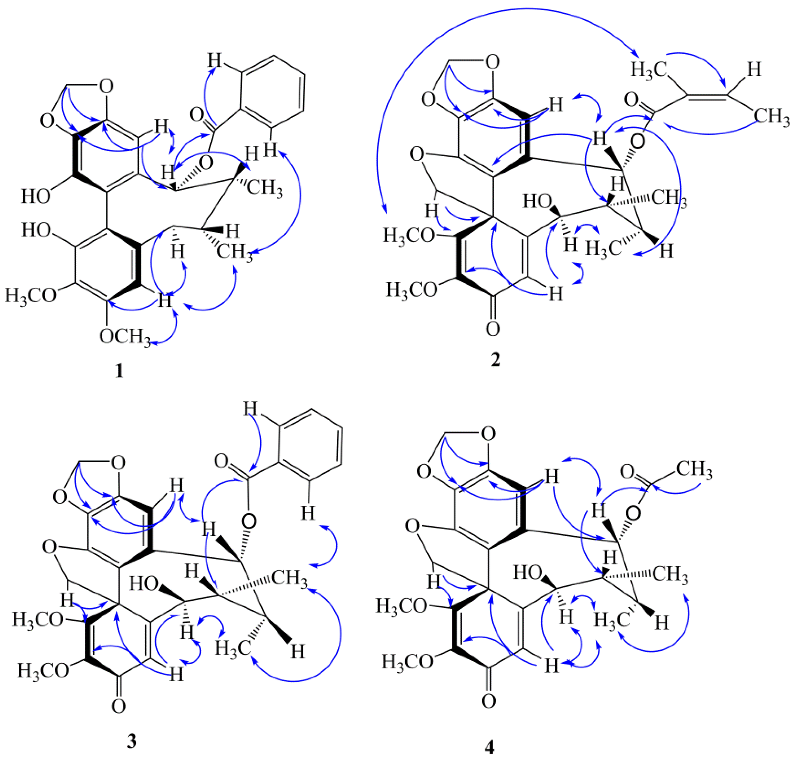
The 1H-NMR spectrum (Table 1) of 1 exhibited two aromatic singlets for a biphenyl moiety at δH 6.49 (H-11) and δH 6.60 (H-4), two singlets for methoxy groups at δH 3.40, 3.96 (3H each), one methylenedioxy (-OCH2O-) group at δH 5.97, 5.98 (1H each, d, J = 1.2 Hz), and three groups with characteristic signals of a benzoyl group at δH 7.48 (1H, dd, J = 7.2, 1.2, Hz, H-5′), 7.43 (2H, dd, J = 7.2, 1.2 Hz, H-3′, 7′) and 7.30 (2H, dd, J = 7.2, 7.2 Hz, H-4′, 6′). A cyclooctadiene ring was recognized from two secondary methyl doublets at δH 1.11 (CH3-17) and δH 1.15 (CH3-18), two methines at δH 2.20 (overlap, H-7, 8), an oxymethine at δH 5.92 (H-9), and a methylene at δH 2.72 and 2.77 (H2-6). The HMBC correlations of H-11 with C-12 (δC 147.9) and C-13 (δC 135.2), H-11 with C-9 (δC 83.5), two aromatic resonances (δH 5.97, 5.98) of the methylenedioxy moiety with C-12 and C-13, indicated that the methylenedioxy moiety was located at C-12 and C-13 (Figure 2). Two methylenedioxy group signals with C-3 (δC 151.2) and C-2 (δC 134.2) showed that two methylenedioxy groups were located at C-3 and C-2, respectively. The presence of a benzoyl group at C-9 was deduced from the HMBC correlation of H-9 with the δC 167.3 (C=O), 117.8 (C-10), 147.6 (C-15), 101.1 (C-11), 42.2 (C-8), and 34.8 (C-7).
The CD spectrum of 1 exhibited a positive Cotton effect around 214 nm and a negative Cotton effect around 242 nm, which suggested that 1 possessed an S-biphenyl conformation [13]. Compound 1 had a twist-boat-chair conformation because the correlated peaks of H-4 with CH3-17, H-9 with CH3-18, H-6a (δH 2.72, 1H, d, J = 13.8 Hz) with H-4, and CH3-17 with H-6a existed in the NOE spectrum (Figure 2). The absolute structure of kadsutherin E (1) was elucidated.
Kadsutherin F (2), which was obtained as a white powder, had the molecular formula C28H28O8, as revealed by its HRESIMS (m/z 521.1791 [M + Na]+). The UV spectrum of 2 showed a maximum absorption at 221 nm. The 1H-NMR and 13C-NMR spectra (Table 1 and Table 2) indicated that 2 was also a dibenzocyclooctene lignan. The characteristic proton signals at δH 4.46, 5.57(2H, d, J = 8.4 Hz) and a quaternary carbon signal at δC 83.7 indicated that 2 possessed a spiroenone ring, similar to kadsutherin D [14]. The 1H-NMR spectrum (Table 1) exhibited two aromatic singlets at δH 6.31 (H-4) and δH 6.39 (H-11), two methoxy groups at δH 3.69, 3.85 (3H each), one methylenedioxy (-OCH2O-) group at δH 5.99 (2H, s). In the cyclooctadiene ring, two doublet methyl groups at δH 1.02 and 1.04 (each 3H, d, J = 7.2 Hz) were located at C-7and C-8, respectively. Furthemore, the characteristic signals of an angeloxy group (δH 5.87, 1.77, 1.66 for H-3′, H-4′ and H-5′) were found in the 1H- and 13C-NMR spectra (Table 1 and Table 2). The HMBC correlations of H-11 with C-12 (δC 150.6) and C-13 (δC 129.8), H-11 with C-9 (δC 79.2), and two H-atoms (δH 5.97, 5.98) of the methylenedioxy moiety with C-12 and C-13, indicated that the methylenedioxy moiety was located at C-12 and C-13 (Figure 2). Moreover, two methoxy groups (1-OCH3, 2-OCH3) were deduced from the HMBC correlations of 1-OCH3 (δH 3.85, 3H, s) with C-1(δC 168.3), two characteristic proton signals (δH 4.46, 5.57, d, J = 8.4 Hz, CH2-20) with C-1, 2-OCH3 (δH 3.69, 3H, s) with C-2 (δC 134.8), H-4 with C-6 (δC 80.8), and H-4 with C-2. The presence of the angeloxy group at C-9 was deduced from the HMBC correlation of H-9 with the δC 167.3 (C=O), 130.1 (C-10), 120.4 (C-15), 100.2 (C-11), 42.3 (C-8), and 38.7 (C-7).
The CD spectrum of 2 exhibited a positive Cotton effect around 276 nm and a negative Cotton effect around 216 nm, which was contrary to the CD spectrum of the 1, which suggested that 2 possessed an R-biphenyl conformation [14]. A twist-boat conformation of the cycloctadiene ring was deduced from the NOE correlations of H-9 with H-11, H-9 with CH3-18, H-6a (δH 4.13, 1H, d, J = 10.8 Hz) with H-4, and CH3-18 with H-4 (Figure 2). According to the above data, the structure of 2 was elucidated as kadsutherin F.
Kadsutherin G (3), which was obtained as a white powder, had the molecular formula C29H28O9, as it was revealed by its HRESIMS (m/z 543.1647 [M + Na]+). The 1H-NMR spectrum of 3 was similar to that of 2, but the prominent difference in the 1H-NMR spectrum (Table 1) was the presence of a benzoyl group in 3, which was substituted the angeloxy group in 2. The data of the 13C-NMR spectrum of 3 (Table 2) also confirmed this deduction. In the HMBC spectrum of 3, H-9 was correlated with the carbonyl carbon (δC 168.5, C=O) of the benzoyl group and δC 122.0 (C-10), 156.4 (C-15), 101.7 (C-11), 39.8 (C-8), and 44.0 (C-7), which clearly indicated that the benzoyl group was located at C-9.
The CD spectrum of 3 exhibited a positive Cotton effect around 311 nm and a negative Cotton effect around 224 nm, which suggested that 3 possessed an R-biphenyl conformation. A twist-boat conformation of 3 was deduced from the NOE correlations of CH3-17 with CH3-18, H-9b (δH 5.72, 1H, d, J = 7.2 Hz) with CH3-18, and H-6a (δH 4.26, 1H, d, J = 10.2 Hz) with CH3-17 (Figure 2). On the basis of the above data, the structure of 3 was educidated as kadsutherin G.
Kadsutherin H (4), which was obtained as a white powder, had the molecular formula C24H26O9, as it was revealed by its HRESIMS (m/z 481.1478 [M + Na]+). The 1H-NMR spectrum of 4 was also similar to that of 2, but the prominent difference in the 1H-NMR spectrum (Table 1) was the presence of an acetoxy group in 4, which was substituted the angeloxy group in 2. The data of the 13C-NMR spectrum of 4 (Table 2) also confirmed this deduction. In the HMBC spectrum of 4, H-9 was correlated with the carbonyl carbon (δC 171.9, C=O) of the acetoxy group and δC 122.2 (C-10), 146.6(C-15), 101.3 (C-11), 43.3 (C-8), and 40.4 (C-7), which clearly indicated that the acetoxy group was located at C-9.
The CD spectrum of 4 exhibited a positive Cotton effect around 245 nm and a negative Cotton effect around 223 nm, which suggested that 4 possessed an R-biphenyl conformation. Compound 4 had a twist-boat conformation, which was deduced from the NOE correlations of H-9 with H-11, H-9 with CH3-18, H-6a (δH 4.11, 1H, d, J = 10.2 Hz) with H-4, and CH3-18 with H-4 (Figure 2). Thus, 4 was elucidated as kadsutherin G.
2.2. Anti-Platelet Effects of Compounds 1–6
The anti-platelet effects of the compounds from the stems of the K. interior plants were tested in vitro, using the turbidimetric method in washed rat platelets that were induced by ADP (100 μM.). The anti-platelet aggregation data are shown in Table 3. The clinically applied anti-platelet agent aspirin, was used as the positive control. From the results of our anti-platelet aggregation tests, kadsutherin E (1), kadsutherin F (2), kadsutherin G (3), kadsutherin H (4), acetoxyl oxokadsurane (5), and heteroclitin D (6) exhibited inhibition (with inhibition in the range of 11.77–49.47%) against the ADP induced platelet aggregation. Among these compounds, kadsutherin F (2) showed the strongest anti-platelet aggregation activity with an inhibition of 49.47 ± 2.93%.
3. Materials and Methods
3.1. General Experimental Procedures
Column chromatography (CC) was performed with silica gel (200–300, 300–400 mesh, Qingdao, China). Thin-layer chromatography (TLC) was carried out with silica gel GF-254 plates (Qingdao, China). Ultraviolet (UV) spectra were recorded on a UV2550 UV/Vis spectrometer (SHIMADZU, Kyoto, Japan). The infrared (IR) spectra (KBr) were measured using a FTIR-8400S spectrophotometer (SHIMADZU, Kyoto, Japan). The optical rotations were determined in MeOH at 20 °C, using a PerkinElmer 341 digital polarimeter (Waltham, MA, USA). Circular dichroism (CD) spectra was carried out on a J-815 spectropolarimeter (JASCO, Kyoto, Japan). The MS data were determined on a LTQ-Obitrap XL (Thermo Scientific, Bremen, Germany) mass spectrometer for HRESIMS. The obtained 1D and 2D nuclear magnetic resonance (NMR) spectra were performed on a AVIII 600 spectrometer with TMS as the internal standard (Bruker Bispin Corporation, Fallanden, Switzerland).
3.2. Plant Material
The stems of the K. interior plants were collected from Fengqing City, Yunnan Province, China, in October 2015 and were identified by Prof. Ben-gang Zhang. A voucher specimen (NO. ID-KT-FQ201510) was deposited in the Resource and Conservation Research Center, Institute of Medicinal Plant Development, Beijing, China.
3.3. Extraction and Isolation
The dried and powdered stems (8 kg) of the K. interior plants were extracted three times with 90% EtOH. The extract was concentrated under a reduced pressure to dryness, which was then partitioned between EtOAc and H2O to provide the EtOAc-soluble fraction. The EtOAc fraction (243 g) was purified by CC on silica gel (200–300 mesh; petroleum ether/acetone gradient) to afford eight fractions, as follows: Fr1–Fr8. Fr5 was applied to silica gel CC with PE/EtOAc and was then separated by Sephadex LH-20 CC with CHCl3/MeOH (3:2 v/v) and preparative HPLC with MeOH/H2O, so as to yield compounds 1 (26 mg), 2 (77 mg), 3 (15 mg), 4 (20 mg), 5 (6 mg), and 6 (120 mg).
3.3.1. Kadsutherin E (1)
3.3.2. Kadsutherin F (2)
White powder: [α = 131.2 (c 0.08, MeOH); UV (MeOH, λmax, nm) (log ε): 221 (0.69); IR (KBr, υmax, cm−1): 3426 (-OH), 2956, 2928 (CH), 1669 (C=O), 1623 (C=O), and 1247 (C-O); and HR-ESIMS: m/z = 521.1791 [M + Na]+ (calculated for C28H28O8Na: 521.1792). 1H and 13C-NMR data (CD3OD) are shown in Table 1 and Table 2.
3.3.3. Kadsutherin G (3)
White amorphous powder: [α = −126.0 (c 0.05, MeOH); UV (MeOH, λmax, log ε): 221 (0.70) nm; IR (KBr, υmax, cm−1): 3411 (-OH), 2946, 2836 (CH), 1714 (C=O), 1648(C=O), 1265 (C-O); and HR-ESIMS: m/z = 543.1647 [M + Na]+ (calculated for C29H28O9Na: 543.1645). 1H and 13C-NMR data (CD3OD) are shown in Table 1 and Table 2.
3.3.4. Kadsutherin H (4)
3.4. Anti-Platelet Aggregation Assay
The platelet aggregation assay was carried out according to the methodology that was reported in previous studies [17]. The blood was collected by catheterization of the abdominal aorta in rats (mean weight: 232.9 ± 4.8 g), anticoagulated with acid citrate dextrose (ACD) (9:1, v/v) and centrifuged for 15 min at 100× g at room temperature, so as to obtain platelet rich plasma (PRP). The platelet numbers were counted by a Coulter counter and were adjusted to 5.0 × 10⁵ platelets/μL. The platelet aggregation was measured at 37 °C using the turbidimetric method. The assays were performed at 37 °C in cuvettes using 300 μL of PRP under stirring, and the aggregation was triggered by the addition of adenosine diphosphate (ADP). All of the tested compounds (1mg/mL) were dissolved in 0.5% dimethyl sulfoxide (DMSO) [18] and were incubated with PRP for 2 min at 37 °C, and then, the platelet aggregation was triggered by adding ADP (10 μM). Aspirin was used as a positive control. The percentages of the inhibition were calculated as follows:
Inhibition (%) = [OD(control) − OD(compound)]/OD(control) × 100%.
The results were expressed as the mean ± SD and all of the anti-platelet aggregation data were statistical analyzed by SPSS (version 19.0, Chicago, IL, USA).
4. Conclusions
Four new lignans, named kadsutherin E–H (1–4), together with two known lignans (5–6), were isolated from the stems of the K. interior plants. The structures of those compounds were established on the basis of spectroscopic data. The anti-platelet effects of the six compounds were evaluated by suppressing the ADP-induced platelet aggregation in washed rat platelets. The results of the anti-platelet aggregation experiments indicated that compounds 1–6 could inhibit ADP-induced platelet aggregation at 100 μM. Kadsutherin F (2) showed the strongest anti-platelet aggregation activity with an inhibition of 49.47%.
Author Contributions
H.T.L. designed the research; J.Z., J.-S.L., Y.-D.Q., X.-G.J., and B.-G.Z. performed the experiments and analyzed the data; J.Z. wrote the paper. All of the authors have read and approved the content of the manuscript.
Funding
This research was funded by [National Natural Science Foundation of China] grant number [8373913], [National Major Scientific and Technological Special Project for ‘Significant New Drugs Development] grant number [2015ZX09501005], and [CAMS Initiative for Innovative Medicine] grant number [CAMS-I2M-1-010].
Acknowledgments
This work is financially supported by the National Natural Science Foundation of China (No. 8373913), the National Major Scientific and Technological Special Project for ‘Significant New Drugs Development’ (No. 2015ZX09501005), and the CAMS Initiative for Innovative Medicine (CAMS-I2M-1-010).
Conflicts of Interest
No potential conflict of interest was reported by the authors.
References
- Pharmacopoeia Commission of PRC. Pharmacopoeia of the People’s Republic of China; China Medical Science Press: Beijing, China, 2015; Volume 1, pp. 522–523. [Google Scholar]
- Chen, D.F.; Zhang, S.X.; Xie, L.; Xie, J.X.; Chen, K.; Kashiwada, Y.; Zhou, B.N.; Wang, P.; Cosentino, L.M.; Lee, K.H. Anti-AIDS agents-XXVI. Structure-activity correlations of gomisin G-related anti-HIV lignans from Kadsura interior and of related synthetic analogues. Bioorg. Med. Chem. 1997, 5, 1715–1723. [Google Scholar] [CrossRef]
- Chen, D.F.; Zhang, S.X.; Kozuka, M.; Sun, Q.Z.; Feng, J.; Wang, Q.; Mukainaka, T.; Nobukuni, Y.; Tokuda, H.; Nishino, H.; et al. Interiotherins C and D, two new lignans from Kadsura interior and antitumor-promoting effects of related neolignans on epstein-barr virus activation. J. Nat. Prod. 2002, 65, 1242–1245. [Google Scholar] [CrossRef] [PubMed]
- Ding, Z.H.; Luo, S.D. Study on new lignans from Kadsura interior A. C. Smith. Acta Chim. Sin. 1990, 48, 1075–1079. [Google Scholar]
- Xu, L.J.; Ma, P.; Li, L.; Wang, W.Y.; Xiao, W.; Peng, Y.; Xiao, P.G. New collection of crude drugs in Chinese Pharmacopoeia 2010 III. Chin. Herb. Med. 2012, 4, 177–182. [Google Scholar]
- Yang, X.W.; Hattori, M.; Namba, T.; Chen, D.F.; Xu, G.J. Anti-lipid peroxidative effect of an extract of the stems of Kadsura heteroclita and its major constituent, kadsurin, in mice. Chem. Pharm. Bull. 1992, 40, 406–409. [Google Scholar] [CrossRef] [PubMed]
- Yang, X.W.; Miyashiro, H.; Hattori, M.; Namba, T.; Tezuka, Y.; Kikuchi, T.; Chen, D.F.; Xu, G.J. Isolation of novel lignans, heteroclitins F and G, from the stems of Kadsura heteroclite, and Anti-lipid peroxidative actions of heteroclitins A-G and related componds in the in vitro rat liver homogenate system. Chem. Pharm. Bull. 1992, 40, 1510–1516. [Google Scholar] [CrossRef] [PubMed]
- Jin, X.L.; Gu, Z.; Hu, T.X.; Wang, M.H.; Chen, D.F. Effects of lignan gomisin J from Kadsura interior on liver mitochondria lipid peroxidation and on the superoxide anion radical. Chin. Pharmacol. Bull. 2000, 16, 26–28. [Google Scholar]
- Chen, D.F.; Zhang, S.X.; Chen, K.; Zhou, B.N.; Wang, P.; Cosentino, L.M.; Lee, K.H. Two new lignans, interiotherins A and B, as anti-HIV principles from Kadsura interior. China J. Nat. Prod. 1996, 59, 1066–1068. [Google Scholar] [CrossRef] [PubMed]
- Jiang, S.L.; Zhang, Y.Y.; Chen, D.F. Effects of heteroclitin D, schisanhenol and (+)-anwulignan on platelet aggregation. Fudan Univ. J. Med. Sci. 2005, 32, 467–470. [Google Scholar]
- Pu, J.X.; Yang, L.M.; Xiao, W.L.; Li, R.T.; Lei, M.; Gao, X.M.; Huang, S.X.; Li, S.H.; Zheng, Y.T.; Huang, H.; et al. Compounds from Kadsura heteroclita and related anti-HIV activity. Phytochemistry 2008, 69, 1266–1272. [Google Scholar] [CrossRef] [PubMed]
- Chen, M.; Luo, Y.P.; Zou, Y.L.; Lang, L.H.; Chen, D.F. Heteroclitins R-S: New dibenzocylooctadiene lignans from Kadsura heteroclita. Chin. J. Nat. Med. 2014, 12, 689–692. [Google Scholar] [CrossRef]
- Li, H.R.; Feng, Y.L.; Yang, Z.G.; Wang, J.; Akihiro, D.; Susumu, K.; Xu, L.Z.; Yang, S.L. New Lignans from Kadsura coccinea and Their Nitric Oxide Inhibitory Activities. Chem. Pharm. Bull. 2006, 54, 1022–1025. [Google Scholar] [CrossRef] [PubMed]
- Lu, Y.; Chen, D.F. Kadsutherin D, a new dibenzocyclooctadiene lignan from Kadsura species. Nat. Prod. Res. 2008, 22, 1344–1349. [Google Scholar] [CrossRef] [PubMed]
- Li, L.N.; Xue, H. Dibenzocyclooctadiene lignans possessing a spirobenzofuranoid skeleton from Kadsura coccinea. Phytochemistry 1990, 29, 2730–2732. [Google Scholar] [CrossRef]
- Chen, D.F.; Xu, G.J.; Yang, X.W.; Hattori, M.; Tezuka, Y.; Kikuchi, T.; Namba, T. Dibenzocyclo-octadiene lignans from Kadsura heteroclita. Phytochemistry 1992, 31, 629–632. [Google Scholar] [CrossRef]
- Fuly, A.L.; Machado, O.L.; Alves, C.R.; Carlini, C.R. Mechanism of inhibitory action on platelet activation of a phospholipase A2 isolated from Lachesis muta (Bushmaster) snake venom. J. Thromb. Haemost. 1997, 78, 1372–1380. [Google Scholar]
- Chen, J.J.; Tsai, T.H.; Liao, H.R.; Chen, L.C.; Kuo, Y.H.; Sung, P.J.; Chen, C.L.; Wei, C.S. New sesquiterpenoids and anti-platelet aggregation constituents from the rhizomes of Curcuma zedoaria. Molecules 2016, 21, 1385–1396. [Google Scholar] [CrossRef] [PubMed]
Sample Availability: Not Available. |
Figure 1.
Structures of compounds 1–6.
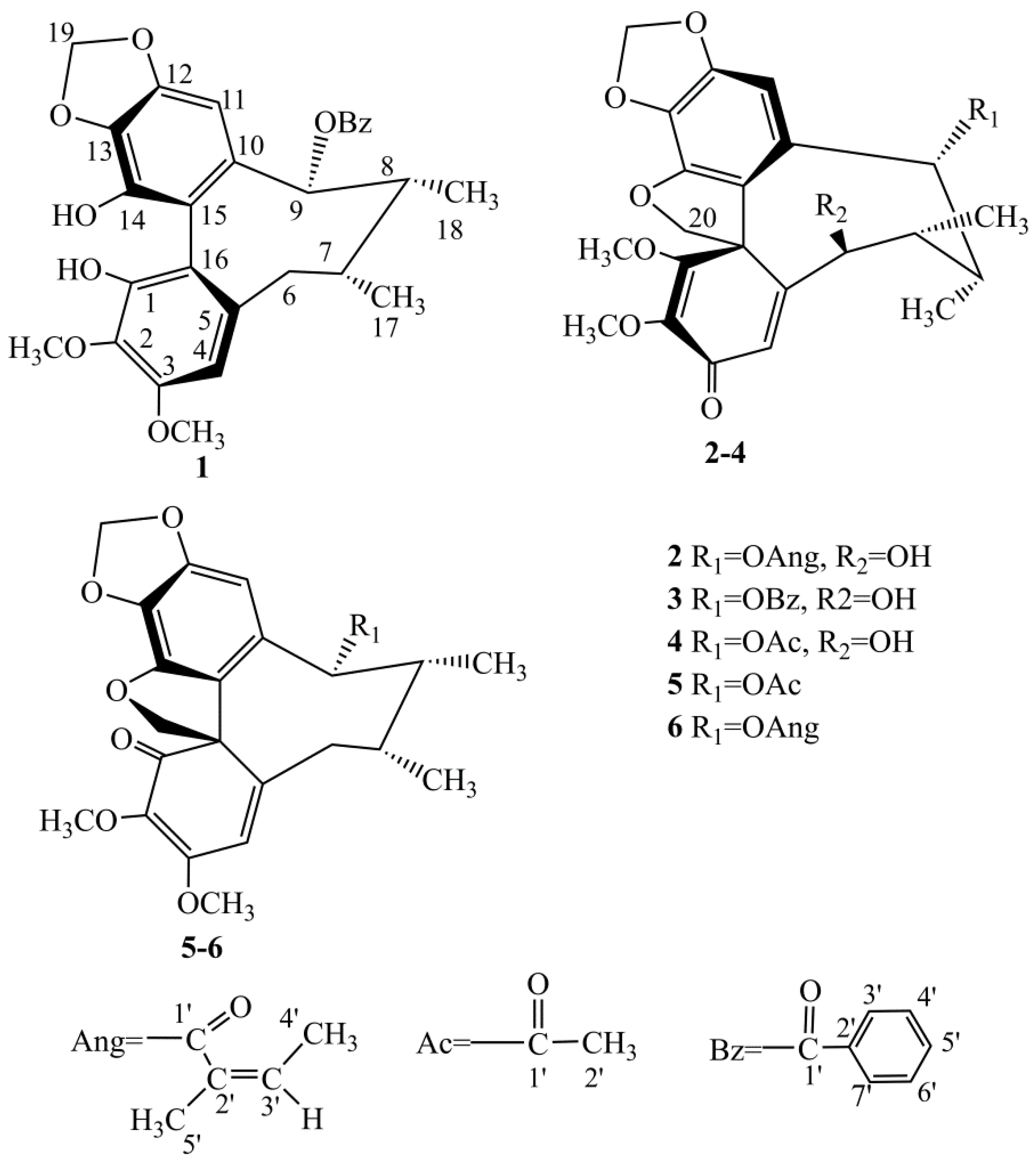
Figure 2.
Key HMBC (H→C) correlations and selected NOESY (H ↔H) correlations of compounds 1–4.

Table 1.
1H-NMR data of compounds 1–4 (CD3OD, δ in ppm, J/Hz, 600 MHz).
| Position | 1 | 2 | 3 | 4 |
|---|---|---|---|---|
| 4 | 6.60 (1H, s) | 6.31 (1H, s) | 6.49 (1H, s) | 6.34 (1H, s) |
| 6a | 2.72 (1H, d, 13.8) | 4.13 (1H, d, 10.8) | 4.26 (1H, d, 10.2) | 4.11 (1H, d, 10.2) |
| 6b | 2.77 (1H, dd, 13.8, 6.6) | - | - | - |
| 7 | 2.20 (1H, overlap) | 1.56 (1H, m) | 1.66 (1H, m) | 1.49 (1H, m) |
| 8 | 2.20 (1H, overlap) | 2.11 (1H, m) | 2.18 (1H, m) | 2.05 (1H, m) |
| 9 | 5.92 (1H, d, 7.2) | 5.61 (1H, d, 7.8) | 5.72 (1H, d, 7.2) | 5.86 (1H, d, 7.8) |
| 11 | 6.49 (1H, s) | 6.39 (1H, s) | 6.48 (1H, s) | 6.34 (1H, s) |
| 17 | 1.11 (3H, d, 7.2) | 1.02 (3H, d, 7.2) | 1.08 (3H, d, 6.6) | 0.92 (3H, d, 7.2) |
| 18 | 1.15 (3H, d, 7.2) | 1.04 (3H, d, 7.2) | 1.17 (3H, d, 7.8) | 1.02 (3H, d, 7.2) |
| 19 | 5.97 (2H, d, 1.2) | 5.99 (2H, s) | 6.02 (2H, s) | 6.00 (2H, s) |
| 20 | - | 4.46, 5.57 (2H, ABq, 8.4) | 4.40, 5.54 (2H, ABq, 8.4) | 4.57, 5.62 (2H, ABq, 8.4) |
| 1-OMe | - | 3.85 (3H, s) | 3.60 (3H, s) | 3.89 (3H, s) |
| 2-OMe | 3.40 (3H, s) | 3.69 (3H, s) | 2.86 (3H, s) | 3.76 (3H, s) |
| 3-OMe | 3.96 (3H, s) | - | - | - |
| 1-OH | 5,74 (1H, s) | - | - | - |
| 6-OH | - | 4.85 (1H, br s) | 4.68 (1H, br s) | 4.67 (1H, br s) |
| 14-OH | 5.26 (1H, s) | - | - | - |
| Acetoxy | ||||
| 2′ | 1.79 (3H, s) | |||
| Angeloyl | ||||
| 3′ | 5.87 (1H, m) | |||
| 4′ | 1.77 (3H, dd, 7.2, 1.2) | |||
| 5′ | 1.66 (3H, t, 4.8) | |||
| Benzoyl | ||||
| 3′,7′ | 7.43 (2H, dd,7.2, 1.2) | 7.76 (2H, dd, 7.8, 1.2) | ||
| 4′,6′ | 7.30 (2H, dd, 7.2, 7.2) | 7.40 (2H, dd, 7.8, 7.8) | ||
| 5′ | 7.48 (1H, dd, 7.2, 7.2) | 7.57 (1H, dd, 7.8, 7.8) |
Table 2.
13C-NMR data of compounds 1–4 (CD3OD, δ in ppm, J/Hz, 150 MHz).
| Position | 1 | 2 | 3 | 4 |
|---|---|---|---|---|
| 1 | 138.2 | 168.3 | 169.5 | 169.8 |
| 2 | 134.2 | 134.8 | 136.0 | 136.2 |
| 3 | 151.2 | 185.5 | 186.6 | 187.0 |
| 4 | 107.0 | 131.8 | 134.6 | 156.8 |
| 5 | 133.8 | 155.3 | 131.6 | 131.5 |
| 6 | 38.3 | 80.8 | 81.8 | 78.9 |
| 7 | 34.8 | 38.7 | 44.0 | 40.4 |
| 8 | 42.2 | 42.3 | 39.8 | 43.3 |
| 9 | 83.5 | 79.2 | 82.1 | 82.2 |
| 10 | 117.8 | 130.1 | 122.0 | 122.2 |
| 11 | 101.1 | 100.2 | 101.7 | 101.3 |
| 12 | 147.9 | 150.6 | 152.0 | 156.8 |
| 13 | 135.2 | 129.8 | 131.3 | 130.0 |
| 14 | 134.4 | 145.3 | 146.5 | 156.8 |
| 15 | 147.6 | 120.4 | 156.4 | 146.6 |
| 16 | 117.3 | 57.9 | 59.2 | 59.0 |
| 17 | 18.5 | 18.2 | 19.6 | 19.4 |
| 18 | 14.3 | 9.11 | 10.4 | 9.1 |
| 19 | 100.2 | 102.0 | 103.4 | 103.2 |
| 20 | - | 83.7 | 85.1 | 84.2 |
| 1-OMe | - | 60.6 | 61.4 | 61.7 |
| 2-OMe | 55.2 | 59.4 | 59.4 | 60.7 |
| 3-OMe | 59.3 | - | - | - |
| 1′ | 167.3 | 168.1 | 168.5 | 171.9 |
| 2′ | 129.7 | 136.8 | 130.9 | 20.8 |
| 3′ | 129.3 | 127.7 | 130.8 | - |
| 4′ | 127.8 | 14.6 | 130.0 | - |
| 5′ | 132.4 | 19.8 | 132.8 | - |
| 6′ | 127.8 | - | 130.0 | - |
| 7′ | 129.3 | - | 130.8 | - |
Table 3.
Inhibitory effects of compounds on the aggregation of rat platelets induced by adenosine diphosphate (ADP) (100 μM, n = 9).
Table 3.
Inhibitory effects of compounds on the aggregation of rat platelets induced by adenosine diphosphate (ADP) (100 μM, n = 9).
| Compounds | Inhibition% |
|---|---|
| Kadsutherin E (1) | 23.64 ± 1.12 |
| Kadsutherin F (2) | 49.47 ± 2.93 |
| Kadsutherin G (3) | 33.10 ± 2.67 |
| Kadsutherin H (4) | 21.75 ± 2.37 |
| Acetoxyl oxokadsurane (5) | 34.31 ± 0.73 |
| Heteroclitin D (6) | 11.77 ± 2.30 |
| Aspirin | 59.94 ± 2.44 |
© 2018 by the authors. Licensee MDPI, Basel, Switzerland. This article is an open access article distributed under the terms and conditions of the Creative Commons Attribution (CC BY) license (http://creativecommons.org/licenses/by/4.0/).
Share and Cite
MDPI and ACS Style
Liu, J.-S.; Zhang, J.; Qi, Y.-D.; Jia, X.-G.; Zhang, B.-G.; Liu, H.-T. Four New Lignans from Kadsura Interior and Their Bioactivity. Molecules 2018, 23, 1279. https://doi.org/10.3390/molecules23061279
AMA Style
Liu J-S, Zhang J, Qi Y-D, Jia X-G, Zhang B-G, Liu H-T. Four New Lignans from Kadsura Interior and Their Bioactivity. Molecules. 2018; 23(6):1279. https://doi.org/10.3390/molecules23061279
Chicago/Turabian StyleLiu, Jiu-Shi, Jin Zhang, Yao-Dong Qi, Xiao-Guang Jia, Ben-Gang Zhang, and Hai-Tao Liu. 2018. "Four New Lignans from Kadsura Interior and Their Bioactivity" Molecules 23, no. 6: 1279. https://doi.org/10.3390/molecules23061279