Article

Fingerprint-Based Machine Learning Approach to Identify Potent and Selective 5-HT2BR Ligands

by
Krzysztof Rataj
1,
Ádám Andor Kelemen
2,
José Brea
3,
María Isabel Loza
3,
Andrzej J. Bojarski
1,* and
György Miklós Keserű
2,*
1
Department of Medicinal Chemistry, Institute of Pharmacology, Polish Academy of Sciences, 12 Smętna Street, 31-343 Krakow, Poland
2
Medicinal Chemistry Research Group, Research Centre for Natural Sciences, Hungarian Academy of Sciences, Magyar tudósok krt. 2, H1117 Budapest, Hungary
3
Grupo de Investigación “BioFarma” USC, Centro de Investigación CIMUS, Planta 3ª, Avd. de Barcelona s/n, 15782 Santiago de Compostela, Spain
*
Authors to whom correspondence should be addressed.
Molecules 2018, 23(5), 1137; https://doi.org/10.3390/molecules23051137
Submission received: 16 April 2018 / Revised: 5 May 2018 / Accepted: 7 May 2018 / Published: 10 May 2018
(This article belongs to the Special Issue Frontiers in Computational Chemistry for Drug Discovery)

Abstract

:
The identification of subtype-selective GPCR (G-protein coupled receptor) ligands is a challenging task. In this study, we developed a computational protocol to find compounds with 5-HT2BR versus 5-HT1BR selectivity. Our approach employs the hierarchical combination of machine learning methods, docking, and multiple scoring methods. First, we applied machine learning tools to filter a large database of druglike compounds by the new Neighbouring Substructures Fingerprint (NSFP). This two-dimensional fingerprint contains information on the connectivity of the substructural features of a compound. Preselected subsets of the database were then subjected to docking calculations. The main indicators of compounds’ selectivity were their different interactions with the secondary binding pockets of both target proteins, while binding modes within the orthosteric binding pocket were preserved. The combined methodology of ligand-based and structure-based methods was validated prospectively, resulting in the identification of hits with nanomolar affinity and ten-fold to ten thousand-fold selectivities.

Graphical Abstract

1. Introduction

There is an increasing need of efficacious CNS (central nervous system) drugs with reduced off-target activity that often connected to notable subtype selectivity. Modulated by one of the major neurotransmitters, serotonergic receptors play a central role in many neuropsychiatric indications [1]. A significant number of drugs was introduced to the market targeting different 5-HT subtypes ranging from 5-HT1–7R [2]. Rational drug design of subtype selectivity remained a challenge in many cases.
The continuously growing number of available relevant class A GPCR (G-protein coupled receptor) X-ray structures (bovine rhodopsin [3,4], β2AR [5], 5-HT1BR [6], 5-HT2BR [7,8,9], D3R [10], M2R [11], etc.) revealed certain important structural motifs of molecular recognition and ligand binding, including determinants of selectivity across the certain subtypes. GPCRs consist of seven transmembrane helices (and the additional intramembrane helix 8) connected in a bunch through 3-3 intracellular, and extracellular loops. Beyond the highly conserved amino acid residues localized across the helices either in the proximity of the binding pocket, or participating in the well-established DRY [12], NpxxY [13], and P-I-F motifs [14], or in the conserved disulfide bridge [15], the most conspicuous differences are present in the extracellular vestibule near the extracellular loop 2 (ECL2) region, which impacts subtype selectivity [16]. The role of the 5-HT2B receptor has been implicated in a number of diseases including migraines [17,18,19], chronic heart disease [20], and irritable bowel syndrome (IBS) [21]. The chemical space of known 5-HT2BR ligands might be represented by a couple of chemotypes (presented in Figure 1). Examples of high affinity 5-HT2BR compounds showing selectivity against 5-HT1BR are represented by triazines (1) [21], piperidines (2, 3), pyrimidines (e.g., RS-127445 (4)) [22], arylpiperazines (m-CPP [23], EGIS-7625 (5) [24]), tetrahydro-β-carbolines (LY-23728 (6), LY-272015, LY-266097) [25], and aryl ureas (SB-200646A (7), SB-204741, SB-215505) [26]. Interestingly, the contribution of in silico methods to the discovery of novel 5-HT2BR ligands is rather limited [27]. Understanding the key drivers of subtype selectivity, here we report the identification of novel selective 5-HT2BR ligands using a combination of ligand-based and structure-based methods.
At the first stage of screening, we applied machine learning tools [28] trained on the available structural information of known h5-HT1BR and h5-HT2BR ligands. Started from the main principle of fragment-based drug design (FBDD), these compounds were represented by Neighbouring Substructures Fingerprint (NSFP) [29], which opens the possibilities of quickly creating fragment libraries with desired target-specific, class-specific, or even family-specific properties. This new methodology is based on connections between SMARTS (SMiles ARbitrary Target Specification) patterns for finding doublets or triplets of small substructures that constitute a larger fragment. Analyzing these structural moieties, machine learning methods [30,31] are applied to recognize non-typical, activity-specific fragments for a particular target or a group of targets. Key-based substructural fingerprints depict the occurrences of a predefined set of chemical subgroups (keys) [32] within the target molecule. However, the standard key-based representations do not provide sufficient structural information. The substructures may be arranged in various ways, resulting in a vast set of possible outcomes from a single fingerprint. This may lead to ambiguities in the process of classification of active and inactive compounds, resulting in a high false positive rate. These flaws may be overcome by the addition of substructural connectivity data and combining this methodology with structure-based approaches.
A thorough analysis of the crystal structures of aminergic GPCR proteins revealed that most of the receptors have a secondary binding pocket (SBP) [16] that is formed at the extracellular part of the protein by the participation of the ECL2. This site contains a significant proportion of non-conserved amino acids across certain aminergic GPCRs that provides an opportunity to obtain subtype selectivity. Previously, we showed [33] that a docking strategy towards the SBP was able to identify bitopic compounds with improved affinity and selectivity. In our present study, we combined both methodologies, i.e., the fragment-based NSFP fingerprint with docking. Applying this approach for 2B and 1B serotonin receptors, we were able to identify new and selective 5-HT2BR ligands, providing potential chemical starting points for further optimization.

2. Results and Discussion

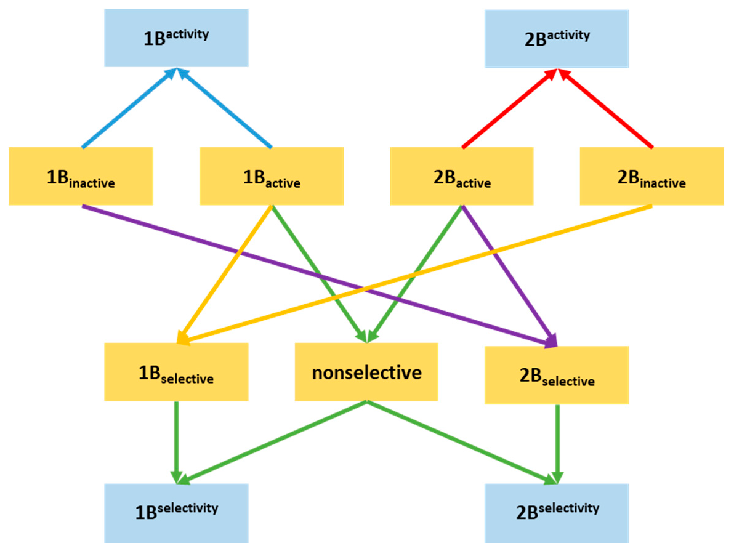
2.1. Compilation of the Training Sets for Machine Learning-Based Classification

In this study, we built a NSFP fingerprint-based machine learning model using in vitro activity data available for human 5-HT1BR and 5-HT2BR receptors in ChEMBL (biochemical database curated by the European Molecular Biology Laboratory) [34]. The compounds were divided into actives and inactives using binding affinity thresholds. Since our design concept was based on the role of the secondary binding pocket in selectivity, only compounds with 22 or more heavy atoms were considered, as they are more likely to bind to both the orthosteric binding pocket (OBP) and SBP of 5-HT1BR and 5-HT2BR.
The active/inactive sets were selected based on binding affinity recalculated from various units reported in the ChEMBL to Ki (actives: Ki ≤ 500 nM, inactives: Ki ≥ 1000 nM). In this way, four sets were acquired (summarized in Table 1): active for 5-HT1B (1Bactive), active for 5-HT2B (2Bactive), inactive for 5-HT1B (1Binactive), and inactive for 5-HT2B (2Binactive). 5-HT1B actives and 5-HT2B inactives, and 5-HT2B actives and 5-HT1B inactives were used for 1Bselective and 2Bselective subsets. The nonselective subset contains compounds that are active on both 5-HT1B and 5-HT2B receptors.
Altogether, seven training sets were used for building machine learning models (1–2. actives, 3–4. inactives, 5–6. selectives for both 1B and 2B receptor subtypes, and 7. nonselectives).

2.2. Building NSFP-Based Machine Learning Model

NSFP fingerprints were calculated for all of the compound sets using Klekota–Roth fingerprint (KRFP) substructure keys. A series of machine learning classifiers were created that were aimed at properly discriminating compounds. Activity classifier for 2B (2Bactivity) was built using 2Bactive and 2Binactive compounds. Selectivity classifier (2Bselectivity) for 2B was built using 2Bselective and nonselective compounds in order to predict the putative selectivity of screened compounds. The same procedure was applied to 1B with its selective sets, resulting in 1Bselectivity and 2Bselectivity classifiers. Altogether, we developed two activity (for 5-HT1BR and 5-HT2BR) and two selectivity (for 5-HT1BR and 5-HT2BR) classifiers using known active, inactive, selective, and non-selective 5-HT1BR and 5-HT2BR ligands. Final models were selected from the total of 4 × 117 classifiers based on the highest acquired Matthews correlation coefficient (MCC) values. These models were used for filtering the in-stock MCule database. The summary of the model development is shown in Figure 2.

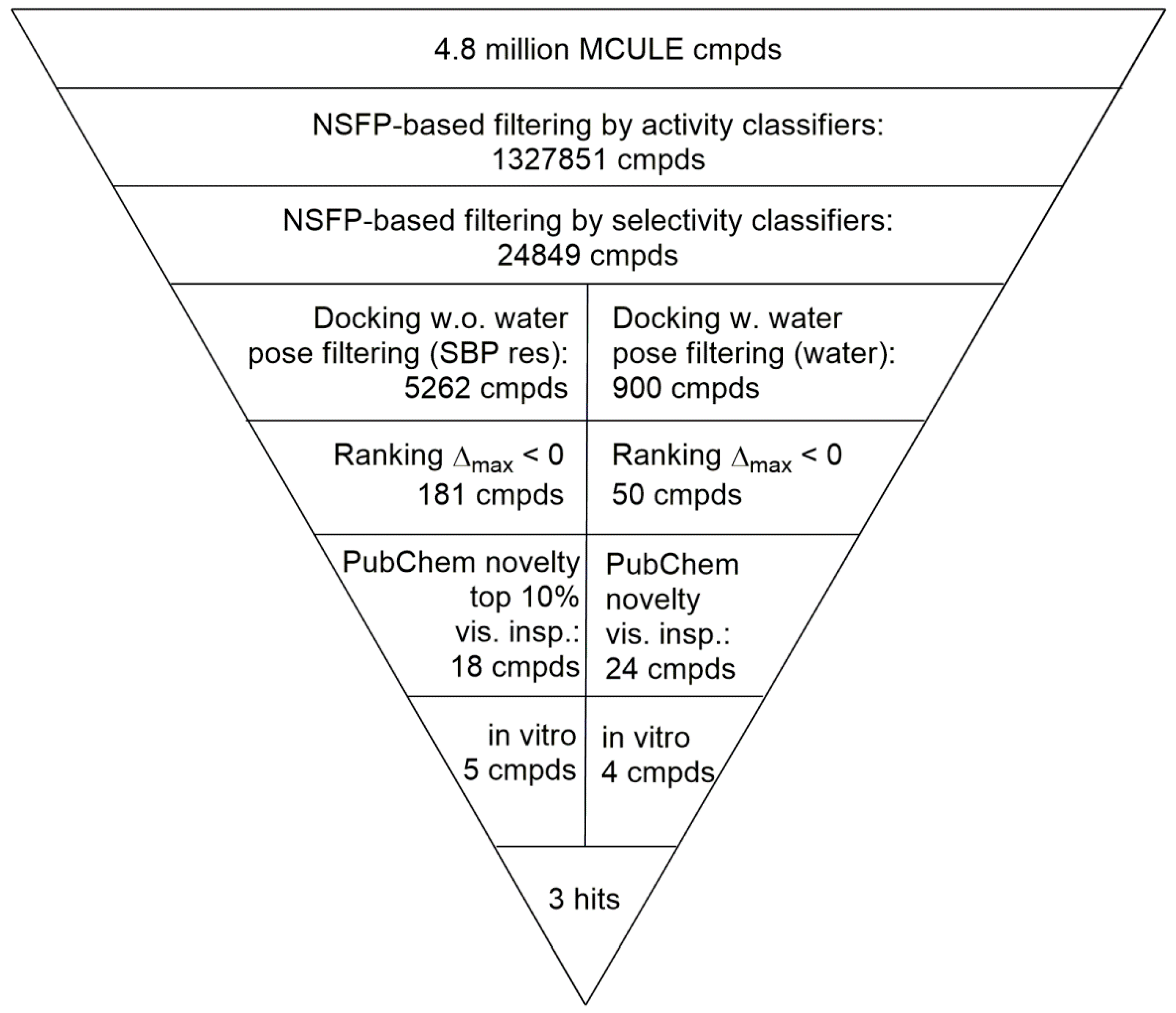
2.3. Prospective Machine Learning-Based Classification

First, NSFP fingerprints have also been calculated for the entire MCule database [35] of commercially available compounds. Next, each compound was classified using the four machine learning models. If a compound’s classification by 2Bactivity was positive (compound classified as 2B active) and classification by 1Bactivity was negative (compound classified as 1B inactive), the compound was treated as a putative 2B selective compound (12Bselective). If the opposite was true (compound classified as 1B active and 2B inactive), the compound was regarded as a putative 1B selective compound (11Bselective). Compounds achieving any other combination of classification results were disregarded from further research.
The second round of classification consisted of validating the compounds against the 1Bselectivity and 2Bselectivity selectivity classifiers. If a 12Bselective compound was classified as positive by the 2Bselectivity and negative by the 1Bselectivity, the compound was regarded as 22Bselective. In any other case, the compound was discarded. The summary of the prospective classification is presented in Figure 3.
The in-stock MCule database containing 4.8 M molecules was filtered prospectively in an activity-classification and consecutively in a selectivity-classification step resulting in 24,849 putative 5-HT2B selective compounds (Figure 4).

2.4. Virtual Screening of the Prefiltered Set Classified by Machine Learning

In the next sequential filtering step, the pre-filtered 5-HT2B selective compound set of 24,849 compounds was subjected to two complementary docking workflows (i) considering non-conserved amino acid pairs and ranking-based consensus scoring; and (ii) accounting for water molecules (Figure 4). The set of 24,849 putative 5-HT2B selective compounds was docked into four crystal structures available to date (PDB (Protein Data Bank) ID: 4IAQ, 4IAR [6] for h5-HT1BR, and 4IB4 [7], 4NC3 [8] for h5-HT2BR). Our docking constraints involved forming a hydrogen bond to Asp3.32 (Ballesteros–Weinstein numbering in superscript) [36] that anchors the charged amines of aminergic ligands [37] and any interactions with the designated non-conserved amino acid pairs present in the SBPs of the two receptors based on the work of Michino et al. [38] (see Figure 5A,B). Each pose-filtered ligand was ranked at the four receptors based on their Glide scores. The consensus scoring [39] resulted in the selection of 181 compounds showing preference to 5-HT2BR based on their rankings. The set of 181 ligands was sorted by their Δmax values = [(RANK at 4IB4 receptor) + (RANK at 4NC3 receptor)] ‒ [(RANK at 4IAQ receptor) + (RANK at 4IAR receptor)]. The top 10% (18 ligands) were subjected to a novelty check against PubChem, and were selected for visual inspection, analyzing their binding modes. This nominated five compounds (8, 10, 11, 12, and 13 marked with “a” in Table 2) with the largest difference of ranks obtained on 5-HT2B and 5-HT1B structures for in vitro tests.
As a complementary approach, we have filtered a second pool of compounds, considering the structural waters found in the X-ray structures of the receptors (PDB ID: 4IAQ [6] for h5-HT1BR, and 4IB4 [7] for h5-HT2BR). In both structures, one water molecule anchoring the oxo group of the ergoline ligands’ amide moiety with the Asp3.32 in TM3 was considered. Whereas the waters settle the actual position and conformation of the co-crystallized ligand, they might also govern other ligands to reach the SBP (see Figure 5). Interactions to these water molecules or directly to the protein were used for pose filtering, yielding 900 ligands with satisfactory binding modes. The 900 pose-filtered compounds were ranked, and Δmax = (RANK at 4IB4 receptor) ‒ (RANK at 4IAQ receptor) values were calculated. The ligands were sorted by Δmax values, and 50 structurally diverse compounds with high rank differences (i.e., Δmax << 0) were selected for novelty screening. We identified 24 compounds with no known similar and active analogues in PubChem [40] that were subjected to visual inspection selecting four hits (9, 14, 15, and 16 marked with “b” in Table 2) with feasible binding mode (compounds marked with “b” in Table 2).
Altogether nine compounds that were predicted to be 2B selective were selected for competition binding assay for 5-HT1BR and 5-HT2BR (Table 2). Three hit compounds were identified (compounds 810), showing preference towards the desired 5-HT2BR target over the 5-HT1BR off-target. Moreover, selectivity of two hit 5-HT2BR ligands 8 and 9 over four serotonin receptors (5-HT1A, 5-HT2A, 5-HT6, and 5-HT7) was confirmed (Table 3).
Our sequential screening protocol was able to identify potent and selective compounds with a 33% hit rate, including a highly active (subnanomolar activity) compound with almost 104 selectivity factor. The 5-HT2B versus 5-HT1B selectivity of the closest literature analogue (compound 17 shown in Figure 6) was not confirmed by Moss et al. [41]. Lacking the relevant 5-HT1B affinity, 17 was not part of the ChEMBL training set that underlines the efficiency of the NSFP-based classification models.

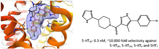
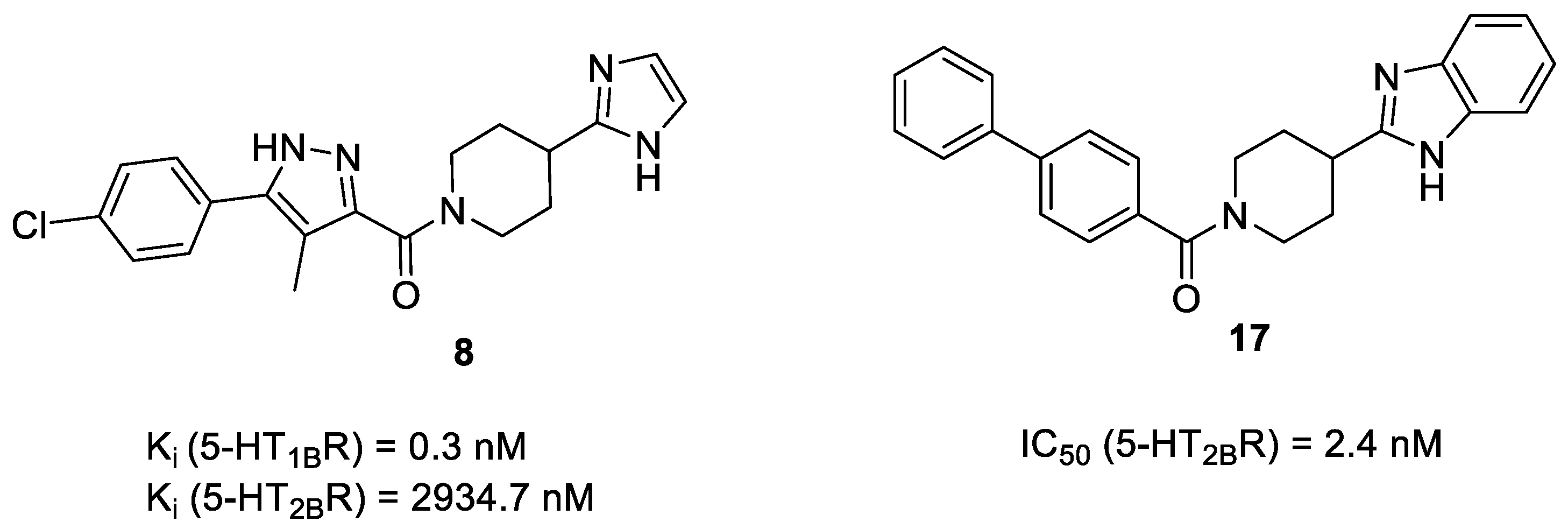
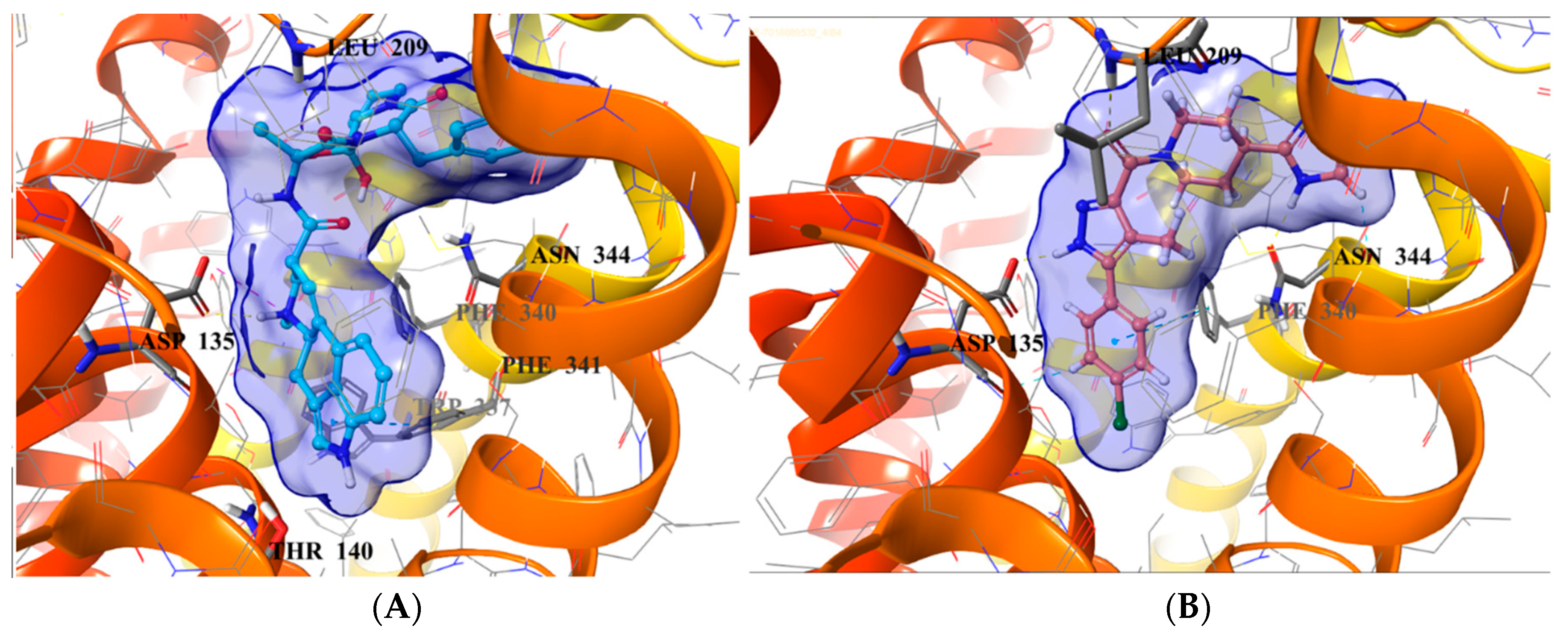
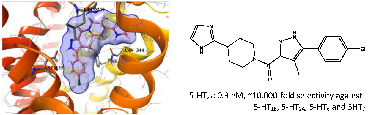
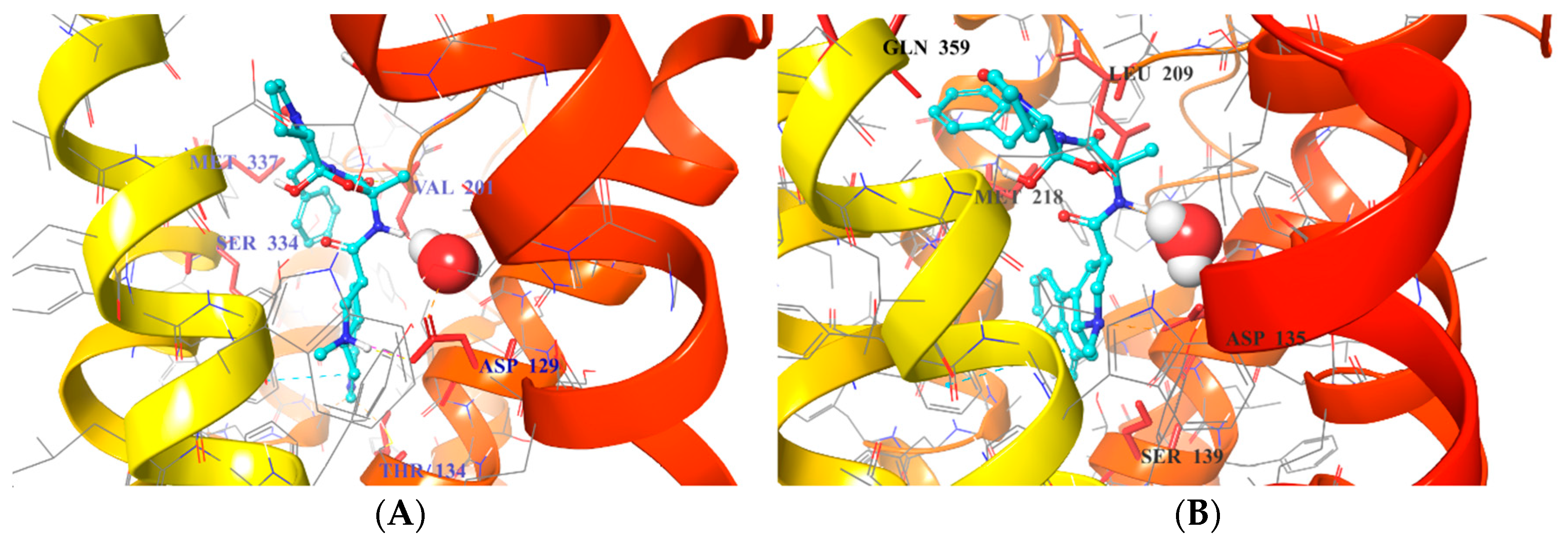
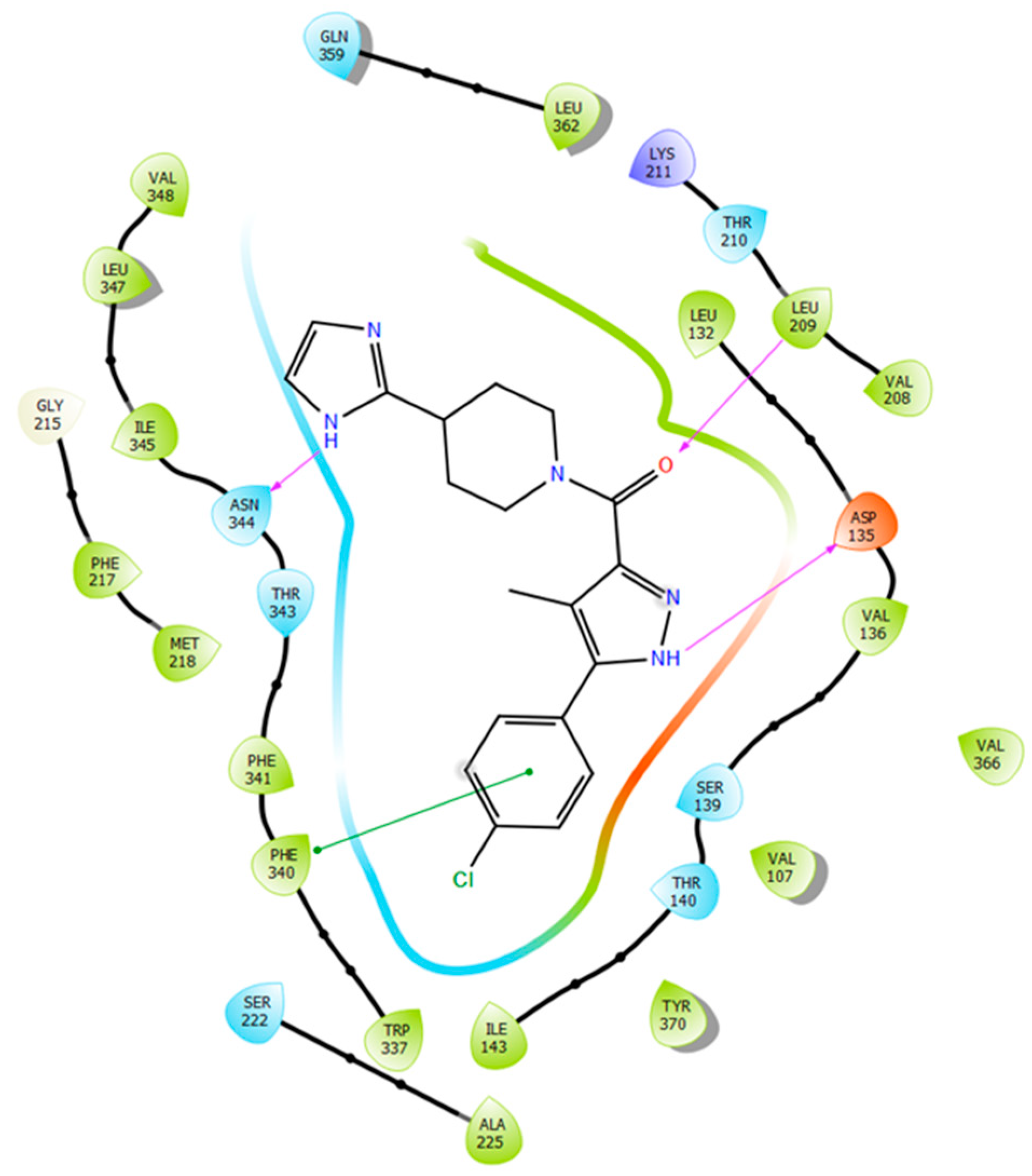
2.5. Binding Mode of Compound 8

The SBP of 5-HT2BR, which was originally occupied by the peptide portion of the ergoline ligand, is defined by several residues of ECL2, TM5, TM7, and TM6. The 4-chlorophenyl tail of 8 occupies the same orthosteric hydrophobic cavity formed by Phe3406.51, Phe3416.52, Ile1433.40, Trp3376.48, and is stacked by π–π interactions similarly to the indole ring of ergotamine (Figure 7).
Additionally, the imidazolyl–piperidine part of the ligand is supposed to reach toward the SBP, and form a hydrogen–bond interaction with the backbone oxo group (Val208) of the same hydrophobic cleft at ECL2, such as the peptide region of ergotamine (see Figure 8). Residues Asn3446.55 and Met2185.39 are narrowing the upper chamber of the 5-HT2BR binding pocket, compared with 5-HT1BR. It has been shown [21] that disturbance of the water-network around the Asn3446.55 is the consequence of ligand binding to this chamber. Interestingly, the imidazole NH of the ligand formed a polar–polar interaction with this residue.

3. Conclusions

In this study, we aimed identifying selective 5-HT2BR ligands by virtual screening. Given the sequential and structural similarities of serotonergic receptors, we hypothesized that receptor selectivity is basically driven from the SBP. Consequently, we developed a screening strategy combining both ligand-based and structure-based approaches. For the first level of our hierarchical approach, we used NSFP and developed machine learning tools to select potential 5-HT2BR selective molecules from a large database of druglike compounds. Next, this subset was subjected to docking calculations, and we identified compounds showing different interactions with the secondary binding pockets of 5-HT2BR and 5-HT1BR. Careful analysis of the binding mode allowed us to select nine compounds for biological testing. Out of these, three compounds showed significant 5-HT2BR affinity, one in the low micromolar (10), one in the submicromolar (9), and one in the subnanomolar range (8). Compounds with submicromolar 5-HT2BR affinity were further profiled against a set of serotonin receptors including 5-HT1AR, 5-HT2AR, 5-HT6R, and 5-HT7R. The best compound (8) showed Ki = 0.3 nM affinity and ten thousand-fold selectivity for 5-HT2BR, which nominates it for in vivo testing.

4. Materials and Methods

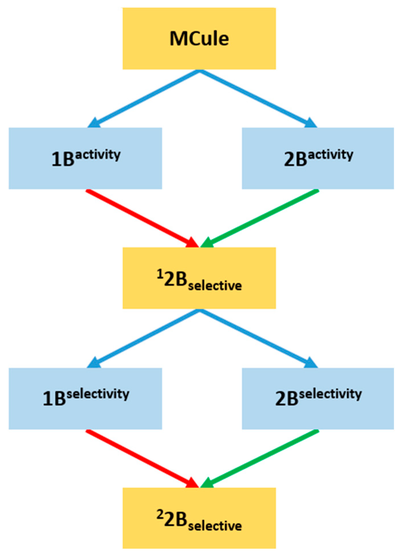
4.1. Procedures of Machine Learning-Based Classification Model Building

Basing on the KR-NSFP representations of compounds sets, four discrimination models were built using machine learning methods: 1Bactivity (based on 1Bactive and 1Binactive sets), 2Bactivity (using 2Bactive and 2Binactive sets), 1Bselectivity (using 1Bselective and nonselective sets), and 2Bselectivity (using 2Bselective and nonselective sets). For each case, a series of initial models was created, using various machine learning techniques including Support Vector Machines with Radial Basis Factor and Tanimoto kernels, Naïve Bayes and Extreme Entropy Machines with Tanimoto and Sorensen kernels [30,31]. Additionally, for each method–kernel pair, a set of hyperparameters was tested, resulting in 117 initial models acquired for each final classification model. The classifiers were tested using five-fold cross-validation methodology, and for each of the compound sets, the model with the highest MCC value (see Table 4) was selected for final classification study. The entire machine learning methodology was implemented in Python programming language using scikit-learn libraries.
The equation of the MCC values calculated for the ChEMBL training set is:
MCC ( TP , TN , FP , FN ) = TP × TN FP × FN ( TP + FP ) ( TP + FN ) ( TN + FP ) ( TN + FN )
where TP—True Positive, FP—False Positive, TN—True Negative, and FN—False Negative.

4.2. Docking-Based Evaluation of NSFP-Classified MCule Sets

4.2.1. Preparation of Ligand Sets, and Receptor Structures for Docking

Compounds used for selective ligand discovery were minimized using Schrödinger’s LigPrep algorithm [42], with the pH set to 7.4 and the number of stereoisomers set to 1. For this study, crystal structures of 5-HT1BR and 5-HT2BR were extracted from the PDB database [43] (PDB ID: 4IAQ and 4IAR [6] for 1B, and 4IB4 [7] and 4NC3 [8] for 2B). Proteins were prepared using PrepWiz software (version 2017-4) from Schrödinger [44] and transformed into a receptor grid using Schrödinger software package [45], with the binding pocket center set to the Asp3.32 residue. All of the docking procedures have been conducted using Schrödinger’s Glide software [46], with precision set to standard precision (SP) and varying the number of reported docking poses (5 or 10).

4.2.2. Evaluation of the Docking Results

The 22Bselective set was docked to the crystal structures of both 5-HT1B and 5-HT2B receptors (PDB ID: 4IAQ, 4IAR for 5-HT1BR and 4IB4, 4NC3 for 5-HT2BR, respectively), and the number of reported poses was set to 10. Additionally, poses were filtered for interactions with the SBP characteristic amino acids (see Table 5).
Scoring of reported poses was performed using custom scoring function [39], taking into consideration the rankings of compounds based on the assigned Glide docking score. It compares the numerical sum of the ranks of all of the poses acquired by a compound in both crystals of the target protein with those found in both crystals of the antitarget:
Δmax = [∑(ranktarget) − ∑(rankantitarget)]
Since better rankings have lower numerical values (a pose with rank 1 is better than a pose with rank 5), if the compound’s Δmax < 0, it was considered target-selective. For further research, the compounds with the lowest Δmax were considered, as the difference in ranks between target and antitarget were the highest. Finally, the docking binding modes of the top 10% (18 ligands) were visually inspected, and five compounds (8, 10, 11, 12, and 13 marked with “a” in Table 2) were hand-picked for in vitro validation.
The second approach included screening for interactions with water molecules buried within the binding pockets of target receptors. Of all four crystal structures of the receptors, only two contained water within the three-dimensional (3D) model: 4IAQ (1B) [6] and 4IB4 (2B) [7]. Therefore, these two structures were used in a second docking study conducted with constraints set for either a hydrogen bond/salt bridge to Asp3.32, or a hydrogen bond with water 2020 (4IAQ) or 2004 (4IB4). Docked compounds were filtered for proper interactions with the water molecule/Asp3.32 residue, and their specificity. The binding mode of the filtered compounds was checked manually, and they were subjected to a novelty check by PubChem [40] before testing with the NSFP.
The 900 pose-filtered docked ligands were first clustered by structural similarity, and a diverse set of 50 cluster centroids were subjected to novelty check using the PubChem engine. Compounds having no similar compounds or no tested analogues, or having tested but inactive analogues, were prioritized. This process yielded 24 ligands in total, out of which four compounds were selected (9, 14, 15, and 16, marked with “b” in Table 2) after visual inspection for in vitro testing.

4.3. Procedures of In Vitro Screening Assays

All of the tested compounds were purchased via MCule, Inc. (Palo Alto, CA, USA). The compounds were tested for their ability to displace radioligand from the membrane of 5-HT1BR and 5-HT2BR-expressing cells. The first test was performed with a 10-µM concentration of a compound, and the inhibition percentage was calculated. If the value was above 50%, a full affinity screening was performed, and the data was gathered as Ki with units reported as nM.

4.3.1. Competition Binding in Human 5-HT1B Receptor

Serotonin 5-HT1BR competition binding experiments were carried out in a polypropylene 96-well plate. In each well, 5 μg of membranes from a Hela-5-HT1B cell line that was prepared in our laboratory (Lot: A001/14-11-2011, protein concentration = 3179 μg/mL), 1.5 nM [3H]-GR125743 (83.9 Ci/mmol, 0.1 mCi/mL, Perkin Elmer NET1172100UC, KD = 0.74 nM), the studied compounds and the reference compound were incubated. Non-specific binding was determined in the presence of GR55562 10 μM (TOCRIS 1054), and total binding was determined in the absence of any unlabeled compound. The reaction mixture (Vt: 250 μL/well) was incubated at 25 °C for 90 min. Then, 200 μL was transferred to a GF/C 96-well plate (Millipore, Madrid, Spain) pretreated with 0.5% of PEI and treated with binding buffer (Tris-HCl 50 mM, EDTA 1 mM, MgCl2 10 mM, pH = 7.4). Afterwards, it was filtered and washed four times with 250 μL of wash buffer (Tris-HCl 50 mM, pH = 7.4), before measuring in a microplate beta scintillation counter (Microbeta Trilux, PerkinElmer, Madrid, Spain). 5-carboxytryptamine was included as a reference compound in all of the assays. Compounds were first tested at 10 micromolar; those compounds showing a percentage of displacement of specific binding higher than 50% were classified as active compounds, and Ki values were determined by means of concentration-response curves.

4.3.2. Competition Binding in Human 5-HT2B Receptor

Serotonin 5-HT2BR competition binding experiments were carried out in a polypropylene 96-well plate. In each well, 5 μg of membranes from a CHO-5-HT2B cell line prepared in our laboratory (Lot: A003/27-03-2012, protein concentration = 3431 μg/mL), 1 nM [3H]-LSD (82.4 Ci/mmol, 1 mCi/mL, PerkinElmer NET638250UC, KD = 0.57 nM), the studied compounds and the reference compound were incubated. Non-specific binding was determined in the presence of 5-HT 50 μM (Sigma H9523), and total binding was determined in the absence of any unlabeled compound. The reaction mixture (Vt: 250 μL/well) was incubated at 37 °C for 30 min. Then, 200 μL was transferred to a GF/C 96-well plate (Millipore, Madrid, Spain) pretreated with 0.5% of PEI and treated with binding buffer (Tris-HCl 50 mM, Ascorbic acid 0.1%, CaCl2 4 mM, pH = 7.4). Afterwards, it was filtered and washed four times with 250 μL of wash buffer (Tris-HCl 50 mM, pH = 7.4) before measuring in a microplate beta scintillation counter (Microbeta Trilux, PerkinElmer, Madrid, Spain). Methysergide was included as a reference compound in all of the assays. Compounds were first tested at 10 micromolar, and those compounds showing a percentage of displacement of specific binding higher than 50% were classified as active compounds, and Ki values were determined by means of concentration-response curves.

4.3.3. Competition Binding in Human 5-HT1A, 5-HT2A 5-HT6 and 5-HT7 Receptors

HEK293 cells with stable expression of human serotonin 5-HT1AR, 5-HT2A, 5-HT6, or 5-HT7b receptor (all prepared with the use of Lipofectamine 2000) were maintained at 37 °C in a humidified atmosphere with 5% CO2, and were grown in Dulbeco’s Modifier Eagle Medium containing 10% dialyzed fetal bovine serum and 500 mg/mL G418 sulfate. For the preparation of membranes, cells were subcultured in 10-cm diameter dishes, grown to 90% confluence, washed twice with prewarmed to 37 °C phosphate-buffered saline (PBS), and were pelleted by centrifugation (200 g) in PBS containing 0.1 mM of EDTA and 1 mM of dithiothreitol. Prior to membrane preparations, pellets were stored at −80 °C.
Cell pellets were thawed and homogenized in 20 volumes of assay buffer using an Ultra Turrax tissue homogenizer and centrifuged twice at 35,000 g for 20 min at 4 °C, with incubation for 15 min at 37 °C in between. The composition of the assay buffers was as follows: for 5-HT1AR: 50 mM Tris-HCl, 0.1 mM EDTA, 4 mM MgCl2, 10 µM pargyline, and 0.1% ascorbate; for 5-HT2AR: 50 mM Tris-HCl, 0.1 mM EDTA, 4 mM MgCl2, and 0.1% ascorbate; for 5-HT6R: 50 mM Tris-HCl, 0.5 mM EDTA, and 4 mM MgCl2, for 5-HT7bR: 50 mM Tris-HCl, 4 mM MgCl2, 10 µM pargyline, and 0.1% ascorbate. All of the assays were incubated in a total volume of 200 µL in 96-well microtiter plates for 1 h at 37 °C, except for 5-HT1AR and 5-HT2AR, which were incubated at room temperature for 1 h and 1.5 h, respectively. The process of equilibration was terminated by rapid filtration through Unifilter plates with a 96-well cell harvester, and the radioactivity retained on the filters was quantified on a Microbeta plate reader (PerkinElmer, Waltham, MA, USA).
For displacement studies, the assay samples contained as radioligands: 1.5 nM of [3H]-8-OH-DPAT (135.2 Ci/mmol, KD = 1.5 nM) for 5-HT1AR; 2 nM of [3H]-Ketanserin (53.4 Ci/mmol, KD = 2 nM) for 5-HT2AR; 2 nM of [3H]-LSD (83.6 Ci/mmol, KD = 2 nM) for 5-HT6R, or 0.6 nM of [3H]-5-CT (39.2 Ci/mmol, KD = 0.6 nM) for 5-HT7R. Non-specific binding is defined with 10 µM of 5-HT in 5-HT1AR and 5-HT7R binding experiments, whereas 10 µM of chlorpromazine or 10 µM of methiothepin were used in 5-HT2AR and 5-HT6R assays, respectively. Each compound was tested in triplicate at seven concentrations (10−10–10−4 M). Reference compounds for 5-HT1AR: buspirone Ki = 32.2 nM ± 2.9; for 5-HT2AR: olanzapine Ki = 5.6 nM ± 1.1; for 5-HT6R: olanzapine Ki = 8.8 nM ± 1.3; and for 5-HT7R: 5-CT Ki = 0.8 nM ± 0.2. The inhibition constants (Ki) were calculated from the Cheng-Prusoff equation [47]. Results were expressed as means of at least two separate experiments.

Author Contributions

G.M.K. and A.J.B. conceived the story; K.R. performed and evaluated the fingerprint-based machine learning model; Á.A.K. performed and evaluated the docking experiments; J.B., M.I.L. and S.P. performed the in vitro assay measurements.

Funding

Á.A.K. and G.M.K. were supported by the National Brain Research Program (2017-1.2.1-NKP-2017-00002). K.R. is grateful for the ETIUDA scholarship of the National Science Center, Poland. J.B. and M.I.L. are grateful for the support from the Spanish Ministerio de Economía y Comptetitividad (SAF2017-85225-C3-1-R).

Acknowledgments

Authors are grateful to Grzegorz Satała for testing compounds affinity for the extended panel of serotonin receptors.

Conflicts of Interest

The authors declare no conflict of interest.

References

  1. McCorvy, J.D.; Roth, B.L. Structure and function of serotonin G protein-coupled receptors. Pharmacol. Ther. 2015, 150, 129–142. [Google Scholar] [CrossRef] [PubMed]
  2. Kitson, S.L. 5-hydroxytryptamine (5-HT) receptor ligands. Curr. Pharm. Des. 2007, 13, 2621–2637. [Google Scholar] [CrossRef] [PubMed]
  3. Costanzi, S.; Siegel, J.; Tikhonova, I.; Jacobson, K. Rhodopsin and the Others: A Historical Perspective on Structural Studies of G Protein-Coupled Receptors. Curr. Pharm. Des. 2009, 15, 3994–4002. [Google Scholar] [CrossRef] [PubMed]
  4. Palczewski, K.; Kumasaka, T.; Hori, T.; Behnke, C.A.; Motoshima, H.; Fox, B.A.; Le Trong, I.; Teller, D.C.; Okada, T.; Stenkamp, R.E.; et al. Crystal structure of rhodopsin: A G protein-coupled receptor. Science 2000, 289, 739–745. [Google Scholar] [CrossRef] [PubMed]
  5. Cherezov, V.; Rosenbaum, D.M.; Hanson, M.A.; Rasmussen, S.G.F.; Foon, S.T.; Kobilka, T.S.; Choi, H.J.; Kuhn, P.; Weis, W.I.; Kobilka, B.K.; et al. High-resolution crystal structure of an engineered human β2-adrenergic G protein-coupled receptor. Science 2007, 318, 1258–1265. [Google Scholar] [CrossRef] [PubMed]
  6. Wang, C.; Jiang, Y.; Ma, J.; Wu, H.; Wacker, D.; Katritch, V.; Han, G.W.; Liu, W.; Huang, X.-P.; Vardy, E.; et al. Structural basis for molecular recognition at serotonin receptors. Science 2013, 340, 610–614. [Google Scholar] [CrossRef] [PubMed]
  7. Wacker, D.; Wang, C.; Katritch, V.; Han, G.W.; Huang, X.-P.; Vardy, E.; McCorvy, J.D.; Jiang, Y.; Chu, M.; Siu, F.Y.; et al. Structural Features for Functional Selectivity at Serotonin Receptors. Science 2013, 340, 615–619. [Google Scholar] [CrossRef] [PubMed]
  8. Liu, W.; Wacker, D.; Gati, C.; Han, G.W.; James, D.; Wang, D.; Nelson, G.; Weierstall, U.; Katritch, V.; Barty, A.; et al. Serial Femtosecond Crystallography of G Protein–Coupled Receptors. Science 2013, 342, 1521–1524. [Google Scholar] [CrossRef] [PubMed]
  9. Wacker, D.; Wang, S.; McCorvy, J.D.; Betz, R.M.; Venkatakrishnan, A.J.; Levit, A.; Lansu, K.; Schools, Z.L.; Che, T.; Nichols, D.E.; et al. Crystal Structure of an LSD-Bound Human Serotonin Receptor. Cell 2017, 168, 377–389. [Google Scholar] [CrossRef] [PubMed]
  10. Chien, E.Y.T.; Liu, W.; Zhao, Q.; Katritch, V.; Han, G.W.; Hanson, M.A.; Shi, L.; Newman, A.H.; Javitch, J.A.; Cherezov, V.; et al. Structure of the human dopamine D3 receptor in complex with a D2/D3 selective antagonist. Science 2010, 330, 1091–1095. [Google Scholar] [CrossRef] [PubMed]
  11. Kruse, A.C.; Hu, J.; Pan, A.C.; Arlow, D.H.; Rosenbaum, D.M.; Rosemond, E.; Green, H.F.; Liu, T.; Chae, P.S.; Dror, R.O.; et al. Structure and dynamics of the M3 muscarinic acetylcholine receptor. Nature 2012, 482, 552–556. [Google Scholar] [CrossRef] [PubMed]
  12. Rovati, G.E.; Capra, V.; Neubig, R.R.; Smit, M.J.; Roovers, E.; Cotecchia, S.; Leurs, R. The highly conserved DRY motif of class A G protein-coupled receptors: Beyond the ground state. Mol. Pharmacol. 2007, 71, 959–964. [Google Scholar] [CrossRef] [PubMed]
  13. Nygaard, R.; Zou, Y.; Dror, R.O.; Mildorf, T.J.; Arlow, D.H.; Manglik, A.; Pan, A.C.; Liu, C.W.; Fung, J.J.; Bokoch, M.P.; et al. The dynamic process of β2-adrenergic receptor activation. Cell 2013, 152, 532–542. [Google Scholar] [CrossRef] [PubMed]
  14. Dror, R.O.; Pan, A.C.; Arlow, D.H.; Borhani, D.W.; Maragakis, P.; Shan, Y.; Xu, H.; Shaw, D.E. Pathway and mechanism of drug binding to G-protein-coupled receptors. Proc. Natl. Acad. Sci. USA 2011, 108, 13118–13123. [Google Scholar] [CrossRef] [PubMed]
  15. Rasmussen, S.G.F.; Choi, H.-J.; Rosenbaum, D.M.; Kobilka, T.S.; Thian, F.S.; Edwards, P.C.; Burghammer, M.; Ratnala, V.R.P.; Sanishvili, R.; Fischetti, R.F.; et al. Crystal structure of the human β2 adrenergic G-protein-coupled receptor. Nature 2007, 450, 383–387. [Google Scholar] [CrossRef] [PubMed]
  16. Michino, M.; Beuming, T.; Donthamsetti, P.; Newman, A.H.; Javitch, J.A.; Shi, L. What Can Crystal Structures of Aminergic Receptors Tell Us about Designing Subtype-Selective Ligands? Pharmacol. Rev. 2014, 67, 198–213. [Google Scholar] [CrossRef] [PubMed]
  17. Johnson, K.W.; Phebus, L.A.; Cohen, M.L. Serotonin in migraine: Theories, animal models and emerging therapies. Prog. Drug Res. 1998, 51, 219–244. [Google Scholar] [CrossRef] [PubMed]
  18. Fozard, J.R.; Kalkman, H.O. 5-Hydroxytryptamine (5-HT) and the initiation of migraine: New perspectives. Naunyn-Schmiedebergs Arch. Pharmacol. 1994, 350, 225–229. [Google Scholar] [CrossRef] [PubMed]
  19. Schmuck, K.; Ullmer, C.; Kalkman, H.O.; Probst, A.; Lübbert, H. Activation of Meningeal 5-HT2B Receptors: An Early Step in the Generation of Migraine Headache? Eur. J. Neurosci. 1996, 8, 959–967. [Google Scholar] [CrossRef] [PubMed]
  20. Janssen, W.; Schymura, Y.; Novoyatleva, T.; Kojonazarov, B.; Boehm, M.; Wietelmann, A.; Luitel, H.; Murmann, K.; Krompiec, D.R.; Tretyn, A.; et al. 5-HT2B receptor antagonists inhibit fibrosis and protect from RV heart failure. Biomed. Res. Int. 2015, 2015, 438403. [Google Scholar] [CrossRef] [PubMed]
  21. Zhou, Y.; Ma, J.; Lin, X.; Huang, X.-P.; Wu, K.; Huang, N. Structure-Based Discovery of Novel and Selective 5-Hydroxytryptamine 2B Receptor Antagonists for the Treatment of Irritable Bowel Syndrome. J. Med. Chem. 2016, 59, 707–720. [Google Scholar] [CrossRef] [PubMed]
  22. Bonhaus, D.W.; Flippin, L.A.; Greenhouse, R.J.; Jaime, S.; Rocha, C.; Dawson, M.; Van Natta, K.; Chang, L.K.; Pulido-Rios, T.; Webber, A.; et al. RS-127445: A selective, high affinity, orally bioavailable 5-HT2B receptor antagonist. Br. J. Pharmacol. 1999, 127, 1075–1082. [Google Scholar] [CrossRef] [PubMed]
  23. Thomas, D.R.; Gager, T.L.; Holland, V.; Brown, A.M.; Wood, M.D. m-Chlorophenylpiperazine (mCPP) is an antagonist at the cloned human 5-HT2B receptor. Neuroreport 1996, 7, 1457–1460. [Google Scholar] [CrossRef] [PubMed]
  24. Kovács, A.; Gacsályi, I.; Wellmann, J.; Schmidt, É.; Szücs, Z.; Dubreuil, V.; Nicolas, J.P.; Boutin, J.; Bózsing, D.; Egyed, A.; et al. Effects of EGIS-7625, a Selective and Competitive 5-HT2B Receptor Antagonist. Cardiovasc. Drugs Ther. 2003, 17, 427–434. [Google Scholar] [CrossRef] [PubMed]
  25. Audia, J.E.; Evrard, D.A.; Murdoch, G.R.; Droste, J.J.; Nissen, J.S.; Schenck, K.W.; Fludzinski, P.; Lucaites, V.L.; Nelson, D.L.; Cohen, M.L. Potent, selective tetrahydro-β-carboline antagonists of the serotonin 2B (5HT(2B)) contractile receptor in the rat stomach fundus. J. Med. Chem. 1996, 39, 2773–2780. [Google Scholar] [CrossRef] [PubMed]
  26. Forbes, I.T.; Kennett, G.A.; Gadre, A.; Ham, P.; Hayward, C.J.; Martin, R.T.; Thompson, M.; Wood, M.D.; Baxter, G.S. N-(1-Methyl-5-indolyl)-N’-(3-pyridyl)urea hydrochloride: The first selective 5-HT1C receptor antagonist. J. Med. Chem. 1993, 36, 1104–1107. [Google Scholar] [CrossRef] [PubMed]
  27. Reid, T.-E.; Kumar, K.; Wang, X. Predictive In Silico Studies of Human 5-hydroxytryptamine Receptor Subtype 2B (5-HT2B) and Valvular Heart Disease. Curr. Top. Med. Chem. 2013, 13, 1353–1362. [Google Scholar] [CrossRef] [PubMed]
  28. Lacivita, E.; Podlewska, S.; Speranza, L.; Niso, M.; Satała, G.; Perrone, R.; Perrone-Capano, C.; Bojarski, A.J.; Leopoldo, M. Structural modifications of the serotonin 5-HT7 receptor agonist N-(4-cyanophenylmethyl)-4-(2-biphenyl)-1-piperazinehexanamide (LP-211) to improve in vitro microsomal stability: A case study. Eur. J. Med. Chem. 2016, 120, 363–379. [Google Scholar] [CrossRef] [PubMed]
  29. Rataj, K.; Czarnecki, W.; Podlewska, S.; Bojarski, A.J. Substructural Connectivity Fingerprint and Extreme Entropy Machines—A New Method of Compound Representation and Analysis. Molecules 2018. submitted. [Google Scholar]
  30. Czarnecki, W.M. Weighted Tanimoto Extreme Learning Machine with Case Study in Drug Discovery. IEEE Comput. Intell. Mag. 2015, 10, 19–29. [Google Scholar] [CrossRef]
  31. Cortes, C.; Vapnik, V. Support-vector networks. Mach. Learn. 1995, 20, 273–297. [Google Scholar] [CrossRef]
  32. Klekota, J.; Roth, F.P. Chemical substructures that enrich for biological activity. Bioinformatics 2008, 24, 2518–2525. [Google Scholar] [CrossRef] [PubMed]
  33. Vass, M.; Ágai-Csongor, É.; Horti, F.; Keserű, G.M. Multiple Fragment Docking and Linking in Primary and Secondary Pockets of Dopamine Receptors. ACS Med. Chem. Lett. 2014, 5, 1010–1014. [Google Scholar] [CrossRef] [PubMed] [Green Version]
  34. Bento, A.P.; Gaulton, A.; Hersey, A.; Bellis, L.J.; Chambers, J.; Davies, M.; Krüger, F.A.; Light, Y.; Mak, L.; McGlinchey, S.; et al. The ChEMBL bioactivity database: An update. Nucleic Acids Res. 2014, 42, D1083–D1090. [Google Scholar] [CrossRef] [PubMed]
  35. Kiss, R.; Sandor, M.; Szalai, F.A. http://Mcule.com: A public web service for drug discovery. J. Cheminform. 2012, 4, P17. [Google Scholar] [CrossRef]
  36. Ballesteros, J.A.; Weinstein, H. [19] Integrated methods for the construction of three-dimensional models and computational probing of structure-function relations in G protein-coupled receptors. Methods Neurosci. 1995, 25, 366–428. [Google Scholar] [CrossRef]
  37. Kristiansen, K.; Kroeze, W.K.; Willins, D.L.; Gelber, E.I.; Savage, J.E.; Glennon, R.A.; Roth, B.L. A highly conserved aspartic acid (Asp-155) anchors the terminal amine moiety of tryptamines and is involved in membrane targeting of the 5-HT(2A) serotonin receptor but does not participate in activation via a “salt-bridge disruption” mechanism. J. Pharmacol. Exp. Ther. 2000, 293, 735–746. [Google Scholar] [PubMed]
  38. Michino, M.; Beuming, T.; Donthamsetti, P.; Newman, A.H.; Javitch, J.A.; Shi, L.; Molinoff, P.B.; Ruffolo, R.R.; Trendelenburg, U. What can crystal structures of aminergic receptors tell us about designing subtype-selective ligands? Pharmacol. Rev. 2015, 67, 198–213. [Google Scholar] [CrossRef] [PubMed]
  39. Kelemen, Á.A.; Kiss, R.; Ferenczy, G.G.; Kovács, L.; Flachner, B.; Lőrincz, Z.; Keserű, G.M. Structure-Based Consensus Scoring Scheme for Selecting Class A Aminergic GPCR Fragments. J. Chem. Inf. Model. 2016, 56, 412–422. [Google Scholar] [CrossRef] [PubMed]
  40. The PubChem Project. Available online: https://pubchem.ncbi.nlm.nih.gov/ (accessed on 24 Janunary 2018).
  41. Moss, N.; Choi, Y.; Cogan, D.; Flegg, A.; Kahrs, A.; Loke, P.; Meyn, O.; Nagaraja, R.; Napier, S.; Parker, A.; et al. A new class of 5-HT2B antagonists possesses favorable potency, selectivity, and rat pharmacokinetic properties. Bioorgan. Med. Chem. Lett. 2009, 19, 2206–2210. [Google Scholar] [CrossRef] [PubMed]
  42. Schrödinger Release 2017-4: LigPrep; Schrödinger, LLC: New York, NY, USA, 2017.
  43. Berman, H.M. The Protein Data Bank. Nucleic Acids Res. 2000, 28, 235–242. [Google Scholar] [CrossRef] [PubMed]
  44. Schrödinger Release 2017-4: Schrödinger Suite 2017-4 Protein Preparation Wizard; Schrödinger, LLC: New York, NY, USA, 2017.
  45. Small-Molecule Drug Discovery Suite 2017-4; Schrödinger, LLC: New York, NY, USA, 2017.
  46. Schrödinger Release 2017-4: Glide; Schrödinger, LLC: New York, NY, USA, 2017.
  47. Yung-Chi, C.; Prusoff, W.H. Relationship between the inhibition constant (KI) and the concentration of inhibitor which causes 50 per cent inhibition (I50) of an enzymatic reaction. Biochem. Pharmacol. 1973, 22, 3099–3108. [Google Scholar] [CrossRef]
Sample Availability: Samples of the tested compounds are available from commercial vendors.
Figure 1. Representative 5-HT2BR ligands (compounds 13 with available 5-HT1BR and 5-HT2BR binding affinity data, and examples of clinical candidates 47).
Molecules 23 01137 g001
Figure 2. Depiction of Neighbouring Substructures Fingerprint (NSFP) representations of constructed compound sets (orange) and machine learning models (blue).
Molecules 23 01137 g002
Figure 3. Classification procedure for putative 2B selective compounds. Green arrows represent positive classification and red arrows represent negative classification.
Molecules 23 01137 g003
Figure 4. Flowchart of the virtual screening cascade used for the identification of 5-HT2B selective hits.
Molecules 23 01137 g004
Figure 5. Residues (red) used in pose filtering for 5-HT1BR (PDB ID: 4IAQ) panel (A) and 5-HT2BR X-ray structures (PDB ID: 4IB4) panel (B); Water molecules forming interactions with the Asp3.32 shown as a sphere representation.
Molecules 23 01137 g005
Figure 6. Structure of hit compound (8) and its closest literature analogue (17).
Molecules 23 01137 g006
Figure 7. Two-dimensional (2D) interaction diagram of MCULE-7016689532 (8) in the 5-HT2BR.
Molecules 23 01137 g007
Figure 8. Binding site surfaces (blue spheres) of ergotamine panel (A) and 8 panel (B) in the 5-HT2BR crystal structure. Important ligand-contacting residues are shown in thick tube representation.
Molecules 23 01137 g008
Table 1. Active, inactive, and selective 5-HT1B and 5-HT2B ligands retrieved from the ChEMBL with at least 22 heavy atoms.
ReceptorNumber of Actives 1Number of Inactives 2Number of Selectives 3
5-HT1B858 (1011)339 (477)86
5-HT2B478 (718)259 (351)33
1 Ki ≤ 500 nM; 2 Ki ≥ 1000 nM; 3 Classified as 5-HT2B selective for being active at 5-HT2BR, and inactive at 5-HT1BR; numbers in parenthesis are the total number of all actives without size-filtering.
Table 2. Structures and measured in vitro assay data of top-ranked compounds (sorted by percentage of inhibition and Ki (nM) in h5-HT2BR competition binding assay). Values represent the mean ± SD of three independent assays with duplicate measurements.
CompoundID5-HT1B (%) 15-HT2B (%) 15-HT1B (nM) 25-HT2B (nM) 2
Molecules 23 01137 i0018a--2934.7 ± 321.30.3 ± 0.07
Molecules 23 01137 i0029b--2099 ± 622.2235.1 ± 15.9
Molecules 23 01137 i00310a7 ± 4--2612.9 ± 422.1
Molecules 23 01137 i00411a12 ± 440 ± 3--
Molecules 23 01137 i00512a7 ± 136 ± 4--
Molecules 23 01137 i00613a19 ± 334 ± 4--
Molecules 23 01137 i00714b26 ± 434 ± 4--
Molecules 23 01137 i00815b28 ± 331 ± 4--
Molecules 23 01137 i00916b1 ± 124 ± 4--
1 Percentage of inhibition measured at 10 µM concentration; 2 Ki measured (reported in nM units) if percentage of inhibition exceeded 50%; a Selection not accounting waters, b Selection accounting waters.
Table 3. 5-HT panel screening of the best hits. Values represent the mean ± SD of three independent assays with duplicate measurements.
CompoundID5-HT1A Ki (µM)5-HT2A Ki (µM)5-HT6 Ki (µM)5-HT7 Ki (µM)
Molecules 23 01137 i0108>20>2013.2 ± 1.5>20
Molecules 23 01137 i0119>20>207.5 ± 0.9>20
Table 4. Performance data of the activity and selectivity filters obtained on ChEMBL training set.
ReceptorMCC of Activity ClassifiersMCC of Selectivity Classifiers
5-HT1B0.78670.8057
5-HT2B0.73760.8238
Table 5. All of the interactions considered during the pose filtering phase. The blue cells represent hydrogen bonds, and the red cells represent distance criterion (≤5.0 Å between the compound and any atom of a residue). OBP: orthosteric binding pocket; SBP: secondary binding pocket.
Interactions1B Crystals2B Crystals
OBPD1293.32D1353.32
T1343.37S1393.36
S2125.42
SBP Q3597.32
M3376.58M2185.39
V201ECL2.52L209ECL2.52

