UFLC-Q-TOF-MS/MS-Based Screening and Identification of Flavonoids and Derived Metabolites in Human Urine after Oral Administration of Exocarpium Citri Grandis Extract

 , , , ,
Abstract
:1. Introduction
2. Results and Discussion
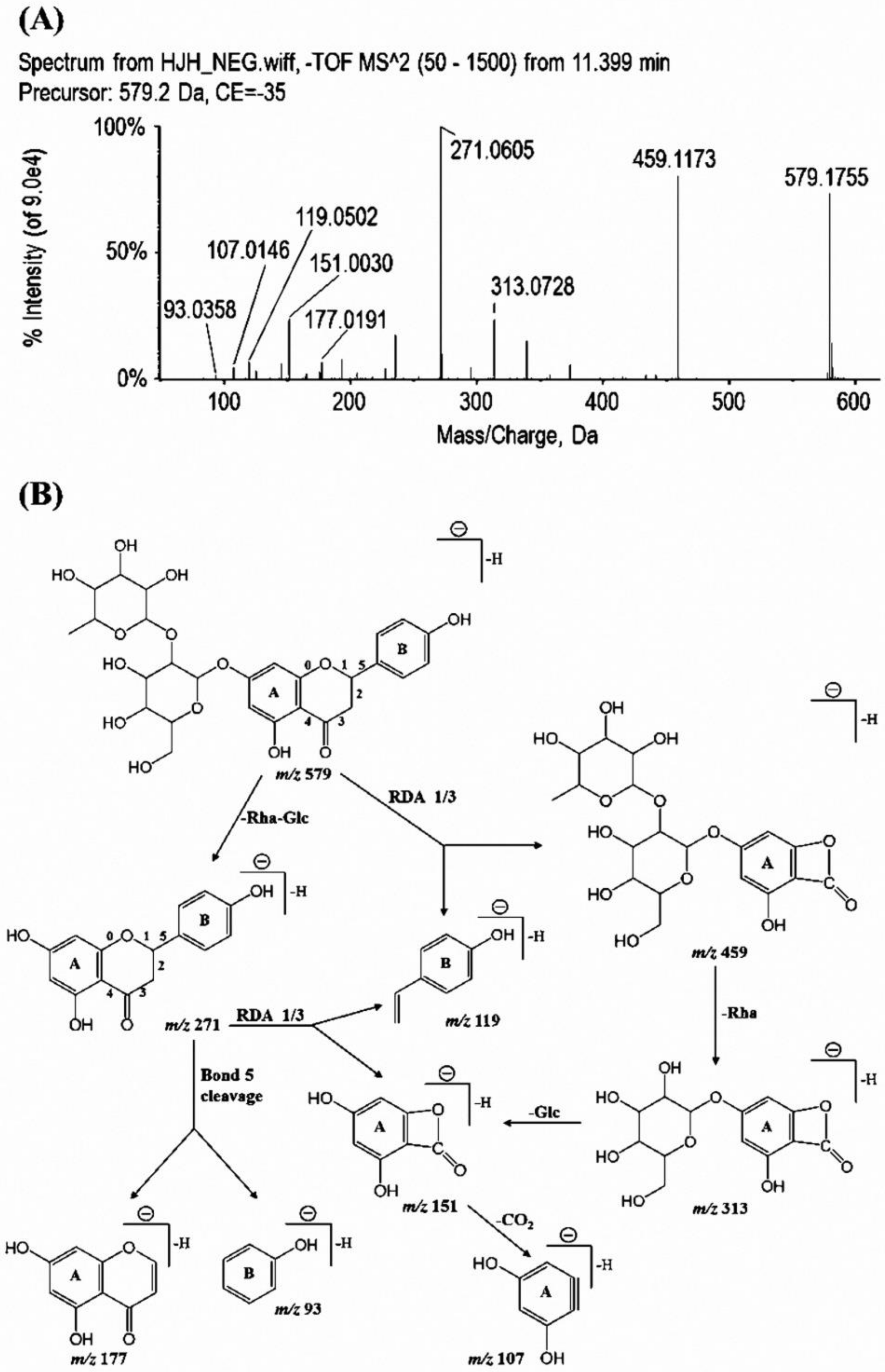
2.1. Identification and Quantification of Flavonoids in ECG Extract
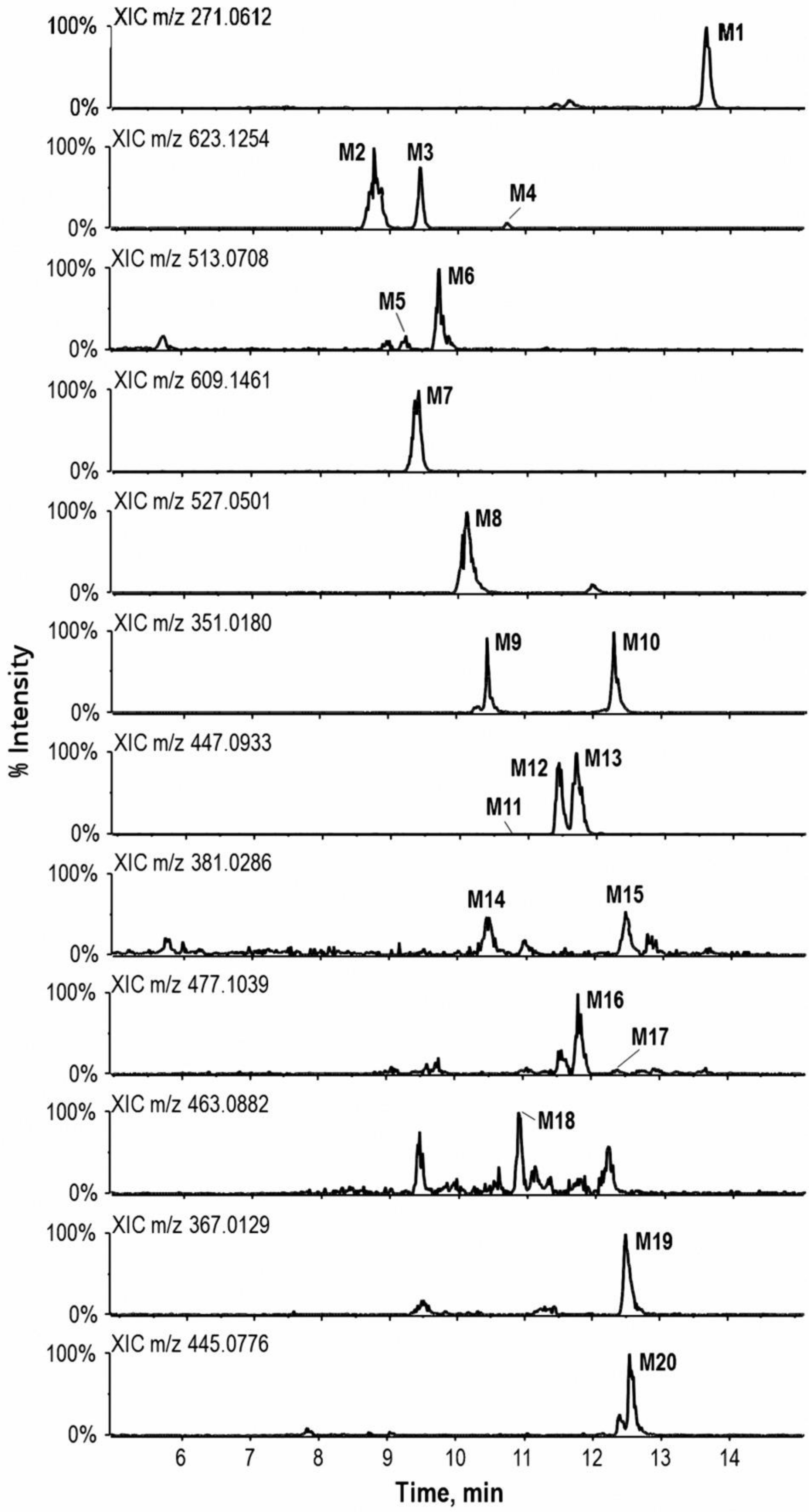
2.2. Identification and Quantification of Metabolites in Urine
3. Experimental
3.1. Chemicals and Reagents
3.2. Preparation of ECG Extract
3.3. Study Design
3.4. Sample Preparation
3.5. UFLC-Q-TOF-MS/MS Analysis
4. Conclusions
Supplementary Materials
Acknowledgments
Author Contributions
Conflicts of Interest
References
- Cushnie, T.P.; Lamb, A.J. Recent advances in understanding the antibacterial properties of flavonoids. Int. J. Antimicrob. Agents 2011, 38, 99–107. [Google Scholar] [CrossRef] [PubMed]
- Friedman, M. Overview of antibacterial, antitoxin, antiviral, and antifungal activities of tea flavonoids and teas. Mol. Nutr. Food Res. 2007, 51, 116–134. [Google Scholar] [CrossRef] [PubMed]
- Witkowska-Banaszczak, E. Flavonoids from Trollius europaeus flowers and evaluation of their biological activity. J. Pharm. Pharmacol. 2018, 70, 550–558. [Google Scholar] [CrossRef] [PubMed]
- Chalet, C.; Hollebrands, B.; Janssen, H.G.; Augustijns, P.; Duchateau, G. Identification of phase-II metabolites of flavonoids by liquid chromatography-ion-mobility spectrometry-mass spectrometry. Anal. Bioanal. Chem. 2017, 410, 471–482. [Google Scholar] [CrossRef] [PubMed]
- Du, L.Y.; Qian, D.W.; Shang, E.X.; Liu, P.; Jiang, S.; Guo, J.M.; Su, S.L.; Duan, J.A.; Xu, J.; Zhao, M. UPLC-Q-TOF/MS-based screening and identification of the main flavonoids and their metabolites in rat bile, urine and faeces after oral administration of Scutellaria baicalensis extract. J. Ethnopharmacol. 2015, 169, 156–162. [Google Scholar] [CrossRef] [PubMed]
- Gad, M.H.; Tuenter, E.; El-Sawi, N.; Younes, S.; El-Ghadban, E.M.; Demeyer, K.; Pieters, L.; Heyden, Y.V.; Mangelings, D. Identification of some Bioactive Metabolites in a Fractionated Methanol Extract from Ipomoea aquatica (Aerial Parts) through TLC, HPLC, UPLC-ESI-QTOF-MS and LC-SPE-NMR Fingerprints Analyses. Phytochem. Anal. 2018, 29, 5–15. [Google Scholar]
- Yuan, Y.; Long, P.; Jiang, C.; Li, M.; Huang, L. Development and characterization of simple sequence repeat (SSR) markers based on a full-length cDNA library of Scutellaria baicalensis. Genomics 2015, 105, 61–67. [Google Scholar] [CrossRef] [PubMed]
- Li, P.; Liu, M.; Hu, J.; Su, W. Systematic chemical profiling of Citrus grandis ‘Tomentosa’ by ultra-fast liquid chromatography/diode-array detector/quadrupole time-of-flight tandem mass spectrometry. J. Pharm. Biomed. Anal. 2014, 90, 167–179. [Google Scholar] [CrossRef] [PubMed]
- Liu, M.; Zou, W.; Yang, C.; Peng, W.; Su, W. Metabolism and excretion studies of oral administered naringin, a putative antitussive, in rats and dogs. Biopharm. Drug Dispos. 2012, 33, 123–134. [Google Scholar] [CrossRef] [PubMed]
- Jiang, K.; Song, Q.; Wang, L.; Xie, T.; Wu, X.; Wang, P.; Yin, G.; Ye, W.; Wang, T. Antitussive, expectorant and anti-inflammatory activities of different extracts from Exocarpium Citri grandis. J. Ethnopharmacol. 2014, 156, 97–101. [Google Scholar] [CrossRef] [PubMed]
- Lin, B.; Li, P.; Wang, Y.; Peng, W.; Wu, Z.; Su, W.; Ji, H. The expectorant activity of naringenin. Pulm. Pharmacol. Ther. 2008, 21, 259–263. [Google Scholar] [CrossRef] [PubMed]
- Luo, Y.; Zhang, C.; Li, P.; Nie, Y.; Wu, H.; Shen, J.; Su, W. Naringin attenuates enhanced cough, airway hyperresponsiveness and airway inflammation in a guinea pig model of chronic bronchitis induced by cigarette smoke. Int. Immunopharmacol. 2012, 13, 301–307. [Google Scholar] [CrossRef] [PubMed]
- Gao, S.; Li, P.; Yang, H.; Fang, S.; Su, W. Antitussive effect of naringin on experimentally induced cough in Guinea pigs. Planta Med. 2011, 77, 16–21. [Google Scholar] [CrossRef] [PubMed]
- Li, P.; Wang, Y.; Wu, Z.; Peng, W.; Yang, C.; Nie, Y.; Liu, M.; Luo, Y.; Zou, W.; Liu, Y. The pre-clinical studies of naringin, an innovative drug, derived from Citri Grandis Exocarpium (Huajuhong). Acta Sci. Nat. Univ. Sunyatseni 2015, 54, 1–5. [Google Scholar]
- Liang, Y.; Huang, Z.; Chen, H.; Zhang, T.; Ito, Y. Preparative Isolation and Purification of Two Closely Related Glycosidic Flavonoids from Exocarpium Citri Grandis by High-Speed Countercurrent Chromatography. J. Liq. Chromatogr. Relat. Technol. 2007, 30, 419–430. [Google Scholar] [CrossRef]
- Wang, Y. Research Progress of Citri Grandis Exocarpium. Mod. Traditional Chin. Med. Mater. Med. World Sci. Technol. 2017, 19, 1076–1082. [Google Scholar]
- Liu, X.; Chen, Y.; Lin, L.; Chen, D.; Zhuang, M. Simultaneous Determination of Naringin and Rhoifolin in Exocarpium Citri Grandis by RP-HPLC. Traditional Chin. Drug Res. Clin. Pharmacol. 2010, 21, 640–642. [Google Scholar]
- Yu, X.; Liu, Q.; Xie, Z.; Lam, S.; Xu, X. Chromatographic Fingerprint Analysis of Exocarpium Citri Grandis by High-Performance Liquid Chromatography Coupled with Diode-Array Detector. Food Anal. Meth. 2015, 8, 1868–1875. [Google Scholar] [CrossRef]
- Yu, X.X.; Liu, Q.D.; Wu, J.W.; Liang, Z.K.; Zhao, M.Q.; Xu, X.J. Simultaneous determination of four major constituents in Citri Grandis Exocarpium by HPLC-DAD. Acta Chromatogr. 2015, 28, 1–15. [Google Scholar] [CrossRef]
- Sun, G.; Qian, D.; Duan, J.; Li, X.; Wan, J.; Gou, J. UPLC-Q-TOF/MS analysis of naringin and naringenin and its metabolites in rat plasma after intragastric administration of alcohol extract of Exocarpium Citri Grandis. China J. Chin. Mater. Med. 2010, 35, 1580–1586. [Google Scholar]
- Sun, G.; Qian, D.; Duan, J.; Li, X.; Wan, J. UPLC-Q-TOF/MS analysis of naringin and naringenin and its metabolites in rat urine and feces after intragastric administration of alcohol extract of Exocarpium Citri Grandis. Acta Pharm. Sin. 2010, 45, 761–766. [Google Scholar]
- Ma, Y.; Li, Q.; Van den Heuvel, H.; Claeys, M. Characterization of flavone and flavonol aglycones by collision-induced dissociation tandem mass spectrometry. Rapid Commun. Mass Spectrom. 1997, 11, 1357–1364. [Google Scholar] [CrossRef]
- Fabre, N.; Rustan, I.; De, H.E.; Quetin-Leclercq, J. Determination of flavone, flavonol, and flavanone aglycones by negative ion liquid chromatography electrospray ion trap mass spectrometry. J. Am. Soc. Mass Spectrom. 2001, 12, 707–715. [Google Scholar] [CrossRef]
- Zhang, J.M.; Brodbelt, J.S. Screening flavonoid metabolites of naringin and narirutin in urine after human consumption of grapefruit juice by LC-MS and LC-MS/MS. Analyst 2004, 129, 1227–1233. [Google Scholar] [CrossRef] [PubMed]
- Zeng, X.; Bai, Y.; Peng, W.; Su, W. Identification of naringin metabolites in human urine and feces. Eur. J. Drug Metab. Pharm. 2017, 42, 647–656. [Google Scholar] [CrossRef] [PubMed]
- Mullen, W.; Archeveque, M.A.; Edwards, C.A.; Matsumoto, H.; Crozier, A. Bioavailability and metabolism of orange juice flavanones in humans: Impact of a full-fat yogurt. J. Agric. Food Chem. 2008, 56, 11157–11164. [Google Scholar] [CrossRef] [PubMed]
- Pereiracaro, G.; Ludwig, I.A.; Polyviou, T.; Malkova, D.; García, A.; Morenorojas, J.M.; Crozier, A. Identification of plasma and urinary metabolites and catabolites derived from orange juice (poly)phenols: Analysis by high-performance liquid chromatography–high-resolution mass spectrometry. J. Agric. Food Chem. 2016, 64, 5724–5735. [Google Scholar] [CrossRef] [PubMed]
- Brand, W.; Boersma, M.G.; Bik, H.; Hoek-van den Hil, E.F.; Vervoort, J.; Barron, D.; Meinl, W.; Glatt, H.; Williamson, G.; van Bladeren, P.J. Phase II metabolism of hesperetin by individual UDP-glucuronosyltransferases and sulfotransferases and rat and human tissue samples. Drug Metab. Dispos. 2010, 38, 617–625. [Google Scholar] [CrossRef] [PubMed]
- Kim, U.; Han, S.B.; Kwon, O.S.; Yoo, H.H. Identfication of phase I and phase II metabolites of hesperetin in rat liver microsomes by liquid chromatography-electrospray ionization-tandem mass spectrometry. Mass Spectrom. Lett. 2011, 2, 53–56. [Google Scholar] [CrossRef]
- Qu, J.; Wang, Y.; Luo, G.; Wu, Z. Identification and determination of glucuronides and their aglycones in Erigeron breviscapus by liquid chromatography-tandem mass spectrometry. J. Chromatogr. A 2001, 928, 155–162. [Google Scholar] [CrossRef]
- Chen, Z.; Zheng, S.; Li, L.; Jiang, H. Metabolism of flavonoids in human: A comprehensive review. Curr. Drug Metab. 2014, 15, 48–61. [Google Scholar] [CrossRef] [PubMed]
- Pereira-Caro, G.; Oliver, C.M.; Weerakkody, R.; Singh, T.; Conlon, M.; Borges, G.; Sanguansri, L.; Lockett, T.; Roberts, S.A.; Crozier, A. Chronic administration of a microencapsulated probiotic enhances the bioavailability of orange juice flavanones in humans. Free Radic. Biol. Med. 2015, 84, 206–214. [Google Scholar] [CrossRef] [PubMed]
- Borges, G.; Mullen, W.; Mullan, A.; Lean, M.E.; Roberts, S.A.; Crozier, A. Bioavailability of multiple components following acute ingestion of a polyphenol-rich juice drink. Mol. Nutr. Food Res. 2010, 54 (Suppl. 2), S268–S277. [Google Scholar] [CrossRef] [PubMed]
- Mullen, W.; Borges, G.; Lean, M.E.J.; Roberts, S.A.; Crozier, A. Identification of metabolites in human plasma and urine after consumption of a polyphenol-rich juice drink. J. Agric. Food Chem. 2010, 58, 2586–2595. [Google Scholar] [CrossRef] [PubMed]
- Zeng, X.; Su, W.; Bai, Y.; Chen, T.; Yan, Z.; Wang, J.; Su, M.; Zheng, Y.; Peng, W.; Yao, H. Urinary metabolite profiling of flavonoids in Chinese volunteers after consumption of orange juice by UFLC-Q-TOF-MS/MS. J. Chromatogr. B 2017, 1061, 79–88. [Google Scholar] [CrossRef] [PubMed]
- Pereiracaro, G.; Borges, G.; van der Hooft, J.; Clifford, M.N.; Del, R.D.; Lean, M.E.; Roberts, S.A.; Kellerhals, M.B.; Crozier, A. Orange juice (poly)phenols are highly bioavailable in humans. Am. J. Clin. Nutr. 2014, 100, 1378–1384. [Google Scholar] [CrossRef] [PubMed]
- Zou, W.; Luo, Y.; Liu, M.; Chen, S.; Wang, S.; Nie, Y.; Cheng, G.; Su, W.; Zhang, K. Human intestinal microbial metabolism of naringin. Eur. J. Drug Metab. Pharmacokinet. 2015, 40, 363–367. [Google Scholar] [CrossRef] [PubMed]
- Jenner, A.M.; Rafter, J.; Halliwell, B. Human fecal water content of phenolics: The extent of colonic exposure to aromatic compounds. Free Radic. Biol. Med. 2005, 38, 763–772. [Google Scholar] [CrossRef] [PubMed]
- Saura-Calixto, F.; Serrano, J.; Goñi, I. Intake and bioaccessibility of total polyphenols in a whole diet. Food Chem. 2007, 101, 492–501. [Google Scholar] [CrossRef]
- Feng, L.; He, Y.-Q.; Xu, G.-H.; Hu, H.; Guo, L.; Wan, Y.-Q. Determination of Tyrosine and Its Metabolites in Human Serum with Application to Cancer Diagnosis. Anal. Lett. 2014, 47, 1275–1289. [Google Scholar] [CrossRef]
Sample Availability: Not Available. |


| No. | Identified Compounds | Formula | RT (min) | [M + H]+ (Error, ppm) | [M − H]− (Error, ppm) | Fragment Ions in Positive (+) Ion Mode a | Fragment Ions in Negative (−) Ion Mode |
|---|---|---|---|---|---|---|---|
| Apigenin derivatives | |||||||
| F1 | Vicenin-2 | C27H30O15 | 9.5 | 595.1604 (−1.7) | 593.1552 (2.4) | 577.1506[M + H-H2O]+, 559.1432[M + H-2H2O]+, 379.1026, 337.0692, 325.0690, 295.0585 | 575.1548[M − H-H2O]−, 503.1236, 473.1113[M − H-C4H8O4]−, 383.1025, 353.0673[M − H-2C4H8O4]−, 325.0730, 297.0821 |
| F2 | Rhoifolin b,c | C27H30O14 | 12.1 | 579.1640 (2.3) | 577.1625 (4.1) | 433.1226[M + H-Rha]+, 271.0569[M + H-Rha-Glc]+ | 413.1364[M − H-Rha-H2O]−, 269.0465[M − H-Rha-Glc]− |
| F3 | Apigenin b | C15H10O5 | 14.9 | 271.0575 (1.9) | 269.0476 (5.1) | 153.0182[M + H-C8H6O]+, 119.0203[M + H-C7H4O4]+, 91.0126 | 227.0379, 151.0231[M − H-C8H6O]−, 11.03567[M − H-C7H4O4]−, 107.0411[M − H-C8H6O-CO2]− |
| Naringenin derivatives | |||||||
| F4 | Narirutin c | C27H32O14 | 9.7 | 581.1848 (3.3) | 579.1782 (2.6) | 527.1632,419.1288[M + H-ORha]+, 383.1061, 339.1104, 315.0832[M + H-Rha-C8H8O]+, 273.0724[M + H-Rha-Glc]+, 195.0269, 153.0924[M + H-Rha-Glc-C8H8O]+, 85.0293 | 459.1205[M − H-C8H8O]−, 313.1028[M − H-C8H8O-Rha]−, 271.0622[M − H-Rha-Glc]−, 151.0035[M − H-Rha-Glc-C8H8O]−, 119.0856[M − H-Rha-Glc-C7H4O4]− |
| F5 | Narirutin-4′-O-glucoside | C33H42O19 | 9.7 | 743.2304 (−2.1) | 741.2358 (4.9) | 581.1742[M + H-Glc]+, 435.1284[M + H-Glc-Rha]+, 315.0793[M + H-Glc-Rha-C8H8O]+, 273.0721[M + H-Rha-2Glc]+, 195.0278, 85.0453 | 579.1770[M − H-Glc]−, 459.1195[M − H-Glc-C8H8O]−, 433.1068[M − H-Glc-Rha]−, 271.0618[M − H-Rha-2Glc]−, 151.0425[M − H-Rha-2Glc-C8H8O]− |
| F6 | Naringin-4′-O-glucoside | C33H42O19 | 10.4 | 743.2147 (-1.8) | 741.2335 (4.3) | 581.1750[M + H-Glc]+, 459.1250, 435.1136[M + H-Glc-Rha]+, 417.1221[M + H-Glc-Rha-H2O]+, 315.0954[M + H-Glc-Rha-C8H8O]+, 297.0732[M + H-Glc-Rha-C8H8O-H2O]+, 273.0726[M + H-Rha-2Glc]+, 219.0273, 153.0451[M + H-Rha-2Glc-C8H8O]+, 129.0577 | 621.1738[M − H-C4H8O4]−, 459.1203[M − H-Glc-C8H8O]−, 271.0624[M − H-Rha-2Glc]−, 177.0857[M − H-Rha-2Glc-C6H6O]−, 151.0031[M − H-Rha-2Glc-C8H8O]− |
| F7 | Naringin b, c | C27H32O14 | 11.4 | 581.1808 (1.3) | 579.1755 (−1.9) | 435.1426[M + H-Rha]+, 419.1300[M + H-ORha]+, 383.1098[M + H-Orha-2H2O]+, 339.0828, 315.0869[M + H-Rha-C4H8O4]+, 273.0725[M + H-Rha-Glc]+, 195.0269, 153.0167[M + H-Rha-Glc-C8H8O]+, 129.0630, 85.0581 | 459.1173[M − H-C8H8O]−, 373.0950, 339.0924, 313.0728[M − H-C8H8O-Rha]−, 271.0605[M − H-Rha-Glc]−, 235.0855, 193.0628, 177.0069[M − H-Rha-Glc-C6H6O]−, 151.0030[M − H-Rha-Glc-C8H8O]−, 119.0502[M − H-Rha-Glc-C7H4O4]−, 107.0421[M − H-Rha-Glc-C8H8O-CO2]−, 93.0087[M − H-Rha-Glc-C9H7O4]− |
| F8 | Melitidin | C33H40O18 | 12.4 | 725.2217 (2.5) | 723.2220 (3.3) | 671.0857[M + H-3H2O]+, 603.1236, 579.1659[M + H-Rha]+, 561.1258[M + H-Rha-H2O]+, 509.1124[M + H-3H2O-ORha]+, 461.1407, 417.0954[M + H-Rha-C6H10O5]+, 381.0322[M + H-Rha-Glc-2H2O-C6H10O5]+, 339.1008, 315.0861, 273.0741[M + H-Rha-Glc-C6H8O4]+, 195.0862, 153.1204[M + H-Rha-Glc-C6H8O4-C8H8O]+, 127.0382, 85.0562 | 661.1856[M − H-CO2-H2O]−, 621.1882, 579.1767[M − H-C6H8O4]−, 541.1564[M − H-Rha-2H2O]−, 501.1284, 459.1204[M − H-C6H8O4-C8H8O]−, 339.0739, 271.0615[M − H-Rha-Glc-C6H8O4]−, 151.0034[M − H-Rha-Glc-C6H8O4-C8H8O]− |
| F9 | Naringenin b,c | C15H12O5 | 13.7 | 273.0734 (−1.7) | 271.0606 (−2.8) | 153.0171[M + H-C8H8O]+, 147.0433, 123.0510, 119.0486, 91.0548 | 177.0187[M − H-C6H6O]−, 151.0029[M − H-C8H8O]−, 119.0501[M − H-C7H4O4]−, 107.0147[M − H-C8H8O-CO2]−, 93.0360[M − H-C9H6O4]−, 83.0522 |
| Diosmetin derivatives | |||||||
| F10 | Lucenin-2,4′-methyl ether | C28H32O16 | 9.8 | 625.1724 (2.9) | 623.1686 (2.6) | 607.1573[M + H-H2O]+, 487.1167[M + H-H2O-C4H8O4]+, 439.1033, 409.0986, 355.0809, 317.0643 | 533.1122[M − H-C3H6O3]−, 503.1242[M − H-C4H8O4]−, 413.0992[M − H-C4H8O4-C3H6O3]−, 383[.0800M − H-2C4H8O4]−, 312.0662 |
| F11 | Neodiosmin | C28H32O15 | 12.2 | 609.1778 (−1.3) | 607.1754 (0.3) | 301.0677[M + H-Rha-Glc]+, 286.0449[M + H-Rha-Glc-CH3]+ | 299.0580[M − H-Rha-Glc]−, 284.0338[M − H-Rha-Glc-CH3]− |
| Eriodictyol derivatives | |||||||
| F12 | Eriocitrin | C27H32O15 | 10.2 | 597.1753 (2.6) | 595.1723 (2.9) | 579.1625[M + H-H2O]+, 451.1230[M + H-Rha]+, 435.1149, 289.0689[M + H-Rha-Glc]+, 235.0622, 169.0093, 147.0524, 85.0323 | 475.1138, 431.1033[M − H-Rha-H2O]−, 287.0576[M − H-Rha-Glc]−, 166.9979 |
| F13 | Neoeriocitrin | C27H32O15 | 10.7 | 597.1748 (3.5) | 595.1709 (2.3) | 451.1224[M + H-Rha]+, 435.1285[M + H-ORha]+, 399.1005[M + H-Orha-2H2O]+, 331.0988, 315.0838[M + H-Orha-C4H8O4]+, 289.0685[M + H-Rha-Glc]+, 273.1004[M + H-Rha-OGlc]+, 219.0854, 195.0270, 153.0162[M + H-Rha-OGlc-C8H8O2]+, 129.0065, 85.0425 | 475.1038[M − H-C4H8O4]−, 459.1162[M − H-C8H8O2]−, 339.0729[M − H-C4H8O4-C8H8O2]−, 287.0564[M − H-Rha-Glc]−, 235.0822, 193.0321, 151.0034[M − H-Rha-Glc-C8H8O2]−, 135.0244[M − H-Rha-Glc-C7H4O4]−, 107.0626[M − H-Rha-Glc-C8H8O2-CO2]− |
| Luteolin derivatives | |||||||
| F14 | Luteolin-6-C-glucoside | C21H20O11 | 10.5 | 449.1021 (−4.3) | 447.0958 (0.9) | 431.1036[M + H-H2O]+, 413.0884[M + H-2H2O]+, 353.0615, 329.0612[M + H-C4H8O4]+, 299.0529, 287.0614[M + H-Glc]+, 243.0261 | 429.0800[M − H-H2O]−, 411.0784[M − H-2H2O]−, 369.0551, 357.0621, 327.0528[M − H-C4H8O4]−, 297.0419, 285.0403[M − H-Glc]−, 229.0552, 133.0311 |
| F15 | Veronicastroside | C27H30O15 | 11.9 | 595.1625 (4.2) | 593.1586 (1.8) | 449.1057[M + H-Rha]+, 287.0533[M + H-Rha-Glc]+ | 285.0422[M − H-Rha-Glc]− |
| Hesperitin derivatives | |||||||
| F16 | Hesperidin b,c | C28H34O15 | 11.6 | ND d | 609.1852 (4.9) | ND | 459.1656[M − H-C9H10O2]−, 301.0713[M − H-Rha-Glc]−, 235.0662, 151.0016[M − H-Rha-Glc-C9H10O2]− |
| Isosakuranetin derivatives | |||||||
| F17 | Poncirin | C28H34O14 | 13.9 | ND | 593.1955 (−3.9) | ND | 549.1722[M − H-CO-CH4]−, 491.1596, 449.1469, 285.0889[M − H-Rha-Glc]−, 227.0724, 143.0356, 125.0249, 99.0468 |
| Kaempferol derivatives | |||||||
| F18 | Kaempferol b | C15H10O6 | 14.7 | ND | 285.0411 (3.3) | ND | 267.0355[M − H-H2O]−, 256.0218, 228.0296 |
| No. | Identified Metabolites | Formula | RT (min) | [M − H]− (Error, ppm) | Fragment Ions in Negative (−) Ion Mode a |
|---|---|---|---|---|---|
| Naringenin metabolites | |||||
| M1 | Naringenin b | C15H12O5 | 13.6 | 271.0623 (3.5) | 177.0184[M − H-C6H6O]−, 151.0031[M − H-C8H8O]−, 119.0505[M − H-C7H4O4]−, 107.0145[M − H-C8H8O-CO2]−, 93.0356[M − H-C9H6O4]− |
| M2 | Naringenin-4′,7-O-diglucuronide | C27H28O17 | 8.8 | 623.1321 (3.7) | 447.0966[M − H-GlcUA]−, 313.0733[M − H-GlcUA-C4H6O5]−, 271.0621[M − H-2GlcUA]−, 175.0301[M − H-NE-GlcUA]−, 151.0046[M − H-2GlcUA-C8H8O]−, 113.0233[M − H-NE-GlcUA-CO2-H2O]− |
| M3 | Naringenin-5,7-O-diglucuronide | C27H28O17 | 9.4 | 623.1348 (-1.2) | 447.0989[M − H-GlcUA]−, 271.0641[M − H-2GlcUA]−, 175.0259[M − H-NE-GlcUA]−, 151.0054[M − H-2GlcUA-C8H8O]−, 113.0258[M − H-NE-GlcUA-CO2-H2O]− |
| M4 | Naringenin-4′,5-O-diglucuronide | C27H28O17 | 10.7 | 623.1338 (2.9) | 447.0966[M − H-GlcUA]−, 313.0709[M − H-GlcUA-C4H6O5]−, 271.0619[M − H-2GlcUA]−, 175.0320[M − H-NE-GlcUA]−, 151.0028[M − H-2GlcUA-C8H8O]−, 113.0198[M − H-NE-GlcUA-CO2-H2O]− |
| M5 | Naringenin-O-glucoside-O-sulfate | C21H22O13S | 9.3 | 513.0744 (0.6) | 433.1198[M − H-SO3]−, 313.0704[M − H-SO3-C4H8O4]−, 271.0635[M − H-SO3-Glc]−, 151.0025[M − H-SO3-Glc-C8H8O]− |
| M6 | Naringenin-O-glucoside-O-sulfate | C21H22O13S | 9.9 | 513.0761 (1.5) | 433.1186[M − H-SO3]−, 351.0199[M − H-Glc]−, 313.0727[M − H-SO3-C4H8O4]−, 271.0624[M − H-SO3-Glc]−, 151.0031[M − H-SO3-Glc-C8H8O]−, 119.0108[M − H-SO3-Glc-C7H4O4]− |
| M7 | Naringenin-O-glucoside-O-glucuronide | C27H30O16 | 9.4 | 609.1517 (2.9) | 489.1078[M − H-C4H8O4]−, 447.0964[M − H-Glc]−, 429.0871[M − H-Glc-H2O]−, 313.0716[M − H-Glc-C4H6O5]−, 271.0622[M − H-Glc-GlcUA]−, 175.0248[M − H-NE-Glc]−, 151.0040[M − H-Glc-GlcUA-C8H8O]−, 113.0249[M − H-NE-Glc-CO2-H2O]−, 99.0554, 85.0336[M − H-NE-Glc-CO2-H2O-CO]− |
| M8 | Naringenin-O-glucuronide-O-sulfate | C21H20O14S | 10.1 | 527.0545 (0.7) | 447.0957[M − H-SO3]−, 351.0192[M − H-GlcUA]−, 271.0611[M − H-SO3-GlcUA]−, 175.0222[M − H-NE-SO3]−, 151.0030[M − H-SO3-GlcUA-C8H8O]−, 113.0250[M − H-NE-SO3-CO2-H2O]− |
| M9 | Naringenin-4′-O-sulfate | C15H12O8S | 10.3 | 351.0192 (-0.8) | 271.0615[M − H-SO3]−, 177.0203[M − H-SO3-C6H6O]−, 151.0034[M − H-SO3-C8H8O]−, 119.0322[M − H-SO3-C7H4O4]−, 107.0156[M − H-SO3-C8H8O-CO2]−, 93.0244[M − H-SO3-C9H6O4]− |
| M10 | Naringenin-7-O-sulfate | C15H12O8S | 12.3 | 351.0198 (1.3) | 271.0620[M − H-SO3]−, 177.0198[M − H-SO3-C6H6O]−, 151.0043[M − H-SO3-C8H8O]−, 119.0509[M − H-SO3-C7H4O4]−, 107.0144[M − H-SO3-C8H8O-CO2]−, 93.0212[M − H-SO3-C9H6O4]− |
| M11 | Naringenin-5-O-glucuronide | C21H20O11 | 10.7 | 447.0942 (2.5) | 326.2070,271.0619[M − H-GlcUA]−, 151.0038[M − H-GlcUA-C8H8O]−, 125.1002, 107.0114[M − H-GlcUA-C8H8O-CO2]− |
| M12 | Naringenin-7-O-glucuronide b | C21H20O11 | 11.4 | 447.0952 (0.9) | 271.0607[M − H-GlcUA]−, 175.0243[M − H-NE]−, 151.0029[M − H-GlcUA-C8H8O]−, 119.0323[M − H-GlcUA-C7H4O4]−, 113.0248[M − H-NE-CO2-H2O]−, 85.0384[M − H-NE-CO2-H2O-CO]− |
| M13 | Naringenin-4′-O-glucuronide b | C21H20O11 | 11.7 | 447.0943 (-1.1) | 429.0864[M − H-H2O]−, 385.0948[M − H-H2O-CO2]−, 271.0608[M − H-GlcUA]−, 175.0242[M − H-NE]−, 151.0025[M − H-GlcUA-C8H8O]−, 119.0354[M − H-GlcUA-C7H4O4]−, 113.0243[M − H-NE-CO2-H2O]−, 85.0311[M − H-NE-CO2-H2O-CO]− |
| Hesperetin metabolites | |||||
| M14 | Hesperetin-3′-O-sulfate b | C16H14O9S | 10.4 | 381.0301 (0.8) | 301.0712[M − H-SO3]−, 286.0487[M − H-SO3-CH3]−, 177.0188[M − H-SO3-C7H8O2]−, 151.0029[M − H-SO3-C9H10O2]−, 107.0145[M − H-SO3-C9H10O2-CO2]−, 83.0308 |
| M15 | Hesperetin-7-O-sulfate | C16H14O9S | 12.6 | 381.0311 (2.3) | 301.0730[M − H-SO3]−, 286.0498[M − H-SO3-CH3]−, 242.0581, 199.0603, 164.0117, 151.0032[M − H-SO3-C9H10O2]−, 134.0373 |
| M16 | Hesperetin-7-O-glucuronide b | C22H22O12 | 11.8 | 477.1083 (-2.2) | 379.0833, 301.0737[M − H-GlcUA]−, 286.0489[M − H-GlcUA-CH3]−, 175.0242[M − H-HE]−, 113.0252[M − H-HE-CO2-H2O]−, 96.0085 |
| M17 | Hesperetin-3′-O-glucuronide b | C22H22O12 | 12.3 | 477.1064 (1.1) | 301.0734[M − H-GlcUA]−, 175.0226[M − H-HE]−, 113.0248[M − H-HE-CO2-H2O]−, 85.0355[M − H-HE-CO2-H2O-CO]− |
| Eriodictyol metabolites | |||||
| M18 | Eriodictyol-O-glucuronide | C21H20O12 | 10.9 | 463.0929 (2.3) | 287.0563[M − H-GlcUA]−, 255.0668, 175.0233[M − H-EY]−, 151.0030[M − H-GlcUA-C8H8O2]−, 135.0451[M − H-GlcUA-C7H4O4]−, 113.0226[M − H-EY-CO2-H2O]−, 85.0326[M − H-EY-CO2-H2O-CO]− |
| M19 | Eriodictyol-O-sulfate | C15H12O9S | 12.5 | 367.0169 (4.2) | 287.0572[M − H-SO3]−, 151.0037[M − H-SO3-C8H8O2]−, 135.0451[M − H-SO3-C7H4O4]−, 107.0144[M − H-SO3-C8H8O2-CO2]− |
| Apigenin metabolites | |||||
| M20 | Apigenin-O-glucuronide | C21H18O11 | 12.6 | 445.2089 (0.6) | 269.0463[M − H-GlcUA]−, 175.0240[M − H-AE]−, 151.0028[M − H-GlcUA-C8H6O]−, 117.0240[M − H-GlcUA-C7H4O4]−, 113.0250[M − H-AE-CO2-H2O]−, 85.0312[M − H-AE-CO2-H2O-CO]− |
| No. | Metabolites (nmol) a | 0–4 h | 4–8 h | 8–12 h | 12–24 h | 24–36 h | 36–48 h | Total |
|---|---|---|---|---|---|---|---|---|
| M1 | Naringenin | 26.4 ± 8.5 | 276 ± 62 | 227 ± 39 | 182 ± 61 | 13.5 ± 6.1 | <LD b | 725 ± 80 |
| M2 | Naringenin-4′,7-O-diglucuronide | 0.6 ± 0.3 | 7.2 ± 2.5 | 146 ± 46 | 32.3 ± 6.1 | <LD | <LD | 186 ± 50 |
| M3 | Naringenin-5,7-O-diglucuronide | 1.2 ± 0.5 | 228 ± 92 | 124 ± 20 | 26.1 ± 8.5 | 2.0 ± 0.9 | <LD | 382 ± 109 |
| M4 | Naringenin-4′,5-O-diglucuronide | <LD | 22.0 ± 9.8 | 7.9 ± 3.0 | <LD | <LD | <LD | 29.9 ± 12.8 |
| M5 | Naringenin-O-glucoside-O-sulfate | <LD | <LD | 7.4 ± 2.2 | <LD | <LD | <LD | 7.4 ± 2.2 |
| M6 | Naringenin-O-glucoside-O-sulfate | <LD | 328 ± 104 | 329 ± 68 | 119 ± 46 | 13.4 ± 6.0 | <LD | 789 ± 126 |
| M7 | Naringenin-O-glucoside-O-glucuronide | <LD | 295 ± 92 | 242 ± 34 | 80.1 ± 29.7 | 7.1 ± 3.2 | <LD | 624 ± 109 |
| M8 | Naringenin-O-glucuronide-O-sulfate | <LD | 429 ± 174 | 221 ± 24 | 55.9 ± 19.7 | 7.1 ± 3.2 | <LD | 713 ± 179 |
| M9 | Naringenin-4′-O-sulfate | <LD | 93.1 ± 26.8 | 114 ± 19 | 20.0 ± 6.9 | 3.0 ± 1.3 | <LD | 230 ± 22 |
| M10 | Naringenin-7-O-sulfate | <LD | 73.4 ± 19.2 | 86.0 ± 15.4 | 15.8 ± 6.1 | <LD | <LD | 175 ± 20 |
| M11 | Naringenin-5-O-glucuronide | <LD | 26.8 ± 10.2 | 18.3 ± 2.2 | 5.2 ± 1.7 | <LD | <LD | 50.4 ± 10.9 |
| M12 | Naringenin-7-O-glucuronide | 43.8 ± 6.8 | 3113 ± 860 | 2564 ± 302 | 829 ± 283 | 87.4 ± 32.3 | 5.8 ± 2.6 | 8296 ± 986 |
| M13 | Naringenin-4′-O-glucuronide | 62.2 ± 8.9 | 3590 ± 885 | 3450 ± 344 | 1068 ± 322 | 115 ± 33 | 10.1 ± 3.0 | 6643 ± 806 |
| Total naringenin metabolites | 134 ± 21 | 8483 ± 2303 | 7536 ± 739 | 2434 ± 785 | 249 ± 85 | 15.8 ± 5.3 | 18852 ± 2370 | |
| % Recovery | 0.039 ± 0.006 | 2.45 ± 0.67 | 2.18 ± 0.21 | 0.70 ± 0.23 | 0.072 ± 0.025 | 0.005 ± 0.002 | 5.45 ± 0.68 | |
| M14 | Hesperetin-3′-O-glucuronide | <LD | 2.8 ± 1.2 | 1.7 ± 0.8 | <LD | <LD | <LD | 4.5 ± 2.0 |
| M15 | Hesperetin-7-O-glucuronide | 3.9 ± 1.8 | 7.0 ± 3.1 | 15.0 ± 1.9 | 2.8 ± 1.2 | <LD | <LD | 28.7 ± 5.7 |
| M16 | Hesperetin -7-O-sulfate | <LD | 1.5 ± 0.7 | 4.1 ± 0.8 | <LD | <LD | <LD | 5.6 ± 0.6 |
| M17 | Hesperetin-3′-O-sulfate | <LD | 1.5 ± 0.7 | 4.9 ± 0.6 | 1.7 ± 0.7 | <LD | <LD | 8.1 ± 1.6 |
| Total hesperetin metabolites | 3.9 ± 1.8 | 12.8 ± 5.7 | 25.8 ± 2.7 | 4.4 ± 1.3 | <LD | <LD | 47.0 ± 9.5 | |
| % Recovery | 4.9 ± 2.2 | 16.0 ± 7.2 | 32.2 ± 3.4 | 5.6 ± 1.6 | <LD | <LD | 58.7 ± 11.9 |
© 2018 by the authors. Licensee MDPI, Basel, Switzerland. This article is an open access article distributed under the terms and conditions of the Creative Commons Attribution (CC BY) license (http://creativecommons.org/licenses/by/4.0/).
Share and Cite
Zeng, X.; Su, W.; Zheng, Y.; Liu, H.; Li, P.; Zhang, W.; Liang, Y.; Bai, Y.; Peng, W.; Yao, H. UFLC-Q-TOF-MS/MS-Based Screening and Identification of Flavonoids and Derived Metabolites in Human Urine after Oral Administration of Exocarpium Citri Grandis Extract. Molecules 2018, 23, 895. https://doi.org/10.3390/molecules23040895
Zeng X, Su W, Zheng Y, Liu H, Li P, Zhang W, Liang Y, Bai Y, Peng W, Yao H. UFLC-Q-TOF-MS/MS-Based Screening and Identification of Flavonoids and Derived Metabolites in Human Urine after Oral Administration of Exocarpium Citri Grandis Extract. Molecules. 2018; 23(4):895. https://doi.org/10.3390/molecules23040895
Chicago/Turabian StyleZeng, Xuan, Weiwei Su, Yuying Zheng, Hong Liu, Panlin Li, Weijian Zhang, Yuting Liang, Yang Bai, Wei Peng, and Hongliang Yao. 2018. "UFLC-Q-TOF-MS/MS-Based Screening and Identification of Flavonoids and Derived Metabolites in Human Urine after Oral Administration of Exocarpium Citri Grandis Extract" Molecules 23, no. 4: 895. https://doi.org/10.3390/molecules23040895