Article

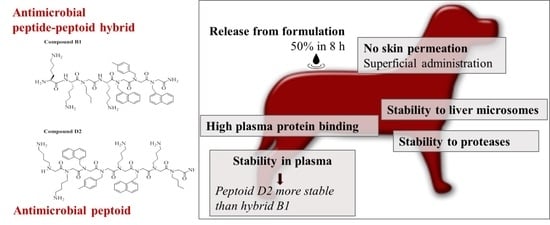
In Vitro ADME Properties of Two Novel Antimicrobial Peptoid-Based Compounds as Potential Agents against Canine Pyoderma

1
Department of Drug Design and Pharmacology, University of Copenhagen, Universitetsparken 2, 2100 Copenhagen Ø, Denmark
2
Department of Food Science, University of Copenhagen, Rolighedsvej 30, DK-1958 Frederiksberg, Denmark
3
Zoetis Inc., 333 Portage St., Kalamazoo, MI 49007, USA
4
Department of Pharmacy, University of Copenhagen, Universitetsparken 2, 2100 Copenhagen Ø, Denmark
5
Department of Veterinary and Animal Sciences, University of Copenhagen, Stigbøjlen 4, 1870 Frederiksberg C, Denmark
*
Authors to whom correspondence should be addressed.
Molecules 2018, 23(3), 630; https://doi.org/10.3390/molecules23030630
Submission received: 17 January 2018 / Revised: 28 February 2018 / Accepted: 5 March 2018 / Published: 10 March 2018
(This article belongs to the Special Issue Antimicrobial Peptides and Peptidomimetics)

Abstract

:
Antimicrobial peptides (AMPs) hold promise as the next generation of antimicrobial agents, but often suffer from rapid degradation in vivo. Modifying AMPs with non-proteinogenic residues such as peptoids (oligomers of N-alkylglycines) provides the potential to improve stability. We have identified two novel peptoid-based compounds, B1 and D2, which are effective against the canine skin pathogen Staphylococcus pseudintermedius, the main cause of antibiotic use in companion animals. We report on their potential to treat infections topically by characterizing their release from formulation and in vitro ADME properties. In vitro ADME assays included skin penetration profiles, stability to proteases and liver microsomes, and plasma protein binding. Both B1 and D2 were resistant to proteases and >98% bound to plasma proteins. While half-lives in liver microsomes for both were >2 h, peptoid D2 showed higher stability to plasma proteases than the peptide-peptoid hybrid B1 (>2 versus 0.5 h). Both compounds were suitable for administration in an oil-in-water cream formulation (50% release in 8 h), and displayed no skin permeation, in the absence or presence of skin permeability modifiers. Our results indicate that these peptoid-based drugs may be suitable as antimicrobials for local treatment of canine superficial pyoderma and that they can overcome the inherent limitations of stability encountered in peptides.

Graphical Abstract

1. Introduction

The emergence of bacterial pathogens resistant to multiple antimicrobial agents over the past decades is a growing public health threat, and there is an urgent need for novel antimicrobial drugs for human and veterinary use [1,2]. In companion animals, canine skin infections constitute the main cause of antibiotic use [3]. Staphylococcus pseudintermedius is the primary pathogen of this disease, as it is isolated from around 90% of canine clinical skin samples [4]. In the recent decade, the worldwide emergence of methicillin-resistant S. pseudintermedius (MRSP) [5] has resulted in infections untreatable with antimicrobial agents licensed for systemic use in dogs. Therefore, there is an urgent need to develop alternative antimicrobial agents to combat this pathogen. Furthermore, systemic administration of antibiotics may promote the development of resistance and current guidelines encourage the use of topical therapy for treatment of MRSP infections [6]. Peptides with antimicrobial activity (AMPs) are the object of rapidly-growing research interest as the next generation of antibacterials [7,8,9]. In general, AMPs are small (<50 amino acids), cationic, amphipathic host defense peptides naturally present in the innate immune systems [10]. AMPs generally show specificity for bacterial targets, broad-spectrum activity and lower affinity for mammalian host cells [11,12,13,14]. Although promising, they display limited stability in vivo being easily degraded by proteases [15]. In recent years, chemical and structural modifications have been used as strategies for improving stability of peptides. Structural analogues of peptides such as peptoids (polymers of N-substituted alkylglycines) [16,17] have been introduced to overcome stability issues. Introduction of peptoid residues in the structure of existing or de novo designed AMPs has also become a successful approach to improve their activity and pharmacokinetic properties [15]. Whereas effort has been made to assess the structure-activity relationship of antimicrobial peptoids to improve activity [18], their ADME (absorption, distribution, metabolism and excretion) profile has not been investigated to the same extent. In the present study, these aspects were characterized for a peptide-peptoid hybrid (B1) and a peptoid (D2) designed as antimicrobials for topical use against S. pseudintermedius. The structures of B1 and D2 are shown in Figure 1. First generation formulations of B1 and D2 for topical administration have been developed and used for testing. The in vitro ADME profile of B1 and D2 in terms of degradation by metabolic enzymes, plasma protein binding and permeation through excised dog skin were investigated in this study.

2. Results

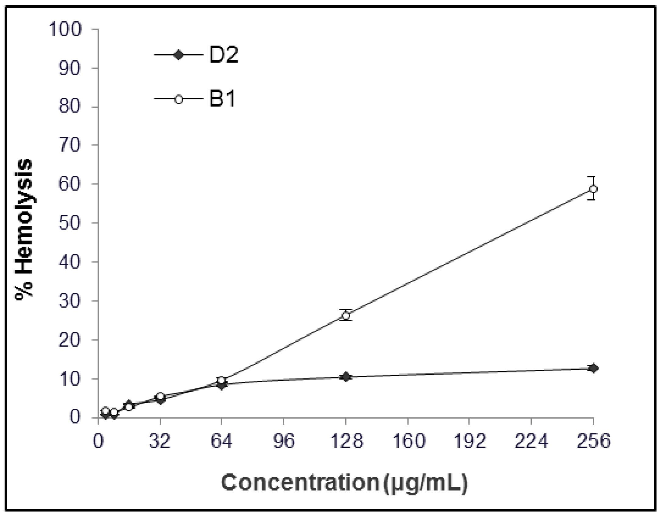
2.1. MIC and Hemolytic Activity In Vitro

B1 and D2 were selected after screening of a combinatorial library of de novo designed compounds for their activity against S. pseudintermedius and host toxicity in vitro measured as lytic activity against red blood cells. B1 and D2 were designed on the basis that the majority of antimicrobial peptides are cationic and hydrophobic. We envisaged that short linear peptide-peptoid hybrids consisting of cationic lysines and hydrophobic peptoid residues would result in derivatives with potent antibacterial activity and low cytotoxicity. Minimum inhibitory concentrations against S. pseudintermedius were 2–4 μg/mL for B1 and 1–2 μg/mL for D2, irrespective of the strain being MSSP or MRSP. Preliminary data suggest that B1 and D2 also are active against a wide range of canine pathogens (data not shown, Greco et al., manuscript in preparation). The hemolytic activity of B1 and D2 as a function of their concentration is shown in Figure 2. Repeated experiments on different days with different human blood samples confirmed the results. The concentration of compound resulting in 10 and 50% hemolysis of erythrocytes (EC10 and EC50) were 64 µg/mL and 219 μg/mL for B1 and 128 µg/mL and >256 μg/mL for D2. In a separate experiment, performing the test with dog blood resulted in hemolysis at 150 μM of 45% and 22% for B1 and D2, respectively.

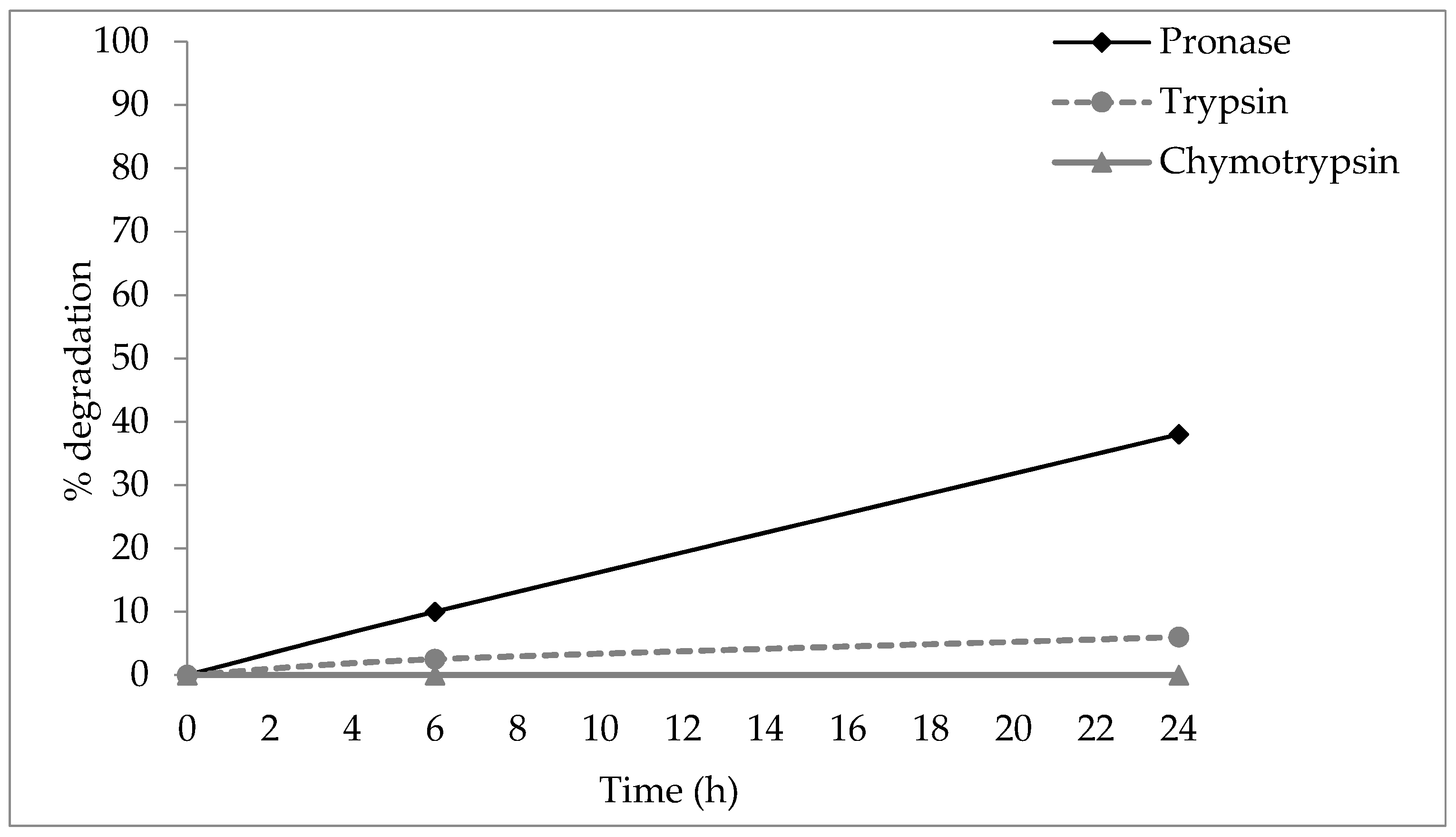
2.2. Stability to Proteases

The stability of B1 and D2 was tested against trypsin and chymotrypsin—proteases of mammalian origin—and pronase, a mix of different enzymes extracted from bacterium Streptomyces griseus. B1 and D2 were incubated singularly in different vials with trypsin, chymotrypsin, and pronase at 37 °C (trypsin and pronase) and 30 °C (chymotrypsin), and the results were analyzed by comparing areas of the resulting RP-HPLC peaks after 6 and 24 h. The peptide-peptoid hybrid B1 showed 6% degradation after 24 h of incubation with trypsin, 38% after pronase exposure, and no degradation after α-chymotrypsin exposure (Figure 3). Peptoid D2 showed 0.2% degradation after 24 h incubation with pronase, and no degradation was observed by trypsin and chymotrypsin (see Supplementary Materials, Section S3).

2.3. Stability in Plasma

Plasma stability of B1 and D2 was tested as an estimation of their half-life in case of systemic absorption or intravenous administration. Plasma stability was determined by incubation of a known concentration of the peptoid/hybrid of interest with dog plasma up to 60 min at 37 °C and analyzing the concentration of the peptide in the supernatant by LC-MS/MS after protein precipitation. Results are presented as average of triplicates from a single donor. Three peptides with known stability profiles (Peptide 1–3) were used as controls in this study (see Table 1). B1 showed some degradation in plasma, with a half-life of 28.9 minutes, while D2 showed no detectable degradation and a half-life > 120 min (see Supplementary Materials, Section S6).

2.4. Stabilty in Liver Microsomes

Liver microsomal assay is used to determine hepatic intrinsic clearance of a drug and identification and profile of metabolites across species. Evaluation of microsomal stability of compounds in different species is common procedure in the drug discovery process to evaluate different metabolites in case of instability. In vitro stability of B1 and D2 in liver microsomes was investigated by incubating B1 and D2 with dog and human microsomes, and analyzing the resulting solution via LC-MS/MS to determine the residual amount of B1 and D2 after microsomal degradation. Both B1 and D2 were rather stable upon incubation with liver microsomes, both displaying in vitro half-lives >120 min (Table 1). After 60 min, 77% of D2 and 100% of B1 were recovered.

2.5. Plasma Protein Binding

The plasma protein binding properties of candidate drugs can influence their efficacy, safety, and distribution. The fraction of peptoid bound to proteins of plasma collected from dogs was determined using rapid equilibrium dialysis. Both B1 and D2 were highly bound (>98%) to dog plasma proteins (Table 2). A free fraction of 0.16% of the peptide-peptoid hybrid B1 and 1.43% of the peptoid D2 were detected via LC-MS/MS from dog plasma. Recoveries for both B1 and D2 were >70%.

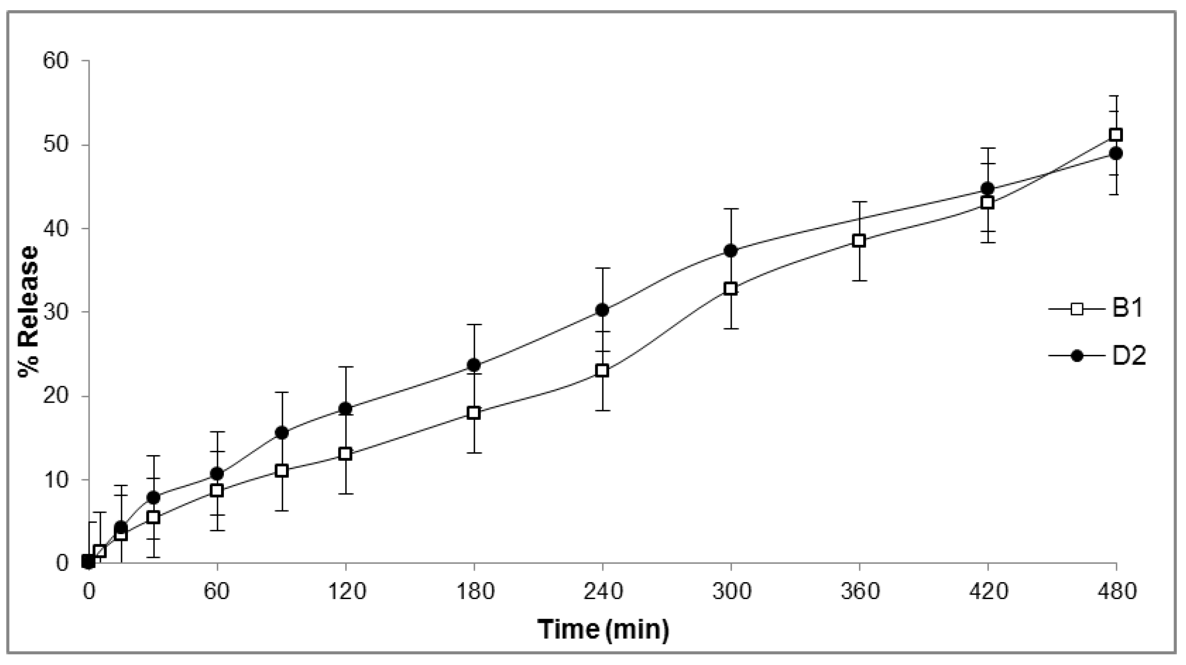
2.6. In Vitro Release

To identify a suitable carrier for B1 and D2, their release from three different formulations (oil-in-water cream, 20% poloxamer gel and ointment, see Table 3 and Section 4.6) was investigated. Release was evaluated as function of the amount of peptide diffused through a cellulose membrane in the receptor of a Franz cell and detected via RP-HPLC from 0 to 8 h. Approximately 50% of the content of both B1 and D2 was released from the oil-in-water cream over 8 h (Figure 4). The release kinetics indicated zero-order kinetics, and rates were comparable for the two compounds. On the contrary, no detectable release of B1 and D2 was observed from the 20% poloxamer gel and the ointment after eight hours (data not shown).

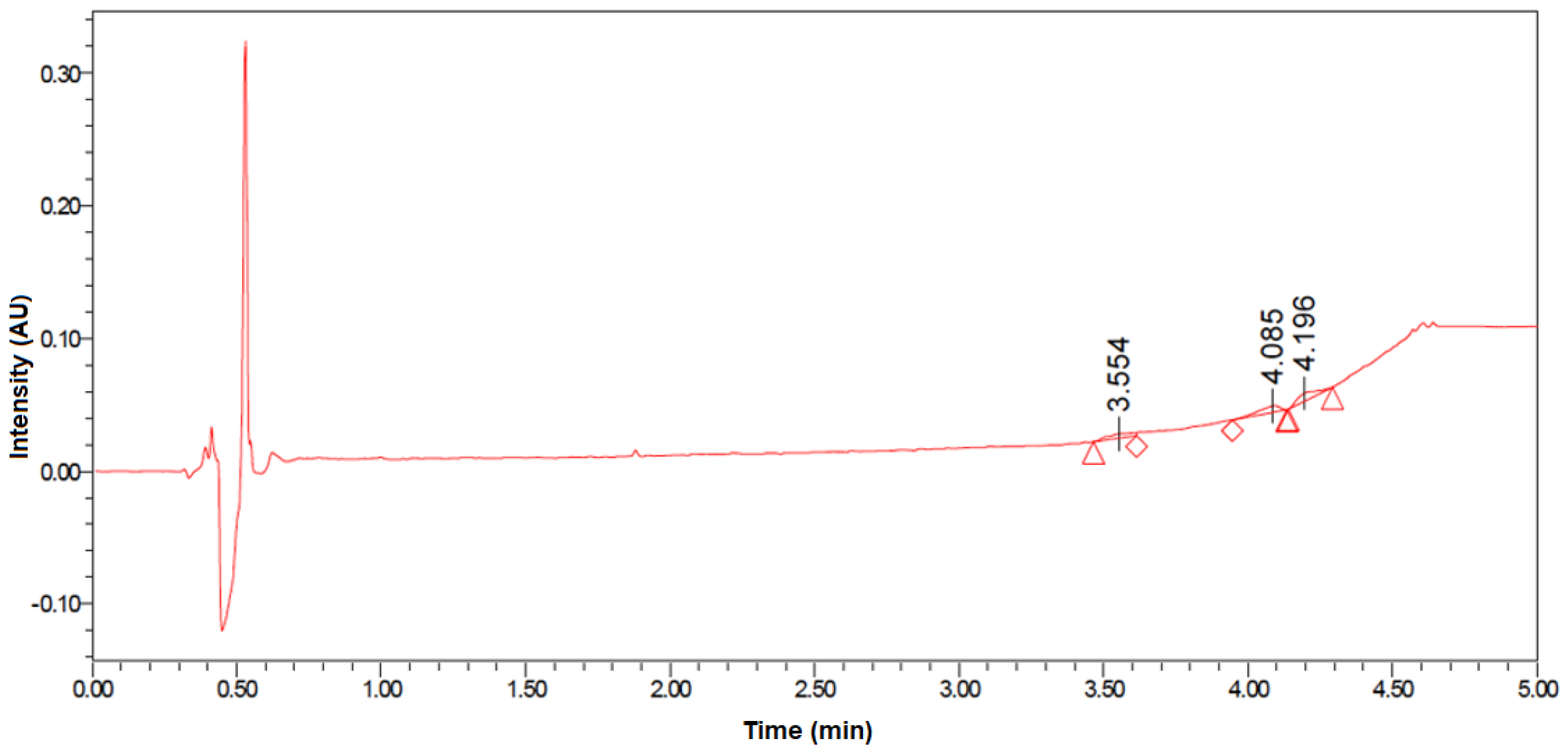
2.7. Skin Permeability

To evaluate the absorption of B1 and D2 into systemic circulation after topical administration, their in vitro permeation profile through excised dog skin was investigated in different excipient conditions, with and without the presence of a skin permeability modifier (transcutol). For each permeation study, donor chambers contained formulation at suitable concentration of peptoid for UPLC detection of penetration of 1% of total content and every sample was present three times. The experiment was repeated twice. Permeation was measured as a function of the concentration detected in the donor chamber of a Franz cell apparatus. A representative chromatogram is shown in Figure 5. No, or negligible, permeation (<1%) of B1 and D2 applied in any of the vehicles was detected after 48 h, suggesting practically no skin penetration of B1 and D2. However, the presented results do not rule out potential partitioning into the stratum corneum.

3. Discussion

There is an urgent need for novel antibacterials specifically tailored for veterinary use only. Developing such agents for veterinary medicine would address the current lack of therapeutic options within this field, and reduce the use of critically important antibiotics approved for humans in the treatment of animal infections [19,20,21].
AMPs act quickly and selectively at micromolar concentrations, are rarely associated with antimicrobial resistance, and can be structurally optimized by chemical synthesis [22,23,24]. Their main limitations as therapeutics are their very short in vivo half-life and high production costs. Despite this, to date several AMPs are under commercial development [25]. De novo design of AMPs and peptidomimetics, such as peptoids [26,27,28,29,30], peptide-peptoid hybrids [31,32,33] and α-peptide/β-peptoids [34,35] have been successful strategies to increase the in vivo stability. The therapeutic potential of peptoids resides in their biological activity comparable to the parent peptide, and their resistance to proteolytic degradation [36]. Despite the large interest in developing peptoid-based antimicrobial agents, though, only a few studies on ADME properties of peptoid exist [37,38,39,40,41]. However, these are mainly limited to short peptoids—up to four residues. In this study, we describe a peptide-peptoid hybrid, B1, and a peptoid, D2, that were highly active against S. pseudintermedius (MIC 2–4 µg/mL). Moreover, the in vitro ADME properties of these peptoid-based antimicrobials were investigated in order to assess their potential as topical drug molecules to treat canine pyoderma.
Topical antimicrobial treatment of canine superficial pyoderma is favorable to systemic treatment, as very high concentrations can be reached at the site of infection. Furthermore, it minimizes the side effects and resistance development in other body sites, including the intestinal tract [6]. Creams and ointments are currently employed for the treatment of staphylococcal infections in veterinary medicine [42] and AMPs formulated in ointments (neomycin, bacitracin and polymyxin B [43]), cream (pexiganan) [44] and poloxamer gel (vancomycin) [45] are known.
To our knowledge there is no evidence in the literature of topical formulation studies of peptoid-based molecules, although they have been indicated as promising compounds for topical application [46]. To characterize the potential of B1 and D2 for topical treatment, we evaluated their release from three potential formulations with different hydrophilicity features (a hydrogel, an ointment and an oil-in-water cream), and assessed their permeation through canine skin. No release was observed when B1 and D2 were formulated in a poloxamer gel or an ointment. Only oil-in-water cream indicated suitability for topical delivery of B1 and D2.
The permeation across excised skin was found to be negligible for both compounds as expected from their physicochemical properties. The skin permeability is often the most difficult challenge to overcome for potential topical drugs [47]. Drugs with a molecular weight >500 Dalton are likely to fail penetrating the SC, the external layer of the skin. Furthermore, the diffusion through skin of positively charged peptides may be prevented by association with negatively charged constituents in the skin [48]. To assess the permeation through skin, B1 and D2 were tested alone and in combination with chemical penetration enhancers (ethanol and transcutol) modifying physical properties of the SC [49,50]. Ethanol is widely used in topical applications and has been previously reported as a skin-penetrating agent for its effect of lipid/protein extraction from the SC and for its “push effect” due to evaporation [51]. Transcutol (diethylene glycol monoethylether) increases the solubility of drugs in the skin and is frequently used as a transdermal penetration enhancer in recent years [49,52]. Even in the presence of enhancers known to modify the physical properties of the SC [50], no permeation was observed. These results overall indicate that B1 and D2 are suitable for topical administration to skin. The lack of permeation likely predicts no systemic absorption of B1 and D2 following topical administration in vivo. Skin penetration of peptides is generally difficult: as an example, a recent paper reports of 5000 publications on formulation and clinical trials for the improvement of penetration of cyclosporine [53], a naturally-occurring multiple N-methylated cyclic peptide with a MW similar to D2. Further experiments are needed to evaluate the extent of the penetration of B1 and D2 into the skin layers, and in case of skin lesions caused by pyoderma, including in vivo studies. However, these initial results indicate that B1 and D2 are suitable for the treatment of superficial infections.
To characterize in vitro the behavior of B1 and D2 if systemically absorbed, their metabolic stability, hemolytic activity, and plasma protein binding were evaluated along with their susceptibility to proteolysis by trypsin, chymotrypsin and pronase. The peptide-peptoid hybrid B1 only showed 6% degradation after 24 h of incubation with trypsin and 38% with pronase, while D2 was stable. Our result is in good agreement with one of the earliest studies on stability of peptoids [54]. In that study, homologous l-amino acid, d-amino acid, and N-substituted glycine peptide and peptoid oligomers were incubated with different proteases from each major class (carboxypeptidase A, chymotrypsin, elastase, papain, pepsin, trypsin). The authors found that the analogues were virtually intact, while the l-peptides were rapidly degraded.
In our study, the peptoid D2 was also stable to plasma proteases, while the initial concentration of the peptide-peptoid hybrid B1 was halved in about 30 min. Results indicate an increased stability in dog plasma conferred by the introduction of peptoid residues, while the presence of peptide bonds results in lower stability. Furthermore, the N-terminus of a peptide correlates with its resistance to proteolysis in plasma [15], and that may correspond to the decreased metabolic stability of the peptide-peptoid hybrid B1 as well. Although the cleavage site is not known with certainty, the presence of two N-terminal Lys residues suggests the site of proteolytic susceptibility. Stability to proteases is also relevant when considering topical applications, due to the presence of proteolytic enzymes of bacteria and skin. In one of the few studies addressing the in vivo pharmacokinetics of peptoids, [37] compared the distribution of a tripeptoid and a tetrapeptide after IV administration. The peptoid was not degraded and rapidly excreted, while the degradation products of the peptide were detected in peripheral body sites [37]. Similar results were reported by [41], who reported complete metabolic degradation of a labelled tripeptide in blood after two hours compared to stability of a labelled tripeptoid.
Both B1 and D2 showed good stability to in vitro degradation by liver microsomes, an assay which has been previously reported to correlate with in vivo data [55]. Half-lives for B1 and D2 were higher than 2 h, indicating a trend of low-medium clearance, compared to controls with known behavior. This suggests that B1 and D2 are not readily susceptible to degradation by liver metabolism. Furthermore, the skin contains metabolizing enzymes similar to the liver (CYP and UDP-glucuronosyltransferase) [56], hence, the assay may aid in predicting stability after cutaneous administration.
Toxicity is an important issue when developing antimicrobial agents. Hemolysis is a standard assay used when screening antimicrobial peptides. The concentration of B1 resulting in 50% of human blood hemolysis was close to 256 μg/mL, comparable to approximately 100× MIC, while hemolysis of D2 at the highest concentration tested did not reach 30%. A similar trend also appeared when the experiment was repeated with dog blood. This could be explained by the higher structural flexibility of peptoid chains compared to peptides and a different type interaction with erythrocyte membranes. The absence of backbone hydrogen bonds in peptoids prevents backbone-driven aggregation and makes peptoids better membrane permeating agents [57]. Compared to B1, the peptoid D2 has a reduced number of hydrogen donors due to the peptoid backbone, higher hydrophilicity (featuring five positive charges, see Figure 1) and a higher degree of rotational flexibility. Rotational flexibility [58] is generally directly proportional to molecular weight, and it counts non-ring bonds to nonterminal non-hydrogen atoms. Despite the promising results, toxicity needs to be further assessed, e.g., by cytotoxicity to fibroblasts or other skin cells, as well as in vivo.
Both B1 and D2 have high protein binding, as both were >98% bound to plasma proteins in equilibrium dialysis in vitro. Plasma protein binding is also likely to occur with administration to a site of infection. Other antimicrobial peptides and drug candidates are also highly protein bound [59], especially apolipoprotein and serum albumin. Despite some inhibition of antimicrobial activity [60], these compounds retained part of their antimicrobial activity and, furthermore, were less cytotoxic at the site of action [61].

4. Materials and Methods

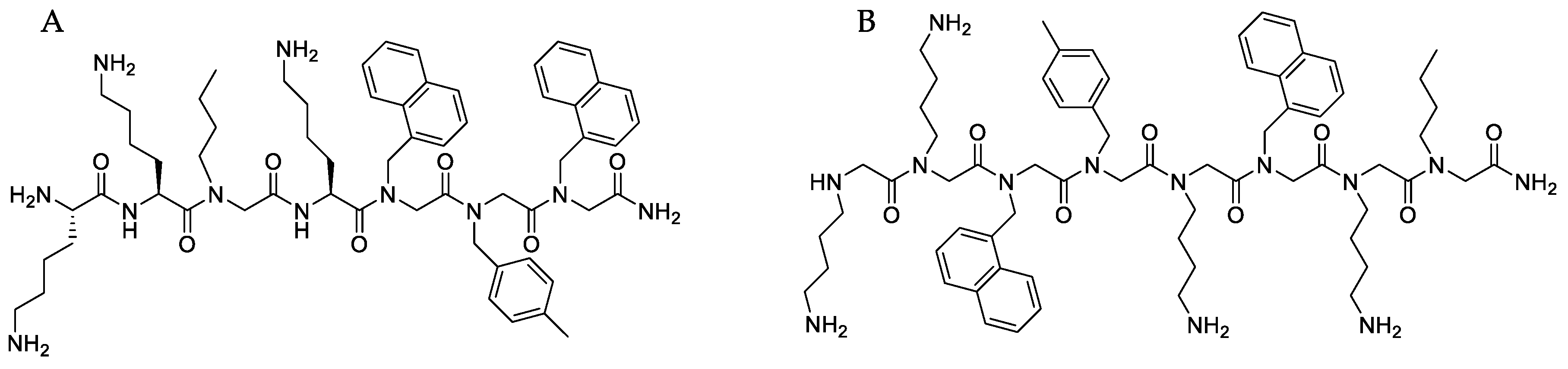
4.1. Synthesis and Characterization of Peptide-Peptoid Hybrid (B1) and Peptoid (D2)

HOAt (1-hydroxy-7-azabenzotriazole) and HATU (1-[Bis(dimethylamino)methylene]-1H-1,2,3-triazolo[4,5-b]pyridinium 3-oxid hexafluorophosphate) were from GL Biochem Shanghai (Shanghai, China). The compounds 1-naphthylmethylamine, 4-methylbenzylamine, butylamine, DIPCDI (N,N-diisopropylcarbodiimide), DIEA (diisopropylamine) and TIS (triisopropylsilane) were from Sigma-Aldrich (Copenhagen, Denmark. TentaGel SRAM (loading 0.2 mmol/g), TFA (trifluoroacetic acid), piperidine, and Fmoc-protected amino acids were purchased from Iris-Biotech GmbH (Marktredwitz, Germany). Disposable 5-mL polypropylene reactors fitted with a PTFE filter were acquired from Thermo Scientific (Hvidovre, Denmark).
Acetic acid and DMF (dimethylformamide), DCM (dichloromethane), ACN (acetonitrile) were purchased from VWR (Copenhagen, Denmark). All reagents and solvents were used without further purification. The manual solid-phase synthesis of peptide-peptoid hybrid B1 and peptoid D2 were carried out in disposable syringes on 100 mg of TentaGel S RAM (loading 0.2 mmol/g) using Fmoc chemistry for amino acids and the submonomer approach for peptoid building blocks as previously described [31]. Following synthesis, the product was cleaved from the resin using TFA/H2O/triisopopylsilane (95:2.5:2.5). Crude products were purified by preparative RP-HPLC until ≥97% purity was obtained (determined by analytical RP-HPLC (Waters Corporation, Milford, MA USA)). The identity of the products was verified by MALDI-TOF-MS (Bruker Daltonik GmbH, Bremen, Germany) (see supplementary information, Section S2).

4.2. Minimum Inhibitory Concentration

The antimicrobial activity of B1 and D2 was investigated by determining their minimum inhibitory concentration (MIC) against a methicillin-susceptible and a methicillin-resistant strain of Staphylococcus pseudintermedius isolated from canine skin and ears at the Veterinary Diagnostic laboratory at University of Copenhagen, Denmark. Assays were performed by microbroth dilution of test compounds in Mueller-Hinton broth (MHB) in microtiter plates (Corning 96-well clear polypropylene round bottom plates, non-treated) according to the Clinical and Laboratory Standard Institute guidelines [62]. The compounds were tested in concentrations ranging from 1 to 64 μg/mL (1, 2, 4, 8, 16, 32, 64 μg/mL). Briefly, bacteria were streaked on blood agar plates and incubated overnight at 37 °C. From these plates, colonies were suspended in saline solution and adjusted by nephelometer to obtain turbidity equivalent to a 0.5 McFarland standard. The suspension was diluted 1:100 in sterile 0.9% (w/v) saline, and 50 μL of this suspension was transferred to each well of the microtiter plate containing 50 μL of peptide solution in water, resulting in a concentration of 5 × 10⁵ CFU/well. The microtiter plate was sealed and incubated at 35 °C for 16–20 h. Results were read by visual inspection.

4.3. Hemolytic Activity

EC10 and EC50 values represent concentrations that result in 10 and 50% hemolysis, respectively. The EC10 and EC50 values were determined for B1 and D2, as previously described [63]. Briefly, two-fold serial dilutions (1 to 256 μg/mL) were incubated in PBS with a suspension of freshly withdrawn human red blood cells in PBS in 96-well v-shaped microtiter plates at 37 °C for 1 h covered with Microseal foil to avoid evaporation, and transferred to 96-well flat bottomed microtiter plates. The release of hemoglobin was detected by measuring the absorbance at 414 nm by a UV spectrophotometer (VERSAmax™ Tunable Microplate Reader, Molecular Devices, San José, CA, USA). PBS and melittin were used as negative and positive controls, respectively. All human blood samples were obtained after informed consent of the voluntary donors upon understanding of the research purposes, within local hospitals and according to local guidelines.

4.4. Stability of Peptide-Peptoid Hybrid (B1) and Peptoid (D2)

4.4.1. Stability to Proteases

The effect of proteases on B1 and D2 was tested by incubation of the compounds at 37 °C with three commercially-available proteases (trypsin and α-chymotrypsin from bovine pancreas, and pronase from Streptomyces griseus, all purchased from Sigma Aldrich). Solutions, concentrations, and reaction temperatures were selected according to manufacturer’s recommendations for total peptide digestion. A stock solution of 1 mM trypsin was prepared in 1 mM Tris HCl buffer solution containing 20 mM Ca2+. For the proteolytic stability assay, a 10 µg/mL trypsin concentration and 1 mg/mL concentration of B1 and D2 in 0.1 M Tris buffer were used to obtain a 1:100 enzyme:peptide ratio. Reaction was conducted at 37 °C for 6 h. Chymotrypsin was solubilized at 10 mg/mL in 1 mM HCl solution containing 2 mM CaCl2. A ratio of 1:60 chymotrypsin: peptide was used (17 µg/mL chymotrypsin, 1 mg/mL peptide). Chymotrypsin reaction was conducted in 100 mM Tris HCl containing 10 mM CaCl2, pH 7.8 at 30 °C. The reaction with pronase was conducted, following the enzyme producer’s recommendation, using 0.2 µM B1 and D2 and 1% pronase powder (w/w) in 50 mM ammonium bicarbonate buffer at pH 8 at 37 °C. At time 0, 3, and 6 h, 50 µL aliquots were removed for RP-HPLC analysis (see supplementary Materials, Section S3).

4.4.2. Stability in Plasma

Dog plasma was collected from a single Beagle dog at the research facilities of Zoetis Animal Health (Kalamazoo, MI, USA) and frozen immediately until use. Proprietary linear peptides with a short and medium half-life and a cyclic peptide with a long half-life were used as controls (Table 2). B1, D2, and controls were diluted to 10 μM in 2% (w/v) BSA containing 0.2% (v/v) formic acid to prevent non-specific binding to materials. Test compounds were added to plasma diluted 1:1 with PBS buffer pH 7.4 and preheated for 5 min at 37 °C. Samples were removed at 0, 5, 10, 20, 30, and 60 min. The reaction was stopped by protein precipitation by adding a 9:1 (v/v) ACN:MeOH mixture followed by centrifugation at 10,000 rpm for 5 min, and the remaining amount of intact test compounds in the supernatant was quantified via API Sciex 4000Q LC-MS/MS (Framingham, MA, USA) (coupled with an HPLC column to filter off the plasma proteins—see Supplementary Materials, Section S6) upon completion of the assay. Experiments were performed in quadruplicate. The half-life values were calculated on the basis of the rate of substrate disappearance (Equation (1)) and calculating the in vitro half-lives (t½) (Equation (2)):
% compound remaining = Ae−kt,
where k is the rate constant for disappearance of parent compound, t is time and A is the starting concentration and is close to 100 when data is normalized to the zero time point:
t1/2 = 0.693/k,

4.4.3. Stability to Liver Microsomes

Evaluation of stability of B1 and D2 to the enzymes in liver microsomes was performed with a robotic system for liquid handling (Hamilton Microlab STAR, Hamilton Robotics, Reno, NV, USA). Dog and human microsomes were obtained from XenoTech LLC (Kansas City, KS, USA) and reconstituted in buffer containing d-(l)-isocitrate trisodium salt and 10 U/mL isocitrate dehydrogenase type IV glycerol solution (procine). A stock solution of 10 mM substrate (B1, D2 or controls, proprietary peptides of Zoetis, Kalamazoo, MI, USA) was diluted 1:1000 in 1% (v/v) ACN before adding to the reaction mixture. The reaction was initiated by adding NADPH and substrate to reconstituted microsomes. A final concentration of 1 µM substrate was incubated with 0.5 mg/mL microsomes in the presence of 1 mM NADP+ in 100 mM PBS with 1 mM MgCl2. Samples were collected at 0, 5, 10, 20, 30, and 60 min. The reaction was conducted at 37 °C before termination by addition of ACN, and analysis was conducted by LC-MS/MS (similar to the method described above). Controls with no drug and with no microsome regeneration system (buffer only) were included. All samples were prepared in triplicate. The rate of degradation by metabolism was measured on the basis of the difference of substrate concentration remaining at every time point. Metabolic half-lives were calculated as mentioned above.

4.5. Plasma Protein Binding

The in vitro plasma protein binding properties of B1 and D2 were investigated with the use of Thermo Scientific™ rapid equilibrium dialysis polypropylene 96-well plates. Plate inserts comprised two side-by-side chambers separated by a dialysis membrane (8K MWCO). The chambers contained 1100 µL plasma or 350 µL dialysis buffer (PBS buffer pH 7.4), respectively. Dogplasma lot was identical to the one used in the plasma stability assay. Ten mM stock solutions of B1 and D2 were diluted 1:50 in water, and 200 µL of diluted B1, D2, and control compound (sarolaner, >99% protein bound) were added to the wells containing plasma in an automated robot for liquid handling. Plates were sealed and incubated at 37 °C for 4 h on a shaker. Each test was run in quadruplicate in dog plasma. Upon completion of the assay, the plasma protein binding was determined by protein precipitation with ACN of both plasma and PBS, and drug levels were quantified via LC-MS/MS as described above. The percentage of bound compounds was determined from peak areas using Equations (3) and (4):
% free = (concentration buffer chamber/concentration plasma chamber) × 100%
% bound = 100% − % free

4.6. Formulation Preparation

An oil-in-water cream and an ointment were prepared as described by Danish Health and Medicines Authority [64]. For the oil-in-water cream, ingredients of the oil phase (Table 3) were melted on a water bath at 70 °C and mixed with the water phase, which was previously brought to boil and cooled down to 65–70 °C. The mixture was stirred until cooled. The ointment formulation was prepared by mixing melted Vaseline (80%) with paraffin oil (20%) on a water bath and allowed to cool under stirring. The cold method was used for preparation of the 20% (w/w) poloxamer gel [65], where the poloxamer powder was dissolved in purified water at 4 °C and subsequently left overnight at 2–8 °C to stabilize. All formulations were stored at 4 °C for maximum seven days before use. Low doses of B1 or D2 were loaded into the formulations just before initializing the release experiment by first dissolving an appropriate amount of compound in 40 to 60 µL of water (or 96% ethanol in case of the ointment) and mixing this into 1 mL of the formulation to obtain a final concentration of 600 µg/mL. For the hydrogel, the compounds dissolved in water were mixed with the cold poloxamer solution before gelling at room temperature.

4.7. In Vitro Drug Release

The release of B1 and D2 from the formulations was determined by utilizing the Franz cell apparatus. Approximately 200 µL of each formulation was filled into the donor chambers of the Franz cells via a 1 mL syringe ensuring contact with the cellulose membrane separating the donor compartment from the receptor compartment. The donor compartment was maintained at room temperature (25 ± 1 °C) and the membrane diffusion area was 1.57 cm2. Activated cellulose membranes (washed 3× with cold water and 3× with 100 °C warm water under stirring for 5 min) were used. The receptor medium (3 mL PBS, pH 7.4) was stirred using magnetic stirring and thermo-regulated to 36 °C by a water jacket. Samples (200 μL) were collected from the receptor compartment through the sampling port over 8 h, and the withdrawn volume was replaced with an equal volume of buffer at every time point. The samples were analyzed by a Waters 600E RP-HPLC equipped with Empower 3 software (Waters Corporation, Milford, MA, USA): 30 min run time with 20 min linear gradient from 0.1% (v/v) TFA in water to 30% (v/v) ACN:H2O with 0.1% (v/v) TFA (90:10). The amounts of B1 and D2 released and diffused through the cellulose membrane were determined using the calibration curve as previously described. Experiments were performed in duplicate.

4.8. Skin Permeation

The amount of B1 and D2 that permeated excised dog skin using three different vehicles was determined ex vivo on an automated Franz cell apparatus. B1 and D2 were dissolved (10 mg/mL) in 20 mM sodium acetate buffer pH 5, in 50% (v/v) ethanol and in 50% (v/v) diethylene glycol monoethyl ether (Transcutol®) in sodium acetate buffer. Skin obtained from the dorsal side of Beagle dogs was pre-treated and stored at −20 °C until use. Pre-treated skin was partially shaved and consisted of stratum corneum (SC) and epidermis. On the day of the experiment, the skin was thawed at room temperature (RT) for one hour, cut in circles (diameter ≈ 2 cm), and stored at RT for 1 h prior to experiments. A circulating water bath was used to keep the cells at a temperature of 37 °C. Polysorbate 80 was used in the receptor chambers (containing 10 mL) to avoid non-specific binding of B1 and D2 to the glass. Prior to the experiment, the receptor chamber solution was degassed for 30 min at RT. Cells were primed and rinsed three times with 10 mL water and three times with 10 mL polysorbate 80 solution. The skin was then mounted on the diffusion cells with the SC facing upwards towards the donor compartment. Transepidermal water loss (TEWL) was measured for each piece of skin independently at the start of the experiment by utilizing Delfin VapoMeter in contact for 10 s with the skin mounted on the chamber. The receptor chambers were subsequently filled with further 10 mL polysorbate 80 solution and left to equilibrate for 150 min. Donor chambers contained 100 µL of formulation added in two steps (2 × 50 µL). Permeation experiments were conducted for 48 h, and donor chambers containing Transcutol® were covered with tin foil to avoid any light exposure. At predetermined time points (2, 6, 12, 24, and 48 h), 1 mL of the receptor phase was withdrawn and replaced with the same amount of receptor medium. The amount of B1 and D2 in the withdrawn samples was determined by UPLC analysis. The experiment was conducted in duplicate.

5. Conclusions

In conclusion, the novel antimicrobial peptide-peptoid hybrid B1 and peptoid D2 displayed properties suitable for the topical treatment of S. pseudintermedius infections in, e.g., an oil-in-water cream and metabolic stability. The positively-charged peptoid-based molecules were found to not penetrate skin, thus, rendering them suitable for topical application when no systemic absorption is desired (e.g., to treat superficial pyoderma). Although in vitro results need to be confirmed by in vivo data, our results show how the presence of peptoid residues correlates with increased stability at conditions resembling in vivo metabolism and possibly with a decreased degree of hemolysis. To the authors’ knowledge, this study represents the first characterization of in vitro early ADME properties of peptoid-based antimicrobials.

Supplementary Materials

Supplementary materials are available on line. Table S1: Gradient elution used for purification of compounds on preparative RP-HPLC. Table S2: Gradient elution used for analytical RP-HPLC , Table S3: Retention times, peak areas and relative peak areas of D2 as detected by RP-HPLC., Table S4: Final gradient elution UV-UPLC method used for the LC-MS/MS experiments., Table S5: Half-lives of peptide-peptoid hybrid B1 and peptoid D2 measured after 60 minutes of incubation at 37°C with dog plasma; Figure S1: Analytical HPLC chromatogram of compound B1, Figure S2: MALDI-TOF MS spectrum of B1, Figure S3: Analytical HPLC chromatogram of compound D2, Figure S4: MALDI-TOF MS spectrum of D2, Figure S5: Stability of D2 to pronase E, Figure S6: B1 in 50% Transcutol in acetate buffer pH 5 detected via UV-UPLC (day 0), Figure S7: B1 in 50% Transcutol in acetate buffer pH 5 detected via UV-UPLC (Day 1), Figure S8: Concentration-time profile in plasma of the compounds B1 and D2.

Acknowledgments

We thank the reviewers for very relevant and useful comments. This work was supported by Marie Curie Actions under the Seventh Frame Programme (grant number 289285) and The Augustinus Foundation (grant number 41825).

Author Contributions

Ines Greco, Jason Koch, and Johannes E. Hansen have performed the experimental work (acquisition, analysis, and/or interpretation of data). Ines Greco, Paul R. Hansen, Hanne Mørck Nielsen, Peter Damborg, Jeffrey L. Watts, Jaspreet Vasir, and Bernard D. Hummel have participated in the conception of the work and experimental design. Ines Greco has drafted the manuscript and all authors have revised it critically for important intellectual content, contributed to the final draft, and approved the final version.

Conflicts of Interest

The authors declare no conflicts of interest.

References

  1. WHO. Antimicrobial Resistance; Global Report on Surveillance; WHO: Geneva, Switzerland, 2014. [Google Scholar]
  2. European Centre for Disease, Prevention and Control. Antimicrobial Resistance in Europe in 2014; Annual Report of the European Antimicrobial Resistance Surveillance Network (EARS-Net); ECDC: Stockholm, Sweden, 2015. [Google Scholar]
  3. Rantala, M.; Holso, K.; Lillas, A.; Huovinen, P.; Kaartinen, L. Survey of condition-based prescribing of antimicrobial drugs for dogs at a veterinary teaching hospital. Vet. Rec. 2004, 155, 259–262. [Google Scholar] [CrossRef] [PubMed]
  4. Petersen, A.D.; Walker, R.D.; Bowman, M.M., II; Schott, H.C.; Rosser, E.J., Jr. Frequency of Isolation and Antimicrobial Susceptibility Patterns of Staphylococcus intermedius and Pseudomonas aeruginosa Isolates From Canine Skin and Ear Samples Over a 6-Year Period (1992–1997). J. Am. Anim. Hosp. Assoc. 2002, 38, 407–413. [Google Scholar] [CrossRef] [PubMed]
  5. Perreten, V.; Kadlec, K.; Schwarz, S.; Grönlund Andersson, U.; Finn, M.; Greko, C.; Moodley, A.; Kania, S.A.; Frank, L.A.; Bemis, D.A.; et al. Clonal spread of methicillin-resistant Staphylococcus pseudintermedius in Europe and North America: An international multicentre study. J. Antimicrob. Chemother. 2010, 65, 1145–1154. [Google Scholar] [CrossRef] [PubMed]
  6. Hillier, A.; Lloyd, D.H.; Weese, J.S.; Blondeau, J.M.; Boothe, D.; Breitschwerdt, E.; Guardabassi, L.; Papich, M.G.; Rankin, S.; Turnidge, J.D.; et al. Guidelines for the diagnosis and antimicrobial therapy of canine superficial bacterial folliculitis (Antimicrobial Guidelines Working Group of the International Society for Companion Animal Infectious Diseases). Vet. Dermatol. 2014, 25, 163–175. [Google Scholar] [CrossRef] [PubMed]
  7. Molchanova, N.; Hansen, P.R.; Franzyk, H. Advances in Development of Antimicrobial Peptidomimetics as Potential Drugs. Molecules 2017, 22, 1430. [Google Scholar] [CrossRef] [PubMed]
  8. Fosgerau, K.; Hoffmann, T. Peptide therapeutics: Current status and future directions. Drug Discov. Today 2015, 20, 122–128. [Google Scholar] [CrossRef] [PubMed]
  9. Mercer, D.K.; O’Neil, D.A. Peptides as the next generation of anti-infectives. Futur. Med. Chem. 2013, 5, 315–337. [Google Scholar] [CrossRef] [PubMed]
  10. Pasupuleti, M.; Schmidtchen, A.; Malmsten, M. Antimicrobial peptides: Key components of the innate immune system. Crit. Rev. Biotechnol. 2012, 32, 143–171. [Google Scholar] [CrossRef] [PubMed]
  11. Haney, E.F.; Mansour, S.C.; Hancock, R.E.W. Antimicrobial Peptides: An Introduction. In Antimicrobial Peptides: Methods and Protocols; Hansen, P.R., Ed.; Springer: New York, NY, USA, 2017; pp. 3–22. [Google Scholar] [CrossRef]
  12. Aoki, W.; Ueda, M. Characterization of Antimicrobial Peptides toward the Development of Novel Antibiotics. Pharmaceuticals 2013, 6, 1055–1081. [Google Scholar] [CrossRef] [PubMed]
  13. Wang, G.; Mishra, B.; Lau, K.; Lushnikova, T.; Golla, R.; Wang, X. Antimicrobial Peptides in 2014. Pharmaceuticals 2015, 8, 123–150. [Google Scholar] [CrossRef] [PubMed]
  14. Zasloff, M. Antimicrobial peptides of multicellular organisms. Nature 2002, 415, 389–395. [Google Scholar] [CrossRef] [PubMed]
  15. Di, L. Strategic Approaches to Optimizing Peptide ADME Properties. AAPS J. 2015, 17, 134–143. [Google Scholar] [CrossRef] [PubMed]
  16. Chongsiriwatana, N.P.; Wetzler, M.; Barron, A.E. Functional Synergy between Antimicrobial Peptoids and Peptides against Gram-Negative Bacteria. Antimicrob. Agents Chemother. 2011, 55, 5399–5402. [Google Scholar] [CrossRef] [PubMed]
  17. Zuckermann, R.N.; Kodadek, T. Peptoids as potential therapeutics. Curr. Opin. Mol. Ther. 2009, 11, 299–307. [Google Scholar] [PubMed]
  18. Fowler, S.A.; Blackwell, H.E. Structure–function relationships in peptoids: Recent advances toward deciphering the structural requirements for biological function. Org. Biomol. Chem. 2009, 7, 1508–1524. [Google Scholar] [CrossRef] [PubMed]
  19. Bengtsson, B.; Greko, C. Antibiotic resistance-consequences for animal health, welfare, and food production. Upsala J. Med. Sci. 2014, 119, 96–102. [Google Scholar] [CrossRef] [PubMed]
  20. Guardabassi, L.; Prescott, J.F. Antimicrobial Stewardship in Small Animal Veterinary Practice: From Theory to Practice. Vet. Clin. N. Am. Small Anim. Pract. 2015, 45, 361–376. [Google Scholar] [CrossRef] [PubMed]
  21. Monath, T.P.; Kahn, L.H.; Kaplan, B. One Health Perspective. ILAR J. 2010, 51, 193–198. [Google Scholar] [CrossRef] [PubMed]
  22. Giuliani, A.; Pirri, G.; Bozzi, A.; Di Giulio, A.; Aschi, M.; Rinaldi, A.C. Antimicrobial peptides: Natural templates for synthetic membrane-active compounds. Cell. Mol. Life Sci. 2008, 65, 2450–2460. [Google Scholar] [CrossRef] [PubMed]
  23. Chen, Y.; Mant, C.T.; Farmer, S.W.; Hancock, R.E.W.; Vasil, M.L.; Hodges, R.S. Rational Design of α-Helical Antimicrobial Peptides with Enhanced Activities and Specificity/Therapeutic Index. J. Biol. Chem. 2005, 280, 12316–12329. [Google Scholar] [CrossRef] [PubMed]
  24. Eckert, R.; Qi, F.X.; Yarbrough, D.K.; He, J.; Anderson, M.H.; Shi, W.Y. Adding selectivity to antimicrobial peptides: Rational design of a multidomain peptide against Pseudomonas spp. Antimicrob. Agents Chemother. 2006, 50, 1480–1488. [Google Scholar] [CrossRef] [PubMed]
  25. Czaplewski, L.; Bax, R.; Clokie, M.; Dawson, M.; Fairhead, H.; Fischetti, V.A.; Foster, S.; Gilmore, B.F.; Hancock, R.E.W.; Harper, D.; et al. Alternatives to antibiotics-a pipeline portfolio review. Lancet Infect. Dis. 2016, 16, 239–251. [Google Scholar] [CrossRef]
  26. Kessler, H. Peptoids—A new approach to the development of pharmaceuticals. Angew. Chem. Int. Ed. 1993, 32, 543–544. [Google Scholar] [CrossRef]
  27. Dohm, M.T.; Kapoor, R.; Barron, A.E. Peptoids: Bio-Inspired Polymers as Potential Pharmaceuticals. Curr. Pharm. Des. 2011, 17, 2732–2747. [Google Scholar] [CrossRef] [PubMed]
  28. Chongsiriwatana, N.P.; Patch, J.A.; Czyzewski, A.M.; Dohm, M.T.; Ivankin, A.; Gidalevitz, D.; Zuckermann, R.N.; Barron, A.E. Peptoids that mimic the structure, function, and mechanism of helical antimicrobial peptides. Proc. Natl. Acad. Sci. USA 2008, 105, 2794–2799. [Google Scholar] [CrossRef] [PubMed]
  29. Martelli, G.; Monsignori, A.; Orena, M.; Rinaldi, S.; Castellucci, N.; Tomasini, C. beta-Peptoids: Synthesis of a novel dimer having a fully extended conformation. Amino Acids 2012, 43, 2005–2014. [Google Scholar] [CrossRef] [PubMed]
  30. Mojsoska, B.; Zuckermann, R.N.; Jenssen, H. Structure-Activity Relationship Study of Novel Peptoids That Mimic the Structure of Antimicrobial Peptides. Antimicrob. Agents Chemother. 2015, 59, 4112–4120. [Google Scholar] [CrossRef] [PubMed]
  31. Ryge, T.S.; Frimodt-Moller, N.; Hansen, P.R. Antimicrobial activities of twenty lysine-peptoid hybrids against clinically relevant bacteria and fungi. Chemotherapy 2008, 54, 152–156. [Google Scholar] [CrossRef] [PubMed]
  32. Ryge, T.S.; Hansen, P.R. Potent antibacterial lysine-peptoid hybrids identified from a positional scanning combinatorial library. Bioorg. Med. Chem. 2006, 14, 4444–4451. [Google Scholar] [CrossRef] [PubMed]
  33. Godballe, T.; Nilsson, L.L.; Petersen, P.D.; Jenssen, H. Antimicrobial beta-Peptides and alpha-Peptoids. Chem. Biol. Drug Des. 2011, 77, 107–116. [Google Scholar] [CrossRef] [PubMed]
  34. Molchanova, N.; Hansen, P.R.; Damborg, P.; Nielsen, H.M.; Franzyk, H. Lysine-Based α-peptide/β-peptoid peptidomimetics: Influence of hydrophobicity, fluorination and distribution of cationic charge on antimicrobial activity and cytotoxicity. ChemMedChem 2017, 20, 312–318. [Google Scholar] [CrossRef] [PubMed]
  35. Liu, Y.; Knapp, K.M.; Yang, L.; Molin, S.; Franzyk, H.; Folkesson, A. High in vitro antimicrobial activity of beta-peptoid-peptide hybrid oligomers against planktonic and biofilm cultures of Staphylococcus epidermidis. Int. J. Antimicrob. Agents 2013, 41, 20–27. [Google Scholar] [CrossRef] [PubMed]
  36. Bolt, H.L.; Eggimann, G.A.; Jahoda, C.A.B.; Zuckermann, R.N.; Sharples, G.J.; Cobb, S.L. Exploring the links between peptoid antibacterial activity and toxicity. MedChemComm 2017, 8, 886–896. [Google Scholar] [CrossRef]
  37. Seo, J.; Ren, G.; Liu, H.G.; Miao, Z.; Park, M.; Wang, Y.H.; Miller, T.M.; Barron, A.E.; Cheng, Z. In vivo Biodistribution and Small Animal PET of Cu-64-Labeled Antimicrobial Peptoids. Bioconjugate Chem. 2012, 23, 1069–1079. [Google Scholar] [CrossRef] [PubMed]
  38. Ross, T.M.; Zuckermann, R.N.; Reinhard, C.; Frey, W.H. Intranasal administration delivers peptoids to the rat central nervous system. Neurosci. Lett. 2008, 439, 30–33. [Google Scholar] [CrossRef] [PubMed]
  39. Lim, H.S.; Cai, D.; Archer, C.T.; Kodadek, T. Periodate-triggered cross-linking reveals Sug2/Rpt4 as the molecular target of a peptoid inhibitor of the 19S proteasome regulatory particle. J. Am. Chem. Soc. 2007, 129, 12936–12937. [Google Scholar] [CrossRef] [PubMed]
  40. Hao, G.; Hajibeigi, A.; León-Rodríguez, L.M.D.; Öz, O.K.; Sun, X. Peptoid-based PET imaging of vascular endothelial growth factor receptor (VEGFR) expression. Am. J. Nucl. Med. Mol. Imaging 2011, 1, 65–75. [Google Scholar] [PubMed]
  41. Wang, Y.F.; Lin, H.; Tullman, R.; Jewell, C.F.; Weetall, M.L.; Tse, F.L.S. Absorption and disposition of a tripeptoid and a tetrapeptide in the rat. Biopharm. Drug Dispos. 1999, 20, 69–75. [Google Scholar] [CrossRef]
  42. Werner, A.H.; Russell, A.D. Mupirocin, fusidic acid and bacitracin: Activity, action and clinical uses of three topical antibiotics. Vet. Dermatol. 1999, 10, 225–240. [Google Scholar] [CrossRef]
  43. Bonomo, R.A.; Van Zile, P.S.; Li, Q.; Shermock, K.M.; McCormick, W.G.; Kohut, B. Topical triple-anti biotic ointment as a novel therapeutic choice in wound management and infection prevention: A practical perspective. Expert Rev. Anti-Infect. Ther. 2007, 5, 773–782. [Google Scholar] [CrossRef] [PubMed]
  44. Lipsky, B.A.; Holroyd, K.J.; Zasloff, M. Topical versus Systemic Antimicrobial Therapy for Treating Mildly Infected Diabetic Foot Ulcers: A Randomized, Controlled, Double-Blinded, Multicenter Trial of Pexiganan Cream. Clin. Infect. Dis. 2008, 47, 1537–1545. [Google Scholar] [CrossRef] [PubMed]
  45. Veyries, M.L.; Couarraze, G.; Geiger, S.; Agnely, F.; Massias, L.; Kunzli, B.; Faurisson, F.; Rouveix, B. Controlled release of vancomycin from Poloxamer 407 gels. Int. J. Pharm. 1999, 192, 183–193. [Google Scholar] [CrossRef]
  46. Czyzewski, A.M.; Jenssen, H.; Fjell, C.D.; Waldbrook, M.; Chongsiriwatana, N.P.; Yuen, E.; Hancock, R.E.W.; Barron, A.E. In vivo, In Vitro, and In Silico Characterization of Peptoids as Antimicrobial Agents. PLoS ONE 2016, 11, e0135961. [Google Scholar] [CrossRef] [PubMed]
  47. Bos, J.D.; Meinardi, M. The 500 Dalton rule for the skin penetration of chemical compounds and drugs. Exp. Dermatol. 2000, 9, 165–169. [Google Scholar] [CrossRef] [PubMed]
  48. Wise, D. Handbook of Pharmaceutical Controlled Release Technology; CRC Press: Boca Raton, FL, USA, 2000; ISBN 9780824703691. [Google Scholar]
  49. Mohammed, D.; Hirata, K.; Hadgraft, J.; Lane, M.E. Influence of skin penetration enhancers on skin barrier function and skin protease activity. Eur. J. Pharm. Sci. 2014, 51, 118–122. [Google Scholar] [CrossRef] [PubMed]
  50. Lane, M.E. Skin penetration enhancers. Int. J. Pharm. 2013, 447, 12–21. [Google Scholar] [CrossRef] [PubMed]
  51. Verma, D.D.; Fahr, A. Synergistic penetration enhancement effect of ethanol and phospholipids on the topical delivery of cyclosporin A. J. Controll. Release 2004, 97, 55–66. [Google Scholar] [CrossRef] [PubMed]
  52. Fini, A.; Bergamante, V.; Ceschel, G.C.; Ronchi, C.; De Moraes, C.A.F. Control of Transdermal Permeation of Hydrocortisone Acetate from Hydrophilic and Lipophilic Formulations. AAPS PharmSciTech 2008, 9, 762–768. [Google Scholar] [CrossRef] [PubMed]
  53. Carmona-Ribeiro, A.; de Melo Carrasco, L. Novel Formulations for Antimicrobial Peptides. Int. J. Mol. Sci. 2014, 15, 18040–18083. [Google Scholar] [CrossRef] [PubMed]
  54. Miller, S.M.; Simon, R.J.; Ng, S.; Zuckermann, R.N.; Kerr, J.M.; Moos, W.H. Proteolytic studies of homologous peptide and N-substituted glycine. Bioorg. Med. Chem. Lett. 1994, 4, 2657–2662. [Google Scholar] [CrossRef]
  55. Riley, R.J.; McGinnity, D.F.; Austin, R.P. A unified model for predicting human hepatic, metabolic clearance from in vitro intrinsic clearance data in hepatocytes and microsomes. Drug Metab. Dispos. 2005, 33, 1304–1311. [Google Scholar] [CrossRef] [PubMed]
  56. Oesch, F.; Fabian, E.; Oesch-Bartlomowicz, B.; Werner, C.; Landsiedel, R. Drug-metabolizing enzymes in the skin of man, rat, and pig. Drug Metab. Rev. 2007, 39, 659–698. [Google Scholar] [CrossRef] [PubMed]
  57. Simon, R.J.; Kania, R.S.; Zuckermann, R.N.; Huebner, V.D.; Jewell, D.A.; Banville, S.; Ng, S.; Wang, L.; Rosenberg, S.; Marlowe, C.K.; et al. Peptoids: A modular approach to drug discovery. Proc. Natl. Acad. Sci. USA 1992, 89, 9367–9371. [Google Scholar] [CrossRef] [PubMed]
  58. Barbour, N.P.; Lipper, R.A. Introduction to Biopharmaceutics and its Role in Drug Development. In Biopharmaceutics Applications in Drug Development; Krishna, R., Yu, L., Eds.; Springer: Boston, MA, USA, 2008; pp. 1–25. [Google Scholar] [CrossRef]
  59. Beer, J.; Wagner, C.C.; Zeitlinger, M. Protein Binding of Antimicrobials: Methods for Quantification and for Investigation of its Impact on Bacterial Killing. AAPS J. 2009, 11, 1–12. [Google Scholar] [CrossRef] [PubMed]
  60. Burian, A.; Wagner, C.; Stanek, J.; Manafi, M.; Bohmdorfer, M.; Jager, W.; Zeitlinger, M. Plasma protein binding may reduce antimicrobial activity by preventing intra-bacterial uptake of antibiotics, for example clindamycin. J. Antimicrob. Chemother. 2011, 66, 134–137. [Google Scholar] [CrossRef] [PubMed]
  61. Wang, Y.Q.; Agerberth, B.; Lothgren, A.; Almstedt, A.; Johansson, J. Apolipoprotein A-I binds and inhibits the human antibacterial/cytotolic peptide LL-37. J. Biol. Chem. 1998, 273, 33115–33118. [Google Scholar] [CrossRef] [PubMed]
  62. CLSI. Performance Standards for Antimicrobial Disk and Dilution Susceptibility Tests for Bacteria Isolated from Animals; Approved Standard—Fourth Edition; VET01-A4; CLSI: Wayne, PA, USA, 2013. [Google Scholar]
  63. Oddo, A.; Nyberg, N.T.; Frimodt-Moller, N.; Thulstrup, P.W.; Hansen, P.R. The effect of glycine replacement with flexible omega-amino acids on the antimicrobial and haemolytic activity of an amphipathic cyclic heptapeptide. Eur. J. Med. Chem. 2015, 102, 574–581. [Google Scholar] [CrossRef] [PubMed]
  64. Danish Health and Medicines Authority. 2, Appendix 3, Quality of Medicinal Products, Addition to drug form standards. In Danish Medicinal Standards; Danish Health and Medicines Authority: Copenhagen, Denmark, 2013. [Google Scholar]
  65. Dumortier, G.; Grossiord, J.L.; Agnely, F.; Chaumeil, J.C. A Review of Poloxamer 407 Pharmaceutical and Pharmacological Characteristics. Pharm. Res. 2006, 23, 2709–2728. [Google Scholar] [CrossRef] [PubMed]
Sample Availability: Samples of the compounds are not available from the authors.
Figure 1. Structure of B1 (A) and D2 (B) both isolated as trifluoacetic acid salts.
Molecules 23 00630 g001
Figure 2. Hemolytic activity of peptide-peptoid hybrid B1 and peptoid D2 in human erythrocytes as a function of concentration. Mean ± SEM, n = 3.
Molecules 23 00630 g002
Figure 3. Proteolytic degradation of peptide-peptoid hybrid B1 by chymotrypsin, trypsin, and pronase.
Molecules 23 00630 g003
Figure 4. In vitro release of B1 and D2 from oil-in-water cream formulation. Mean ± SEM, n = 2.
Molecules 23 00630 g004
Figure 5. Skin penetration of B1 from 50% transcutol formulation detected via UV-UPLC after 24 h. No penetration of B1 through dog skin is observed (UV-UPLC detection limit: 1 µg/L). Since B1 and D2 showed similar skin penetration pattern in all tested formulations, only representative data for B1 are shown.
Molecules 23 00630 g005
Table 1. Half-lives (minutes) of B1 and D2 and control peptides 1 in plasma and liver microsomes.
CompoundPlasma (Dog)Liver Microsomes (Dog)Liver Microsomes (Human)
B128.9>120>120
D2>120>120>120
Peptide-1>120--
Peptide-2<3--
Peptide-381.2--
1 Peptide-1, -2, and -3 are control peptides with long, medium and short plasma half-life, respectively.
Table 2. Protein binding of B1, D2, and control peptide after 4 h incubation in plasma.
Entry% Average RecoverySD 1% UnboundBoundSD 1
B175.620.70.1699.80.000
D270.86.51.4398.60.005
Control67.112.60.2799.70.001
1 Mean ± SD (n = 4).
Table 3. Composition of the tested compounds.
FormulationIngredients% (w/w)
Oil-in-water creamOil Phase
Polysorbate 800.5
Cetostearyl alcohol5
Paraffin oil5
Glycerol monostearate6
Water Phase
Methyl parahydroxy-benzoate0.1
85% glycerol4
Sorbitol7
Purified water72.4
Oinment
Paraffin oil20
Vaselin80
Hydrogel
Poloxamer 40720
Purified water80

