Selenides and Diselenides: A Review of Their Anticancer and Chemopreventive Activity
by
 , ,
, ,
Mónica Álvarez-Pérez
1 ,
,
Wesam Ali
2,3,
Małgorzata Anna Marć
3 ,
,
Jadwiga Handzlik
3 and
Enrique Domínguez-Álvarez
1,* 1
Instituto de Química Orgánica General, Consejo Superior de Investigaciones Científicas (IQOG, CSIC), Juan de la Cierva 3, 28006 Madrid, Spain
2
Division of Bioorganic Chemistry, School of Pharmacy, Saarland University, Campus B2 1, D-66123 Saarbruecken, Germany
3
Department of Technology and Biotechnology of Drugs, Faculty of Pharmacy, Jagiellonian University Medical College, Medyczna 9, 30-688 Cracow, Poland
*
Author to whom correspondence should be addressed.
Molecules 2018, 23(3), 628; https://doi.org/10.3390/molecules23030628
Submission received: 3 January 2018
/
Revised: 7 March 2018
/
Accepted: 9 March 2018
/
Published: 10 March 2018
(This article belongs to the Special Issue Small Molecule Catalysts with Therapeutic Potential)
Abstract
:Selenium and selenocompounds have attracted the attention and the efforts of scientists worldwide due to their promising potential applications in cancer prevention and/or treatment. Different organic selenocompounds, with diverse functional groups that contain selenium, have been reported to exhibit anticancer and/or chemopreventive activity. Among them, selenocyanates, selenoureas, selenoesters, selenium-containing heterocycles, selenium nanoparticles, selenides and diselenides have been considered in the search for efficiency in prevention and treatment of cancer and other related diseases. In this review, we focus our attention on the potential applications of selenides and diselenides in cancer prevention and treatment that have been reported so far. The around 80 selenides and diselenides selected herein as representative compounds include promising antioxidant, prooxidant, redox-modulating, chemopreventive, anticancer, cytotoxic and radioprotective compounds, among other activities. The aim of this work is to highlight the possibilities that these novel organic selenocompounds can offer in an effort to contribute to inspire medicinal chemists in their search of new promising derivatives.
1. Introduction
Various authors have extensively reviewed the different applications of selenium in human health [1,2,3,4]. Selenium is an essential element that forms part of the selenoaminoacids selenomethionine (1, Figure 1a) and selenocysteine (2). The latest is an essential constituent of the selenoproteins [5,6,7] and its deficiency can cause serious disorders [1,2]. Selenoproteins have crucial functions for human health. The most known are the gluthathione peroxidases, the iodothyronine deiodinases, the thioredoxin reductases and the selenoprotein P. The different glutathione peroxidases (GPx) are involved in the elimination of free radicals; the iodothyronine deiodinases enable the activation and deactivation of thyroid hormones. Thioredoxin reductases regenerate the thioredoxin (an important antioxidant class of proteins), and the selenoprotein P is involved in the transport of selenium in plasma and in the antioxidant defense against free radicals in lipids [5,6,7].
An intense research in the chemopreventive and/or anticancer activity of organic and inorganic selenium-containing compounds has been developed, as has been reviewed by different authors [8,9,10,11,12,13,14]. The duality of selenium as an antioxidant and as a prooxidant agent provides the basis for its potential applications in both cancer prevention and treatment. This dual chemopreventive and anticancer action has been extensively discussed or reported by different authors [15,16,17,18]. Regarding the chemopreventive activity, a mechanism usually exerted by different selenocompounds is the glutathione peroxidase-like activity [19]. Although inorganic selenocompounds may be superior chemopreventive agents than organic ones, current research is focused on the latter group due to their lower toxicity risk [20]. Among the inorganic selenocompounds, selenite can be highlighted by its chemopreventive [21] and anticancer action [22].
On the other hand, the mechanisms of action of the organic selenocompounds are very diverse [23]. Some of the most frequent are the reduction of oxidative stress through the elimination of free radicals [24,25], i.e., the induction of mutations [26], the cytotoxic activity [27] and the triggering of apoptotic events [28,29]. Nevertheless, certain selenocompounds can act through other less usual mechanisms, such as the inhibition of angiogenesis [30], the inhibition of the efflux pumps in cancer multidrug resistant cell lines [31] and the enhancement of the activity of chemotherapeutic drugs [32]. The chemical structure of anticancer and chemopreventive organic selenocompounds also shows a great variety, including selenocyanates [33], selenoureas [34], selenoesters [35], heterocycles with endocyclic selenium [36], selenium nanoparticles [37], selenides and diselenides.
Herein, we review an anticancer and a chemopreventive activity of both selenides and diselenides, with a special attention to mechanisms of their biological action. The question of diselenides symmetry, as a feature favorable for anticancer activity [38], is also under consideration.
2. Selenides and Diselenides with Therapeutic Perspective
2.1. Naturally Occurring Selenides and Diselenides
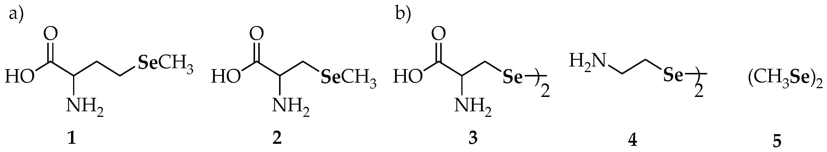
Diselenides have received significant attention, not only because this moiety is usually found among the metabolites of the dietary compounds but also particularly because they have been postulated as one of the most important species in redox-cycling. In line with this, selenocystine, a naturally occurring diselenide (3, Figure 1b), exhibits strong antioxidant properties [39] and has been considered in several studies related to chemoprevention. Thus, it was effective against 4-(methylnitrosamino)-1-(3-pyridyl)-1-butanone (NNK) induced lung tumors in a specific mouse model sensitive to chemical carcinogenesis (the A/J mouse model) when administered in a pre-initiation period to tumor development, or, alternatively, when supplementation was restricted to a post-initiation period [40].
In a different study, 3 was found to trigger apoptosis in human breast adenocarcinoma MCF-7 cells through kinase regulation [41]. Induction of apoptosis was also attributed to 3 in HepG2 human hepatoma cells and in isolated rat liver mitochondria [42]. In this case, the proposed mechanism of apoptosis involved the opening of the mitochondrial permeability transition pores (and the associated loss of Δψm, mitochondrial swelling and cytochrome c release), which is mediated by the cross-linking of protein thiol groups and by the generation of superoxide radical anions (O2•−) through reaction with glutathione (GSH) [42]. Selenocystamine (4) also oxidized the protein thiol groups in isolated mitochondria and in crude extracts of HepG2 cells, but not in intact HepG2 cells. These lines of evidence suggest that the action was similar to that exerted by 3, precluding its inability to cross the cell membrane. Additionally, dimethyldiselenide (5), another naturally occurring diselenide, has been reported to be active against oxidative damage [43].
2.2. Synthetic Diselenides
In addition to biologically existing diselenides, a variety of synthetic diselenides has exhibited protective potential. Due to the high number of diselenides, they are reviewed herein in function of their biological activity: antioxidant activity, prooxidant/redox-modulating, kinase-modulating, antiproliferative/cytotoxic and apoptotic. The chemical names of mentioned diselenides are summarized in Table 1.
2.2.1. Antioxidant Activities of Synthetic Diselenides: GPx-Like, Metal-Binding, Chemopreventive and Free Radical Scavenging Potential
Diphenyl diselenide (6, Figure 2) has been evaluated in several studies and it is considered a reference compound in the evaluation of the antioxidant properties and of the glutathione peroxidase-like (GPx-like) activity [44,45,46,47,48]. Especially, 6 displayed a higher antioxidant activity than both dibenzyl diselenide (7) and a group of asymmetric selenides represented by 8 [44]. Concerning the protective role of diselenides on lipid peroxidation, 6 behaved as the most antioxidant compound among the different diaryl and dialkyl diselenides tested, regardless of the species (mice and rats) [46]. Dialkyl selenides, e.g., diethyl (9) and substituted diaryl diselenides such as di(p-clorophenyl) diselenide (10), were effective against lipid peroxidation but showed differential antioxidant potential in mice and rats, being more potent in mice [46].
The ability of diphenyl diselenide (6) to protect murine J774 macrophage-like cells from reactive oxygen species (ROS) generated by the oxidation of low density lipoproteins (LDL) was proved by Straliotto et al. [49]. The redox signaling effects of 6 were accompanied by the down-regulation of the nuclear factor-κB (NF-κB), a transcription factor involved in pro-apoptotic pathways activated by ROS. Melo et al. found that 6 was also able to protect breast (MCF-7) cancer cells from oxidative damage induced by tamoxifen hormone therapy without interfering with its cytotoxicity [50]. They concluded that, if used at low concentrations, 6 could be employed as a potent antigenotoxic agent to prevent the risk of cancer induction.
The meta-CF3 substituted diphenyl diselenide (11) has been reported as an anti-mutagenic and anti-genotoxic selenium derivative [51]. It protected Chinese hamster lung fibroblasts (V79 cells) against H2O2-induced DNA damage at doses as low as 12.5 µM. In addition, 11 mimics catalase activity better than 6, both compounds giving rise to different products upon reaction with H2O2.
The dimethyl- (5), diphenyl- (6) and dibenzyl diselenides (7) induced quinone reductase and glutathione-S-transferase activity [52]. The generated selenol derivatives (i.e., selenolates at physiological pH) can be considered as the metabolites responsible for such responses.
Selenolates can be easily generated from selenols due to the high acidity of the Se-H bond (which is more labile than the equivalent S-H and O-H bonds because of the higher polarizability of selenium), that enables the rapid and almost complete ionization at physiological pH [53]. One of the first selenols assayed for inhibition of carcinogenesis has been p-methoxyphenylselenol (12), which showed effectiveness in the inhibition of azoxymethane-induced liver [54], colon and kidney [55] tumors in rats.
On the other hand, the antioxidant and GPx activity of 3,3′-diselenodipropionic acid (13) has been evaluated by Kunwar et al. [56]. The treatment with 13 prevented the depletion of the levels of endogenous antioxidants (GSH) in red blood cells through free-radical-induced stress. However, in comparison to sodium selenite or ebselen, its GPx activity was lower. Compound 13 has been also evaluated as a radio-protective agent against whole-body γ-radiation on Swiss albino mice [57]. In this in vivo study, authors concluded that 13 can attenuate radiation-induced DNA damage. Furthermore, it can inhibit radiation-induced apoptosis in the spleen and can reverse radiation-induced alterations in the expression of the pro-apoptotic BAX and the anti-apoptotic Bcl-2 genes.
Adding a second phenyl group to diphenyl selenide, converting it in binaphthyl diselenide (14), revealed that it showed a significant antioxidant activity [58]. Compound 14 could exert a protective action against 2-nitropropane (2-NP)-induced hepatoxicity in rats.
Including nitrogen into the phenyl ring, converting it in a pyridine ring (15), has provided cytoprotective effects [59]. The 2-pyridyl derivative 15 caused the largest shift in the Cu+2 reduction potential and was also the most promising in the metallothionein (MT) assay for the measurement of catalytic activity. Similarly, the N-exocyclic aniline derivative 16 exhibited a noteworthy activity in the antioxidant assays considered. Indeed, 16 behaved as a better antioxidant than ebselen in human skin fibroblast (FCP7) cell culture under UVA-induced stress, thus suggesting that 16 could depict a leading compound for the development of more effective [46] multi-functional antioxidants. According to the authors, these different activities (GPx-like and metal binding characteristics of their nitrogen and chalcogen groups) could explain the extraordinary protection exerted by these compounds against the damage caused by ROS. The role of selenium-metal coordination in DNA damage inhibition was reported to be essential in a parallel study [60].
The naphthylamine derivative 17 did not show GPx-like activity when this activity was evaluated for selected intramolecularly-coordinated diorganyl diselenides [45]. In contrast, compounds with phenyl or naphthyl rings substituted with a (dimethylamino)methyl group (18–20) were active and these observations were attributed to the presence of better electron-donating groups in close proximity to selenium [61]. Likewise, a good activity was found for 21, with an electron-withdrawing group (a nitro group) in ortho position to selenium. The same authors had previously evaluated the thiol peroxidase-like activity of related diselenides based on ferrocene, such as 22 [62]. The introduction of the ferrocene moiety increased the activity, although the most important effect was suspected to be exerted through the efficacy of the intramolecular N…Se coordination, as 23, with an amide group and, thus, a less coordinating nitrogen atom, was only superior to 6 and 17 (GPx-like activity: 22 > 20 > 21 >> 18 > 23 >> 6 > 17).
Bhabak and Mugesh [63] reported the enhancement of the antioxidant activity of diselenides compounds such as 18 by the introduction of a methoxy group as a substituent in ortho to selenium (19). The observed increment of activity in compound 19 was investigated through kinetic and computational methods and ascribed to the role of the methoxy group in altering the steric and electronic environment around selenium, in particular, preventing over-oxidation by peroxides and preventing thiol-exchange reactions in the selenenylsulfide intermediate of the catalytic cycle. The same authors also found an enhanced GPx activity of sec-amino-substituted diselenides analogous versus the analogous tert-amine-based compounds [64].
The p-methoxyphenyl- (24a) and p-chlorophenyl-substituted (24b) compounds were the most active molecules in a series of related imidazole-containing derivatives assayed for their GPx-like activity and their capacity to be reduced by glutathione (GSH) [65]. Regarding GPx-like activity, they were 4-fold more potent than ebselen, whilst they showed 3–5-fold more reactivity toward GSH than 6. In general, the most effective compounds were those substituted at 5-position of the imidazole ring, close to the selenium atom. Apart from this fact, no other clear structure–activity relationship emerged from these antioxidant studies.
An interesting study by Romano et al. [66] compared the capability of new selenocarbamate derivatives to scavenge the radical 1,1-diphenyl-2-picryl-hydrazyl (DPPH) and 2,2-azinobis(3-ethylbenzothiazoline-6-sulfonic acid) (ABTS), as well as their ability to mimic GPx activity. Two families containing common active fragments, consisting of selenocyanate and diselenide derivatives, did not differ much in their activities despite double amount of selenium (on a molar basis) and symmetry in the case of diselenides. Among diselenides, 25a, 25b and 25c (Figure 2) displayed potential radical scavenging activity for DPPH and ABTS, although they did not exhibit GPx-like activity.
When evaluating the GPx-like activity of a group of selenosteroids (26 and 27, among others), Fuentes-Aguilar et al. [67] found that the GPx-like activity was under that of ebselen for all the compounds considered.
On some occasions, the chemoprotective activity of diselenides has been related to conformational restriction. Thus, certain peri-diselenides (28a and 28b) have shown superior GPx-like activity than acyclic diaryl diselenides [68]. It should be expected that several properties of the compounds worked synergistically, however, as the introduction of methoxy groups in the naphthalene moiety made chemoprevention increase for derivative 28b in comparison to 28a.
2.2.2. Prooxidant Activity and Redox Modulation Activity of Synthetic Diselenides
Dialkyl diselenides (29 and 30), in contrast to diaryl ones (6, 10, and 11), proved to behave as prooxidants at low concentrations [46]. According to in vivo studies and despites its above-mentioned antioxidant potential, 6 can also be toxic in certain conditions, depending on dose, species and route of administration [69,70].
The intracellular redox modulation by selenocompounds has been deeply studied. Nevertheless, the extracellular redox control has been only recently linked to the cellular uptake and to the cytotoxicity of selected selenium-containing derivatives [71,72]. The extracellular redox status, in whose regulation both cell surface and secreted redox-active proteins are involved, seems to be correlated with several biological responses such as cell proliferation and apoptosis. The cysteine/cystine couple is attributed to play a key-role in the extracellular redox state, whereas thioredoxin (Trx), thioredoxin reductase (TrxR) and protein disulfide isomerase (PDI) are among the proteins whose expression may increase the production of cysteine. Apart from phenylselenol (PhSeH) and ebselen, diphenyl diselenide (6, Figure 2) and dibenzyl diselenide (7) have been pointed as inductors of extracellular cysteine accumulation in murine macrophage RAW 264.7 cells and in differentiated human THP-1 monocytes [72]. Nevertheless, neither selenocystine (3, Figure 1) nor methylselenocysteine could exert the same effects. The authors proposed a model where production of extracellular cysteine increases because of the induction of TrxR1 expression on the cell surface by certain organoselenium compounds. Again, selenolates are pointed as the putative bioactive species, as 6 and PhSeH showed nearly identical cellular effects on a molar basis [72].
Diethyl- (9) and dipropyl (29) diselenides demonstrated a higher potential for –SH group oxidation than diaryl diselenides, presumably because the generated selenolates would be more nucleophilic. Dibutyl diselenide (30) was prooxidant at low concentrations [46].
In contrast to endocyclic (15) and exocyclic (16) N-containing monocyclic diselenides, the diquinolyl-8-yl diselenide (31) containing fused-ring systems acted as a prooxidant at 5 µM, although it exerted a slightly protective action at 1 µM [59].
2.2.3. Kinase Modulation Activity of Synthetic Diselenides
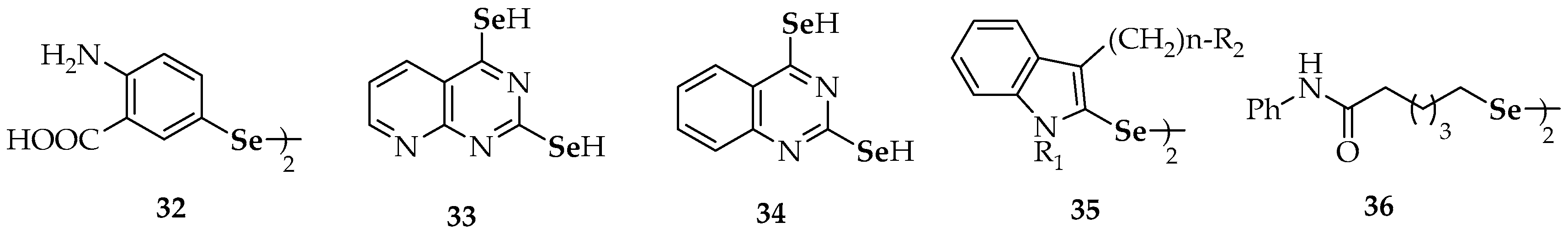
In the frame of kinase modulation as a possible pathway in the mechanisms of action of selenocompounds as anticancer agents [73], a library of organoselenium compounds was analyzed by Plano et al. [74]. Compounds with a variety of aromatic, heteroaromatic and aliphatic nitrogen-containing moieties (32–36, Figure 3) showed a noteworthy influence over different kinases. Among them, the bis(4-amino-3-carboxyphenyl) diselenide derivative 32 showed a mild inhibition of cyclin kinase (CDKs) activity. On the other hand, selenol-disubstituted pyrido[2,3-d]pyrimidine (33) and quinazoline (34) derivatives were proposed as promising candidates against cancer and other diseases: they are capable to produce a potent activation of the insulin-like growth factor 1 receptor (IGF-1R), playing thus a major role in cancer growth, tumor cell survival and resistance to therapy [75]. In line with the former compounds, the indol-containing diselenide 35 acted through inhibition of tyrosine kinase [73].
The antitumoral effect of the amide-containing derivative 36 [76] has been partially attributed to its capability for inhibiting the PI3 kinase pathway (see Section 2.2.4).
2.2.4. Antiproliferative and Cytotoxic Activity of Synthetic Diselenides
There are several Se-based compounds that can inhibit the growth of malignant cells. The challenge of such compounds is selectivity, in the sense that the effect on normal cells should ideally be marginal. Among the compounds with antiproliferative potential, 5 (Figure 1), 6 and 7 (Figure 2), which have been already mentioned with regard to their antioxidant activity, inhibited cell growth in murine hepatoma (Hepa 1c1c7) cells [52]. The authors pointed out that the inhibition of cell growth correlated with their potential to induce quinone reductase and glutathione-S-transferase activity. By contrast, the 3,3′-diselenodipropionic acid 13, described as a radio-protective agent and a free-radical scavenger, was not found to be toxic toward lymphocytes and EL4 tumor cells at the concentrations used [56].
Regarding the phenylcarbamate diselenides 25a, 25b and 25c (Figure 2) [66], which showed radical scavenging activity but lacked in GPx activity (mentioned above), they displayed the best in vitro antiproliferative activities among several tested diselenides against a panel of human tumor cell lines including lung carcinoma (HTB-54), colon carcinoma (HT-29), lymphocytic leukemia (CCRF-CEM), breast adenocarcinoma (MCF-7) and prostate adenocarcinoma (PC-3). Unfortunately, most compounds exhibited similar activities in both tumor and non-tumor cell lines. By a subsequent molecular modeling analysis [77], it was concluded that the cytotoxic activity correlated with the greater or lesser ability to release the active fragments: the central common fragment, the alcohol or phenols moieties, or the corresponding selenol (RSeH) and selenenic acid (RSeOH) derivatives.
For estrone and diosgenin-based diselenides (26 and 27), the in vitro antiproliferative activity against a panel of six human solid tumor cell lines, when tested up to a 100 µM concentration, was negligible [67]. Cancer cell lines included non-small cell lung (A549 and SW1573), breast (HBL-100 and T-47D), cervix (HeLa) and colon (WiDr) [67].
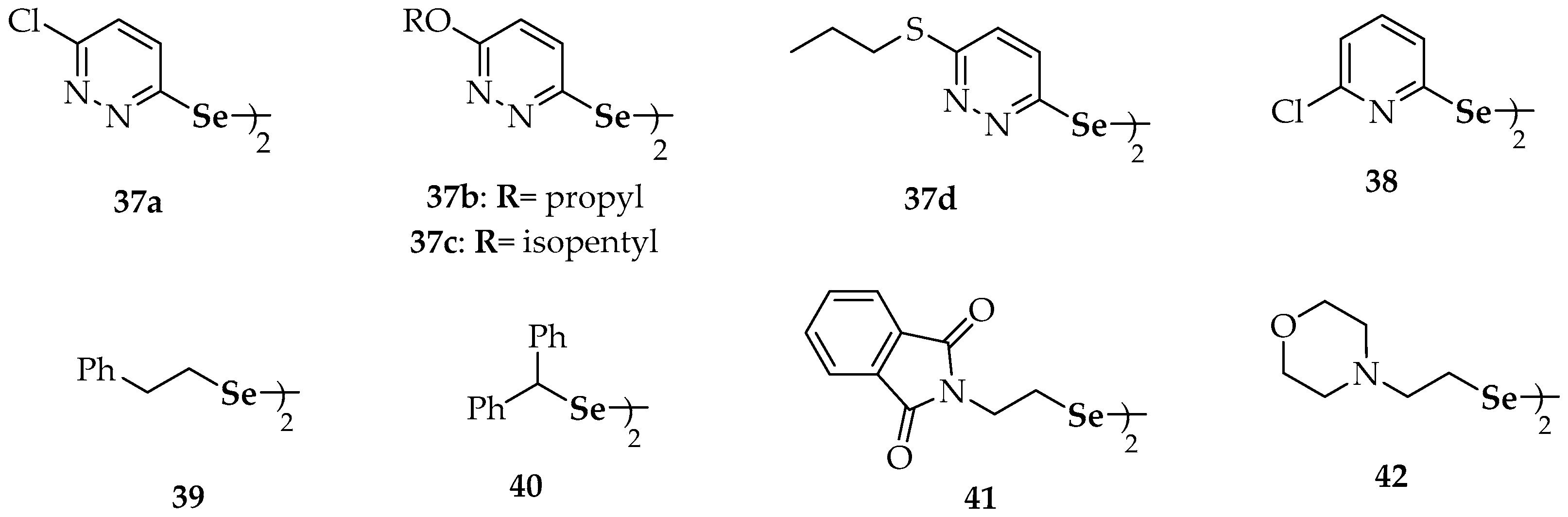
Substituted dipyridazinyl diselenides 37a–d (Figure 4), substituted diaralkyl diselenides (7), substituted dipyridinyl diselenides 15 and 38, di(phenylethyl) diselenide (39), dibenzhydryl diselenide (40), the phthalimide-containing diselenide 41 and the morpholine-containing diselenide 42 (Figure 2 and Figure 4, respectively) have been recently evaluated on their antiproliferative ability against human breast cancer MCF-7 cells [78]. The chloro-substituted pyridazine derivative 37a showed the highest potency, markedly inhibiting MCF-7 cell growth with an IC50 value of 10.34 µM, in a dose-dependent manner. In addition, other seven diselenides (37b, 37c, 37d, 38, 15, 41 and 42) showed higher potency, in terms of IC50, than the positive control (5-fluorouracil) against MCF-7 cells, suggesting the potential anticancer activity of these selenocompounds. The relatively broad diversity of structures that gave rise to antiproliferative activities makes it difficult to deduce a structure–activity relationship valid to explain the results, apart from the consideration of the symmetry that implies the presence of the diselenide moiety in all of these compounds [78].
Two of the active compounds in the abovementioned study of Kim et al. [78], phthalimide (41) and morpholine (42) derivatives, depicted sp3 carbon atoms directly bonded to selenium. As indicated above, most part of synthetic selenide-derivatives with reported anticancer activity depict Csp2-Se bonds, even though methylselenol has been proposed as the critical selenium metabolite of the dietary seleno-amino acids for anticancer activity [79]. Nevertheless, also compound 36 (Figure 3), described by Gowda et al. [76], has alkylic C-Se bonds. A promising anticancer activity has been reported for this symmetrical diarylalkyl diselenide. Compound 36 was tested in the prevention of early melanocytic lesion development in laboratory-generated skin reconstructs of WM35 or WM115 melanoma cell lines [76]. The topical application of 36 inhibited melanoma by up to 87% with negligible toxicological effects. Mechanistically, its action corresponds to a histone deacetylase (HDAC) and PI3 kinase pathway inhibitor. This combination of actions explains its efficacy in comparison to suberoylanilide hydroxamic acid (SAHA), the best-known HDAC inhibitor at the time. Furthermore, results suggested the efficacy of the indole derivative 35 (Figure 3) against other cancer types, as it was also able to inhibit cell growth in pancreatic (MIA-PA-Ca-2), breast (MDA-MB-231), prostate (PC-3) or sarcoma (HT-1080) cell lines.
2.2.5. Apoptotic Activity of Synthetic Diselenides
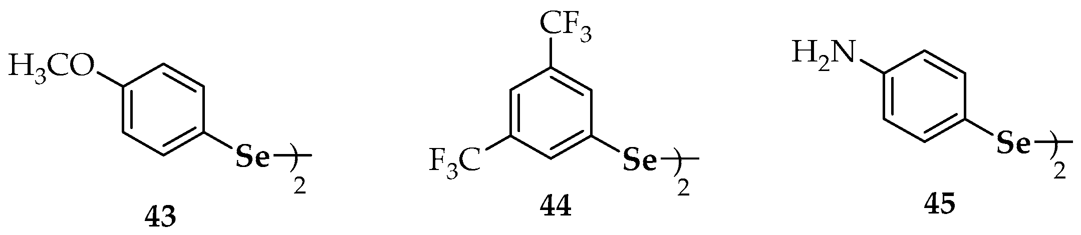
Diphenyl diselenide (6) can down-regulate the expression of the nuclear factor-κB (NF-κB), which is a transcription factor involved in pro-apoptotic pathways activated by ROS [80]. Similarly, substituted diaryl diselenides 11 (Figure 2) and 43 (Figure 5) were shown to induce apoptosis in human colon adenocarcinoma cells (HT-29) [80]. Nedel et al. used the 3-(4,5-dimethylthiazol-2-yl)-2,5-diphenyl-tetrazolium bromide (MTT) assay and flow cytometry analyses to show the effect of 11 and 43. They proposed that such action was produced through activation of caspase-dependent and independent pathways, in addition to cell-cycle arrest mediated by the p53, p21 and MYC genes.
The cytotoxic and apoptotic potential of diselenide 44 (Figure 5) has been confirmed though DNA cell cycle analysis and through microscopic monitoring of the formation of apoptotic bodies [81]. Among the 13 symmetric aromatic diselenides with a variety of substitution patterns evaluated by Rizvi et al. [81], compound 44 showed the highest inhibition of cell growth in human leukemia HL-60 cells (IC50 = 8 µM). Its cytotoxic potential against prostate (PC-3), breast (MCF-7), pancreatic (MIA-PA-Ca-2) and colon (HCT-116) cancer cell lines was also demonstrated (IC50 values of 13, 18, 25 and 27 µM, respectively). The antiproliferative activity of 44 on HL-60 cells involved the inhibition of the S phase of the cell cycle and the induction of apoptosis through the mitochondrial-dependent pathway. The authors also determined the DNA binding ability of 44 using molecular docking, showing that binding of the compound occurs selectively to the minor groove of DNA through hydrogen bonds involving the fluorine atoms. This binding was reinforced through hydrophobic interactions mediated by the benzene rings. Interestingly, pyridine-containing compound 15 (Figure 2) did not show any remarkable pro-apoptotic potential against the series of cell lines mentioned above [81].
A comprehensive library including nine symmetrical diarylalkyl diselenides (Ar-(CH2)n-Se-Se-(CH2)n-Ar; n = 0, 1) has been assayed for apoptotic potential in several cell lines [82]. Breast adenocarcinoma (MCF-7) cells were considered, as well as human prostate (PC-3) cells, lymphocytic leukemia (CCRF-CEM) cells and human colorectal adenocarcinoma (HT-29) cells. Again, it was a diaryl diselenide (45, Figure 5) the compound which displayed the best therapeutic profile regarding superoxide generation and cell cytotoxicity. Thus, aniline derivative (45) was the most potent organoselenium compound (among 59 candidates) against human prostate (PC-3) cells (IC50 = 1.7 µM) and was 8-fold more active than etoposide (IC50 = 13.6 µM), an agent commonly used in the treatment of prostate cancer. Compound 45 also exhibited more cytotoxic potency (IC50 = 4.3 µM) than etoposide (IC50 = 17.5 µM) in MCF-7 cells and showed noteworthy cytotoxic effects both in CCRF-CEM cells (IC50 = 9.0 µM) and HT-29 cells (IC50 = 9.8 µM). Additionally, compound 45 affected the cell cycle distribution of MCF-7 cells in the G2/M and S-phases. Moreover, 45 developed an apoptotic activity in CCRF-CEM cells, mediated by reactive oxygen species (ROS) and similar to camptothecin, the pro-apoptotic compound used as positive control.
2.3. Synthetic Selenides
Organic selenides are less reported in the literature of anticancer reagents in comparison to diselenide derivatives. Herein, the studies involving selenides are divided in two main activities: antioxidant (redox modulating, antioxidant and chemopreventive compounds) and antitumoral compounds (derivatives with antiproliferative, cytotoxic and apoptotic activity). The chemical names of the mentioned selenides are summarized in Table 2.
2.3.1. Redox-Modulating and Antioxidant and Chemo-Preventive Activities of Synthetic Selenides
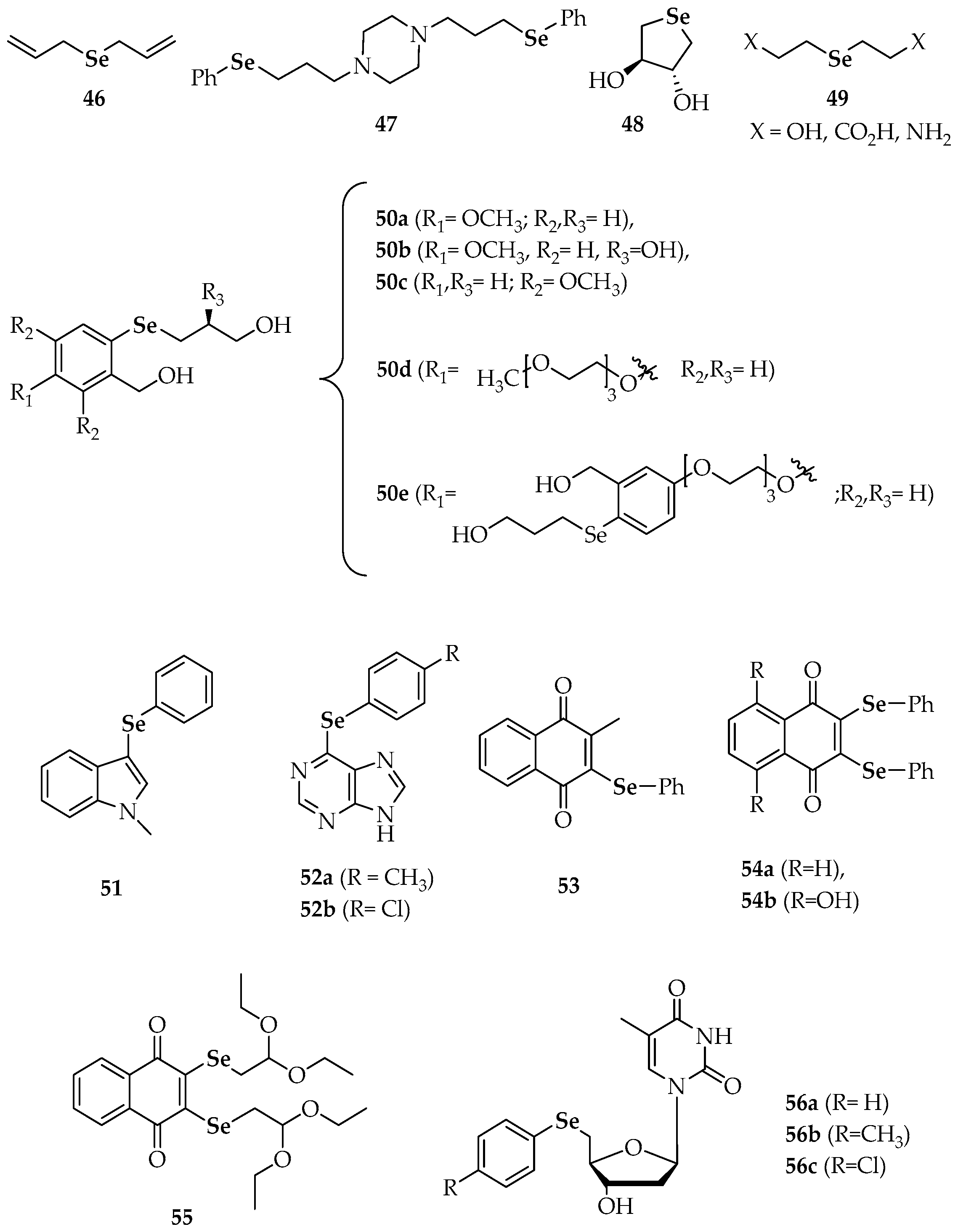
The diallyl selenide 46 (Figure 5) exhibited significant anti-carcinogenic activity when assayed in a murine (DMBA)-induced mammary tumor model in a study performed to compare the chemopreventive potential of organoselenium compounds versus the corresponding sulfur analogs against cancer [83]. Through administration in a pre-initiation period to tumor development, the protective activity of this symmetric selenide resulted around 300 times superior to its sulfur isoster (diallyl sulfide). In a more recent research work by Mecklenburg et al. [84], the piperazine compound 47, which combines redox catalysis with metal binding properties, was virtually non-toxic (at concentrations up to 50 µM) against human SK-Mel-5 melanoma cells in the absence of H2O2 (91% survival at 25 µM), whereas it showed a toxicity in the presence of H2O2 2–3-fold higher than in its absence (65% survival at 25 µM). As compound 47 (Figure 5) was somehow able to recognize the particular redox state of the cells and, according to this state, exert either an antioxidative or a prooxidative action, it has been considered a sensor/effector agent.
Some additional selenides have been studied from the point of view of their antioxidant activity, but on these occasions, they have not been explicitly connected to anticancer research. For instance, Kumakura et al. [85] evaluated the GPx-like activity [86] of the water-soluble cyclic selenide 48 (Figure 5). In the same way as it occurred with cyclic diselenides 28 (Figure 2), the derivative 48 exerted higher antioxidant catalytic activity than its linear analogs 49. According to the authors, conformational restriction increased the HOMO energy level and made the selenium atom more exposed. Other selenides, such as 50a–c (Figure 5), provided fast reaction rates for the reduction of H2O2 with thiols by both high-performance liquid chromatography (HPLC) and nuclear magnetic resonance (NMR)-based assays [87]. The fact that rates were more elevated than for their unsubstituted or otherwise methoxy-substituted analogues was attributed to resonance stabilization of increased positive charge on the selenium atom during the rate determining oxidation step. The polyethylene glycol (PEG) derivatives 50d and 50e were prepared to increase aqueous solubility and they also afforded remarkable activity. Noteworthy, both the non-PEGylated and PEGylated compounds that revealed the best activities gave rise spirodioxyselenuranes adducts upon oxidation, while derivatives with diminished activity generated pincer compounds.
Prevention of oxidative damage and radical scavenging potential were found for the indol-selenide derivative 51 [88] and the 6-arylselanylpurines 52a and 52b [89], respectively. Apart from preventing oxidative stress, compound 51 did not show hepatic, renal or cerebral toxicity. Regarding derivatives 52a and 52b, both presented a significant ABTS radical scavenging activity at concentrations equal/higher than 100 µM, although only the latter displayed moderated DPPH radical scavenging activity, at a concentration of 200 µM.
The multifunctional redox naphthalene-1,4-dione selenides (53, 54a, 54b and 55), which combine more than one redox site in one molecule including a quinone moiety, are particularly suitable to target (cancer) cells under ROS. Compound 53 seemed to trigger the formation of superoxide in the mitochondria, since a yeast mutant deficient in superoxide dismutase 2 (SOD2) was hypersusceptible to it [90]. On the other hand, compound 54b exhibited similar GPx-like activity to ebselen [91], increasing the initial rate of the reaction by 1.95-fold (ebselen increased the rate by 1.87-fold).
The in vitro antioxidant activities of several chalcogen-based zidovudine (3′-Azido-3′-deoxythymidine, AZT) analogues have been explored [92]. Firstly, the thiobarbituric acid reactive substance (TBARS) assay allowed the quantification of the final products of lipid peroxidation in brain homogenates and showed excellent results for compounds 56b and 56c at two different concentrations (100 and 200 µM). Additionally, the same compounds were able to decompose H2O2 efficiently and this decomposition involved a 4–5-fold decrease in times required to oxidize 50% of the benzenethiol in comparison to DMSO (control) and to therapeutically employed AZT.
2.3.2. Antitumoral Activity of Synthetic Selenides
To gain an initial overview of possible cytotoxicity of the naphthalene-1,4-dione derivatives (53, 54a, 54b and 55, Figure 6), an activity screening was performed in human myeloid leukemia K562 cells [91]. Compound 55 was found to be particularly active at 10 µM, with an average remaining cell viability of just 8.6%. However, the effect of the latter might also be related to the protected aldehyde (acetal) function: the reactive aldehyde could be liberated in cells and exert its own toxic effects. When tested in chronic lymphocytic leukemia (CLL) B-cells isolated from the peripheral blood (PB) of leukemia patients, compounds 54a and 54b showed strong activity in reducing CLL B-cell viability at low to submicromolar concentrations [91]. At the same concentrations, viability of healthy donor peripheral blood mononuclear cells (PBMC) was unaffected by 54a whereas a high toxicity to healthy control cells was observed for compound 54b. Thus, compound 54a was the most effective in selectively reducing the viability of CLL cells. This effect was particularly pronounced at 500 nM and 1 µM. Fludarabine, the major single chemotherapeutic component used in CLL at the moment, only afforded around 30% reduction of CLL B-cell survival in vitro when used at clinically feasible doses. The significant reduction of cell survival achieved by 54a at concentrations as low as 500 nM means that it compares well with such established cytostatics, at least in vitro.
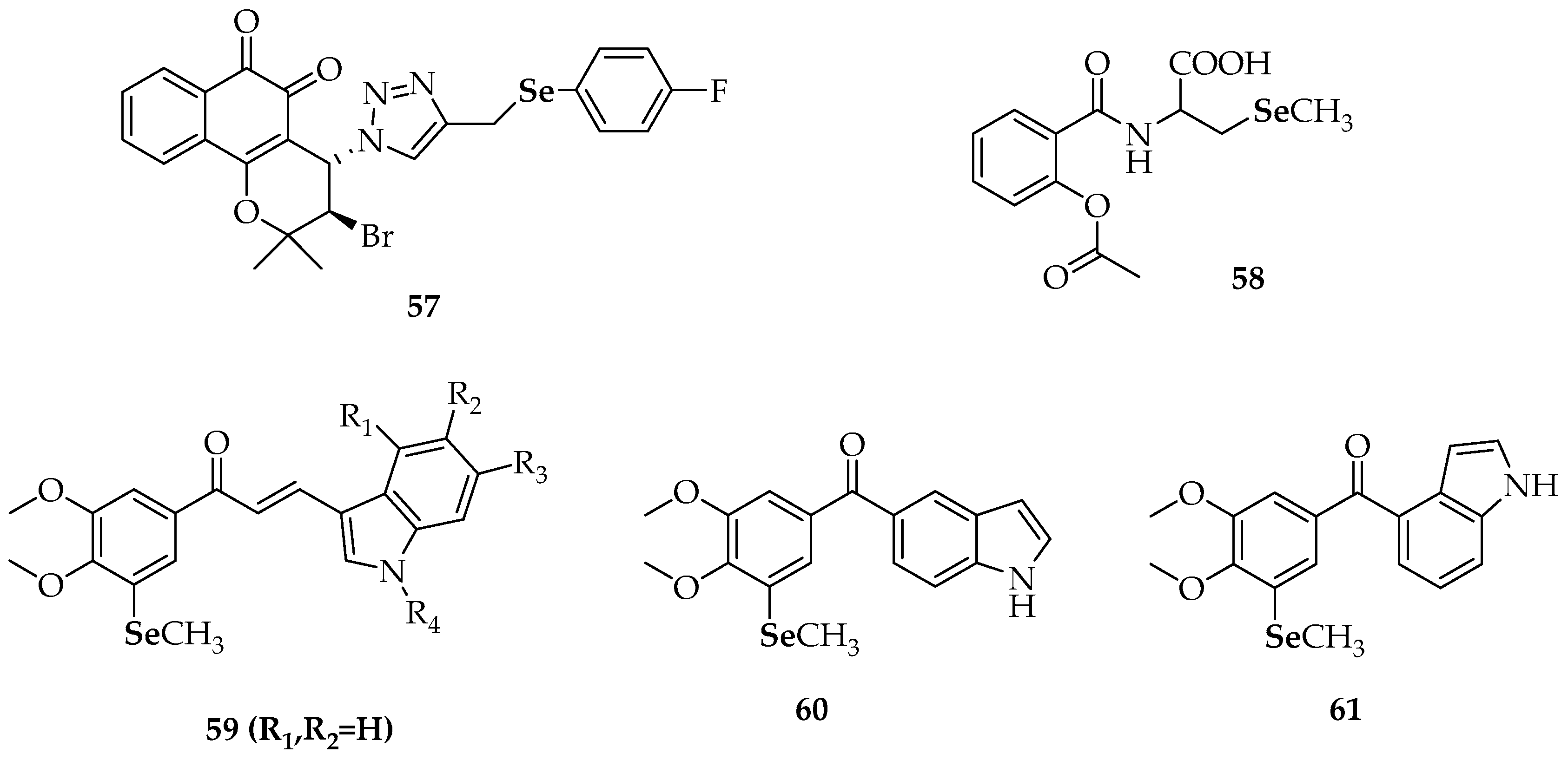
In relation to the above mentioned quinone-containing selenides, the antiproliferative activity of the 1,2-naphthoquinone-containing selenide (57, Figure 7) was found to be superior to the activity of other 1,2- and 1,4-naphthoquinone-based chalcogen derivatives [93]. While the IC50 values of the latter were between 10 and 100 µM and for other 1,2-naphthoquinone derivatives were between 1 and 10 µM, compound 57 was active even in sub-micromolar concentrations against human promyelocytic leukemia cells (HL-60) and human colon carcinoma cells (HCT-116). The remarkable activity of 57 was attributed to three factors: the 1,2-naphthoquinone moiety, the Se atom and the electron-withdrawing substituent in para-position to Se. Unfortunately, no selective cytotoxicity against cancer versus normal cells was detected.
One of the major limitations in the clinical use of AZT is associated with hepatotoxicity. In fact, AZT caused a significant reduction in hepatic cell viability at concentrations of 200, 100 and 50 µM compared with the above-mentioned compound 56c (Figure 6) at 200 µM [92]. When the cytotoxicity against bladder carcinoma cells (5637) was tested, AZT proved to be more effective than its derivatives in inhibiting tumor growth at the lowest concentrations. Nevertheless, at the highest concentrations, 56a–c were more effective and did not cause cellular injury. Derivatives 56a–c also showed better pro-apoptotic properties than AZT in 5637 cells according to annexin V-binding assays. Thus, at 50 µM, 56c induced around 70% of apoptosis whereas AZT caused around 8% of apoptosis. Additionally, the percentage of apoptosis reached 90% by exposure of 5637 cells to 56a–c at 100 µM. The analysis of DNA fragmentation revealed that 56a–c significantly increased apoptosis-associated DNA fragmentation comparing to AZT. When the gene expression of anti-apoptotic Bcl-2 and pro-apoptotic BAX was investigated, the authors found that the BAX: Bcl-2 ratio was affected by these compounds. Whereas Bcl-2 mRNA levels were significantly lower (compared to control) in cells exposed to both AZT and 56a–c, BAX mRNA levels were significantly higher except for cells exposed to AZT. Moreover, a predominance of BAX to Bcl-2 levels, as well as significant increases in caspase-9 mRNA levels, were observed for compounds 56a and 56b in comparison with the controls.
Plano et al. [94] evaluated the anticancer activity of several synthetic anti-inflammatory drug-hybrid molecules against four cancer cell lines. Among the compounds tested, the derivative 58 (Figure 7), which is a conjugate of selenocysteine with aspirin, was only moderately cytotoxic to human pancreatic carcinoma cell line (PANC-1 cells). The most active compound was an aspirin analogue N-substituted with a 2-selenocyanoethyl group, which was particularly effective in reducing the viability of colorectal cancer (CRC) cells.
Very recently, Zhang et al. [95] studied the antiproliferative activity of several synthetic indole-chalcone derivatives 59 and the phenylindolyl ketone derivatives 60 and 61 (Figure 7). Most of these compounds exhibited good to excellent activity (with IC50 values in the sub-micromolar level) in six human cancer cell lines: non-small-cell-lung cancer cells (A549), human breast cancer cells (MDAMB-231), human liver carcinoma cells (HepG2), human epithelial cervical cancer cells (HeLa), human colon carcinoma cells (HCT-116 and RKO). Among the indole-chalcone derivatives, the non-substituted indole-3-carbaldehyde 59 exhibited the highest activity (23 to 79 nM). The authors found that an N-methyl group in the indole moiety was unfavorable and that the activity was related to the position of substitution on the indole rings. Compound 61 (Figure 7), a phenylindolyl ketone derivative, exhibited the most potent antiproliferative activities against the six human cancer cell lines with IC50 values between 4 nM for the A549 cell line and 22 nM for RKO cells. A microtubule dynamics assay and an immunofluorescence assay confirmed that 61 could effectively inhibit tubulin polymerization in HeLa cells (IC50 = 2.1 ± 0.27 µM). Further cellular mechanism studies revealed that 61 induced G2/M phase arrest and that apoptosis was associated with a decrease in the mitochondrial membrane potential Δψm. Compared to the reference compound showing an oxygen atom instead a Se atom, (1H-indol-4-yl)(3,4,5-trimethoxyphenyl)methanone, a remarkable effect of substitution by selenium was observed, suggesting that the introduction of selenium was beneficial to the antitumor activity. The activity of 61 also pointed out that the joint position of benzoyl on the indole ring was important.
3. Conclusions
Our overview of current selenium compounds with anticancer and chemopreventive properties indicates the predominant role for the diaryldiselenide family that is also wider investigated than both selenides and dialkyldiselenides.
An analysis of the comprehensive studies on (di)selenide compounds shows some structure–activity relationship that can be useful for further search of new selenium anticancer agents with therapeutic perspective. Thus, the most potent anticancer properties, with the widest spectrum of protein targets and mechanisms involved in their action, can be observed for symmetric diselenides with direct aromatic moieties. The alkyl spacer between an aryl ring and Se usually causes a decrease of the desirable biological actions.
The role of the aromatic ring properties, including their size, presence of heteroatoms as well as the number and kind of substituents, is crucial for potency and decisive for mechanisms of anticancer/chemopreventive action. Various lines of evidence underline an importance of nitrogen atom, occurred either as exo-amine substituents or as endocyclic heteroatoms of pyridine, pyridazine, quinolines, indol or purines. More or less electron-withdrawing substituents, i.e., halogens, trifluoromethyl or nitro groups, at the diphenyl diselenides aromatic rings, play also an important role and can be found within the structures of apoptosis agents of particularly dangerous human cancer lines. However, unsubstituted aromatic rings of diphenyl diselenides have been confirmed as beneficial for various mechanisms of anticancer and chemopreventive activities, including protection of murine J774 macrophage-like cells from ROS, an action involving down-regulation of NF-κB or anti-genotoxic one.
On the other hand, some studies confirm a favorable effect of the electro-donating methoxy-substituent that causes the chemoprevention increase. The extensive results of anti-cancer properties of a significant number of selenocompounds show that their symmetric topology is of particular importance. This is observed not only for diaryl, but also for dialkyl diselenides and for the less extended family of the selenide agents. Among selenides, the most potent anticancer properties were identified for the symmetric diallylselenide, although the cyclic compounds are confirmed as more potent than linear ones. The recent search for anticancer agents has also provided some data for niche groups of selenium compounds, i.e., the active ferrocene- and peri-diselenides and the less active selenosteroids.
Considering both the potent anticancer/chemopreventive activity and various mechanisms of the biological action, selenides and, especially, diselenides described to date, create a library of “small molecule catalysts” that can be useful for further stages of the antitumor drug research and development process to give a high probability of finding new therapies against cancer.
Acknowledgments
This study is supported by the Spanish “Consejo Superior de Investigaciones Científicas” (CSIC, Spanish National Research Council), through the project 201780I027.
Author Contributions
M.A.-P., W.A., M.A.M. and E.D.-A. performed the bibliography search and contributed to the writing of the manuscript. E.D.-A. coordinated the work of the different authors. J.H. wrote the Conclusions paragraph.
Conflicts of Interest
The authors declare no conflict of interest.
Abbreviations
| NNK | 4-(methylnitrosamino)-1-(3-pyridyl)-1-butanone |
| GSH | Reduced glutathione |
| GPx-like | Glutathione peroxidase-like |
| ROS | Reactive oxygen species |
| LDL | Low density lipoproteins |
| NF-κB | Nuclear factor-κB DNA Binding |
| MTT | 3-(4,5-dimethylthiazol-2-yl)-2,5-diphenyl-tetrazolium bromide |
| Trx | Thioredoxin |
| TrxR | Thioredoxin reductase |
| PDI | Disulfide isomerase |
| 2-NP | 2-Nitropropane |
| MT | Metallothionein |
| CDKs | Cyclin kinases |
| IGF-1R | Insulin-like growth factor 1 receptor |
| HDAC | Histone deacetylase |
| SAHA | Suberoylanilide hydroxamic acid |
| DPPH | 1,1-Diphenyl-2-picryl-hydrazyl |
| ABTS | 2,2-Azinobis(3-ethylbenzothiazoline- 6-sulfonic acid) |
| DMBA | 7, 12-dimethylbenz(a)anthracene |
| HPLC | High-performance liquid chromatography |
| NMR | Nuclear magnetic resonance |
| PEG | Polyethylene glycol |
| SOD2 | superoxide dismutase 2 |
| PB | peripheral blood |
| PBMC | peripheral blood mononuclear cells |
References
- Fairweather-Tait, S.J.; Bao, Y.; Broadley, M.R.; Collings, R.; Ford, D.; Hesketh, J.E.; Hurst, R. Selenium in human health and disease. Antioxid. Redox Signal. 2011, 14, 1337–1383. [Google Scholar] [CrossRef] [PubMed]
- Rayman, M.P. The importance of selenium to human health. Lancet 2000, 356, 233–241. [Google Scholar] [CrossRef] [Green Version]
- Roman, M.; Jitaru, P.; Barbante, C. Selenium biochemistry and its role for human health. Metallomics 2014, 6, 25–54. [Google Scholar] [CrossRef] [PubMed] [Green Version]
- Rayman, M.P. Selenium and human health. Lancet 2012, 379, 1256–1268. [Google Scholar] [CrossRef]
- Lu, J.; Holmgren, A. Selenoproteins. J. Biol. Chem. 2009, 284, 723–727. [Google Scholar] [CrossRef] [PubMed]
- Rose, A.H.; Hoffmann, P.R. Selenoproteins and cardiovascular stress. Throm. Haemost. 2015, 113, 494–504. [Google Scholar] [CrossRef] [PubMed]
- Steinbrenner, H.; Speckmann, B.; Klotz, L.O. Selenoproteins: Antioxidant selenoenzymes and beyond. Arch. Biochem. Biophys. 2016, 595, 113–119. [Google Scholar] [CrossRef] [PubMed]
- Sanmartín, C.; Plano, D.; Sharma, A.K.; Palop, J.A. Selenium compounds, apoptosis and other types of cell death: An overview for cancer therapy. Int. J. Mol. Sci. 2012, 13, 9649–9672. [Google Scholar] [CrossRef] [PubMed]
- Fernandes, A.P.; Gandin, V. Selenium compounds as therapeutic agents in cancer. Biophys. Acta 2014, 1850, 1642–1660. [Google Scholar] [CrossRef] [PubMed]
- Sanmartín, C.; Plano, D.; Font, M.; Palop, J.A. Selenium and clinical trials: New therapeutic evidence for multiple diseases. Curr. Med. Chem. 2011, 18, 4635–4650. [Google Scholar] [CrossRef] [PubMed]
- Mugesh, G.; du Mont, W.W.; Sies, H. Chemistry of biologically important synthetic organoselenium compounds. Chem. Rev. 2001, 101, 2125–2179. [Google Scholar] [CrossRef] [PubMed]
- Banerjee, B.; Koketsu, M. Recent developments in the synthesis of biologically relevant selenium-containing scaffolds. Coord. Chem. Rev. 2017, 339, 104–127. [Google Scholar] [CrossRef]
- Nogueira, C.W.; Zeni, G.; Rocha, J.B. Organoselenium and organotellurium compounds: Toxicology and pharmacology. Chem. Rev. 2004, 104, 6255–6285. [Google Scholar] [CrossRef] [PubMed]
- Alberto, E.E.; Nascimiento, V.; Braga, A.L. Catalytic application of selenium and tellurium compounds as glutathione peroxidase enzyme mimetics. J. Braz. Chem. Soc. 2010, 21, 2032–2041. [Google Scholar] [CrossRef]
- Brozmanová, J.; Mániková, D.; Vlčková, V.; Chovanec, M. Selenium: A double-edged sword for defense and offence in cancer. Arch. Toxicol. 2010, 84, 919–938. [Google Scholar] [CrossRef] [PubMed]
- Misra, S.; Boylan, M.; Selvam, A.; Spallholz, J.E.; Björnstedt, M. Redox-active selenium compounds—From toxicity and cell death to cancer treatment. Nutrients 2016, 7, 3536. [Google Scholar] [CrossRef] [PubMed]
- Domínguez-Álvarez, E.; Plano, D.; Font, M.; Calvo, A.; Prior, C.; Jacob, C.; Palop, J.A.; Sanmartín, C. Synthesis and antiproliferative activity of novel selenoester derivatives. Eur. J. Med. Chem. 2014, 73, 153–156. [Google Scholar] [CrossRef] [PubMed]
- Sanmartín, C.; Plano, D.; Domínguez, E.; Font, M.; Calvo, A.; Prior, C.; Encío, I.; Palop, J.A. Synthesis and pharmacological screening of several aroyl and heteroaroyl selenylacetic acid derivatives as cytotoxic and antiproliferative agents. Molecules 2009, 14, 3313–3338. [Google Scholar] [CrossRef] [PubMed]
- Orian, L.; Toppo, S. Organochalcogen peroxidase mimetics as potential drugs: A long story of a promise still unfulfilled. Free Radic. Biol. Med. 2014, 66, 65–74. [Google Scholar] [CrossRef] [PubMed]
- Ronai, Z.; Tillotson, J.K.; Traganos, F.; Darzynkiewicz, Z.; Conaway, C.C.; Upadhyaya, P.; El-Bayoumy, K. Effects of organic and inorganic selenium compounds on rat mammary tumour cells. Int. J. Cancer 1995, 63, 428–434. [Google Scholar] [CrossRef] [PubMed]
- Kieliszek, M.; Lipinski, B.; Błażejak, S. Application of Sodium Selenite in the Prevention and Treatment of Cancers. Cells 2017, 6, 39. [Google Scholar] [CrossRef] [PubMed]
- Lipinski, B. Sodium selenite as an anticancer agent. Anticancer Agents Med. Chem. 2017, 17, 658–661. [Google Scholar] [CrossRef] [PubMed]
- Evans, S.O.; Khairuddin, P.F.; Jameson, M.B. Optimising Selenium for Modulation of Cancer Treatments. Anticancer Res. 2017, 37, 6497–6509. [Google Scholar] [PubMed]
- Jamier, V.; Ba, L.A.; Jacob, C. Selenium- and tellurium-containing multifunctional redox agents as biochemical redox modulators with selective cytotoxicity. Chemistry 2010, 16, 10920–10928. [Google Scholar] [CrossRef] [PubMed]
- Storkey, C.; Davies, M.J.; White, J.M.; Schiesser, C.H. Synthesis and antioxidant capacity of 5-selenopyranose derivatives. Chem. Commun. 2011, 47, 9693–9695. [Google Scholar] [CrossRef] [PubMed]
- Mániková, D.; Vlasáková, D.; Loduhová, J.; Letavayová, L.; Vigasová, D.; Krascsenitsová, E.; Vlcková, V.; Brozmanová, J.; Chovanec, M. Investigations on the role of base excision repair and non-homologous end-joining pathways in sodium selenite-induced toxicity and mutagenicity in Saccharomyces cerevisiae. Mutagenesis 2010, 25, 155–162. [Google Scholar] [CrossRef] [PubMed]
- Pang, Y.; An, B.; Lou, L.; Zhang, J.; Yan, J.; Huang, L.; Li, X.; Yin, S. Design, Synthesis, and Biological Evaluation of Novel Selenium-Containing Isocombretastatins and Phenstatins as Antitumor Agents. J. Med. Chem. 2017, 60, 7300–7314. [Google Scholar] [CrossRef] [PubMed]
- Domracheva, I.; Kanepe-Lapsa, I.; Jackevica, L.; Vasiljeva, J.; Arsenyan, P. Selenopheno quinolinones and coumarins promote cancer cell apoptosis by ROS depletion and caspase-7 activation. factor 2. Life Sci. 2017, 186, 92–101. [Google Scholar] [CrossRef] [PubMed]
- Domínguez-Álvarez, E.; Gajdács, M.; Spengler, G.; Palop, J.A.; Marć, M.A.; Kieć-Kononowicz, K.; Amaral, L.; Molnár, J.; Jacob, C.; Handzlik, J.; et al. Identification of selenocompounds with promising properties to reverse cancer multidrug resistance. Bioorg. Med. Chem. Lett. 2016, 26, 2821–2824. [Google Scholar] [CrossRef] [PubMed]
- Li, W.; Guo, M.; Liu, Y.; Mu, W.; Deng, G.; Li, C.; Qiu, C. Selenium induces an anti-tumor effect via inhibiting intratumoral angiogenesis in a mouse model of transplanted canine mammary tumor cells. Biol. Trace Elem. Res. 2016, 171, 371–379. [Google Scholar] [CrossRef] [PubMed]
- Gajdács, M.; Spengler, G.; Sanmartín, C.; Marć, M.A.; Handzlik, J.; Domínguez-Álvarez, E. Selenoesters and selenoanhydrides as novel multidrug resistance reversing agents: A confirmation study in a colon cancer MDR cell line. Bioorg. Med. Chem. Lett. 2017, 27, 797–802. [Google Scholar] [CrossRef] [PubMed]
- Sakalli Çetin, E.; Nazıroğlu, M.; Çiğ, B.; Övey, İ.S.; Aslan Koşar, P. Selenium potentiates the anticancer effect of cisplatin against oxidative stress and calcium ion signaling-induced intracellular toxicity in MCF-7 breast cancer cells: Involvement of the TRPV1 channel. J. Recept. Signal. Transduct. Res. 2017, 37, 84–93. [Google Scholar] [CrossRef] [PubMed]
- Emmert, S.W.; El-Bayoumy, K.; Das, A.; Sun, Y.W.; Amin, S.; Desai, D.; Aliaga, C.; Richie, J.P., Jr. Induction of lung glutathione and glutamylcysteine ligase by 1,4-phenylenebis(methylene)selenocyanate and its glutathione conjugate: Role of nuclear factor-erythroid 2-related factor 2. Free Radic. Biol. Med. 2012, 52, 2064–2071. [Google Scholar] [CrossRef] [PubMed]
- Alcolea, V.; Plano, D.; Karelia, D.N.; Palop, J.A.; Amin, S.; Sanmartín, C.; Sharma, A.K. Novel seleno- and thio-urea derivatives with potent in vitro activities against several cancer cell lines. Eur. J. Med. Chem. 2016, 113, 134–144. [Google Scholar] [CrossRef] [PubMed]
- Díaz-Argelich, N.; Encío, I.; Plano, D.; Fernandes, A.P.; Palop, J.; Sanmartín, C.; Sanmartín, C. Novel Methylselenoesters as Antiproliferative Agents. Molecules 2017, 22, 1288. [Google Scholar] [CrossRef] [PubMed]
- Nakamura, Y.; Feng, Q.; Kumagai, T.; Torikai, K.; Ohigashi, H.; Osawa, T.; Noguchi, N.; Niki, E.; Uchida, K. Ebselen, a glutathione peroxidase mimetic seleno-organic compound, as a multifunctional antioxidant. Implication for inflammation-associated carcinogenesis. J. Biol. Chem. 2002, 277, 2687–2694. [Google Scholar] [CrossRef] [PubMed]
- Estevam, E.C.; Griffin, S.; Nasim, M.J.; Denezhkin, P.; Schneider, R.; Lilischkis, R.; Dominguez-Alvarez, E.; Witek, K.; Latacz, G.; Keck, C.; et al. Natural selenium particles from Staphylococcus carnosus: Hazards or particles with particular promise? J. Hazard. Mater. 2017, 324, 22–30. [Google Scholar] [CrossRef] [PubMed]
- Sanmartín, C.; Font, M.; Palop, J.A. Molecular symmetry: A structural property frequently present in new cytotoxic and proapoptotic drugs. Mini Rev. Med. Chem. 2006, 6, 639–650. [Google Scholar] [CrossRef] [PubMed]
- Sanmartín, C.; Plano, D.; Palop, J.A. Selenium compounds and apoptotic modulation: A new perspective in cancer therapy. Mini Rev. Med. Chem. 2008, 8, 1020–1031. [Google Scholar] [CrossRef] [PubMed]
- Li, L.; Xie, Y.; El-Sayed, W.M.; Szakacs, J.G.; Franklin, M.R.; Roberts, J.C. Chemopreventive activity of selenocysteine prodrugs against tobacco-derived nitrosamine (NNK) induced lung tumors in the A/J mouse. J. Biochem. Mol. Toxicol. 2005, 19, 396–405. [Google Scholar] [CrossRef] [PubMed]
- Chen, T.F.; Wong, Y.S. Selenocystine induces S-phase arrest and apoptosis in human breast adenocarcinoma MCF-7 cells by modulating ERK and Akt phosphorylation. J. Agric. Food Chem. 2008, 56, 10574–10581. [Google Scholar] [CrossRef] [PubMed]
- Kim, T.-S.; Yun, B.Y.; Kim, I.Y. Induction of the mitochondrial permeability transition by selenium compounds mediated by oxidation of the protein thiol groups and generation of the superoxide. Biochem. Pharmacol. 2003, 66, 2301–2311. [Google Scholar] [CrossRef] [PubMed]
- Li, G.X.; Hu, H.; Jiang, C.; Schuster, T.; Lü, J. Differential involvement of reactive oxygen species in apoptosis induced by two classes of selenium compounds in human prostate cancer cells. Int. J. Cancer 2007, 120, 2034–2043. [Google Scholar] [CrossRef] [PubMed]
- Saito, Y.; Umemoto, D.; Matsunaga, A.; Sato, T.; Chikuma, M. Antioxidant activities of synthesized selenocompounds without selenol groups. Biomed. Res. Trace Elem. 2006, 17, 423–426. [Google Scholar] [CrossRef]
- Kumar, S.; Singh, H.B. Thiol peroxidase-like activity of some intramolecularly coordinated diorganyl diselenides. J. Chem. Sci. 2005, 117, 621–628. [Google Scholar] [CrossRef]
- Meotti, F.C.; Stangherlin, E.C.; Zeni, G.; Nogueira, C.W.; Rocha, J.B. Protective role of aryl and alkyl diselenides on lipid peroxidation. Environ. Res. 2004, 94, 276–282. [Google Scholar] [CrossRef]
- Rosa, R.M.; Moura, D.J.; Romano e Silva, A.C.; Saffi, J.; Pegas-Henriques, J.A. Antioxidant activity of diphenyl diselenide prevents the genotoxicity of several mutagens in Chinese hamster V79 cells. Mutat. Res. 2007, 631, 44–54. [Google Scholar] [CrossRef] [PubMed]
- Brandão, R.; Acker, C.I.; Leite, M.R.; Barbosa, N.B.; Nogueira, C.W. Diphenyl diselenide protects against glycerol-induced renal damage in rats. J. Appl. Toxicol. 2009, 29, 612–618. [Google Scholar] [CrossRef] [PubMed]
- Straliotto, M.R.; Hort, M.A.; Fiuza, B.; Rocha, J.B.; Farina, M.; Chiabrando, G.; de Bem, A.F. Diphenyl diselenide modulates oxLDL-induced cytotoxicity in macrophage by improving the redox signaling. Biochimie 2013, 95, 1544–1551. [Google Scholar] [CrossRef] [PubMed]
- Melo, M.T.; de Oliveira, I.M.; Grivicich, I.; Guecheva, T.N.; Saffi, J.; Henriques, J.A.; Rosa, R.M. Diphenyl diselenide protects cultured MCF-7 cells against tamoxifen-induced oxidative DNA damage. Biomed. Pharmacother. 2013, 67, 329–335. [Google Scholar] [CrossRef] [PubMed]
- Da Silva Machado, M.; Villela, I.V.; Moura, D.J.; Rosa, R.M.; Salvador, M.; Lopes, N.P.; Braga, A.L.; Roesler, R.; Saffi, J.; Henriques, J.A. 3,3-Ditrifluoromethyldiphenyl diselenide: A new organoselenium compound with interesting antigenotoxic and antimutagenic activities. Mutat. Res. 2009, 673, 133–140. [Google Scholar] [CrossRef] [PubMed]
- Xiao, H.; Parkin, K.L. Induction of phase II enzyme activity by various selenium compounds. Nutr. Cancer 2006, 55, 210–223. [Google Scholar] [CrossRef] [PubMed]
- Reich, H.J.; Hondal, R.J. Why Nature Chose Selenium. ACS Chem. Biol. 2016, 11, 821–841. [Google Scholar] [CrossRef] [PubMed]
- Tanaka, T.; Reddy, B.S.; el-Bayoumy, K. Inhibition by dietary organoselenium, p-methoxybenzene-selenol, of hepatocarcinogenesis induced by azoxymethane in rats. Jpn. J. Cancer Res. 1985, 76, 462–467. [Google Scholar] [CrossRef] [PubMed]
- Reddy, B.S.; Tanaka, T.; el-Bayoumy, K. Inhibitory effect of dietary p-methoxybenzeneselenol on azoxymethane-induced colon and kidney carcinogenesis in female F344 rats. J. Natl. Cancer Inst. 1985, 74, 1325–1328. [Google Scholar] [CrossRef] [PubMed]
- Kunwar, A.; Mishra, B.; Barik, A.; Kumbhare, L.B.; Pandey, R.; Jain, V.K.; Priyadarsini, K.I. 3,3’-Diselenodipropionic acid, an efficient peroxyl radical scavenger and GPx mimic, protects erythrocytes (RBCs) from AAPH-induced hemolysis. Chem. Res. Toxicol. 2007, 20, 1482–1487. [Google Scholar] [CrossRef] [PubMed]
- Kunwar, A.; Bansal, P.; Kumar, S.J.; Bag, P.P.; Paul, P.; Reddy, N.D.; Kumbhare, L.B.; Jain, V.K.; Chaubey, R.C.; Unnikrishnan, M.K.; et al. In vivo radioprotection studies of 3,3′-diselenodipropionic acid, a selenocystine derivative. Free Radic. Biol. Med. 2010, 48, 399–410. [Google Scholar] [CrossRef] [PubMed]
- Ibrahim, M.; Prigol, M.; Hassan, W.; Nogueira, C.W.; Rocha, J.B. Protective effect of binaphthyl diselenide, a synthetic organoselenium compound, on 2-nitropropane-induced hepatotoxicity in rats. Cell Biochem. Funct. 2010, 28, 258–265. [Google Scholar] [CrossRef] [PubMed]
- Collins, C.A.; Fry, F.H.; Holme, A.L.; Yiakouvaki, A.; Al-Qenaei, A.; Pourzand, C.; Jacob, C. Towards multifunctional antioxidants: Synthesis, electrochemistry, in vitro and cell culture evaluation of compounds with ligand/catalytic properties. Org. Biomol. Chem. 2005, 3, 1541–1546. [Google Scholar] [CrossRef] [PubMed]
- Battin, E.E.; Perron, N.E.; Brumaghim, J.L. The central role of metal coordination in selenium antioxidant activity. Inorg. Chem. 2006, 45, 499–501. [Google Scholar] [CrossRef] [PubMed]
- Mugesh, G.; Panda, A.; Singh, H.B.; Punekar, N.S.; Butcher, R.J. Glutathione peroxidase-like antioxidant activity of diaryl diselenides: A Mechanistic Study. J. Am. Chem. Soc. 2001, 123, 839–850. [Google Scholar] [CrossRef] [PubMed]
- Mugesh, G.; Panda, A.; Singh, H.B.; Punekar, N.S.; Butcher, R.J. Diferrocenyl diselenides: Excellent thiol peroxidase-like antioxidants. Chem. Commun. 1998, 2227–2228. [Google Scholar] [CrossRef]
- Bhabak, K.P.; Mugesh, G. A simple and efficient strategy to enhance the antioxidant activities of amino-substituted glutathione peroxidase mimics. Chem. Eur. J. 2008, 14, 8640–8651. [Google Scholar] [CrossRef] [PubMed]
- Bhabak, K.P.; Mugesh, G. Synthesis and structure–activity correlation studies of secondary-and tertiary-amine-based glutathione peroxidase mimics. Chem. Eur. J. 2009, 15, 9846–9854. [Google Scholar] [CrossRef] [PubMed]
- Bailly, F.; Azaroual, N.; Bernier, J.L. Design, synthesis and glutathione peroxidase-like properties of ovothiol-derived diselenides. Bioorg. Med. Chem. 2003, 11, 4623–4630. [Google Scholar] [CrossRef]
- Romano, B.; Plano, D.; Encio, I.; Palop, J.A.; Sanmartín, C. In vitro radical scavenging and cytotoxic activities of novel hybrid selenocarbamates. Bioorg. Med. Chem. 2015, 23, 1716–1727. [Google Scholar] [CrossRef] [PubMed]
- Fuentes-Aguilar, A.; Romero-Hernández, L.L.; Arenas-González, A.; Merino-Montiel, P.; Montiel-Smith, S.; Meza-Reyes, S.; Vega-Báez, J.L.; Plata, G.B.; Padrón, J.M.; López, Ó.; et al. New selenosteroids as antiproliferative agents. Org. Biomol. Chem. 2017, 15, 5041–5054. [Google Scholar] [CrossRef] [PubMed]
- Press, D.J.; Back, T.G. Enhanced glutathione peroxidase activity of conformationally restricted naphthalene peri-dichalcogenides. Org. Lett. 2011, 13, 4104–4107. [Google Scholar] [CrossRef] [PubMed]
- Rosa, R.M.; Picada, J.D.; Saffi, J.; Henriques, J.A.P. Cytotoxic, genotoxic and mutagenic effects of diphenyl diselenide in Chinese hamster lung fibroblasts. Mutat. Res. Genet. Toxicol. Environ. Mutagen 2007, 628, 87–98. [Google Scholar] [CrossRef] [PubMed]
- Nogueira, C.W.; Meotti, F.C.; Curte, E.; Pilissão, C.; Zeni, G.; Rocha, J.B. Investigations into the potential neurotoxicity induced by diselenides in mice and rats. Toxicology 2003, 183, 29–37. [Google Scholar] [CrossRef]
- Olm, E.; Fernandes, A.P.; Hebert, C.; Rundlöf, A.K.; Larsen, E.H.; Danielsson, O.; Björnstedt, M. Extracellular thiol-assisted selenium uptake dependent on the x(c)- cystine transporter explains the cancer-specific cytotoxicity of selenite. Proc. Natl. Acad. Sci. USA 2009, 106, 11400–11405. [Google Scholar] [CrossRef] [PubMed]
- Zhang, G.; Nitteranon, V.; Guo, S.; Qiu, P.; Wu, X.; Li, F.; Xiao, H.; Hu, Q.; Parkin, K.L. Organoselenium compounds modulate extracellular redox by induction of extracellular cysteine and cell surface thioredoxin reductase. Chem. Res. Toxicol. 2013, 26, 456–464. [Google Scholar] [CrossRef] [PubMed]
- Sanmartín, C.; Plano, D.; Font, M.; Palop, J.A. Kinase regulation by sulfur and selenium containing compounds. Curr. Cancer Drug Targets 2011, 11, 496–523. [Google Scholar] [CrossRef] [PubMed]
- Plano, D.; Ibáñez, E.; Calvo, A.; Palop, J.A.; Sanmartín, C. Novel library of selenocompounds as kinase modulators. Molecules 2011, 16, 6349–6364. [Google Scholar] [CrossRef] [PubMed]
- Yanochko, G.M.; Eckhart, W. Type I insulin-like growth factor receptor over-expression induces proliferation and anti-apoptotic signaling in a three-dimensional culture model of breast epithelial cells. Breast Cancer Res. 2006, 8, R18. [Google Scholar] [CrossRef] [PubMed]
- Gowda, R.; Madhunapantula, S.V.; Desai, D.; Amin, S.; Robertson, G.F. Selenium-containing histone deacetylase inhibitors for melanoma management. Cancer Biol. Ther. 2012, 13, 756–765. [Google Scholar] [CrossRef] [PubMed]
- Font, M.; Plano, D.; Sanmartín, C.; Palop, J.A. Topological and quantum molecular descriptors as effective tools for analyzing cytotoxic activity achieved by a series of (diselanediyldibenzene-4,1 diylnide)biscarbamate derivatives. J. Mol. Graph. Model. 2017, 73, 62–73. [Google Scholar] [CrossRef] [PubMed]
- Kim, C.; Lee, J.; Park, M.S. Synthesis of new diorganodiselenides from organic halides: Their antiproliferative effects against human breast cancer MCF-7 cells. Arch. Pharm. Res. 2015, 38, 659–665. [Google Scholar] [CrossRef] [PubMed]
- Weekley, C.M.; Harris, H.H. Which form is that? The importance of selenium speciation and metabolism in the prevention and treatment of disease. Chem. Soc. Rev. 2013, 42, 8870–8894. [Google Scholar] [CrossRef] [PubMed]
- Nedel, F.; Campos, V.F.; Alves, D.; McBride, A.J.; Dellagostin, O.A.; Collares, T.; Savegnago, L.; Seixas, F.K. Substituted diaryl diselenides: Cytotoxic and apoptotic effect in human colon adenocarcinoma cells. Life Sci. 2012, 91, 345–352. [Google Scholar] [CrossRef] [PubMed]
- Rizvi, M.A.; Guru, S.; Naqvi, T.; Kumar, M.; Kumbhar, N.; Akhoon, S.; Banday, S.; Singh, S.K.; Bhushan, S.; Mustafa Peerzada, G.; et al. An investigation of in vitro cytotoxicity and apoptotic potential of aromatic diselenides. Bioorg. Med. Chem. Lett. 2014, 24, 3440–3446. [Google Scholar] [CrossRef] [PubMed]
- Plano, D.; Baquedano, Y.; Ibáñez, E.; Jiménez, I.; Palop, J.A.; Spallholz, J.E.; Sanmartín, C. Antioxidant-prooxidant properties of a new organoselenium compound. Molecules 2010, 15, 7292–7312. [Google Scholar] [CrossRef] [PubMed]
- El-Bayoumy, K.; Chae, Y.H.; Upadhyaya, P.; Ip, C. Chemoprevention of mammary cancer by diallyl selenide, a novel organoselenium compound. Anticancer Res. 1996, 16, 2911–2915. [Google Scholar] [PubMed]
- Mecklenburg, S.; Shaaban, S.; Ba, L.A.; Burkholz, T.; Schneider, T.; Diesel, B.; Kiemer, A.K.; Röseler, A.; Becker, K.; Reichrath, J.; et al. Exploring synthetic avenues for the effective synthesis of selenium-and tellurium-containing multifunctional redox agents. Org. Biomol. Chem. 2009, 7, 4753–4762. [Google Scholar] [CrossRef] [PubMed]
- Kumakura, F.; Mishra, B.; Priyadarsini, K.I.; Iwaoka, M. A water-soluble cyclic selenide with enhanced glutathione peroxidase-like catalytic activities. Eur. J. Org. Chem. 2010, 440–445. [Google Scholar] [CrossRef]
- Nascimento, V.; Alberto, E.E.; Tondo, D.W.; Dambrowski, D.; Detty, M.R.; Nome, F.; Braga, A.L. GPx-Like activity of selenides and selenoxides: Experimental evidence for the involvement of hydroxy perhydroxy selenane as the active species. J. Am. Chem. Soc. 2012, 134, 138–141. [Google Scholar] [CrossRef] [PubMed]
- McNeil, N.M.; Press, D.J.; Mayder, D.M.; Garnica, P.; Doyle, L.M.; Back, T.G. Enhanced Glutathione Peroxidase Activity of Water-Soluble and Polyethylene Glycol-Supported Selenides, Related Spirodioxyselenuranes and Pincer Selenuranes. J. Org. Chem. 2016, 81, 7884–7897. [Google Scholar] [CrossRef] [PubMed]
- Casaril, A.M.; Domingues, M.; Fronza, M.; Vieira, B.; Begnini, K.; Lenardão, E.J.; Seixas, F.K.; Collares, T.; Nogueira, C.W.; Savegnago, L. Antidepressant-like effect of a new selenium-containing compound is accompanied by a reduction of neuroinflammation and oxidative stress in lipopolysaccharide-challenged mice. J. Psychopharmacol. 2017, 31, 1263–1273. [Google Scholar] [CrossRef] [PubMed]
- Duarte, L.F.B.; Oliveira, R.L.; Rodrigues, K.C.; Voss, G.T.; Godoi, B.; Schumacher, R.F.; Perin, G.; Wilhelm, E.A.; Luchese, C.; Alves, D. Organoselenium compounds from purines: Synthesis of 6-arylselanylpurines with antioxidant and anticholinesterase activities and memory improvement effect. Bioorg. Med. Chem. 2017, 25, 6718–6723. [Google Scholar] [CrossRef] [PubMed]
- Mániková, D.; Letavayová, L.M.; Vlasáková, D.; Košík, P.; Estevam, E.C.; Nasim, M.J.; Gruhlke, M.; Slusarenko, A.; Burkholz, T.; Jacob, C.; et al. Intracellular diagnostics: Hunting for the mode of action of redox-modulating selenium compounds in selected model systems. Molecules 2014, 19, 12258–12279. [Google Scholar] [CrossRef] [PubMed] [Green Version]
- Doering, M.; Ba, L.A.; Lilienthal, N.; Nicco, C.; Scherer, C.; Abbas, M.; Zada, A.A.; Coriat, R.; Burkholz, T.; Wessjohann, L.; et al. Synthesis and selective anticancer activity of organochalcogen based redox catalysts. J. Med. Chem. 2010, 53, 6954–6963. [Google Scholar] [CrossRef] [PubMed]
- De Souza, D.; Mariano, D.O.; Nedel, F.; Schultze, E.; Campos, V.F.; Seixas, F.; da Silva, R.S.; Munchen, T.S.; Ilha, V.; Dornelles, L.; et al. New organochalcogen multitarget drug: Synthesis and antioxidant and antitumoral activities of chalcogenozidovudine derivatives. J. Med. Chem. 2015, 58, 3329–3339. [Google Scholar] [CrossRef] [PubMed]
- Lima, D.J.B.; Valença, W.O.; Lima, D.J.B.; Cavalcanti, B.C.; Pessoa, C.; Rafique, J.; Braga, A.L.; Jacob, C.; da Silva Júnior, E.N.; et al. Synthesis of selenium-quinone hybrid compounds with potential antitumor activity via Rh-catalyzed C-H bond activation and click reactions. Molecules. 2018, 23, 83. [Google Scholar] [CrossRef]
- Plano, D.; Karelia, D.N.; Pandey, M.K.; Spallholz, J.E.; Amin, S.; Sharma, A.K. Design, synthesis and biological evaluation of novel selenium (Se-NSAID) molecules as anticancer agents. J. Med. Chem. 2016, 59, 1946–1959. [Google Scholar] [CrossRef] [PubMed]
- Zhang, S.; An, B.; Li, J.; Hu, J.; Huang, L.; Li, X.; Chan, A.S.C. Synthesis and evaluation of selenium-containing indole chalcone and diarylketone derivatives as tubulin polymerization inhibition agents. Org. Biomol. Chem. 2017, 15, 7404–7410. [Google Scholar] [CrossRef] [PubMed]
Figure 1.
Naturally occurring selenides (a) and diselenides (b).

Figure 2.
Synthetic diselenides and related selenides or selenols with antioxidant and/or prooxidant studies [44,45,46,47,48,49,50,51,52,53,54,55,56,57,58,59,60,61,62,63,64,65,66,67,68].
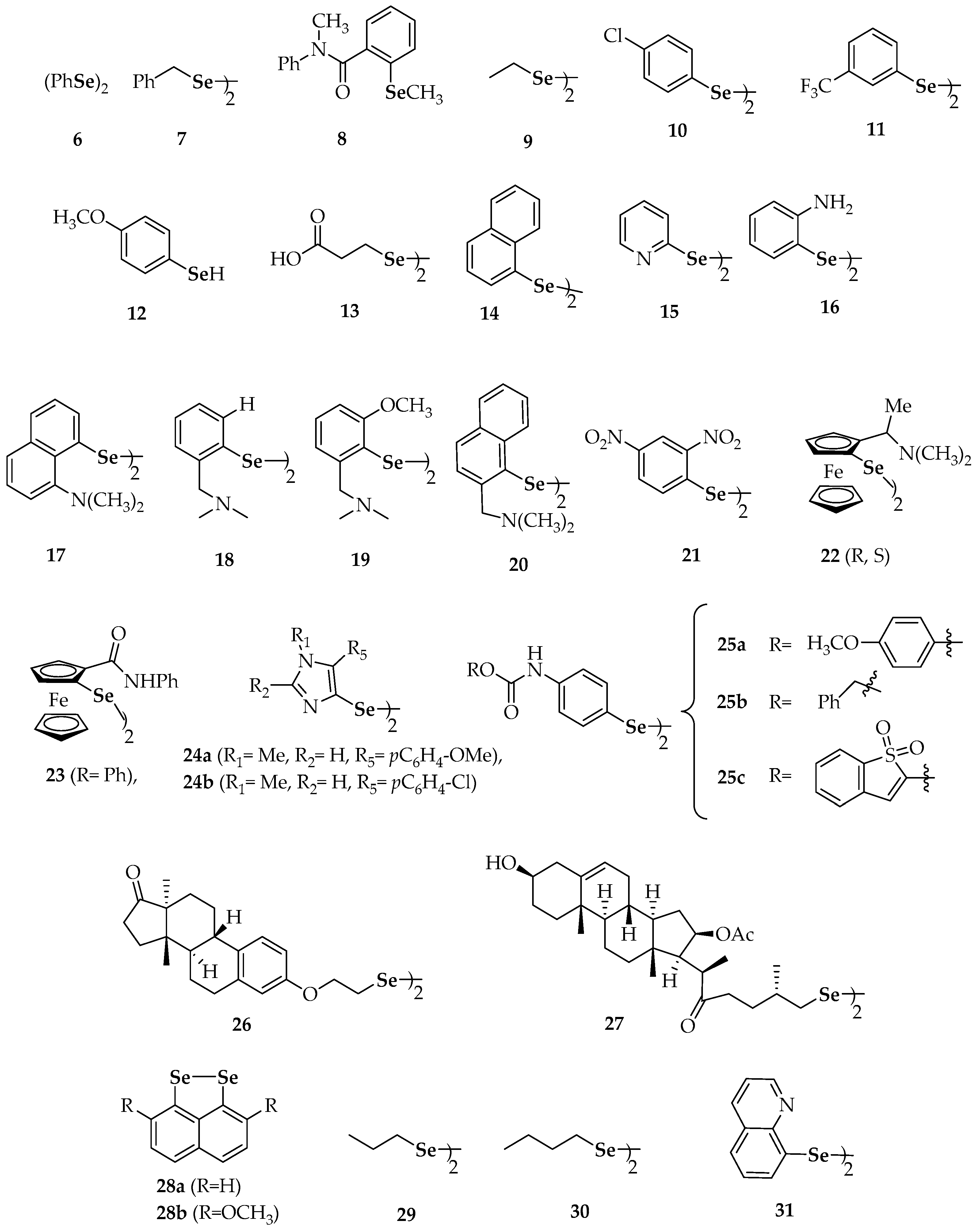
Figure 3.
Synthetic diselenides and related selenols with kinase modulation properties [73,74,75,76].


Table 1.
Synthetic diselenides mentioned.
| Compound | Compound | ||
|---|---|---|---|
| 6 | diphenyl diselenide | 26 | bis[2-(estra-1,3,5(10)-trien-1 7-one-3-yl)oxyethyl]diselenide |
| 7 | dibenzyl diselenide | 27 | bis[(25R)-16β-acetoxy-3β-hydroxy-22-oxocholest-5-en-26-yl]diselenide |
| 9 | diethyl diselenide, | 28a | naphtho[1,8-cd][1,2]diselenole |
| 10 | bis(4-clorophenyl) diselenide | 28b | 3,8-dimethoxynaphtho[1,8-cd][1,2]diselenole |
| 11 | 3′3-ditrifluoromethyldiphenyl diselenide | 29 | dipropyl diselenide |
| 12 | p-methoxybenzeneselenol | 30 | dibutyl diselenide |
| 13 | 3,3′-diselenodipropionic acid | 31 | diquinolyl-8-yl diselenide |
| 14 | binaphthyl diselenide | 32 | 4,4′-diamino-3′,3-dicarboxydiphenyl diselenide |
| 15 | dipyrid-2-yl diselenide | 36 | 6,6′-diselanediylbis(N-phenylhexanamide) |
| 16 | 2′2-diaminodiphenyl diselenide | 37a | 6,6′-dichlorodipyridazyn-2-yl diselenide |
| 17 | 8-(2-(8-(dimethylamino)naphthalen-1-yl)diselanyl)-N,N-dimethylnaphthalen-1-amine | 37b | 6,6′-di(propoxy)dipyridazyn-2-yl diselenide |
| 18 | (2-(2-(2-((dimethylamino)methyl)phenyl)diselanyl) phenyl)-N,N-dimethylmethanamine | 37c | 6,6′-di(isopentyloxy)dipyridazyn-2-yl diselenide |
| 19 | 2-(2-(2-((dimethylamino)methyl)-6-methoxyphenyl) diselanyl)-3-methoxyphenyl)-N,N-dimethylmethanamine | 37d | 6,6′-di(propylthio)dipyridazyn-2-yl diselenide |
| 20 | (1-(2-(2-((dimethylamino)methyl)naphthalen-1-yl)diselanyl)naphthalen-2-yl)-N,N-dimethylmethanamine | 38 | 2-chloro-6-(2-(6-chloropyridin-2-yl)diselanyl)pyridine |
| 21 | 1,2-bis(2,4-dinitrophenyl)diselane | 39 | 2,2′- di(phenylethyl) diselenide |
| 22 | bis(2-(1(R,S)-(N,N-dimethylamino)ethyl)ferrocen-1-yl) diselenide | 40 | dibenzhydryl diselenide |
| 23 | bis(2-(N-phenylcarbamoyl)ferrocen-1-yl) diselenide | 41 | 1,2-bis(isoindoline-1,3-dione-2 ethyl)diselane |
| 24a | 5-(4-methoxyphenyl)-4-(2-(5-(4-methoxyphenyl)-1-methyl-1H-imidazol-4-yl)diselanyl)-1-methyl-1H-imidazole | 42 | 1,2-bis(2-morpholinoethyl)diselane |
| 24b | 5-(4-chlorophenyl)-4-(2-(5-(4-chlorophenyl)-1-methyl-1H-imidazol-4-yl)diselanyl)-1-methyl-1H-imidazole | 43 | 4′,4-dimethoxydiphenyl diselenide |
| 25a | bis-4-[1-[(4′-methoxy)phenyl]-4-seleno-imidazole] | 44 | 3′,5′,3,5-tetratrifluoromethyl-diphenyl diselenide |
| 25b | 4,4′-diselanediylbis(N-benzylbenzamide) | 45 | 4′,4-diaminodiphenyl diselenide |
| 25c | N-(1,1-dioxidobenzo[b]thiophen-2-yl)-4-((4-((1,1-dioxidobenzo[b]thiophen-2-yl)carbamoyl)phenyl) diselaneyl)benzamide | ||
Table 2.
Selenides mentioned in the text.
| Compound | |
|---|---|
| 46 | diallyl selenide |
| 47 | 1,4-bis(3-(phenylselanyl)propyl)piperazine |
| 48 | trans-3,4-dihydroxyselenolane |
| 50 a–e | different substituted phenyl 3-hydroxypropyl selenides |
| 51 | 3-(phenylselanyl)-1-methyl-1H-indole |
| 52a, 52b | 6-(4-methylphenyl)selanylpurine 6-(4-chlorophenyl)selanylpurine |
| 53 | 2-methyl-3-(phenylselanyl) naphthalene-1,4-dione |
| 54a | 2,3-bis(phenylselanyl)naphthalene-1,4-dione |
| 54b | 5,8-dihydroxy-2,3-bis(phenyl-selanyl)naphthalene-1,4-dione |
| 55 | 2,3-bis((2,2-diethoxyethyl)selanyl)naphthalene-1,4-dione |
| 56a | 5′-(phenylseleno)zidovudine |
| 56b | 5′-(4-methylphenylseleno)zidovudine |
| 56c | 5′-(4-chlorophenylseleno)zidovudine |
| 57 | 3-bromo-4-(4-(((4-fluorophenyl)selanyl)methyl)-1H-1,2,3-triazol-1-yl)-2,2-dimethyl-3,4-dihydro-2H-benzo[h]chromene-5,6-dione |
| 58 | 2-(2-acetoxybenzamido)-3-(methylselanyl)propanoic acid |
| 59 | selenide-containing indole chalcone derivatives |
| 60 | (3,4-dimethoxy-5-(methylselanyl)phenyl)(1H-indol-5-yl)methanone |
| 61 | (3,4-dimethoxy-5-(methylselanyl)phenyl)(1H-indol-4-yl)methanone |
© 2018 by the authors. Licensee MDPI, Basel, Switzerland. This article is an open access article distributed under the terms and conditions of the Creative Commons Attribution (CC BY) license (http://creativecommons.org/licenses/by/4.0/).
Share and Cite
MDPI and ACS Style
Álvarez-Pérez, M.; Ali, W.; Marć, M.A.; Handzlik, J.; Domínguez-Álvarez, E. Selenides and Diselenides: A Review of Their Anticancer and Chemopreventive Activity. Molecules 2018, 23, 628. https://doi.org/10.3390/molecules23030628
AMA Style
Álvarez-Pérez M, Ali W, Marć MA, Handzlik J, Domínguez-Álvarez E. Selenides and Diselenides: A Review of Their Anticancer and Chemopreventive Activity. Molecules. 2018; 23(3):628. https://doi.org/10.3390/molecules23030628
Chicago/Turabian StyleÁlvarez-Pérez, Mónica, Wesam Ali, Małgorzata Anna Marć, Jadwiga Handzlik, and Enrique Domínguez-Álvarez. 2018. "Selenides and Diselenides: A Review of Their Anticancer and Chemopreventive Activity" Molecules 23, no. 3: 628. https://doi.org/10.3390/molecules23030628