
Inhibition of Osteoarthritis-Related Molecules by Isomucronulatol 7-O-β-d-glucoside and Ecliptasaponin A in IL-1β-Stimulated Chondrosarcoma Cell Model

 and
and 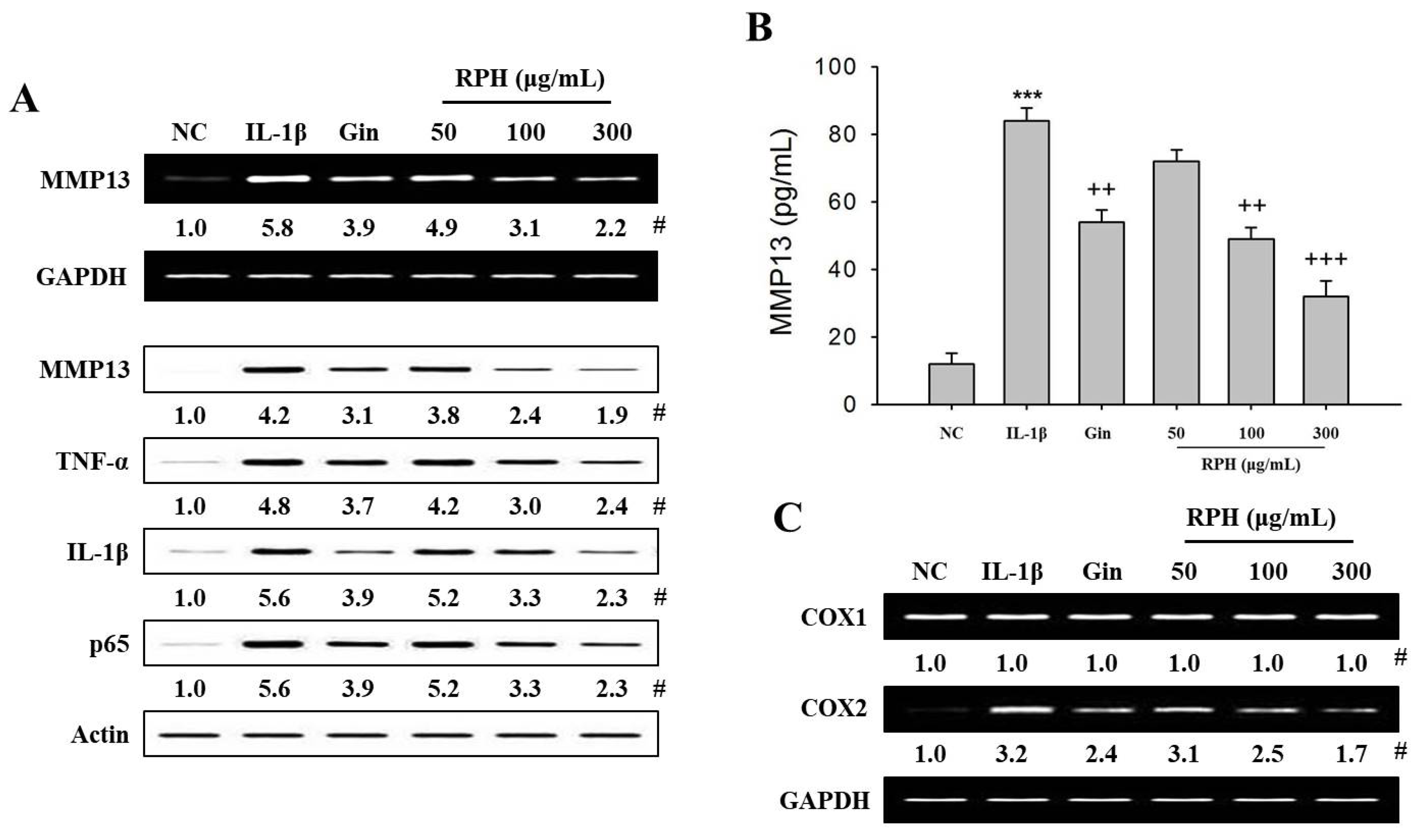
Abstract
:1. Introduction
2. Results
2.1. Effects of Natural Product Mixture (Ryupunghwan, RPH) on the Expression of Osteoarthritis (OA)-Related Molecules or Cyclooxygenase (COX) in IL-1β-Stimulated Chondrosarcoma SW1353 Cells
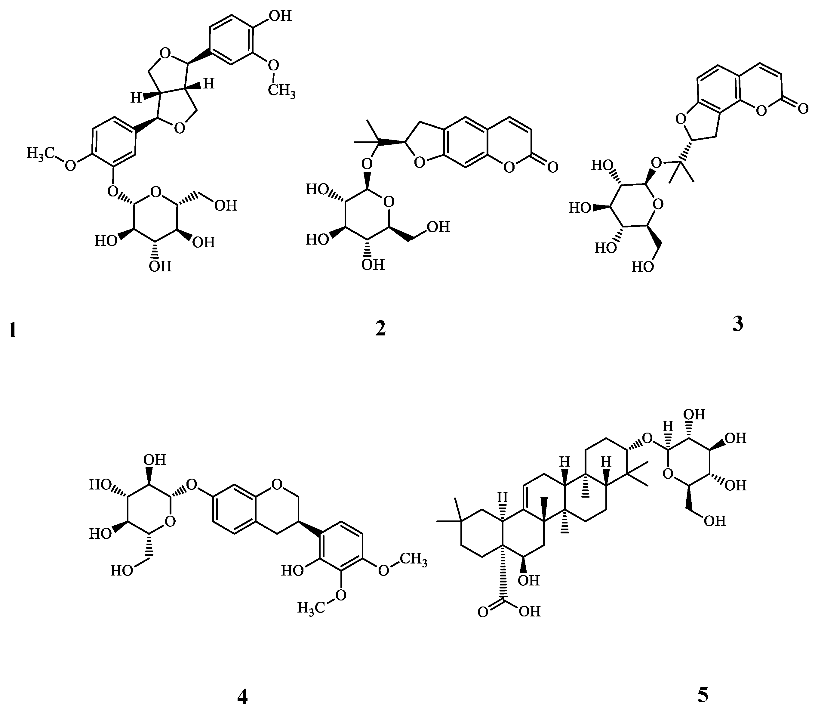
2.2. Purification and Identification of Bioactive Ingredients from RPH
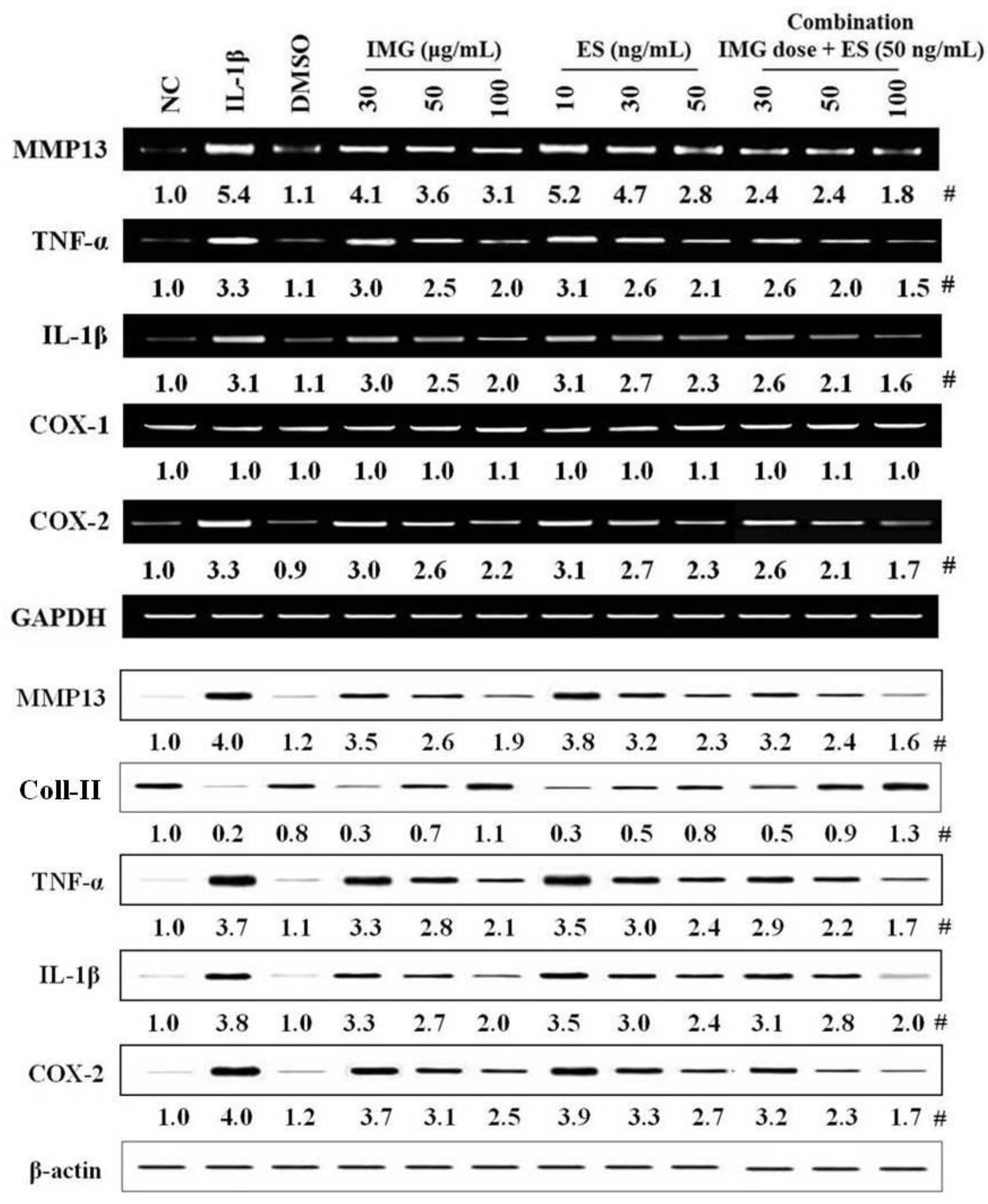
2.3. Effects of Single Component IMG and ES Isolated from Natural Product Mixture (RPH) on the Expression of OA-Related Molecules in IL-1β-Stimulated SW1353 Cells
3. Discussion
4. Materials and Methods
4.1. Materials
4.2. Purification and Identification of Bioactive Ingredients
4.3. Human Chondrosarcoma SW1353 Cells Culture Conditions
4.4. Cell Stimulation and Treatment
4.5. Reverse Transcription-Polymerase Chain Reaction (RT-PCR)
4.6. Preparation of Nuclear Extracts
4.7. Western Blot Analysis
4.8. MMP13 Amount Assay
4.9. Statistical Analysis
Supplementary Materials
Author Contributions
Funding
Conflicts of Interest
References
- Gao, J.; Liu, Z.J.; Chen, T.; Zhao, D. Pharmaceutical properties of calycosin, the major bioactive isoflavonoid in the dry root extract of Radix astragali. Pharm. Biol. 2014, 52, 1217–1222. [Google Scholar] [CrossRef] [PubMed]
- Taha, M.M.; Salga, M.S.; Ali, H.M.; Abdulla, M.A.; Abdelwahab, S.I.; Hadi, A.H. Gastroprotective activities of Turnera diffusa Willd. ex Schult. revisited: Role of arbutin. J. Ethnopharmacol. 2012, 141, 273–281. [Google Scholar] [CrossRef] [PubMed]
- Xu, X.X.; Zhang, X.H.; Diao, Y.; Huang, Y.X. Achyranthes bidentate saponins protect rat articular chondrocytes against interleukin-1β-induced inflammation and apoptosis in vitro. Kaohsiung. J. Med. Sci. 2017, 33, 62–68. [Google Scholar] [CrossRef] [PubMed]
- Wang, K.; Tang, Z.; Zheng, Z.; Cao, P.; Shui, W.; Li, Q.; Zhang, Y. Protective effects of Angelica sinensis polysaccharide against hyperglycemia and liver injury in multiple low-dose streptozotocin-induced type 2 diabetic BALB/c mice. Food Funct. 2016, 7, 4889–4897. [Google Scholar] [CrossRef] [PubMed]
- Morel, L.J.; Azevedo, B.C.; Carmona, F.; Contini, S.H.; Teles, A.M.; Ramalho, F.S.; Bertoni, B.W.; de Castro França, S.; de Carvalho Borges, M.; Pereira, A.M. A standardized methanol extract of Eclipta prostrata (L.) L. (Asteraceae) reduces bronchial hyperresponsiveness and production of Th2 cytokines in a murine model of asthma. J. Ethnopharmacol. 2017, 198, 226–234. [Google Scholar] [CrossRef] [PubMed]
- Wang, J.Y.; Yuan, Y.; Chen, X.J.; Fu, S.G.; Zhang, L.; Hong, Y.L.; You, S.F.; Yang, Y.Q. Extract from Eucommia ulmoides Oliv. ameliorates arthritis via regulation of inflammation, synoviocyte proliferation and osteoclastogenesis in vitro and in vivo. J. Ethnopharmacol. 2016, 194, 609–616. [Google Scholar] [PubMed]
- Pereira, A.A.; Tirapeli, K.G.; Neto, A.H.; da Silva Brasilino, M.; da Rocha, C.Q.; Belló-Klein, A.; Llesuy, S.F.; Dornelles, R.C.; de Melo Stevanato Nakamune, A.C. Ilex paraguariensis supplementation may be an effective nutritional approach to modulate oxidative stress during perimenopause. Exp. Gerontol. 2017, 90, 14–18. [Google Scholar] [CrossRef] [PubMed]
- Palmieri, B.; Lodi, D.; Capone, S. Osteoarthritis and degenerative joint disease: Local treatment options update. Acta Biomed. 2010, 81, 94–100. [Google Scholar] [PubMed]
- Martel-Pelletier, J.; Barr, A.J.; Cicuttini, F.M.; Conaghan, P.G.; Cooper, C.; Goldring, M.B.; Golgring, S.R.; Jones, G.; Teichtahl, A.J.; Pelletier, J.P. Osteoarthritis. Nat. Rev. Dis. Primers 2016, 2, 16072. [Google Scholar] [CrossRef] [PubMed] [Green Version]
- Varela-Eirin, M.; Loureiro, J.; Fonseca, E.; Corrochano, S.; Caeiro, J.R.; Collado, M.; Mayan, M.D. Cartilage regernation and ageing: Targeting cellular plasticity in osteoarthritis. Ageing Res. Rev. 2018, 42, 56–71. [Google Scholar] [CrossRef] [PubMed]
- Van den Berg, W.B. Osteoarthritis year 2010 in review: Pathomechanism. Osteoarthritis Cartilage 2011, 19, 338–341. [Google Scholar] [CrossRef] [PubMed]
- Sun, H.; Wu, Y.; Pan, Z.; Yu, D.; Chen, P.; Zhang, X.; Wu, H.; Zhang, X.; An, C.; Chen, Y.; et al. Gefitinib for epidermal growth factor receptor activated osteoarthritis subpopulation treatment. EBioMedicine 2018, 32, 223–233. [Google Scholar] [CrossRef] [PubMed]
- Boehme, K.A.; Rolauffs, B. Onset and progression of human osteoarthritis-can growth factors, inflammatory cytokines, or differential miRNA expression concomitantly induce proliferation, ECM degradation, and inflammation in articular cartilage? Int. Mol. Sci. 2018, 19, 2282. [Google Scholar] [CrossRef] [PubMed]
- Liu, C.C.; Zhang, Y.; Dai, B.-L.; Ma, Y.-J.; Zhang, Q.; Wang, Y.; Yang, H. Chlorogenic acid prevents inflammatory responses in IL-1β-stimulated human SW-1353 chondrocytes, a model for osteoarthritis. Mol. Med. Rep. 2017, 16, 1369–1375. [Google Scholar] [CrossRef] [PubMed]
- Yan, L.; Pan, M.; Fu, M.; Wang, J.; Huang, W.; Qian, H. Design, Synthesis and biological evaluation of novel analgesic agents targeting both cyclooxygenase and TRPV1. Bioorg. Med. Chem. 2016, 24, 849–857. [Google Scholar] [CrossRef] [PubMed]
- Essex, M.; Bhadra, P.; Sands, G. Efficacy and tolerability of celecoxib versus naproxen in patients with osteoarthritis of the knee: A randomized, double-blind, double-dummy trial. J. Int. Med. Res. 2012, 40, 1357–1370. [Google Scholar] [CrossRef] [PubMed]
- Zhu, X.; Wu, D.; Sang, L.; Wang, Y.; Shen, Y.; Zhuang, X.; Chu, M.; Jiang, L. Comparative effectiveness of glucosamine, chondtoitin, acetaminophen or celecoxib for the treatment of kee and/or hip osteoarthritis: A network meta-analysis. Clin. Exp. Rheumatol. 2018, 36, 595–602. [Google Scholar] [PubMed]
- Singh, R.; Akhtar, N.; Haqqi, T.M. Green tea polyphenol epigallocatechin-3-gallate: Inflammation and arthritis. Life Sci. 2010, 86, 907–918. [Google Scholar] [CrossRef] [PubMed]
- Kalamgam, G.; Memic, A.; Budd, E.; Abbs, M.; Mobasheri, A. A comprehensive review of stem cells for cartilage regeneration in osteoarthritis. Adv. Exp. Med. Biol. 2018. [Google Scholar] [CrossRef]
- Chen, B.; Qin, J.; Wang, H.; Magdalou, J.; Chen, L. Effects of adenovirus-mediated bFGF, IL-1Ra and IGF-1gene transfer on human osteoarthritis chondrocytes and osteoarthritis in rabbits. Exp. Mol. Med. 2010, 42, 684–695. [Google Scholar] [CrossRef] [PubMed]
- Ma, Z.; Wang, Y.; Piao, T.; Liu, J. Echinocystic Acid inhibits IL-1β-Induced COX-2 and iNOS expression in human osteoarthritis chondrocytes. Inflammation 2016, 39, 543–549. [Google Scholar] [CrossRef] [PubMed]
- Ruan, G.; Xu, J.; Wang, K.; Wu, J.; Zhu, Q.; Ren, J.; Bian, F.; Chang, B.; Bai, X.; Han, W.; et al. Association between knee structural measures, circulating inflammatory factors and MMP13 in patients with knee osteoarthritis. Osteoarthritis Cartilage 2018, 26, 1063–1069. [Google Scholar] [CrossRef] [PubMed]
- Bao, G.; Xu, L.; Xu, X.; Zhai, L.; Duan, C.; Xu, D.; Song, J.; Liu, Z.; Tao, R.; Cui, Z.; et al. SGTB promotes the caspase-dependent apoptosis in chondrocytes of osteoarthritis. Inflammation 2016, 39, 601–610. [Google Scholar] [CrossRef] [PubMed]
- Wang, C.; Zeng, L.; Zhang, T.; Liu, J.; Wang, W. Tenuigenin prevents IL-1β-induced inflammation in human osteoarthritis chondrocytes by suppressing PI3K/AKT/NF-κB signaling pathway. Inflammation 2016, 39, 807–812. [Google Scholar] [CrossRef] [PubMed]
- Wang, L.; Gai, P.; Xu, R.; Zheng, Y.; Lv, S.; Li, Y.; Liu, S. Shikonin protects chondrocytes from interleukin-1beta-induced apoptosis by regulating PI3K/Akt signaling pathway. Int. J. Clin. Exp. Pathol. 2015, 8, 298–308. [Google Scholar] [PubMed]
- Ouyang, M.; Wein, Y.; Zhang, Z.K.; Kuo, Y.H. Inhibitory activity against tobacco mosaic virus (TMV) replication of pinoresinol and syringaresinol lignans and their glycosides from the root of Rhus javanica var. roxburghiana. J. Agric. Food Chem. 2007, 55, 6460–6465. [Google Scholar] [CrossRef] [PubMed]
- Kwon, Y.S.; Woo, E.R.; Kim, C.M. A study on the constituents of bioactive fractions of Ostericum koreanum Kitagawa. Korean J. Pharmacogn. 1991, 22, 156–161. [Google Scholar]
- Kim, Y.A.; Lee, J.I.; Kong, C.-S.; Choe, J.C.; Oh, K.S.; Seo, Y. Antioxidant activity of dihydrofurocoumarins from Corydalis heterocarpa. Biotech. Bioproc. Eng. 2014, 19, 771–779. [Google Scholar] [CrossRef]
- Ma, X.; Tu, P.F.; Chen, Y.J.; Zhang, T.Y.; Wei, Y.; Ito, Y.I. Preparative isolation and purification of isoflavan and pterocarpan glycosides from Astragalus membranaceus Bge. var. mongholicus (Bge.) Hsiao by high-speed counter-current chromatography. J. Chromatograph. A 2004, 1023, 311–315. [Google Scholar] [CrossRef]
- Yahara, S.J.; Ding, N.; Nohara, T.H. Oleanane glycosides from Eclipta alba. Chem. Pharmaceu. Bull. 1994, 42, 1336–1338. [Google Scholar] [CrossRef]
- Kim, G.S.; Lee, D.Y.; Lee, S.E.; Noh, H.J.; Choi, J.H.; Park, C.G.; Choi, S.I.; Hong, S.J.; Kim, S.Y. Evaluation of extraction conditions and HPLC analysis method for bioactive compounds of Astragali Radix. Korean J. Med. Crop. Sci. 2013, 21, 486–492. [Google Scholar] [CrossRef]
- Choi, S.I.; Park, S.R.; Heo, T.R. Inhibitory effect of Astragali Radix on matrix degradation in human articular cartilage. J. Microbiol. Biotechnol. 2005, 15, 1258–1266. [Google Scholar]
- Choi, S.I.; Heo, T.R.; Min, B.H.; Cui, J.H.; Choi, B.H.; Park, S.R. Alleviation of osteoarthritis by calycosin-7-O-beta-D-glucopyranoside (CG) isolated from Astragali Radix (AR) in rabbit osteoarthritis (OA) model. Osteoarthritis Cartilage 2007, 15, 1086–1092. [Google Scholar] [CrossRef] [PubMed]
- You, X.Y.; Xue, Q.; Fang, Y.; Liu, Q.; Zhang, C.F.; Zhao, C.; Zhang, M.; Xu, X.H. Preventive effects of Ecliptae Herba extract and its component, ecliptasaponin A, on bleomycin-induced pulmonary fibrosis in mice. J. Ethnopharmacol. 2015, 175, 172–180. [Google Scholar] [CrossRef] [PubMed]
- Takeuchi, K.; Amagase, L. Roles of cyclooxygenase, prostaglandin E2 and EP receptors in mucosal protection and ulcer healing in the gastrointestinal tract. Curr. Pharm. Des. 2018, 24, 2002–2011. [Google Scholar] [CrossRef] [PubMed]
- Chang, C.C.; Hsieh, M.S.; Liao, S.T.; Chen, Y.H.; Cheng, C.W.; Huang, P.T.; Lin, Y.F.; Chen, C.H. Hyaluronan regulates PPARγ and inflammatory responses in IL-1β-stimulated human chondrosarcoma cells, a model for osteoarthritis. Carbohydr. Polym. 2012, 90, 1168–1175. [Google Scholar] [CrossRef] [PubMed]
- Wang, Z.; Ding, L.; Zhang, S.; Jiang, T.; Yang, Y.; Li, R. Effects of icariin on the regulation of the OPG-RANKL-RANK system are mediated through the MAPK pathways in IL-1β-stimulated human SW1353 chondrosarcoma cells. Int. J. Mol. Med. 2014, 34, 1720–1726. [Google Scholar] [CrossRef] [PubMed]
- Hong, G.U.; Kim, N.G.; Kim, T.J.; Ro, J.Y. CD1d expressed in mast cell surface enhances IgE production in B cells by up-regulating CD40L expression and mediator release in allergic asthma in mice. Cell. Signal. 2014, 26, 1105–1117. [Google Scholar] [CrossRef] [PubMed]
- Keifer, J.A.; Guttridge, D.C.; Ashburner, B.P.; Baldwin, A.S., Jr. Inhibition of NF-kappa B activity by thalidomide through suppression of IκappaB kinase activity. J. Biol. Chem. 2001, 276, 22382–22387. [Google Scholar] [CrossRef] [PubMed]
- Ahn, Y.M.; Hong, G.U.; Kim, S.H.; Lee, H.J.; Baek, H.S.; Kim, M.N.; Park, K.Y.; Ro, J.Y. Transglutaminase 2 expressed in mast cells recruited into skin or bone marrow induces the development of pediatric mastocytosis. Pediatr. Allergy Immunol. 2015, 26, 438–445. [Google Scholar] [CrossRef] [PubMed]
- Liao, S.; Zhou, K.; Li, D.; Xie, X.; Jun, F.; Wang, J. Schisantherin A suppresses interleukin-1β-induced inflammation in human chondrocytes via inhibition of NF-κB and MAPKs activation. Eur. J. Pharmacol. 2016, 780, 65–70. [Google Scholar] [CrossRef] [PubMed]
Sample Availability: Samples of the compounds are available from the corresponding authors (Y.I.K.; J.Y.R.). |


© 2018 by the authors. Licensee MDPI, Basel, Switzerland. This article is an open access article distributed under the terms and conditions of the Creative Commons Attribution (CC BY) license (http://creativecommons.org/licenses/by/4.0/).
Share and Cite
Hong, G.U.; Lee, J.-Y.; Kang, H.; Kim, T.Y.; Park, J.Y.; Hong, E.Y.; Shin, Y.H.; Jung, S.H.; Chang, H.-B.; Kim, Y.H.; et al. Inhibition of Osteoarthritis-Related Molecules by Isomucronulatol 7-O-β-d-glucoside and Ecliptasaponin A in IL-1β-Stimulated Chondrosarcoma Cell Model. Molecules 2018, 23, 2807. https://doi.org/10.3390/molecules23112807
Hong GU, Lee J-Y, Kang H, Kim TY, Park JY, Hong EY, Shin YH, Jung SH, Chang H-B, Kim YH, et al. Inhibition of Osteoarthritis-Related Molecules by Isomucronulatol 7-O-β-d-glucoside and Ecliptasaponin A in IL-1β-Stimulated Chondrosarcoma Cell Model. Molecules. 2018; 23(11):2807. https://doi.org/10.3390/molecules23112807
Chicago/Turabian StyleHong, Gwan Ui, Jung-Yun Lee, Hanna Kang, Tae Yang Kim, Jae Yeo Park, Eun Young Hong, Youn Ho Shin, Sung Hoon Jung, Hung-Bae Chang, Young Ho Kim, and et al. 2018. "Inhibition of Osteoarthritis-Related Molecules by Isomucronulatol 7-O-β-d-glucoside and Ecliptasaponin A in IL-1β-Stimulated Chondrosarcoma Cell Model" Molecules 23, no. 11: 2807. https://doi.org/10.3390/molecules23112807