Daphnauranins C–E, Three New Antifeedants from Daphne aurantiaca Roots
by
Sheng Zhuo Huang
1, 
Qing Yun Ma
1,
Qi Wang
1,
Hao Fu Dai
1,
Yu Qing Liu
2,
Jun Zhou
2,* and
You Xing Zhao
1,* 1
Hainan Key Laboratory for Research and Development of Natural Products from Li Folk Medicine, Ministry of Agriculture, Institute of Tropical Bioscience and Biotechnology, Chinese Academy of Tropical Agricultural Sciences, Haikou 571101, China
2
State Key Laboratory of Phytochemistry and Plant Resources in West China, Kunming Institute of Botany, Chinese Academy of Sciences, Kunming 650201, China
*
Authors to whom correspondence should be addressed.
Molecules 2018, 23(10), 2429; https://doi.org/10.3390/molecules23102429
Submission received: 11 September 2018
/
Accepted: 20 September 2018
/
Published: 21 September 2018
(This article belongs to the Collection Bioactive Compounds)
Abstract
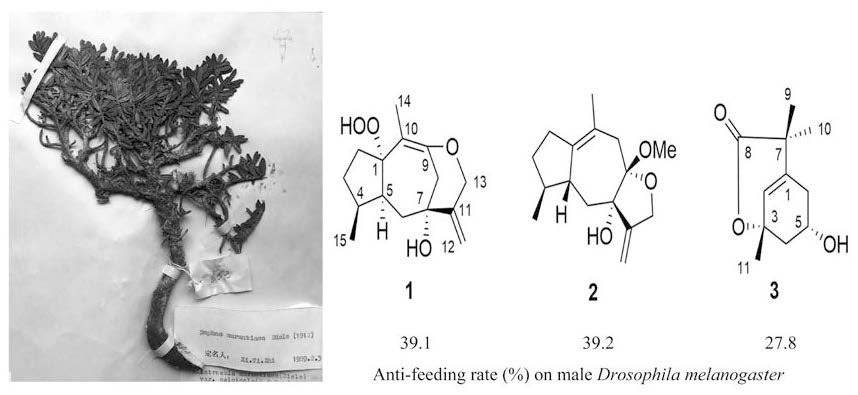
:Daphnauranins C–E (compounds 1–3), two sesquiterpenoids and one monoterpenoid were isolated from the roots of Daphne aurantiaca Diels. Daphnauranin C is a 9-O-13 etherified and hydroperoxy-substituted guaiane sesquiterpenoid, daphnauranin D is a guaiane sesquiterpenoid ketal, and daphnauranin E is a monoterpenoid lactone. Their structures were elucidated by comprehensive analyses of MS, 1D NMR, and 2D NMR spectroscopic data. In an anti-feeding activities test, daphnauranins C–E showed activity against male fruit fly with anti-feeding indexes (AI) up to 39.1, 39.2, and 27.8% respectively, at 1 mM.

1. Introduction
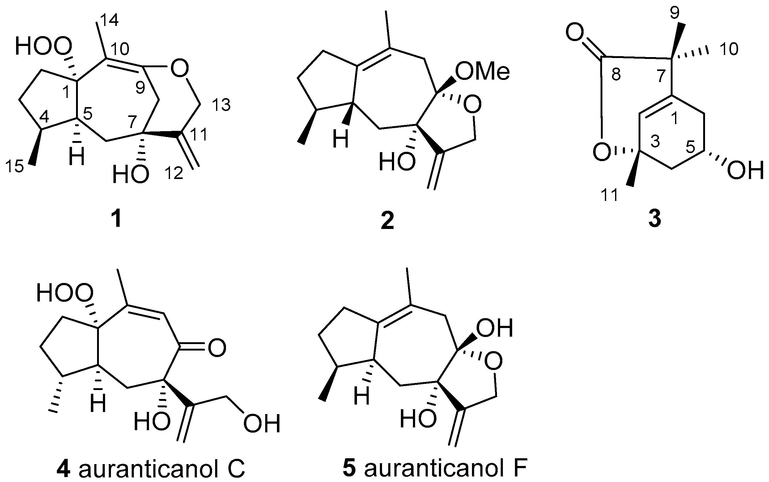
With the increase of global population and the changes in food-consumption habits, the food production industry is under unprecedented pressure. One of the biggest threats in agriculture are insects, which cause enormous production losses. To solve this problem there is a need for continuous effective prophylaxis and treatment. Therefore, finding and developing new effective and ecologically safe insecticides and insect repellents is one of the most important science topics for chemical and biological researchers in agriculture [1]. With multifarious natural products, traditional herbs have a long history of use as insecticides and insect repellents [2,3]. Natural products have become a treasury to explore for new insecticides or lead compounds with incomparable excellent structural diversity, ecological safety, biodegradability, and sustainability properties [4,5,6,7]. Daphne aurantiaca Diels., a traditional herb, was used as the raw material to make insect repellent paper by Tibetan Buddhists [8]. In previous studies, this plant showed some different natural products from other species. A few diterpenoids and several sesquiterpenoids with special skeletons and insect repellent activities were isolated from the stems of this plant [9,10,11,12]. To seek new insecticide and insect repellent from natural products, herein we studied the D. aurantiaca roots and found three new compounds, a 9-O-13 etherifie and hydroperoxy-substituted guaiane sesquiterpenoid [daphnauranin C (1)], a guaiane sesquiterpenoid ketal [daphnauranin D (2)], a monoterpenoid lactone [daphnauranin E (3)], and two known compounds: auranticanol C (4) and auranticanol F (5) (Figure 1). The isolation process and structural elucidation details of these new compounds, as well as their anti-insect assay results, are described in this paper.
2. Results and Discussion
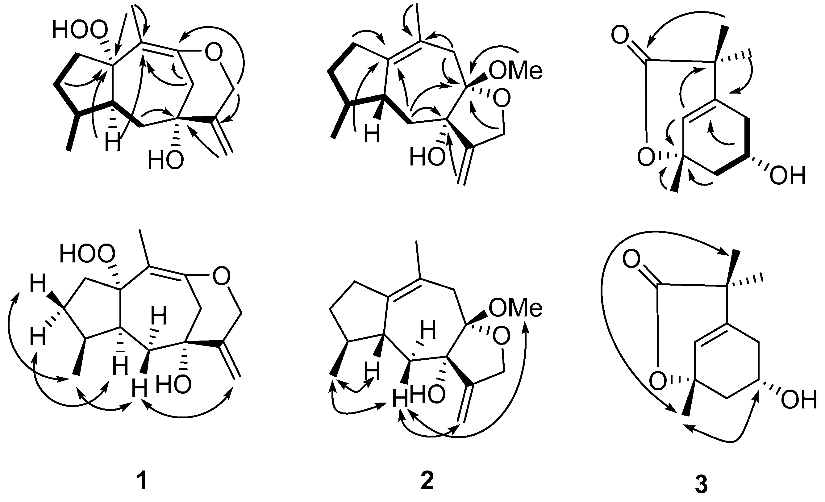
Daphnauranin C (1) was isolated as a colorless amorphous powder. Its molecular formula of C15H22O4 was determined by the HRESIMS ion at m/z 289.1408 [M + Na]+ (calcd for C15H22O4Na, 289.1415) (Supplementary Materials), indicative of five degrees of unsaturation. The IR spectrum revealed the presence of hydroxyl (3441 cm−1) and double bond (1629 and 1661 cm−1) absorptions. The 1H-NMR spectrum (Table 1) of compound 1 exhibited signals of two methyls [δH 1.36 (3H, s, H-14) and 0.92 (3H, d, J = 6.9 Hz, H-15)], two oxygenated methylene protons (δH 4.54 (1H, d, J = 13.2 Hz, Ha-13) and 4.49 (1H, d, J = 13.2 Hz, Hb-13), and two olefinic protons (δH 5.32 (1H, d, J = 1.3 Hz, Ha-12) and 5.11 (1H, d, J = 1.3 Hz, Hb-12). The 13C-NMR and DEPT spectroscopic data (Table 1) showed 15 carbon resonances, including two methyls, six methylenes (one olefinic and one oxygenated), two methines, and five quaternary carbons (three olefinic, two oxygenated). According to a comparison of the corresponding NMR data, compound 1 was similar to auranticanol C (4) [11], a rare peroxyhydroxyl-substituted guaiane sesquiterpenoid, except for the markedly different shifts at δC 75.8 (s, C-1), 43.4 (t, C-8), 112.4 (s, C-9), and 96.3 (s, C-10) instead of δC 106.7 (s, C-1), 199.1 (s, C-8), 126.1 (s, C-9), and 166.8 (s, C-10) in auranticanol C, indicating compound 1 was generated from auranticanol C by reductive deoxygenation of C-8 and etherification of C-9 to C-13. The determined C15H22O4 molecular formula and the key HMBC correlations from H-14 to C-1 and C-10, from H-8 to quaternary carbon C-10, and from H-8 and H-13 to quaternary carbon C-9 verified this hypothesis. The other 1H-1HCOSY and HMBC correlations (Figure 2) also confirmed this atom connectivity. The relative configuration of compound 1 was determined to be the same as that of auranticanol C (Figure 2) by ROESY cross-peaks H-3α [δH 1.32 (1H, m)] /H-5 [δH 2.46 (1H, m)], H-15/H-3β [δH 1.60 (1H, m)], H-15/H-6β (δH 1.69 (1H, dd, J = 5.2, 13.2 Hz), and H-6β/H-12. Thus, the structure of compound 1 was assigned as shown in Figure 1 and it was named daphnauranin C.
Daphnauranin D (2) was obtained as a colorless oil, and possessed the molecular formula C16H24O3 based on HRESIMS (m/z 287.1620 [M + Na]+, calcd for C16H24O3Na, 287.1623) with five degrees of unsaturation (Supplementary Materials). The 13C-NMR and DEPT spectroscopic data (Table 1) showed 16 carbon resonances classified into two methyls, five methylenes (one olefinic and one oxygenated), two methines, and five quaternary carbons (three olefinic and two oxygenated). Detailed inspection of spectral data of 2 suggested that it was similar to auranticanol F (5) (also isolated from stems of D. aurantiaca) [11], except for an additional methoxy group (δC 48.7 q) in compound 2. Compound 2 should thus be generated from auranticanol F (5) via C-8 methoxylation as shown, which was further supported by the HMBC correlations (Figure 1) from H-OMe [δH 3.17 (3 H, s)] to C-8 [δC 107.5 (s)] with the aid of its molecular formula C16H24O3. The configuration 7-αOH and 8-βOMe in 2 was deduced from its ROESY cross-peaks (Figure 2) H-6β [δH 1.81 (1H, dd, J = 1.3, 14.4 Hz)] to H-8-OMe and H-12 [δH 5.17 (1H, d, J = 1.3 Hz)]. The configuration 4-αH, 5-βH were determined by ROESY cross-peaks (Figure 2) H-15 [δH 0.88 (3H, d, J = 7.1 Hz)] to H-6β and H-5 [δH 2.18 (1H, m)] with molecular model and comparison of 1D and 2D NMR data to those of auranticanol F. Thus, the structure of compound 2 was assigned as shown and named daphnauranin D.
The molecular formula of daphnauranin E (3), was determined as C11H16O3, with four degrees of unsaturation, from HREIMS (m/z 196.1102 [M]+, calcd for C11H16O3, 196.1099 (Supplementary Materials). The 13C-NMR and DEPT spectroscopic data (Table 1) showed 11 carbon resonances, including three methyls, two methylenes, two methines (one olefinic and one oxygenated), and four quaternary carbons (one olefinic, one oxygenated, and one carbonyl). By comparing the molecular formula and NMR data (Table 1) with Δ2-8-m-menthenecarboxylic acid [13], compound 3 was proposed to be derived from this compound via the oxygenation of C-3 and C-5 to a hydroxyl or ester group. The key HMBC correlations (Figure 2) from H-5 [δH 4.31 (1H, dddd, J = 2.2, 2.4, 3.2, 3.5 Hz)] to C-1 [δC 178.2 (s)], from H-4 [δH 3.70 (1H, dd, J = 2.4, 15.6 Hz) and 3.26 (1H, dd, J = 3.2, 15.6 Hz)] to C-3 [δC 86.8 (s)], and from H-2 [δH 5.68 (1H, m)] to C-3, and 1H 1H COSY correlations between H-4/H-5 [δH 4.31 (1H, dddd, J = 2.2, 2.4, 3.2, 3.5 Hz)] and H-5/H-6 [δH 2.48 (1H, dd, J = 2.2, 15.4 Hz) and 1.77 (1H, dd, J = 3.5, 15.4 Hz)] in compound 3 (Figure 1) supported the assignment. The linkage of 3-O-8 lactone in compound 3 was determined by the 13C NMR data of C-3 and C-11 [δC 182.6 (s)] and the determined molecular formula C11H16O3. The relative configuration of compound 3 was determined as β-Me-11 and α-6-OH based on its NOESY NMR spectrum (Figure 2) revealing the key NOE of H-5/H-11 [δH 1.26 (3H, s)]. Thus, the structure of compound daphnauranin E (3) was assigned as shown.
Compounds 1–3 were all tested for their anti-feeding activities against male fruit fly (Drosophila melanogaster) as described before [9,14]. As a result, the anti-feeding index (AI) of 1–3 (at the concentration of 1 mM) were 39.1 ± 5.8, 39.2 ± 3.9 and 27.8 ± 6.2%, respectively. Meanwhile AI of the negative control and the positive control (nicotine at 1 mM) were 17.9 ± 2.4 and 28.5 ± 3.9%, respectively (Table 2).
3. Experimental
3.1. General Information
Optical rotations were acquired on a SEAP-300 polarimeter (Horiba, Kyoto, Japan) ECD were obtained on a Chirascan instrument (Applied Photophysics Ltd., Surrey, UK), UV spectra were measured on a UV 210A spectrophotometer (Hitachi, Hong Kong, China), IR spectra were measured on a FTS-135 spectrometer (Bio-Rad, Hercules, CA, USA) with KBr pellets. 1D and 2D NMR spectra were obtained using an AV400 or DRX-500 instrument (Bruker, Billerica, MA, USA) with TMS as an internal standard, and ESIMS, HRESIMS, and HREIMS were recorded with a Bruker HCT/Esquire (Billerica, MA, USA), VG Auto Spec-3000 mass spectrometer (Manchester, UK) or Autospec Premier spectrometer (Waters, Milford, MA, USA). Column chromatography (CC) was performed on silica gel (200–300 mesh, Qingdao Marine Chemical Inc., Qingdao, China), ODS (40–70 μm, Fuji Silysia Chemical Ltd., Nagoya, Japan), and Sephadex LH-20 (GE Healthcare Bio-Sciences AB, Uppsala, Sweden). Fractions were monitored by TLC and heating after spraying with 7% H2SO4 in EtOH.
3.2. Plant Material
The roots of Daphne aurantiaca Diels. were obtained in Shangri-La, Yunnan Province, People′s Republic of China in July 2014,. The voucher specimen (HUANG0008) identified by Prof. Dr. Y. Niu (Kunming Institute of Botany, Chinese Academy of Sciences) was deposited at the Hainan Key Laboratory for Research and Development of Natural Products from Li Folk Medicine, Institute of Tropical Bioscience and Biotechnology, Chinese Academy of Tropical Agriculture Sciences, Haikou, People’s Republic of China.
3.3. Extraction and Isolation
The air-dried roots of D. aurantiaca (2.5 kg) were powdered and extracted with 95% EtOH by refluxing for three hours (3 × 13 L). The combined EtOH solution was concentrated with a rotary evaporator followed by suspension in water (2 L) and successive extraction with EtOAc (3 × 5 L). The EtOAc extract (143 g) was first subjected to silica gel (200–300 mesh, φ 16 × 150 cm) CC eluted with CHCl3/MeOH (from 50:1 to 1:1, v/v) to obtain fractions A–C. Fraction B (3.8) was chromatographed repeatedly over a ODS (40–70 μm, φ 4 × 18 cm) with MeOH/H2O (gradient elution with 20, 30, 40, 50, 60, 70, 80, and 90%, each 500 mL) and Sephadex LH-20 CC (MeOH as solvent) to yield auranticanol F (5, 23.2 mg), respectively. Fraction C (43 g) was then subjected to CC over silica gel (200–300 mesh, φ 6 × 45 cm) eluted with petroleum ether/acetone (from 3:1 to 0.5:1, v/v) to give four fractions C1–C4. Fraction C2 (3.8) was chromatographed repeatedly over a ODS (40–70 μm, φ 4 × 18 cm) with MeOH/H2O (gradient elution with 20, 30, 40, 50, 60, 70, 80, and 90%, each 500 mL) to obtain fractions C2a-C2d. Fractions C2a-C2d was chromatographed repeatedly over Sephadex LH-20 CC, using MeOH as solvent to yield 1 (5.9 mg), 2 (7.9 mg), and 3 (4.3 mg), respectively. Fractions C3 (1.6 g) was chromatographed repeatedly over a ODS (40–70 μm, φ 4 × 18 cm) with MeOH/H2O (gradient elution with 20, 30, 40, 50, 60, 70, 80, and 90%, each 500 mL) and Sephadex LH-20 CC (MeOH as solvent) to yield auranticanol C (4, 5.2 mg).
3.3.1. Daphnauranin C (1)
Colorless amorphous powder; [α+ 36.84 (c 0.230, MeOH); UV (MeOH) λmax (logε) 202 (3.46); IR (KBr) νmax 3441, 2961, 2933, 2872, 1629, 1661, 1439, 1377, 1341, 1305, 1284, 1160, 1048, 1034, 975, 931, 897; 1H- and 13C-NMR data see Table 1; ESIMS positive m/z [M + Na]+ 289 (80); HRESIMS m/z [M + Na]+ 289.1408 (calcd for C15H22O4Na, 289.1415).
3.3.2. Daphnauranin D (2)
Colorless oil; [α+2.05 (c 0.114, MeOH); ECD (MeOH) ∆ε 273 (+1.15), 356 (−0.094); UV (MeOH) λmax (logε) 203 (3.70); IR (KBr) νmax 3426, 2955, 2932, 2871, 1712, 1631, 1455, 1434, 1378, 1113, 1035; 1H- and 13C-NMR data see Table 1; ESIMS positive m/z [M + Na]+ 287 (40); HRESIMS m/z [M + Na]+ 287.1620 (calcd for C16H24O3Na, 287.1623).
3.3.3. Daphnauranin E (3)
Colorless amorphous powder; [α-23.43 (c 0.327, MeOH); UV (MeOH) λmax (logε) 361 (1.87), 212 (3.57); IR (KBr) νmax 3433, 2962, 2931, 2875, 1734, 1627, 1454, 1379, 1264, 1232, 1165, 1051, 1030, 965; 1H- and 13C-NMR data see Table 1; ESIMS positive m/z [M + H]+ 197 (45); HREIMS m/z [M]+ 196.1102 (calcd for C11H16O3, 196.1099).
3.4. Anti-Feeding Activity Bioassay
The anti-feeding activity was tested on male fruit fly (Drosophila melanogaster supplied by JoeKai Biotech LLC, Bejing, China) by the feeding counting method as reported in the literature [9,14]. Test compounds, positive control nicotine (98%, Sigma-Aldrich Corporation, St. Louis, MO, USA), and negative control DMSO (dimethylsulfoxide, Sinopharm Chemical Reagent Co., Ltd., Shanghai, China) were dissolved in DMSO to 100 mM, and then diluted to 1 mM with 4% red sugar water. Fifty starved (17 h) male fruit flies were put into one tube for one treatment. Each test was carried out with nine replicates. After 7 min feeding, the number of rubescent abdomen and no-rubescent abdomen fruit fly was counted. The feeding index (FI) was the percentage of rubescent abdomen fruit fly. Thus, the anti-feeding index (AI) was calculated by the following equation AI % = [(1 − FI) × 100].
4. Conclusions
Previously, a series of sesquiterpenoids with skeletal diversity revealed the chemical diversity of the Daphne genus. The special substituted compounds isolated from Daphne aurantiaca also showed the accessibility of biosynthesis. The bioactivity evaluation assay showed that daphnauranins C-E have prominent anti-feeding activities against male fruit fly. Therefore, these three new compounds may be used for potential agricultural chemical development.
Supplementary Materials
The following are available online at https://www.mdpi.com/1420-3049/23/10/2429/s1.
Author Contributions
S.Z.H.: Write and revise the manuscript. Q.Y.M.: Activity test. Q.W.: Compound isolation. H.F.D.: Method adviser. Y.Q.L.: Method adviser. J.Z.: theory and orientation adviser. Y.X.Z.: modification and theory adviser.
Funding
This research received no external funding.
Acknowledgments
This work was supported by China Agriculture Research System (CARS-21), Financial Fund of the Ministry of Agriculture and Rural Affairs, P.R. of China (NFZX2018), National Natural Science Foundation of China (No. 31300294), and Central Public-interest Scientific Institution Basal Research Fund for Chinese Academy of Tropical Agricultural Sciences (17CXTD-15, 1630052016008). The authors thank Y.L. Huang (dept. of clinical development. Pfizer (China). Shanghai) for initial proofreading of this paper.
Conflicts of Interest
The authors declare no conflict of interest.
References
- Hedin, P.A.; Hollingworth, R.M.; Masler, E.P.; Miyamoto, J. Phytochemicals for Pest Control; American Chemical Society: Washington, DC, USA, 1997; Volume 658, p. 388. [Google Scholar]
- Defagó, M.; Valladares, G.; Banchio, E.; Carpinella, C.; Palacios, S. Insecticide and antifeedant activity of different plant parts of Melia azedarach on Xanthogaleruca luteola. Fitoterapia 2006, 77, 500–505. [Google Scholar] [CrossRef] [PubMed]
- Hashim, M.S.; Devi, K.S. Insecticidal action of the polyphenolic rich fractions from the stem bark of Streblus asper on Dysdercus cingulatus. Fitoterapia 2003, 74, 670–676. [Google Scholar] [CrossRef]
- Miyakado, M.; Watanabe, K.; Miyamoto, J. Natural Products as Leads in Structural Modification Studies Yielding New Agrochemicals. In Phytochemicals for Pest Control; American Chemical Society: Washington, DC, USA, 1997; Volume 658, pp. 168–182. [Google Scholar]
- Huang, S.Z.; Kong, F.D.; Ma, Q.Y.; Guo, Z.K.; Zhou, L.M.; Wang, Q.; Dai, H.F.; Zhao, Y.X. Nematicidal Stemona Alkaloids from Stemona parviflora. J. Nat. Prod. 2016, 79, 2599–2605. [Google Scholar] [CrossRef] [PubMed]
- Huang, S.Z.; Zhang, X.; Ma, Q.Y.; Peng, H.; Zheng, Y.T.; Hu, J.M.; Dai, H.F.; Zhou, J.; Zhao, Y.X. Anti-HIV-1 tigliane diterpenoids from Excoecaria acertiflia Didr. Fitoterapia 2014, 95, 34–41. [Google Scholar] [CrossRef] [PubMed]
- Montenegro, I.; Pino, L.; Werner, E.; Madrid, A.; Espinoza, L.; Moreno, L.; Villena, J.; Cuellar, M. Comparative study on the larvicidal activity of drimane sesquiterpenes and nordrimane compounds against Drosophila melanogaster til-til. Molecules 2013, 18, 4192–4208. [Google Scholar] [CrossRef] [PubMed]
- Kunming Institute of Botany, Chinese Academy of Sciences. Flora of Yunnan; Science Press: Kunming, China, 1997; Volume 8, p. 219. [Google Scholar]
- Huang, S.Z.; Li, X.N.; Ma, Q.Y.; Dai, H.F.; Li, L.C.; Cai, X.H.; Liu, Y.Q.; Zhou, J.; Zhao, Y.X. Daphnauranols A–C, new antifeedant sesquiterpenoids with a 5/6/7 ring system from Daphne aurantiaca. Tetrahedron Lett. 2014, 55, 3693–3696. [Google Scholar] [CrossRef]
- Huang, S.Z.; Huang, H.N.; Ma, Q.Y.; Mo, M.H.; Zhu, M.L.; Dai, H.F.; Ji, Y.P.; Wang, Q.H.; Zhao, Y.X. The Phytochemicals with Antagonistic Activities Toward Pathogens of a Disease Complex Caused by Meloidogyne incognita and Ralstonia solanacearum. J. Pure Appl. Microb. 2015, 9, 209–231. [Google Scholar]
- Huang, S.Z.; Zhang, X.; Ma, Q.Y.; Zheng, Y.T.; Dai, H.F.; Wang, Q.; Zhou, J.; Zhao, Y.X. Anti-HIV terpenoids from Daphne aurantiaca Diels. stems. RSC Adv. 2015, 5, 80254–80263. [Google Scholar] [CrossRef]
- Zhao, Y.X.; Huang, S.Z.; Ma, Q.Y.; Mei, W.L.; Dai, H.F. Two new daucane sesquiterpenoids from Daphne aurantiaca. Molecules 2012, 17, 10046–10051. [Google Scholar] [CrossRef] [PubMed]
- Wallach, O. Contribution to Our Knowledge of the Terpenes and the Ethereal Oils. (XCII). The Preparation of Ring Hydrocarbons with Semicyclic Linking and their Application to New Syntheses. Justus Liebig’s Ann. Chem. 1908, 360, 26–81. [Google Scholar] [CrossRef]
- Huang, S.Z.; Ma, Q.Y.; Kong, F.D.; Guo, Z.K.; Wang, Q.; Dai, H.F.; Liu, Y.Q.; Zhou, J.; Zhao, Y.X. Daphnauranins A and B, two new antifeedants Isolated from Daphne aurantiaca roots. Fitoterapia 2017, 122, 11–15. [Google Scholar] [CrossRef] [PubMed]
Sample Availability: Samples of the compounds Daphnauranins B and E, auranticanol F are available from the authors. |
Figure 1.
The structures of compounds 1–5.

Figure 2.
Key 1H-1H COSY (–), HMBC (H→C), and ROESY (↔) correlations of 1–3.


Table 1.
1H (400 MHz) and 13C NMR (100 MHz) Data of Compounds 1–3 (in CDCl3).
| Compound | ||||||
|---|---|---|---|---|---|---|
| 1 | 2 | 3 | ||||
| No. | δH mult. (J in Hz) | δC | δH mult. (J in Hz) | δC | δH mult. (J in Hz) | δC |
| 1 | - | 75.8s | - | 141.0s | 178.2s | |
| 2 | 1.68 m 1.63 m | 31.7t | 1.65 m 1.40 m | 32.5t | 5.68 s | 112.8d |
| 3 | 1.60 m 1.32 m | 31.5t | 2.33 m 2.19 m | 31.7t | 86.8s | |
| 4 | 1.96 m | 37.6d | 1.88 m | 38.4d | 1.96 dd (2.4, 15.6) 1.52 dd (3.2, 15.6) | 47.2t |
| 5 | 2.46 m | 36.8d | 2.18 m | 39.7d | 4.31 dddd (2.2, 2.4, 3.2, 3.5) | 66.7d |
| 6 | 1.69 dd (5.2, 13.2) 1.54 dd (11.6, 13.2) | 32.4t | 1.81 dd (1.3, 14.4) 1.66 m | 29.2d | 2.48 dd (2.2, 15.4) 1.77 dd (3.5, 15.4) | 45.6t |
| 7 | - | 78.9s | - | 78.9s | 35.9s | |
| 8 | 2.49 d (14.0) 1.86 d (14.0) | 43.4t | - | 107.5s | 182.6s | |
| 9 | - | 112.4s | 2.95 d (17.2) 2.26 d (17.2) | 33.6t | 1.77 s | 26.9q |
| 10 | - | 93.6s | - | 120.2s | 1.46 s | 26.4q |
| 11 | - | 154.3s | - | 152.4s | 1.26 s | 30.6q |
| 12 | 5.32 d (1.3) 5.11 d (1.3) | 107.6t | 5.17 d (1.3) 5.00 d (1.3) | 104.6t | ||
| 13 | 4.54 d (13.2) 4.49 d (13.2) | 70.0t | 4.50 d (13.1) 4.23 d (13.1) | 67.1t | ||
| 14 | 1.36 s | 25.9q | 1.76 s | 23.9q | ||
| 15 | 0.92 d (6.9) | 14.4q | 0.88 d (7.1) | 15.6q | ||
| -OMe | 3.17 s | 48.7q | ||||
Table 2.
Anti-feeding index (AI) of compounds 1–3 against to male fruit fly (D. melanogaster).
| Compounds | AI (%) |
|---|---|
| 1 | 39.1 ± 5.8 |
| 2 | 39.2 ± 3.9 |
| 3 | 27.8 ± 6.2 |
| Blank Control | 17.9 ± 2.4 |
| Nicotine (Positive Control) | 28.5 ± 3.9 |
© 2018 by the authors. Licensee MDPI, Basel, Switzerland. This article is an open access article distributed under the terms and conditions of the Creative Commons Attribution (CC BY) license (http://creativecommons.org/licenses/by/4.0/).
Share and Cite
MDPI and ACS Style
Huang, S.Z.; Ma, Q.Y.; Wang, Q.; Dai, H.F.; Liu, Y.Q.; Zhou, J.; Zhao, Y.X. Daphnauranins C–E, Three New Antifeedants from Daphne aurantiaca Roots. Molecules 2018, 23, 2429. https://doi.org/10.3390/molecules23102429
AMA Style
Huang SZ, Ma QY, Wang Q, Dai HF, Liu YQ, Zhou J, Zhao YX. Daphnauranins C–E, Three New Antifeedants from Daphne aurantiaca Roots. Molecules. 2018; 23(10):2429. https://doi.org/10.3390/molecules23102429
Chicago/Turabian StyleHuang, Sheng Zhuo, Qing Yun Ma, Qi Wang, Hao Fu Dai, Yu Qing Liu, Jun Zhou, and You Xing Zhao. 2018. "Daphnauranins C–E, Three New Antifeedants from Daphne aurantiaca Roots" Molecules 23, no. 10: 2429. https://doi.org/10.3390/molecules23102429