Metal-Based Nanoparticles for the Treatment of Infectious Diseases

Department of Chemistry, University of Fort Hare, Alice Campus, Eastern Cape 5700, South Africa
Molecules 2017, 22(8), 1370; https://doi.org/10.3390/molecules22081370
Submission received: 24 July 2017
/
Accepted: 15 August 2017
/
Published: 18 August 2017
(This article belongs to the Special Issue Metal Based Drugs: Opportunities and Challenges)
Abstract
:Infectious diseases can be transmitted and they cause a significant burden on public health globally. They are the greatest world killers and it is estimated that they are responsible for the demise of over 17 million people annually. The impact of these diseases is greater in the developing countries. People with compromised immune systems and children are the most affected. Infectious diseases may be caused by bacteria, viruses, and protozoa. The treatment of infectious diseases is hampered by simultaneous resistance to multiple drugs, indicating that there is a serious and pressing need to develop new therapeutics that can overcome drug resistance. This review will focus on the recent reports of metal-based nanoparticles that are potential therapeutics for the treatment of infectious diseases and their biological efficacy (in vitro and in vivo).
Keywords:
infectious diseases; metal-based therapeutics; influenza; HIV; herpes; malaria; cervical cancer; tuberculosis1. Introduction
Infectious diseases are responsible for many deaths worldwide and are caused by fungi, viruses, bacteria, and parasites. Infectious diseases can be classified as emerging and re-emerging infectious diseases. The diseases which are new are referred to as emerging infectious diseases, whereas re-emerging infectious diseases refer to infections which are not new, but suffer from drug-resistance when they reappear, thereby making them difficult to treat or control [1,2,3]. Although the human immune system has the ability to defend the body from infections, some infections however are easily transmitted, while others are very contagious and virulent in nature [4]. On the other hand, some of infections are not contagious. Overall, infections can be transmitted when microorganisms causing infections enter the host body via natural orifices resulting in the microorganism growing at the site of entry followed by multiplication in the host cells resulting in tissue damage [4]. However, it is important to mention that some microorganisms can replicate in the extracellular spaces within the body resulting in tissue damage.
The treatment of infectious diseases is hampered by drug resistance, indicating that there is a serious need to develop new therapeutics which can overcome drug resistance. Therapeutic agents such as antibody-based therapeutics [5,6], metal-based nanoparticles, etc. have been developed for the treatment of infectious diseases. Metal-based nanoparticles are characterized by small sizes between 10–100 nm which accounts for their good interaction with biomolecules within the cell and on the cell surface. Their high surface area promotes cell permeability [7]. They can also be tailored by conjugating selected ligands, proteins, antibodies, drugs and enzymes which have specific binding activity to selected target cells, thereby improving their targeted drug delivery capability and therapeutic efficacy at the pathological site [7]. Conjugation of drugs, antibodies, proteins etc. onto metal nanoparticles protect them against the body’s immune system, thereby extending their blood circulation time. Metal nanoparticles have good physico-chemical properties and surface charges [7]. These unique properties make metal nanoparticles potential therapeutics for the treatment of infectious diseases. Metal-based nanoparticles designed for biomedical applications must meet a series of conditions, such as being stable and not aggregate, biocompatible, selective to target cells/tissues, non-toxic and affordable. This review will focus on metal-based nanoparticles that are potential therapeutics for the treatment of infectious diseases (i.e., parasitic, viral and bacterial infections) and their biological efficacy (in vitro and in vivo).
2. Bacterial Infections
Bacterial infections can be treated, however, their treatment is hindered by drug resistance resulting in a worldwide public health threat. Their ability to adhere to host cells results in their colonization and they have also developed various molecular strategies which enhance their adhesion to the host cells [8]. One of the molecular strategies includes hair-like organelles on the surface of the bacteria known as pili, via which the bacteria bind to the host cells. Hair-like organelle structures are found in selected Gram-negative and Gram-positive bacteria [8,9,10]. Some bacteria can modify their pili resulting in their detachment from their original site of the colony to selected sites which are far away from this original site [8,11]. Another molecular strategy for bacterial adhesion to host cells is by the formation of biofilms that protect bacteria in a hostile environment [8,12]. Biofilm formation is reported to occur in three main stages, namely attachment to a host cell surface, proliferation and formation of the biofilm structure and detachment [13]. The biofilms provide adhesion between bacterial cells and facilitate the formation of multi-layered biofilms. The biofilm consists of components such as proteins, exopolysaccharides, eDNA, etc. These components provide protection from antibiotics and resistance to antimicrobial agents [13]. There are other molecular strategies via which bacteria adhere to the host cell.
Detailed information is reported by Pizarro-Cerdá and Cossart [10]. The general mechanism of resistance of bacteria to antibacterial agents are reduced binding ability of the antibacterial agents resulting from alteration at the target site, inactivation of the antibacterial agent, increased efflux of the antibacterial agents due to reduced intracellular uptake of the antibacterial agent and reduced antibacterial activity resulting from modified metabolic pathways (Figure 1) [14,15,16,17]. Biofilm resistance to antibiotic treatment reported in vitro has been attributed to the biofilm acting as a diffusion barrier for selected antibiotics, the biofilm minimizing sensitivity to antibiotics and higher expression of specific protective molecules in the biofilm mode of growth that may result in antibiotics promoting the expression of these protective mechanisms [13]. Biofilm production by some bacteria inhibits penetration of antimicrobial agents [18,19,20]. There are several reports on metal-based drugs with enhanced antibacterial activity. This section will focus on the aforementioned classes of metal-based antibacterial compounds with antibacterial activity as discussed below.
2.1. Metal-Based Nanoparticles with Antibacterial Activity
Metal-based nanoparticles are characterized by small size and high surface area [21]. Their size, surface charge, and shape all influence their cellular uptake. The functionalities present on the surface of some metal nanoparticles enhance their cellular interactions. Nanoparticles exhibit antibacterial activity resulting from their ability to produce reactive oxygen species that damage the bacteria and their ability to bind to DNA or RNA, thereby hindering microbial replication processes [21,22]. Due to their small size, they can cross membrane boundaries and can be easily absorbed into the bloodstream [23]. Over the years, different types of metal-based nanoparticles have been reported by several researchers. However, some of them are still very new and their mode of action is not known. The mechanism of toxicity of metal nanoparticles on bacteria varies. Most metals are toxic to all cell types, however, they can discriminate between bacteria. Metal-based nanoparticles that have been reported with antibacterial activity include silver, iron, iron oxide, copper oxide, zinc oxide, aluminum oxide, titanium dioxide, gold and gallium nanoparticles.
2.1.1. Silver-Based Nanoparticles
Several researchers have reported the antibacterial activity of metal-based nanoparticles (Table 1). Silver nanoparticles interact with bacteria and release silver ion resulting in deactivation of the cellular enzymes, hindered membrane permeability and cell death [24,25]. Several researchers have proposed explanations for the antibacterial activity of silver nanoparticles (Figure 2).
It has been reported that silver nanoparticles penetrate bacterial cell walls, resulting in structural damage to the cell walls and cell death [26,27]. Others reported that silver nanoparticles produce free radicals that destroy the cell membrane, resulting in cell death [27,28]. Some other researchers have proposed that the silver nanoparticles release silver ions which interact with the thiol groups of many vital enzymes of the bacteria, thereby inhibiting several functions in the cell [27,29]. Silver nanoparticles have also been proposed to interact with the sulfur and phosphorus atoms of DNA, inhibiting DNA replication of the bacteria, thus causing cell death. Silver nanoparticles alter the phosphotyrosine profile of bacterial peptides, resulting in inhibition of signal transduction and inhibition of cell growth [27,30] (Figure 2).
The shapes and the sizes of the nanoparticles of silver have been reported to influence their antibacterial effects. Different shapes of silver nanoparticles have been reported, such as nanoplates, nanospheres, nanorods, nanoprisms, nanocubes (Figure 3). Raza et al., developed triangle- and sphere-shaped silver nanoparticles of varied sizes by wet chemical routes. The antibacterial activity of the nanoparticles against P. aeruginosa bacteria revealed that smaller nanosized silver particles were more effective than larger sized silver nanoparticles because of their large surface area. The spherical shaped nanoparticles exhibited enhanced antibacterial activity than the triangle-shaped nanoparticles [31]. In a report by Pal et al., triangle-shaped nanoparticles exhibited high antibacterial activity than the spherical and rod shaped silver nanoparticles [32]. Similar findings were reported by Dong et al., in which triangular-shaped silver nanoparticles exhibited enhanced antibacterial activity [33]. The enhanced antibacterial effect was attributed to high-atom-density facets and interaction of the facets with the surface of the bacteria [34], suggesting that vertexes of the triangular-shaped nanoparticle results in easy penetration of the nanoparticles into the cells. Sadeghi et al., prepared hexagonal, nanorods and nanoplates-shaped silver nanoparticles [35]. Hexagonal-shaped silver nanoparticles were effective against S. aureus and E. coli when compared to nanorod- and nanoplate-shaped silver nanoparticles. However, the antibacterial effect on E. coli was reduced when compared to S. aureus and this difference in the antibacterial effects is as a result of the composition of the cell wall [36]. Hong et al., prepared nanocube, nanospheres and nanowire-shaped silver nanoparticles. Their antibacterial activity was influenced by their surface area, effective contact area and facet reactivity. Nanocube-shaped silver nanoparticles were reported to exhibit the highest antibacterial activity [37]. Silver nanoparticles have been reported to be effective for the treatment of bacterial infections such as tuberculosis, gonorrhea, chlamydia, syphilis and urinary tract infections.
Tuberculosis is an infectious disease that affects the lungs and it is caused by the bacterium Mycobacterium tuberculosis. The treatment of tuberculosis is hampered by drug resistance, resulting from the usage of antibiotics over a long period of time. Metal-based nanoparticles have been reported to have the potential to overcome drug resistance. Jafari et al., mixed silver and zinc oxide nanoparticles [38]. Silver nanoparticles alone exhibited low cytotoxic effects and did not inhibit the growth of Mycobacterium tuberculosis in vitro. However, mixing silver and zinc oxide nanoparticles at selected ratios resulted in potent antibacterial activity against Mycobacterium tuberculosis [38]. Praba et al., reported silver nanoparticles that exhibited growth-inhibitory effects of M. tuberculosis which was dependent on the concentration of the nanoparticles. A concentration of 25–50 mM of the nanoparticle inhibited the growth of M. tuberculosis [39]. Singh et al., reported comparative studies of silver and gold nanoparticles as anti-mycobacterial agents. Silver nanoparticles inhibited 90% of the mycobacterial growth at a concentration of 3 μg/mL [40]. Silver nanoparticles were selective towards mycobacteria, indicating their potential as therapeutics for the treatment of tuberculosis.
Apart from tuberculosis, silver nanoparticles have also been evaluated as potential therapeutics for the treatment of sexually transmitted diseases such as chlamydia, gonorrhoea and syphilis. Chlamydia is a sexually transmitted infection caused by Chlamydia trachomatis which usually induce severe inflammatory responses. Yilma et al., prepared silver-polyvinylpyrrolidone nanoparticles with good anti-inflammatory activity [41]. In vitro studies on a mouse J774 macrophage model of C. trachomatis infection revealed that the nanoparticles controlled inflammatory mediators triggered by C. trachomatis infection. The nanoparticles also inhibited cytokines and chemokines produced by the infected macrophages [41]. The nanoparticles were very mobile and stable with no aggregation. Silver nanoparticles have also been reported to be effective for the treatment of gonorrhea and syphilis because of their ability to interact with the bacteria cell membrane, resulting in cell death [42].
Silver nanoparticles have also been employed for the treatment of urinary tract infections. Jacob et al., reported the effect of silver nanoparticles against urinary tract infections caused by P. aeruginosa and Enterobacter [43]. The mechanism of action of silver nanoparticles was suggested to be by the attachment of the nanoparticles to the surface of the cell membrane and hindering the respiration functions of the cell, thereby disrupting the ATP production and DNA replication of the bacteria [43]. The incorporation of silver ions onto urinary catheter has also been reported to result in reduced risk of urinary tract infections [44,45]. The Bardex IC and Dover IC are catheters coated with a silver-alloy layer. The silver ions are released from the catheter to the surrounding tissues, thereby preventing urinary tract infections [46]. However, several studies have proved that silver coated catheters do not prevent urinary tract infections [47,48].
Silver nanoparticles have also been used in combination with selected antibiotics for the treatment of bacterial infections, resulting in enhanced therapeutic efficacy. Wan et al., combined silver nanoparticles with the antibiotics polymixin B and rifampicin for good synergistic effects in the treatment of Acinetobacter baumannii infections which are associated with hospital-acquired infections [49]. In vivo studies showed that the combination of nanoparticles with either of the antibiotics resulted in better survival ratios in A. baumannii-infected mouse peritonitis model. The proposed mechanism of the combination was rapid via permeability of the nanoparticles and via the outer cell membrane onto the bacteria resulting in cell death [49]. Li et al., reported the synergistic effect against Escherichia coli of combining amoxicillin and silver nanoparticles. The effect was attributed to chelation between selected functionalities on amoxicillin with the nanosilver particles, resulting in a formulation with potent antibacterial activity [50]. The functionalities included the hydroxyl- and amido-groups on the amoxicillin. Deng et al., investigated the synergistic effects of combining silver nanoparticles with four classes of antibiotics, namely β-lactams, quinolones, aminoglycosides and polypeptides against the drug-resistant bacteria Salmonella typhimurium [51]. Three classes of antibiotics showed synergistic growth inhibition against the Salmonella bacteria when combined with silver nanoparticles. The synergistic effects of the three classes of antibiotics were attributed to complexes formed between the antibiotics and the silver nanoparticles, resulting in the release of a high concentration of silver ion on the bacteria cell wall, thereby inhibiting bacterial growth [51], whereas the β-lactam class of antibiotics did not show any synergistic effects when combined with silver nanoparticles because of their inability to form such complexes. Smekalova et al., investigated the synergistic effects of combining silver nanoparticles with antibiotics such as gentamicin and penicillin against animal bacterial infections [52]. Silver nanoparticles combined with penicillin showed the highest antibacterial effects against Actinobacillus pleuropneumoniae, A. pleuropneumoniae and Pasteurella multocida [52]. The synergistic effects revealed the selectivity of the nanoparticles towards selected bacteria when combined with antibiotics. Akram et al., reported the antimicrobial effects of combinations of silver nanoparticles with visible blue light and antibiotics, namely amoxicillin, azithromycin, clarithromycin, linezolid, and vancomycin, against methicillin-resistant Staphylococcus aureus (MRSA) [53]. The antimicrobial activity of the combination of silver nanoparticles with light and antibiotics against MRSA isolates was enhanced with the highest antimicrobial activity when azithromycin or clarithromycin was included in the combination. The combination resulted in good synergistic antibacterial effects when compared to the antibiotic combined with either light or silver nanoparticles. The possible mechanism for the synergistic effect was the production of cytotoxic reactive oxygen species light by the visible blue light resulting in thermal destruction of the bacteria and the release of silver ions resulting in the destruction of the cell membrane of the bacterial [53]. Thirumurugan et al., evaluated the effects of combining silver nanoparticles with antibiotics such as cefazolin, mupirocin, gentamycin, neomycin, tetracycline and vancomycin against Staphylococcus aureus, Pseudomonas aeruginosa and Escherichia coli [54]. The synergistic antibacterial effect was proposed to be as a result of Reactive Oxygen Species (ROS) generated membrane cell damage [54]. Balaji et al., reported the synergistic effect of cephalexin, an antibiotic conjugated onto silver nanoparticles against E. coli and S. aureus [55]. The good synergistic effects of the formulation on Gram negative bacteria were proposed to be due to their ability to bind to the cell wall resulting in the destruction of the outer cell membrane. Destruction of the outer cell membrane resulted in cephalexin binding to the peptidoglycan structure and hence cell death [55].
Silver nanoparticles have also been prepared using plant extracts. Plant extracts contain reducing agents such as the flavonoids, carbohydrates, steroid, sapogenins, and polyphenols which are responsible for the formation of stable silver nanoparticles. Some of the plant extracts used include: Parkia speciosa Hassk pods [56], neem leaves [57,58,59], Catharanthus roseus (C. roseus) (L.) G. Don, [60], aloe vera [61], apple extract [62], phlomis [63], Lycopersicon esculentum [64], buchu [65], Ocimum sanctum [66] etc. These nanoparticles were reported to exhibit antibacterial activity.
Silver nanoparticles have also been prepared by biological methods using virus, bacteria, and fungi. The biological method of preparing silver nanoparticles offers several advantages such as: it is environmentally friendly, can be scaled up for large-scale synthesis and can control the nanostructural topography of metal ions. The antibacterial effects of these silver nanoparticles have also been reported by several researchers [67,68,69,70,71,72,73].
Based on the different biological results of silver nanoparticles in vitro and in vivo, the antibacterial activity is dependent on several factors such as the size, shape, concentration and method of preparation. The synergistic effect of antibiotics when combined with silver nanoparticles against various strains of bacteria further revealed the potential of silver nanoparticles as an antibacterial agent. The synergistic effects of silver nanoparticles when used in combination with antibiotics is dependent on the chelation of nanoparticles with the functionalities on the antibiotics suggesting that the nanoparticles act as drug carriers.
2.1.2. Iron Oxide-Based Nanoparticles
Other metal-based nanoparticles include iron and iron oxide nanoparticles that have been reported by some researchers (Table 2). The mechanism of action of iron oxide nanoparticles has also been reported by several researchers. Prabhu et al., prepared iron oxide-based nanoparticles with good antibacterial activity on E. coli and P. vulgaris when compared to S. aureus bacterial strains [74]. The antibacterial activity was as a result of the sensitivity of the bacteria. Similar findings were reported by other researchers [75,76,77,78]. The antibacterial activity of iron oxide nanoparticles is via oxidative stress generated by ROS, resulting in the damage of the proteins and DNA in the bacteria [79]. Behera et al., prepared iron oxide nanoparticles that were effective against Gram-positive and Gram-negative bacteria [80]. The antibacterial activity of the nanoparticles was dependent on the concentration and selective towards Gram-positive bacteria. Ismail et al., prepared magnetic iron oxide nanoparticles by pulsed laser ablation [81]. The nanoparticles inhibited growth of Staphylococcus aureus, Escherichia coli, Pseudomonas aeruginosa and Serratia marcescens. Their antibacterial activity was influenced by their preparation conditions [81]. Arakah et al., prepared iron oxide nanoparticles coated with chitosan biomolecules and their antibacterial activity against a Gram positive (Bacillus subtilis) and a Gram negative bacterium (Escherichia coli) were evaluated [82]. The positively charged iron oxide nanoparticles exhibited higher antimicrobial activity than the negatively charged iron oxide nanoparticles. It was hypothesized that the interaction between negatively charged iron oxide nanoparticles and bacteria was weak due to dominant electrostatic repulsion at the interface resulting in non-attachment of the nanoparticles on the bacterial cell. However, at higher concentration, the negatively charged iron oxide nanoparticles exhibited antimicrobial activity, suggesting ROS production [82]. Irshad et al., prepared iron oxide nanoparticles using Punica granatum peel extract [83]. The size, morphology and antibacterial activity of the nanoparticles was influenced by the concentration of the extract used. The highest concentration produced nanoparticles with strong antibacterial activity against Pseudomonas aeruginosa [83]. Aparicio-Caamaño et al., evaluated the synergistic effect of combining iron oxide nanoparticles with erythromycin against S. pneumonia [84]. The combination inhibited the bacterial growth and viability [84]. The presence of iron oxide nanoparticles enhanced the entry of erythromycin into the bacteria revealing that nanoparticles act as carriers by delivering the drug to the bacteria resulting in inhibition of bacteria growth [84]. Tran et al., reported the effects of iron oxide nanoparticles on Staphylococcus aureus [85]. The nanoparticles inhibited S. aureus growth at a high concentration of 3 mg/mL, indicating that the antibacterial activity was influenced by the concentration [85]. Ibaraj et al., prepared chitosan coated iron nanoparticles that inhibited the growth of Escherichia coli and Salmonella enteritidis [86]. The nanoparticles generated reactive oxygen species leading to lipid peroxidation, DNA damage and protein oxidation. The positively charged surface of the nanoparticles interacted strongly with the negatively charged cell membranes via electrostatic interaction resulting in disruption of bacterial functions [86]. However, Massadeh et al., reported that iron oxide nanoparticles are not good antibacterial agents even when combined with ciprofloxacin [87]. The poor antibacterial activity of the nanoparticles suggested that their interaction with ciprofloxacin hindered the absorption of the nanoparticles on the bacterial cell. The interaction of the nanoparticles with ciprofloxacin may also have interfered with ciprofloxacin activity on bacterial DNA inside the bacterial cell [87]. Ahmad et al., prepared iron oxide nanoparticles via co-precipitation method and then modified with Ocimum sanctum leaf extract [88]. The nanoparticles were hexagonal in shape. The modification of the nanoparticles with the leaf extract resulted in a strong inhibition of S. aureus [88]. Iron oxide nanoparticles have been prepared using other plant extracts. Their shapes varied from spherical [89,90,91], chain shaped [92], cubic [93], irregular [94,95,96], hexagonal [97] and rock shaped [97].
The antibacterial activity of iron oxide nanoparticles is dependent on factors such as concentration, preparation methods, the type of charge on the nanoparticles, modification of the nanoparticles and shapes. They are highly selective and their mode of action on bacteria is reported to be attributable to the generation of reactive oxygen species leading to lipid peroxidation, DNA damage, protein oxidation and interaction with bacteria cell membranes via electrostatic interaction resulting in disruption of bacterial functions. However, the poor antibacterial activity of the iron oxide nanoparticles when combined with ciprofloxacin revealed that iron oxide nanoparticles’ interaction with the antibiotics hindered the absorption of the nanoparticles on the bacterial cell and disrupted the antibacterial activity of ciprofloxacin on the bacterial DNA inside the bacterial cell. This report suggest the need for more studies so as to fully understand the interaction of iron nanoparticles with antibiotics.
2.1.3. Copper Oxide Nanoparticles
Copper oxide nanoparticles also exhibit antibacterial activity. Ahamed et al., reported copper oxide nanoparticles that were effective against a wide range of bacterial strains. However, the nanoparticles were highly active towards E. coli and E. faecalis and less active on K. pneumonia [98]. Pandey et al., developed copper oxide nanoparticles, namely copper oxide nanorods and multi-armed nanoparticles, by wet and electrochemical routes. The nanoparticles exhibited good antibacterial activity which was attributed to their shapes. The multi-armed nanoparticles were characterized by tapered spears, suggesting that they were in contact with the bacterial cell, they can penetrate easily into the bacterial cell, resulting in cell death. The nanoparticles killed the cells by destroying the cell membranes. Excellent bactericidal activity against Gram-negative E. coli bacteria was observed in which 2.3 × 10⁷ CFU/mL cells were killed within 2 h of exposure to 1 mg/mL copper oxide nanorods while 1.4 × 10⁷ CFU/mL cells were killed within a period of 30 min of exposure to 0.5 mg/mL copper oxide multi-armed nanoparticles [99]. The antibacterial activity of copper oxides is reported to be due to lipid peroxidation, generation of reactive oxygen species, protein oxidation and DNA degradation in bacteria cells [100]. Other researchers have reported similar findings in which the antibacterial activity of the nanoparticles was dependent on the particle sizes [101,102]. The smaller the particle sizes, the higher the antibacterial activity. Alswat et al., prepared copper oxide nanoparticles by green co-precipitation method [103]. The nanoparticles exhibited spherical shapes with high antibacterial activities against Bacillus subtilis and Salmonella choleraesuis, which was influenced by the good dispersion of nanoparticles on the zeolite surface [103]. Rani et al., reported copper oxide nanoparticles prepared by a reverse micelle technique. The antibacterial activity of the nanoparticles against K. pneumoniae, S. typhimurium, and E. aerogenes was significant, revealing that the nanoparticles penetrated the cell membrane, resulting in inhibition of bacterial cell growth and multiplication [104]. Hseuh et al., evaluated antimicrobial properties of copper oxides nanoparticles against different strains of Staphylococcus aureus. The release of copper (II) ions from the nanoparticles penetrated the bacterial cells [105]. The nanoparticles altered the reductase activity of the bacterial and also destroyed the cell membrane of the bacteria [105]. Azam et al., reported good antibacterial activity of copper oxide nanoparticles against Gram-positive (B. subtilis and S. aureus) and Gram-negative (E. coli and P. aeruginosa) bacteria [106]. The antibacterial activity of the nanoparticles was dependent on the particle size. Small sized nanoparticles exhibited good antibacterial activity. However, the nanoparticles were toxic to E. coli regardless of the sizes of the nanoparticles [106]. Das et al., reported the antimicrobial activity of copper oxide nanoparticle against Escherichia coli and Pseudomonas aeruginosa. The antibacterial activity of the nanoparticles was attributed to the interaction of the nanoparticles with the membrane of the bacteria [107]. Other researchers have reported the antibacterial activity of copper oxide nanoparticles [108,109,110,111,112,113]. Meghana et al., further revealed that cuprous oxide and copper oxide nanoparticles toxicity towards E. coli followed different mechanisms. Cuprous oxide antibacterial activity was influenced by its binding to bacteria protein, resulting in the inactivation of fumarase A which was not observed in copper oxide nanoparticles. Copper oxide nanoparticles produce significant amounts of ROS when compared to cuprous oxide nanoparticles [114]. The different mode of action of the nanoparticles was therefore influenced by their oxidation numbers [114]. Copper oxide-based nanoparticles are thus effective against Gram-positive and Gram-negative bacteria. Their antibacterial activity is reported to be attributed to generation of reactive oxygen species, protein oxidation, lipid peroxidation, destruction of cell membrane and DNA degradation in bacteria cells. Their antibacterial activity is influenced by their shapes, oxidation number and particle size.
2.1.4. Zinc Oxide Nanoparticles
Zinc oxide nanoparticles exhibit good antibacterial activity against a wide range of bacteria (Table 3). Reddy et al., developed zinc oxide nanoparticles with good antibacterial activity on Klebsiella pneumonia that causes respiratory infection by a precipitation method [115]. The nanoparticles acted by destroying the bacterial cell wall membrane. The nanoparticles also inhibited invasion internalization by non-phagocytic cells [115]. Raghupathi et al., reported zinc oxide nanoparticles with antibacterial activity which was dependent on the size of the nanoparticles [116]. Hsueh et al., investigated the effects of zinc oxide nanoparticles on the growth of B. subtilis [117]. The inhibition effects on the growth of B. subtilis was dependent on the concentration of the nanoparticles. The accumulation of nanoparticles in the cytoplasm or on the outer membranes of the bacteria resulted in cell death [117]. Narasimha et al., reported zinc oxide nanoparticles prepared by a chemical method. The antibacterial effect against clinical isolates of Staphylococcus aureus was excellent [118]. Zinc oxide nanoparticles are reported to act as antibacterial agents by altering the permeability of the membrane and inducing oxidative stress resulting in bacterial cell death [118,119]. Other researchers reported similar findings [120,121,122,123]. Mirhosseini et al., reported that the antibacterial activity of zinc oxide nanoparticles was dependent on the concentration, the antibacterial effect was higher against the Gram-positive (S. aureus) than the Gram-negative (E. coli) bacteria. However, uniform distribution of the nanoparticles was crucial for enhanced antibacterial efficacy [124]. Liu et al., reported similar findings in which zinc oxide nanoparticles were effective against E. coli. The inhibitory effect increased with increase in the concentration of the nanoparticles. The nanoparticles damaged the bacterial cell membrane, resulting bacterial cell death [125]. Seil et al., investigated the antibacterial effects of zinc oxide nanoparticles against S. aureus when combined with ultrasound stimulation. The antibacterial effect of zinc oxide nanoparticles increased with a decrease in diameter. The antibacterial activity of the nanoparticles was further enhanced when combined with ultrasound stimulation which was attributed to a significant generation of hydrogen peroxide [126]. Rago et al., investigated the antibacterial effects of zinc oxide microrods and nanorods against Gram-positive bacteria. Both formulations exhibited significant antibacterial activity, however, the antibacterial activity of the nanorods was higher and more effective. No ROS generation was observed but the damage of the bacterial cells was high in B. subtilis cells when compared to S. aureus, suggesting that the morphology of the cells influenced the antibacterial activity of the formulation [127]. Voicu et al., coated zinc oxide nanoparticles with gentamicin, and its antibacterial effects against Escherichia coli, Pseudomonas aeruginosa, Staphylococcus aureus, Bacillus cereus and Listeria monocytogenes were significant [128]. Stan et al., synthesize zinc oxide nanoparticles with excellent antibacterial activity using aqueous extracts of P. crispum [129]. The antibacterial activity of nanoparticles was enhanced when compared to nanoparticles prepared by chemical synthesis. The enhanced antibacterial effects of the nanoparticles prepared using plant extract was attributed to zinc vacancy-interstitial zinc complexes and oxygen vacancies [129]. Zinc oxide nanoparticles prepared by green method have been reported to exhibit enhanced antibacterial effects against selected strain of bacteria when compared to nanoparticles prepared by chemical method [130,131]. The difference in antibacterial activity is attributed to the particle size of the nanoparticles. The mechanism of antibacterial action of zinc oxide nanoparticles is attributed to damage of bacterial cell, the release of zinc (II) ions and ROS formation [130,131].
Zinc nanoparticles are effective against bacteria when used alone, in combination with antibiotics or when used in combination with ultrasound stimulation. Their antibacterial activity is influenced by their particle size, shapes, method of preparation and concentration. The mode of action of the nanoparticles is by accumulation of in the cytoplasm or on the outer membranes of the bacteria and inducing oxidative stress resulting in bacterial cell death.
2.1.5. Aluminium Oxide-Based Nanoparticles
Aluminium oxide nanoparticles’ antibacterial activity is attributed to the attachment of the nanoparticles to the cell surface of the bacteria, resulting in cell death [132]. There are few reports on the antibacterial activity of aluminium oxide nanoparticles (Table 3). Jalal et al., prepared aluminium oxide nanoparticles using leaf extract of Cymbopogon citratus. The nanoparticles penetrated Candida cells, disrupting the morphological and physiological activity of the cells, thus resulting in cell death [133]. Ansari et al., prepared aluminium oxide nanoparticles using leaf extracts of lemongrass [134]. Their antibacterial activity against extended-spectrum β-lactamases and metallo-β-lactamases of clinical isolates of P. aeruginosa was significant [134]. Other researchers also reported similar antibacterial activity of aluminium oxide nanoparticles against Gram-positive and Gram-negative bacteria [135,136]. The antibacterial activity of the nanoparticles was suggested to be due to a combination of factors such as the cationic size of nanoparticles, reactive oxygen species formation and self-promoted uptake mechanism of the nanoparticles across the outer membranes of bacteria resulting in cell death [135]. Sadiq et al., reported that aluminium oxide nanoparticles do not exhibit strong antibacterial activity, even at high concentrations, suggesting that the nanoparticles have mild toxicity toward selected strains of bacteria [132]. Aluminium oxide nanoparticles are very stable over a wide range of temperatures. Their antibacterial activity is attributed to their interaction with the cell membrane, resulting in distortion of the cell membrane and bacterial cell death. However, the antibacterial effects of aluminium oxide nanoparticles is mild and dependent on the concentration of the nanoparticles and there is a need for a good understanding of its mechanism of action on bacteria.
2.1.6. Gold-Based Nanoparticles
Gold-based nanoparticles have also been reported to be effective antibacterial agents (Table 4). Mohamed et al., prepared gold-based nanoparticles which were effective against Corynebacterium pseudotuberculosis, a bacterial infection that affects sheep [137]. The antibacterial activity of gold nanoparticles is due to the penetration ability of the nanoparticles via the cell wall [137]. Zhou et al., reported gold-based nanoparticles that inhibited growth of E. coli [138]. Li et al., reported functionalized gold nanoparticles effective against Gram-negative and Gram-positive uropathogens and multi-drug resistant pathogens. Cationic hydrophobic gold nanoparticles were effective at disrupting the integrity of bacterial membrane and causing toxicity to bacterial cells. They exhibited mild toxicity to mammalian cells [139]. Other researchers reported the antibacterial efficacy of gold nanoparticles [140,141,142,143,144,145,146,147,148,149,150,151,152]. The antibacterial activity of gold nanoparticles against gram-positive and gram-negative bacteria is different because of the structure of the bacteria membrane suggesting the need for higher dose of the nanoparticles. It was reported that gold nanoparticles antibacterial activity occur due to reduced adenosine triphosphate synthase activities which disrupt the metabolic process and a decline of the subunit of the ribosome for tRNA binding, thus resulting in a collapse of the biological mechanism [142]. The high surface area enhanced the direct interaction of the nanoparticles with the bacteria. The nanoparticle interfered with the bacteria protein and cytoplasm, causing cell death [142,143]. Gold nanoparticles combined with antibiotics have resulted in enhanced antibacterial activity [153,154,155,156,157,158,159,160,161,162]. Selected antibiotics have been used such as gentamicin [153], antibodies [154,155], vancomycin [156,157], ampicillin [158], streptomycin [158], kanamycin [158] and levofloxacin [159]. The prepared gold nanoparticles did not exhibit antibacterial activity, however, they acted as a drug carrier for the delivery of gentamicin due to their large surface area. The antibacterial activity of nanoparticles combined with gentamicin was not significant when compared to gentamicin alone [153]. Gold particles conjugated with anti-protein A antibodies for selective antibacterial activity resulted in enhanced cell permeability and irreparable damage to the cell membrane [154]. The combination of anti-protein A antibody, and gold nanorods with laser energy resulted in reduced MRSA cells viability in vitro and in vivo [155]. Gold nanoparticles combined with vancomycin and illuminated with near infra-red light resulted in selective binding to the cell wall of selected bacteria. This approach was effective for drug resistant Gram-positive bacteria, Gram-negative bacteria and antibiotic-resistant bacteria [156,157]. Saha et al., combined gold nanoparticles with ampicillin, streptomycin and kanamycin [158]. The combination was effective against E. coli DH5α, Micrococcus luteus and Staphylococcus aureus. Combining gold nanoparticles with streptomycin or kanamycin showed significant antibacterial activity when compared to gold nanoparticles combined with ampicillin [158]. Conjugating the antibiotics to the nanoparticles resulted in enhance antibacterial activity when compared to the free antibiotics suggesting that gold nanoparticles enhanced the interaction of the formulation with the bacterial cell [158]. The mild antibacterial activity of gold nanoparticle-ampicillin was due to its poor stability [158]. However, the free gold nanoparticles did not exhibit antibacterial activity. A combination of gold nanoparticles with levofloxacin was effective against S. aureus and E. coli. The antibacterial activity was due to the disruptions of the cell wall resulting in cell death. The formulation was highly stable and was able to target bacteria selectively [159].
Free gold nanoparticles are ineffective against bacteria. However, when modified they act as drug carriers and their combination with antibiotics results in synergistic antibacterial activity. Gold nanoparticles are very stable and their ability to interact with bacteria cells make them potential antibacterial agent. Their mode of action on bacteria is attributed to reduced adenosine triphosphate synthase activities thereby disrupting the metabolic process and a decline of the subunit of the ribosome for tRNA binding, their interference with bacterial proteins and cytoplasm and targeted mechanism in which they can target selected bacteria.
2.1.7. Titanium Dioxide-Based Nanoparticles
Titanium dioxide have also been reported to be a potential antibacterial agent (Table 4). Jesline et al., reported the efficacy of titanium dioxide nanoparticles against biofilm-producing methicillin-resistant S. aureus. The antibacterial efficacy of titanium dioxide was attributed to the electromagnetic attraction between the bacteria and the nanoparticles resulting in cell death [160]. Thomas et al., reported the antibacterial efficacy of titanium dioxide nanoparticles against bacteria that cause dental plaque [161]. Santhoshkumar et al., prepared titanium dioxide from aqueous leaf extract of Psidium guajava with good antibacterial effects [162]. Other researchers reported the antibacterial activity of titanium dioxide nanoparticles [163,164]. Titanium dioxide nanoparticles’ antibacterial activity is attributed to their interaction with the bacteria cell upon which photocatalytic action results in enhanced cell permeability, causing accelerated photo-oxidation of the intracellular components and cell death [165]. This process usually results in delayed bacterial activity of titanium dioxide nanoparticles [165]. Lin et al., prepared TiO2 NPs with smaller particle sizes which produced high contents of intracellular reactive oxygen species. The small surface area of nanoparticles resulted in membrane damage and internalization [166]. Planchon et al., reported that toxicity of titanium dioxide nanoparticles was not attributable to the adsorption of the particles onto the cell surface at selected pH [167]. However, the antibacterial effect of titanium dioxide nanoparticles is attributed to production of reactive oxygen species generation, direct interaction of the nanoparticles with the cell wall resulting in the penetration of the nanoparticles [167]. Tong et al., evaluated the toxicity of titanium dioxide nanoparticles against bacteria in natural aquatic systems. The size of nature of aggregation of the nanoparticles in combination with environmental factors enhanced its toxicity towards bacteria [168]. The cytotoxicity of the nanoparticles on E. coli was greater compared to the combination of the nanoparticles with light or light illumination alone [168].
Titanium dioxide nanoparticles antibacterial activity is attributed to their interaction with the bacteria cell upon which photocatalytic action results in enhanced cell permeability, causing accelerated photo oxidation of the intracellular components and cell death which is characterized by delayed bacterial activity and production of reactive oxygen species generation.
2.1.8. Gallium-Based Nanoparticles
Gallium-based nanoparticles have been reported to exhibit antibacterial activity (Table 4). Gallium exhibits many similarities compared to iron metal. However, gallium ion cannot be reduced, suggesting that it can bind to the iron binding sites in the enzyme protein, thereby rendering the enzyme inactive and inhibiting bacterial growth [169]. Narayanasamy et al., reported that gallium nanoparticles reduced the growth of Mycobacterium for 15 days after a single drug loading [169]. Kurtjak et al., developed gallium nanospheres with good antibacterial properties against Pseudomonas aeruginosa [170]. This finding revealed the potential application of gallium nanoparticles to prevent infections caused by P. aeruginosa [170]. Choi et al., prepared gallium nanoparticles for enhanced targeting of M.tb infected-macrophages [171]. The release of gallium inhibited M.tb growth significantly in vitro. The nanoparticles promoted maturation of the phagosome, indicating their potential application as therapeutic anti-tuberculous drugs [171]. The nanoparticles inhibited iron acquisition by mycobacteria [171]. Olakanmi et al., reported that gallium nanoparticles disrupt F. tularensis Fe metabolism thereby inhibiting infection [172]. However, at low concentrations, the antibacterial effect of the nanoparticles was low [172].
3. Viral Infections
Some viral infections can be cleared from the body via the immune system. However, some are persistent and the infections can last for years such as herpes, hepatitis, HIV, etc. Presently, very few drugs can be used to hinder the spread of viral invaders, indicating that there is a pressing need to develop new drug systems which can be effective for the treatment of viral infections. The treatment of viral infections is hampered by drug resistance as a result of viral replication and extended period of exposure to the resistant strain of virus to the drug [173]. Drug resistance results in drug toxicity, severe disease and death. Examples of viral infections are HIV, herpes, cervical cancer, influenza, hepatitis etc. There are reports on the application of metal-based therapeutics for the treatment of viral infections which will be discussed below in detail.
3.1. HIV
HIV is a viral infection with 36.7 million people living with HIV/AIDS. In 2015, it was estimated that there were 2.1 million new infections worldwide [174]. Metal-based nanoparticles (Table 5) have been found to be effective against HIV virus, resulting in decreased viral growth and replication. Examples are silver, gallium and gold nanoparticles (Figure 4). The mode of action of these nanoparticles on viruses varies (Figure 5).
3.1.1. Silver Nanoparticles
Silver nanoparticles are also effective potential therapeutics for the treatment of HIV infections. Lara et al., reported the potential of silver nanoparticles in exerting anti-HIV activity at both the early stage of viral replication and at the post entry stage of the HIV-1 life cycle [175]. In the early stage, the nanoparticles acted by binding to gp120, resulting in the inhibition of CD4-dependent virion binding, fusion, and infectivity [175]. However, the antiviral mode of action of silver nanoparticles is not fully understood. There are also other reports of silver nanoparticles as potential antiviral agents effective against post-infected HIV-1 activity [176]. HIV infection of host cell occurs when gp120 binds to the CD4 receptor site on the host cell. The binding to CD4 results in a conformational change induced in gp120, whereby new binding sites for a chemokine receptor, are exposed [177]. The interaction of silver nanoparticles with gp120 glycoprotein hinders the virus from binding with host cells [177].
3.1.2. Gallium Nanoparticles
Gallium nanoparticles have also been found to be effective against HIV virus. Choi et al., reported the potential of gallium nanoparticles in supressing co-infection of HIV and tuberculosis [178]. The nanoparticles interacted with the CD4 membrane, resulting in endocytosis. pH-dependent endosomal escape of the nanoparticles into the cell cytoplasm resulted in inhibition of viral protease [179]. Soto et al., prepared glucan particles loaded with gallium nanoparticles for delivery of gallium and inhibition of HIV infection in macrophages [180]. The formulation inhibited 95% HIV growth when compared to the free gallium nanoparticles. Gallium nanoparticles lack specificity and loading the nanoparticles onto carriers resulted in efficient targeted delivery of the nanoparticles to macrophages by a receptor-mediated uptake mechanism [180].
3.1.3. Gold Nanoparticles
Gold nanoparticles are employed as delivery systems for enhanced efficacy of anti-HIV drugs. Free gold nanoparticles are ineffective against HIV infection. Kesarkar stabilized gold nanoparticles with amino acid l-Cysteine for enhanced cell internalization and delivery of azidothymidine against HIV-1Ba-L virus in vitro [181]. The nanoparticles exerted anti-HIV activity at early stages of viral replication. The anti-HIV activity of the nanoparticles is due to their polyanionic surface which can bind to the positively charged amino acids in the binding site of the viral envelope glycoprotein gp120. Post-entry inhibition studies further revealed that gold nanoparticles blocked HIV-1 proteins such as reverse transcriptase enzyme etc. [181,182]. Garrido conjugated gold nanoparticles with raltegravir with good HIV activity. The free gold nanoparticles did not exhibit anti-HIV activity [183]. Kesarkar coated gold nanoparticles with polyethylene glycol. The nanoparticles showed greater antiviral activity when allowed to interact with the virus. The formulations at a concentration of 2 ppm and 4 ppm were more effective in inhibiting viral entry. Gold nanoparticles acted as virus entry inhibitors and virus neutralizing agent [184]. Chiodo et al., reported carbohydrate-coated gold nanoparticles conjugated with nucleoside reverse transcriptase inhibitors namely, abacavir and lamivudine [185]. The nanoparticles inhibited HIV viral replication in vitro similar to the free drugs. The delivery of the drug from the nanoparticles inhibited viral replication thereby terminating the growth of viral DNA [185]. Bowman et al., conjugated a fragment of a potent HIV inhibitor, TAK-779, to gold nanoparticles [186]. Drug-gold nanoparticle exhibited anti-HIV activity similar to TAK-779. However, the fragment did not exhibit antiviral activity suggesting that incorporation of inactive drug onto gold nanoparticle surfaces can make the drug a potent therapeutics [186]. Peptide triazoles have been conjugated onto gold nanoparticles, resulting in potent antiviral effects against HIV-1 when compared to the free peptide triazoles [187]. Increasing the nanoparticles diameter and the density of peptide triazoles conjugated on the nanoparticle surface enhanced inhibition of infection [187].
3.2. Herpes
Herpes is a disease caused by herpes simplex virus HSV-1 and HSV-2. However, HSV-2 is associated with sexually transmitted diseases. Herpes simplex virus can replicate various tissues and escape anti-HSV antibodies [188]. Herpes simplex virus causes invasive cervical carcinoma [189]. The available anti-HSV drugs do not eliminate the virus and can result in serious complications such as encephalitis. There are few reports which demonstrate the application of metal-based nanoparticles for the treatment of herpes infections such as, tin nanoparticles, silver, zinc oxide and gold nanoparticles (Table 5).
3.2.1. Tin Nanoparticles
Tin nanoparticles are potential anti-HIV agent. Trigilio et al., developed tin nanoparticles by flame transport synthesis with antiviral activity and studied their potential to trap HSV-1 before entry into the host cell. The nanoparticles were negatively charged and inhibited cell entry suggesting reduced replication, improved viral clearance and good antiviral effects [190]. The nanoparticles were able to compete with the virus at the attachment step by acting like the natural target [190].
3.2.2. Silver Nanoparticles
Interaction between silver nanoparticles and HSV-2 have resulted in significant reduction of progeny viruses with weak cytotoxicity in vitro [191]. In vitro studies on Vero cells revealed that higher concentration of silver nanoparticles was toxic to Vero cells [191]. Virus replication was inhibited at a concentration of 100 μg/mL of silver nanoparticles. The nanoparticles formed bonds with glycoprotein membrane of HSV-2 resulting in interaction that hindered internalization of the virus. This was attributed to the interaction between the glycoprotein and a receptor [191]. Tannic acid modified with silver nanoparticles reduced HSV-2 infection vitro and in vivo [192]. The antiviral activity of the formulation was influenced by the particle size and the dose of the formulation. The nanoparticles hindered virus attachment and penetration. Smaller-sized nanoparticles were characterized by the production of cytokines and chemokines useful for anti-viral response [192]. Silver nanoparticles capped with mercaptoethane sulfonate compete for binding to cellular cell surface heparan sulfate via sulfonate end groups resulting in hindrance of viral entry into the cell and prevention of subsequent infection [193]. Polyurethane condoms coated with silver nanoparticles inhibited HSV-1 and HSV-2 infection. Nanoparticles were very stable on the condom [194]. Silver nanoparticles interaction with herpes simplex virus types 1 and 2 is influenced by their size and method of preparation [195]. They reduce viral infection by inhibiting the interaction of the virus with the cell. Smaller-sized nanoparticles inhibit viral infection significantly [195].
3.2.3. Zinc Oxide Nanoparticles
Zinc oxide nanoparticles are characterized by negatively charged surfaces that can interact with herpes simplex virus-2, thereby prevent viral entry [196]. HSV-2 virus bound to the nanoparticles cannot infect cells because the dendritic cells in the vaginal lining produce antibodies that identify and destroy the infected cells thereby hindering the spread of the infection [196]. Zinc oxide micro-nano structures capped with multiple nanoscopic spikes mimicking cell induced filopodia have been reported [197]. These formulations target the virus to compete for its binding to cellular surface heparan sulfate via partially negatively charged oxygen vacancies on their nanoscopic spikes thereby inhibiting viral entry and subsequent infection. The negatively charged nanoparticles trapped the virions hindering HSV-1 infection [197].
3.2.4. Gold Nanoparticle
Presently, there are limited research reports on the application of gold nanoparticles for the treatment of herpes infections. Sarid et al., invented water-soluble sulfonate-protected gold nanoparticles for the prevention herpes infections. The nanoparticles interacted with the virus by inhibiting viral attachment and penetration into the cells thereby preventing infections [198]. Baram-Pinto et al., reported gold-based mercarptoethene sulfonate nanoparticles that inhibited HSV-1 virus. The formulation was non-toxic and useful for topical application as prophylactic and therapeutic applications. The nanoparticles blocked attachment of virus to cell thereby inhibiting cell-to-cell spread of virus and altering cell susceptibility to viral infection [199].
3.3. Hepatitis
Hepatitis is a viral infection that affects the liver. It can be classified as hepatitis A, B, C, D, E and G. Hepatitis A is caused by an RNA virus and it is found mostly in the faeces of infected individuals. The virus spreads via the faecal-oral route. The virus may also be spread through sexual contact [200]. The infection is common in developing countries and in regions with poor sanitation [200]. Hepatitis B infection is caused by DNA virus. It is transmitted parenterally and sexually. It can be transmitted via blood transfusion or by sharing injection needles. Hepatitis C is caused by RNA virus and it is also transmitted parenterally, perinatally, sexually or when exposed to infected blood [200]. Despite the available antiviral agent for the treatment of hepatitis infections, the available drugs suffer from drug resistance, indicating that there is a need to design therapeutics that can overcome this drug resistance. Metal-based nanoparticles such as gold, silver, iron oxide and cuprous oxide nanoparticles have been found to be potential therapeutics for the treatment of hepatitis infections (Table 5).
3.3.1. Silver Nanoparticles
The effects of silver nanoparticles on hepatitis B virus have been reported using silver nanoparticles with mean particle diameters of 10 nm and 50 nm [201]. In vitro anti-HBV evaluation of these particles on HepAD38 cell line revealed that nanoparticles reduced the extracellular HBV DNA formation of HepAD38 cells by over 50%. The nanoparticles interaction with the HBV viral particles resulted in the inhibition of the production of HBV RNA and extracellular virions [201].
3.3.2. Iron Oxide Nanoparticles
Iron nanoparticles have been employed for targeted delivery systems for the delivery of DNAzyme for the treatment of hepatitis C [202]. The nanoparticles induced the knockdown of hepatitis C virus gene, NS3. HCV NS3 gene encodes helicase and protease which are useful for viral replication. The nanoformulation did not suffer from severe immune responses [202]. In vivo evaluation on mice showed that after administration on the animal models, the nanoparticles accumulated in the hepatocytes and macrophages in the liver suggesting their potential application for the treatment of hepatitis C [202].
3.3.3. Cuprous Oxide Nanoparticles
Cuprous nanoparticles efficacy against hepatitis C have been evaluated in vitro [203]. The nanoparticles inhibited infection at a concentration of 2 µg/mL. They inhibited the entry of virus which included genotypes such as, 1a, 1b and 2a thereby hindering viral replication. The nanoparticles inhibited infection at attachment and entry stages indicating their potential in the treatment of chronic hepatitis C [203]. The nanoparticles interacted with the surface of virion thereby inhibiting the receptor binding sites on the HCV envelope and hence, prevention of viral attachment [203].
3.3.4. Gold Nanoparticles
The effect of gold nanoparticles on viral load of hepatitis C virus has been reported [204]. In vitro evaluation on blood samples infected with HCV for HCV viral load revealed that a 1:1 ratio and gold nanoparticles exhibited no effects on the virus [204]. Despite the non-viral activity of gold nanoparticles, they have been employed as drug delivery systems. Hyaluronic acid-gold nanoparticles were designed for the delivery of interferon α for the treatment of hepatitis C infection [205]. The formulation was delivered to the liver by hyaluronic acid receptor mediated endocytosis enhancing the release of interferon α induced cytokine [205]. The accumulation of the drug in the liver in vivo was specific and enhanced revealing the potential of gold nanoparticles for targeted drug delivery [205].
3.4. Influenza
Influenza is a respiratory viral infection in human that is acute and the cause of high death rate globally especially in children, the elderly and people living with chronic diseases [206]. Metal-based nanoparticles such as gold and silver nanoparticles have been designed and reported to be effective against influenza virus (Table 5). Thiosialoside molecules have been tethered to silver and gold nanoparticles resulting in glycoclusters via artificial thioglycoside bonds to overcome high molecular weight that can limit their effectiveness. The formulation was effective against influenza. A virus [207]. Xiang et al., demonstrated the potential application of silver nanoparticles for the treatment of H3N2 influenza viruses, resulting in damage of the morphological structure of the viruses. The damage was dependent on time [208]. Mori et al., developed chitosan composites loaded with silver nanoparticles. The antiviral activity of the composites increased with increase in the concentration of silver nanoparticles [209]. The chitosan matrix reduced the interaction between the silver nanoparticles and the virions, suggesting that the interaction increased with increase in the concentration of silver nanoparticles in the composites [209]. Li et al., reported the delivery of silver nanoparticles with amantadine. The formulation hindered HINI infection by hindering reactive oxygen species accumulation and infection of the virus on the host cell [210]. Sametband et al., demonstrated the therapeutic efficacy of anionic gold nanoparticles against influenza virus [211]. The antiviral activity of the nanoparticles was by inhibition of the virus from attachment to cell surface. However, the functional group also played an importance role in the antiviral activity of the formulation [211]. Another report also revealed the anti-influenza activity of silver nanoparticles which was influenced by the size of the nanoparticle [212]. Mehrbod et al., reported that nanosilver inhibited interaction between glycoprotein knobs and antibodies. At a concentration of at 0.5 µg/mL, viral infectivity was reduced whereas pre and postpenetration were more effective (p < 0.05). The ability of nanosilver to target the disulfide bonds plays an important role in their antiviral activity thereby inhibiting the host receptor binding sites of the virus [213].
4. Parasitic Infections
Parasitic infections are caused by protozoa, helminths, etc. Some examples of parasitic infections are malaria, leishmaniasis, trypanosomiasis, schistosomiasis, etc. The treatment of parasitic infections is hampered by drug toxicity and resistance, resulting in the application of two or more selected antiparasitic drugs for enhanced therapeutic outcomes [214]. Parasitic infection such as malaria suffer from drug resistance as a result of poor treatment compliance [215].
4.1. Malaria
Malaria is a serious disease caused by the protozoan parasite Plasmodium. The major challenges in the treatment of malaria infections include drug resistance, which is attributed to poor treatment compliance, rate of mutation of the parasite, overall parasite load, efficacy of the selected drugs and the co-infection of different strains of malaria parasites [215]. There are reports on metal nanoparticles with antimalarial activity such as silver, gold, iron oxide, magnesium oxide, aluminium oxide, etc. (Table 6).
4.1.1. Silver Nanoparticles
Silver nanoparticles’ antiplasmodial activity has been reported by a few researchers. The antiplasmodial activity of silver nanoparticles prepared from plant extracts is attributed to the presence of bioactive metabolites which act as anti-oxidative activity against oxidative stress induced in the host parasitized red blood cells by the malarial parasites. Mishra et al., investigated the ability of silver nanoparticles prepared from leaf extracts to inhibit the growth of P. falciparum in ex vivo human red blood cell culture [216]. These nanoparticles prepared from the plant extract were found to have antiplasmodial effects with IC50 (g/mL) of 3.75 for Amylase, 8 for Ashoka and 30 for Neem. The plant extracts did not show any activity up to 40 g/mL. The nanoparticles did not show any sign of hemolysis [216]. Jaganathan et al., prepared silver nanoparticles using Eudrilus eugeniae earthworms as reducing and stabilizing agent [217]. The antiplasmodial activity of the nanoparticles against chloroquine-resistant and chloroquine-sensitive strains of Plasmodium falciparum IC50 were 55.5 μg/mL and 49.3 μg/mL, respectively [217]. The inhibition effect of the nanoparticles on P. falciparum was higher than chloroquine. The antiplamodial efficacy of the nanoparticles was attributed to their ability to inhibit P. falciparum merozoite invasion into the erythrocytes [217]. Murugan et al., prepared silver nanoparticles using C. tomentosum and spongeweed as a reducing and capping agent [218]. The antiplasmodial activity of nanoparticles prepared from C. tomentosum extract against chloroquine-resistant and chloroquine-sensitive strains of Plasmodium falciparum (IC50) was 76.08 and 72.45 μg/mL, respectively [218]. Murugan et al., also synthesized silver nanoparticles using Azadirachta indica seed kernel extract as reducing and stabilizing agent [219]. The antiplasmodial activity IC50 of the nanoparticles against chloroquine-resistant and chloroquine-sensitive strains of Plasmodium falciparum was 86.12 and 82.41 μg/mL, respectively. In vivo antiplasmodial evaluation of the nanoparticles on Plasmodium berghei infected albino mice revealed moderate activity [219].
4.1.2. Metal Oxide Nanoparticles
The only report on antiplasmodial activity of metal oxide nanoparticles is by Inbaneson et al., Metal oxide nanoparticles such as, Fe3O4, MgO, ZrO2, Al2O3 and CeO2 were coated with PDDS and their antiplasmodial activity against P. falciparum was evaluated using chloroquine and artemether as control. Antiplasmodial (IC50) activity of synthesised PDDS coated nanoparticles were 69.97, 67.07, 48.66, 79.66 and 60.28 for PDDS-Al2O3, PDDS-CeO2, PDDS-Fe3O4, PDDS-ZrO2 and PDDS-MgO, respectively, when compared to chloroquine and artemeter which were 19.59 and 4.09, respectively. However, the PDDS-coated metal oxide nanoparticles showed superior antiplasmodial activity than the non-PDDS-coated metal oxide nanoparticles [220].
4.1.3. Gold Nanoparticles
Karthik et al., developed Streptomyces sp. LK-3 (JF710608)-mediated gold nanoparticles with particle size range between 5–50 nm [221]. In vivo evaluation on Plasmodium berghei infected mice delayed the parasitemia rise by 6% over a period of 8 days after infection. The results obtained suggest that the gold nanoparticles are potential therapeutics for the treatment of malarial [221]. Dutta et al., synthesized gold nanoparticles using leaf and bark extract of Syzygium jambos (L.) Alston (Myrtaceae). A [222]. Gold nanoparticles synthesized by bark and leaf extract of S. jambos showed antiplasmodial activity with IC50 values of 49.54 and 45.49 µg/mL against chloroquine sensitive strain P. falciparum and 51.63 and 49.38 µgm/L against chloroquine resistant strain of P. falciparum [222].
4.2. Leishmaniasis
Leishmaniasis is a disease caused by a protozoa parasite, Leishmania, transmitted by the bite of a female sandfly (Phlebotomus species) [223]. Poverty and malnutrition play a major role in the increased transmission of the disease. The disease is classified as visceral, cutaneous and post-kalaazar dermal leishmaniasis, mucocutaneous leishmaniasis. Mucocutaneous leishmaniasis is chronic, fatal and progressive [224]. It affects the mucous membranes of the mouth, nose, and soft palate, and result in severe midfacial mutilation [223]. Visceral leishmaniasis affects individuals with poor states of health and poor nutritional status [223]. Coinfection of a patient with human immunodeficiency virus with leishmaniasis accelerates the onset of acquired immunodeficiency syndrome by cumulative immunosuppression and by stimulating the replication of the virus [223]. In the developing countries the high cost and side effects associated with the anti-leishmanial hinders patient compliance. Some of the anti-leishmanial drugs suffer from drug resistance and some of the mechanism of resistance to some the currently used anti-leishmanial drugs include: increased efflux mechanism, decreased drug concentration inside the parasite, inhibition of drug activation, inactivation of active drug, etc. [224]. Some researchers have designed metal-based nanoparticles such as silver, gold, titanium dioxide, zinc oxide and magnesium oxide nanoparticles that have potential to overcome the mechanism of resistance of the currently used anti-leishmanial (Table 6).
4.2.1. Silver Nanoparticles
Some researchers have investigated the efficacy of silver nanoparticles as anti-leishmanial agents. Ameneh et al., demonstrated that combination of UV light with silver nanoparticles resulted in inhibition of the proliferation and metabolic activity of promastigotes by 2- to 6.5-fold [225]. The combination also inhibited the survival of amastigotes in host cells significantly. In similar studies, silver nanoparticle combined with UV light resulted in good synergistic anti-leishmanial effects in vivo [225]. The nanoparticles attachment to sulfur and phosphorus groups increased its anti-leishmanial effects providing a high capacity of ROS production [225]. The toxic effects of the nanoparticles were enhanced by irradiation of UV photons resulting in the release of releasing ions [225]. Rossi-Bergmann et al., compared in vitro and in vivo antileishmanial activity of the silver nanoparticles prepared by chemical process and by biosynthesis from Fusarium oxysporum [226]. In vitro antipromastigote activity of L. amazonensis showed that chemically synthesized silver nanoparticles, biosynthesized silver nanoparticles and amphotericin B, and anti-leishmanial drug, decreased the parasite load up to 13%, 61% and 68%, respectively. The IC50 values were 103.5 ± 11.5 μM and 31.6 ± 8.2 μM for chemically synthesized and biosynthesized silver nanoparticles, respectively. In vivo studies on BALB/c mice further revealed that the parasitemia inhibition of the silver nanoparticles was the same with amphotericin B at lower concentration than amphotericin B [226]. The results suggested that nanoparticles can be used for the treatment of cutaneous leishmaniasis [226]. Lima et al., prepared chitosan-based silver nanoparticles with induced antileishmanial activity against promastigote forms of Leishmania amazonensis at minimum inhibitory concentrations between 1.69 and 3.38 µg Ag/mL [227]. The nanoparticles were active against L. amazonensis when compared to chitosan [227]. Ahmad et al., used phytochemicals from Sargentodoxa cuneata to reduce and stabilize the silver and gold ions into metallic nanoparticles with sizes between 3–8 nm. In vitro antileishmanial activity revealed the IC50 value of 4.37 and 5.29 μg mL−1 for silver and gold nanoparticles, respectively. The application of Sargentodoxa cuneata as a reducing and capping agent retained the biological activity of the nanoparticles [228]. Zahir et al., reported silver nanoparticles that were effective against Leishmania parasites with IC50 of 14.94 μg/mL and 3.89 μg/mL in promastigotes and intracellular amastigotes, respectively [229]. The growth inhibitory effect of synthesized nanoparticle was attributed to increased length of S phase, a reduced reactive oxygen species level with an inhibition of trypanothione/trypanothione reductase system of Leishmania cells [229].
4.2.2. Gold Nanoparticles
Gold nanoparticles are effective against drug resistant strains of leishmaniasis. Halder et al., prepared monodispersed kaempferol-stabilized gold nanoparticles with particle size of 18.24 nm. The nanoparticles were effective against drug resistant organisms [230]. The resistance index for the nanoparticles was higher in resistant strains suggesting specific efficacy when compared to kaempferol. The nanoparticles were effective against both wild-type and drug-resistant strains. The selectivity index for the nanoparticles was lower than for amphotericin B [230]. Ahmad reported gold nanoparticles with good anti-leishmanial activity influenced by their surface macromolecules [229]. Torabi et al., assessed nanogold efficacy against cutaneous leishmaniasis [231]. In vivo studies on Iranian strain of Leishmania in BALB/c mice model induced by inoculation with Iranian Leishmania major promastigote showed that topical application of the formulation reduced amastigote number into the lesions significantly. The nanogold had therapeutic effect on cutaneous leishmaniaisis and decreased progression of the disease in BALB/c animal model [231].
4.2.3. Metal Oxide Nanoparticles
Jebali and Kazemi reported the antileishmanial activity of nanoparticles when used in combination with UV/IR light [232]. Metal oxide nanoparticles such as titanium dioxide nanoparticles, zinc oxide nanoparticles and magnesium oxide nanoparticles antileishmanial activity increased from titanium dioxide nanoparticles, followed by zinc oxide and magnesium oxide nanoparticles. UV and IR light enhanced antileishmanial properties of all nanoparticles [232]. Zinc oxide have been evaluated as a potential antileishmanial agent [233,234]. Delavari et al., reported zinc oxide nanoparticles with dose dependent anti-leishmanial activity. The IC50 after 24 h of incubation was 37.8 μg/mL. The nanoparticles exerted cytotoxic effects on promastigotes of L. major via induction of apoptosis. A concentration of 120 μg/mL of ZnO nanoparticles induced 93.76% apoptosis in L. major after 72 h [233].
4.3. Helminth Infections
Helminth infections are considered neglected tropical diseases [235]. Helminth are parasitic worms which are invertebrate, elongated, round or flat bodies [235,236]. The most common helminth are intestinal nematodes, schistosomes and filarial worms [235]. Helminth infection causes morbidity and mortality [235,236]. It affects mostly children and it can compromise nutritional status resulting in stunted growth and impaired memory. Helminth infections is treated by combined drug regimen, improved sanitation and health education [235,236]. They are treated using anthelmintics agents however, some of these infections suffer from drug resistance. Some researchers have explored the potential of metal nanoparticles in treating these infections such as, silver and gold nanoparticles and metal-based oxide nanoparticles such as, zinc oxide and iron oxide (Table 6).
4.3.1. Silver Nanoparticles
Silver nanoparticles have been combined with plant extracts resulting in good anthelmintic activity. Rashid et al., reported the anthelmintic activity of polyaniline-coated silver nanoparticles synthesized from Momordica charantia fruit extract [237]. The silver nanoparticles kill times of worms were 35.12 ± 0.5 and 59.3 ± 0.3 min for plant extract and the nanoparticles, respectively. Combining the nanoparticles and the plant extract resulted in enhanced anthelmintic activity against worm [237]. The positive charge on the silver ion can attract to the negatively charged cell membrane of microorganisms via electrostatic interaction. The plant extract contains phytochemicals that can attach with the free proteins in the gastrointestinal tract on the parasite cuticle resulting in death [237].
4.3.2. Gold Nanoparticles
Apart from silver nanoparticles, gold nanoparticles are potential anthelmintic agents. Kar et al., evaluated gold nanoparticles anthelmintic activity [238]. Gold nanoparticles were prepared by treating gold chloride with mycelia-free culture filtrate of the phytopathogenic fungus. The diameter was between 6 and 18 nm. The gold nanoparticles affected the physiological functioning of the parasite causing paralysis and subsequent death. Alterations in the enzyme activity of the parasite after treatment with gold nanoparticles was significant, revealing the potential of gold nanoparticles [238].
4.3.3. Metal Oxide Nanoparticles
Metal oxide nanoparticles such as, zinc oxide and iron oxide exhibit anthelminthic activity on helminth parasites. Zinc oxide nanoparticles anthelmintic effect on helminth parasite which infects Indian livestock was reported by Khan et al. [239]. Low concentrations of the nanoparticles at 80 μg/mL and 160 μg/mL produced oxidative stress by the production of ROS in the parasites. The flukes exhibited survival effort by increasing the activity of antioxidant enzymes to scavenge the ROS. The survival effort of the parasite was disrupted when the worms were treated with high concentration of 240 μg/mL of the nanoparticles. Saturation of antioxidant enzymes of the worm rendered the detoxification mechanism in G. explanatum ineffective. The elevated intracellular ROS level is believed to the permeability of the cell membrane, disrupt the electron transport system inhibiting ATP production and affect the contractile movement of the parasite [239]. Dorostkar et al., evaluated the antihelmintic activity of zinc oxide and iron oxide nanoparticles against T. vitulorum [240]. Iron oxide nanoparticles were more effective than zinc oxide nanoparticles which was attributed to the nature of the nanoparticles. Treatment with low dose of 0.004%, w/v of the both nanoparticles resulted in elevation of Superoxide Dismutase activity (SOD). At high concentration of 0.012%, w/v of the nanoparticles, a reduction of the SOD activity in the T. vitulorum was significant resulting from the saturation of the enzyme [240]. Oxidative stress caused by the nanoparticles at high concentration resulted in structural damage and overwhelms ATP production. The anthelmintic activity of the metal oxides nanoparticles is via induction of oxidative stress [240].
5. Conclusions and Future Trend
Infectious diseases are classified as bacterial, viral and parasitic and their treatment is hampered by drug resistance. Most of the drugs currently used for the treatment of infectious diseases suffer from drug toxicity and they are non-selective. Metal-based nanoparticles have been reported to have the potential to overcome these limitations. Metal-based nanoparticles exhibit good cellular interactions with biomolecules within the cell and on the cell surfaces. They can also be engineered by introducing selected biological moieties with specific binding activity to selected target cells thereby improving their therapeutic efficacy at the pathological site. Their excellent cellular interaction has been employed by several researchers to develop therapeutics for the treatment of several diseases, including infectious diseases. Most of the research reports on the metal-based nanoparticles for the treatment of infectious disease are based on preclinical evaluations. There is a serious need for these nanoparticles to reach clinical trials. The reported nanoparticles are characterized by enhanced cell uptake, good interaction with cell and some exhibit good selectivity when modified with selected functionalities. The antibacterial activity of metal-based nanoparticles is attributed to their ability to produce reactive oxygen species that damage the bacteria cell wall and their ability to bind to DNA or RNA, thereby hindering microbial replication process; disruption of bacterial enzyme and mitochondrial functions; and hindrance of electron transmembrane transport. The antibacterial activities of the metal-based nanoparticles were selective towards some strains of bacteria and were dependent on factors such as the size, concentration, shape, and method of preparation. The combination of the antibacterial agents with nanoparticles often results in good synergistic effects, except in the combination of ciprofloxacin with iron oxide nanoparticles that resulted in a poorer antibacterial activity of the nanoparticles suggesting that metal nanoparticles’ interaction with antibacterial agents may hinder the therapeutic efficacy of the antibacterial agents. More research is needed to understand the interaction of metal based nanoparticles with antibacterial drugs.
Metal-based nanoparticles have also been reported to have the potential to suppress co-viral infection, enhance the efficacy of anti-viral drugs, useful for prophylactic and therapeutic applications. They exhibit antiviral activity by interacting with the surface of virion thereby inhibiting the receptor binding sites on the virus and inhibit early stages of viral replication. However, there are very few reports, indicating that there is the need for more research in the design of metal nanoparticles for the treatment of viral infections. The potential of metal nanoparticles to treat parasitic infection have also been reported for infections such as malaria, leishmaniasis, and helminths. The antimalaria activity of metal nanoparticles has been reported to be good to moderate when compared to the available antimalarials. The antimalarial activity of the nanoparticles prepared from plant extracts is attributed to the presence of bioactive metabolites which act as anti-oxidative activity against oxidative stress induced in the host parasitized red blood cells by the malarial parasites. In the treatment of helminth infections, combining metal nanoparticles and plant extract resulted in enhanced anthelmintic activity. The positive charge on the metal ion was attracted to the negatively charged cell membrane of microorganisms via electrostatic interaction while the plant extract contains phytochemicals that attach with the free proteins in the gastrointestinal tract on the parasite cuticle resulting in parasitic death. The nanoparticles also altered the enzyme activity of the parasite resulting in parasitic death. High concentration of the nanoparticles was reported to be effective, resulting in an elevated level of intracellular ROS that permeates the cell membrane, disrupt the electron transport system and inhibit ATP production of the parasite.
Despite the aforementioned potential therapeutic efficacy of metal nanoparticles, some of the nanoparticles exhibited reduced biological activity which was attributed their design, nature of the metal and poor selectivity towards the target cells. These limitations were overcome in cases where the metal compounds were incorporated into selected drug delivery systems. Reports on the application of metal-based nanoparticles for the treatment of parasitic infection is low when compared to other infectious diseases, indicating there is an urgent need to develop metal-based nanoparticles that are affordable with high therapeutic outcomes. There is also a need to study the toxicological properties and pharmacokinetics of metal-based compounds. Metal-based nanoparticles have the potential to overcome drug resistance which is common with most organic molecules. There is no doubt that metal-based nanoparticles are promising future therapeutics for the treatment of infectious diseases.
Acknowledgments
The financial assistance of the Medical Research Council (Self-Initiated Research), GMRDC and National Research Foundation, South Africa towards this research are hereby acknowledged. The views and opinions expressed in this manuscript are those of the author and not of MRC or NRF.
Conflicts of Interest
The author declares no conflict of interest.
References
- Morens, D.M.; Fauci, A.S. Emerging infectious diseases: Threats to human health and global stability. PLoS Pathog. 2013, 9, e1003467. [Google Scholar] [CrossRef] [PubMed]
- Fauci, A.S.; Morens, D.M. The perpetual challenge of infectious diseases. N. Eng. J. Med. 2012, 366, 454–461. [Google Scholar] [CrossRef] [PubMed]
- Parrish, C.R.; Holmes, E.C.; Morens, D.M.; Park, E.C.; Burke, D.S.; Calisher, C.H.; Laughlin, C.A.; Saif, L.J.; Daszak, P. Cross-species virus transmission and the emergence of new epidemic diseases. Microbiol. Mol. Biol. Rev. 2008, 72, 457–470. [Google Scholar] [CrossRef] [PubMed]
- National Institutes of Health (US). Biological sciences curriculum study NIH curriculum supplement series. In Understanding Emerging and Re-emerging Infectious Diseases; National Institutes of Health (US): Bethesda, MD, USA, 2007. Available online: https://www.ncbi.nlm.nih.gov/books/NBK20370/ (accessed on 20 April 2017).
- Casadevall, A. Antibody-based therapies for emerging infectious diseases. Emerg. Infect. Dis. 1996, 2, 200. [Google Scholar] [CrossRef] [PubMed]
- Saylor, C.; Dadachova, E.; Casadevall, A. Monoclonal antibody-based therapies for microbial diseases. Vaccine 2009, 27, G38–G46. [Google Scholar] [CrossRef] [PubMed]
- Mody, V.V.; Siwale, R.; Singh, A.; Mody, H.R. Introduction to metallic nanoparticles. J. Pharm. Bioall. Sci. 2010, 2, 282–289. [Google Scholar] [CrossRef] [PubMed]
- Ribet, D.; Cossart, P. How bacterial pathogens colonize their hosts and invade deeper tissues. Microbes Infect. 2015, 17, 173–183. [Google Scholar] [CrossRef] [PubMed]
- Melville, S.; Craig, L. Type IV pili in Gram-positive bacteria. Microbiol. Mol. Biol. Rev. 2013, 77, 323–341. [Google Scholar] [CrossRef] [PubMed]
- Pizarro-Cerda, J.; Cossart, P. Bacterial adhesion and entry into host cells. Cell 2006, 124, 715–727. [Google Scholar] [CrossRef] [PubMed]
- Chamot-Rooke, J.; Mikaty, G.; Malosse, C.; Soyer, M.; Dumont, A.; Gault, J.; Imhaus, A.F.; Martin, P.; Trellet, M.; Clary, G.; et al. Posttranslational modification of pili upon cell contact triggers N. meningitis dissemination. Science 2011, 331, 778–782. [Google Scholar] [CrossRef] [PubMed]
- Hall-Stoodley, L.; Costerton, J.W.; Stoodley, P. Bacterial biofilms: From the natural environment to infectious diseases. Nat. Rev. Microbiol. 2004, 2, 95–108. [Google Scholar] [CrossRef] [PubMed]
- Joo, H.S.; Otto, M. Molecular basis of in vivo biofilm formation by bacterial pathogens. Chem. Biol. 2012, 19, 1503–1513. [Google Scholar] [CrossRef] [PubMed]
- McManus, M.C. Mechanisms of bacterial resistance to antimicrobial agents. Am. J. Health Syst. Pharm. 1997, 54, 1420–1433. [Google Scholar] [PubMed]
- Munita, J.M.; Arias, C.A. Mechanisms of antibiotic resistance. Microbiol. Spectr. 2016, 4. [Google Scholar] [CrossRef]
- Courvalin, P. Antimicrobial drug resistance: “Prediction is very difficult, especially about the future”. Emerg. Infect. Dis. 2005, 11, 1503–1506. [Google Scholar] [CrossRef] [PubMed]
- Kumar, S.; Varela, M.F. Molecular mechanisms of bacterial resistance to antimicrobial agents. In Microbial Pathogens and Strategies for Combating Them: Science, Technology and Education; Méndez-Vilas, A., Ed.; Formatex Research Centre: Malaga, Spain, 2013; Volume 1, pp. 522–534. [Google Scholar]
- Roberts, M.C. Update on acquired tetracycline resistance genes. FEMS Microbiol. Lett. 2005, 245, 195–203. [Google Scholar] [CrossRef] [PubMed]
- Roberts, M.C. Tetracycline therapy: Update. Clin. Infect. Dis. 2003, 36, 462–467. [Google Scholar] [CrossRef] [PubMed]
- Ammons, M.C. Anti-biofilm strategies and the need for innovations in wound care. Recent Pat. Anti Infect. Drug Dis. 2010, 5, 10–17. [Google Scholar] [CrossRef]
- Arvizo, R.R.; Bhattacharyya, S.; Kudgus, R.A.; Giri, K.; Bhattacharya, R.; Mukherjee, P. Intrinsic therapeutic applications of noble metal nanoparticles: Past, present and future. Chem. Soc. Rev. 2012, 41, 2943–2970. [Google Scholar] [CrossRef] [PubMed]
- Zain, N.M.; Stapley, A.G.; Shama, G. Green synthesis of silver and copper nanoparticles using ascorbic acid and chitosan for antimicrobial applications. Carbohydr. Polym. 2014, 112, 195–202. [Google Scholar] [CrossRef] [PubMed] [Green Version]
- Singh, M.; Manikandan, S.; Kumaraguru, A.K. Nanoparticles: A new technology with wide applications. Res. J. Nanosci. Nanotechnol. 2011, 1, 1–11. [Google Scholar] [CrossRef]
- Choi, O.; Deng, K.K.; Kim, N.J.; Ross, L.; Surampalli, R.Y.; Hu, Z.Q. The inhibitory effects of silver nanoparticles, silver ions, and silver chloride colloids on microbial growth. Water Res. 2008, 42, 3066–3074. [Google Scholar] [CrossRef] [PubMed]
- Feng, Q.L.; Wu, J.; Chen, G.Q.; Cui, F.Z.; Kim, T.N.; Kim, J.O. A mechanistic study of the antibacterial effect of silver ions on Escherichia coli and Staphylococcus aureus. J. Biomed. Mater. Res. 2000, 52, 662–668. [Google Scholar] [CrossRef]
- Sondi, I.; Salopek-Sondi, B. Silver nanoparticles as antimicrobial agent: A case study on E. coli as a model for Gram-negative bacteria. J. Colloid Interface Sci. 2004, 275, 177–182. [Google Scholar] [CrossRef] [PubMed]
- Prabhu, S.; Poulose, E.K. Silver nanoparticles: Mechanism of antimicrobial action, synthesis, medical applications, and toxicity effects. Int. Nano Lett. 2012, 2, 32. [Google Scholar] [CrossRef]
- Kim, J.S.; Kuk, E.; Yu, K.; Kim, J.H.; Park, S.J.; Lee, H.J.; Kim, S.H.; Park, Y.K.; Park, Y.H.; Hwang, C.-Y.; et al. Antimicrobial effects of silver nanoparticles. Nanomedicine 2007, 3, 95–101. [Google Scholar] [CrossRef] [PubMed]
- Lu, Z.; Rong, K.; Li, J.; Yang, H.; Chen, R. Size-dependent antibacterial activities of silver nanoparticles against oral anaerobic pathogenic bacteria. J. Mater. Sci. Mater. Med. 2013, 24, 1465–1471. [Google Scholar] [CrossRef] [PubMed]
- Morones, J.R.; Elechiguerra, J.L.; Camacho, A.; Holt, K.; Kouri, J.B.; Ramirez, J.T.; Yacaman, M.J. The bactericidal effect of silver nanoparticles. Nanotechnol 2005, 16, 2346–2353. [Google Scholar] [CrossRef] [PubMed]
- Raza, M.A.; Kanwal, Z.; Rauf, A.; Sabri, A.N.; Riaz, S.; Naseem, S. Size-and shape-dependent antibacterial studies of silver nanoparticles synthesized by wet chemical routes. Nanomater 2016, 6. [Google Scholar] [CrossRef] [PubMed]
- Pal, S.; Tak, Y.K.; Song, J.M. Does the antibacterial activity of silver nanoparticles depend on the shape of the nanoparticle? A study of the gram-negative bacterium Escherichia coli. Appl. Environ. Microbiol. 2007, 73, 1712–1720. [Google Scholar] [CrossRef] [PubMed]
- Van Dong, P.; Ha, C.H.; Kasbohm, J. Chemical synthesis and antibacterial activity of novel-shaped silver nanoparticles. Int. Nano Lett. 2012, 2, 9. [Google Scholar] [CrossRef]
- Rai, M.; Yadav, A.; Gade, A. Silver nanoparticles as a new generation of antimicrobials. Biotechnol. Adv. 2009, 27, 76–83. [Google Scholar] [CrossRef] [PubMed]
- Sadeghi, B.; Garmaroudi, F.S.; Hashemi, M.; Nezhad, H.R.; Nasrollahi, A.; Ardalan, S.; Ardalan, S. Comparison of the anti-bacterial activity on the nanosilver shapes: Nanoparticles, nanorods and nanoplates. Adv. Powder Technol. 2012, 23, 22–26. [Google Scholar] [CrossRef]
- Romaniuk, J.A.; Cegelski, L. Bacterial cell wall composition and the influence of antibiotics by cell-wall and whole-cell NMR. Philos. Trans. R. Soc. B 2015, 370. [Google Scholar] [CrossRef] [PubMed]
- Hong, X.; Wen, J.; Xiong, X.; Hu, Y. Shape effect on the antibacterial activity of silver nanoparticles synthesized via a microwave-assisted method. Environ. Sci. Pollut. Res. 2016, 23, 4489–4497. [Google Scholar] [CrossRef] [PubMed]
- Jafari, A.R.; Mosavi, T.; Mosavari, N.; Majid, A.; Movahedzade, F.; Tebyaniyan, M.; Arastoo, S. Mixed metal oxide nanoparticles inhibit growth of Mycobacterium tuberculosis into THP-1 cells. Int. J. Mycobacteriol. 2016, 5, S181–S183. [Google Scholar] [CrossRef] [PubMed]
- Praba, V.L.; Kathirvel, M.; Vallayyachari, K.; Surendar, K.; Muthuraj, M.; Jesuraj, P.J.; Govindarajan, S.; Raman, K.V. Bactericidal effect of silver nanoparticles against Mycobacterium tuberculosis. J. Bionanosci. 2013, 3, 282–287. [Google Scholar] [CrossRef]
- Singh, R.; Nawale, L.U.; Arkile, M.; Shedbalkar, U.U.; Wadhwani, S.A.; Sarkar, D.; Chopade, B.A. Chemical and biological metal nanoparticles as antimycobacterial agents: A comparative study. Int. J. Antimicrob. Agents. 2015, 46, 183–188. [Google Scholar] [CrossRef] [PubMed]
- Yilma, A.N.; Singh, S.R.; Dixit, S.; Dennis, V.A. Anti-inflammatory effects of silver-polyvinyl pyrrolidone (Ag-PVP) nanoparticles in mouse macrophages infected with live Chlamydia trachomatis. Int. J. Nanomed. 2013, 8, 2421–2432. [Google Scholar]
- Alexander, J.W. History of the medical use of a silver. Surg. Infect. 2009, 10, 289–292. [Google Scholar] [CrossRef] [PubMed]
- Jacob Inbaneson, S.; Ravikumar, S.; Manikandan, N. Antibacterial potential of silver nanoparticles against isolated urinary tract infectious bacterial pathogens. Appl. Nanosci. 2011, 1, 231–236. [Google Scholar] [CrossRef]
- Pickard, R.; Lam, T.; MacLennan, G.; Starr, K.; Kilonzo, M.; McPherson, G.; Gillies, K.; McDonald, A.; Walton, K.; Buckley, B.; et al. Antimicrobial catheters for reduction of symptomatic urinary tract infection in adults requiring short-term catheterisation in hospital: A multicentre randomised controlled trial. Lancet 2012, 380, 1927–1935. [Google Scholar] [CrossRef]
- Ritter, J.; Thomas, L.; Lederer, J.; Jarvis, W.R. Effectiveness of a silver-alloy and hydrogel coated urinary catheter on symptomatic catheter-associated urinary tract infections. Am. J. Infect. Control. 2013, 41, S143–S144. [Google Scholar] [CrossRef]
- Ford, J.; Hughes, G.; Phillips, P. Literature Review of Silver-Coated Urinary Catheters—Draft SMTL January 2014. Available online: http://www.medidex.com/research/830-silver-coated-catheters-full-article.html (accessed on 7 July 2017).
- Thibon, P.; Le Coutour, X.; Leroyer, R.; Fabry, J. Randomized multi-centre trial of the effects of a catheter coated with hydrogel and silver salts on the incidence of hospital-acquired urinary tract infections. J. Hosp. Infect. 2000, 45, 117–124. [Google Scholar] [CrossRef] [PubMed]
- Srinivasan, A.; Karchmer, T.; Richards, A.; Song, X.; Perl, T.M. A prospective trial of a novel, silicone-based, silver-coated foley catheter for the prevention of nosocomial urinary tract infections. Infect. Control Hosp. Epidemiol. 2006, 27, 38–43. [Google Scholar] [CrossRef] [PubMed]
- Wan, G.; Ruan, L.; Yin, Y.; Yang, T.; Ge, M.; Cheng, X. Effects of silver nanoparticles in combination with antibiotics on the resistant bacteria Acinetobacter baumannii. Int. J. Nanomed. 2016, 11, 3789–3800. [Google Scholar] [CrossRef] [PubMed]
- Li, P.; Li, J.; Wu, C.; Wu, Q.; Li, J. Synergistic antibacterial effects of β-lactam antibiotic combined with silver nanoparticles. Nanotechnol 2005, 16, 1912–1917. [Google Scholar] [CrossRef]
- Deng, H.; McShan, D.; Zhang, Y.; Sinha, S.S.; Arslan, Z.; Ray, P.C.; Yu, H. Mechanistic study of the synergistic antibacterial activity of combined silver nanoparticles and common antibiotics. Environ. Sci. Technol. 2016, 50, 8840–8848. [Google Scholar] [CrossRef] [PubMed]
- Smekalova, M.; Aragon, V.; Panacek, A.; Prucek, R.; Zboril, R.; Kvitek, L. Enhanced antibacterial effect of antibiotics in combination with silver nanoparticles against animal pathogens. Vet. J. 2016, 209, 174–179. [Google Scholar] [CrossRef] [PubMed]
- Akram, F.E.; El-Tayeb, T.; Abou-Aisha, K.; El-Azizi, M. A combination of silver nanoparticles and visible blue light enhances the antibacterial efficacy of ineffective antibiotics against methicillin-resistant Staphylococcus aureus (MRSA). Ann. Clin. Microb. Antimicrob. 2016, 15, 48. [Google Scholar] [CrossRef] [PubMed]
- Thirumurugan, G.; Rao, J.S.; Dhanaraju, M.D. Elucidating pharmacodynamic interaction of silver nanoparticle-topical deliverable antibiotics. Sci. Rep. 2016, 6, 29982. [Google Scholar] [CrossRef] [PubMed]
- Balaji Raja, R.; Singh, P. Synergistic effect of silver nanoparticles with the cephalexin antibiotic against the test strains. Biores. Bull. 2012, 2, 171–179. [Google Scholar]
- Fatimah, I. Green synthesis of silver nanoparticles using extract of Parkia speciosa Hassk pods assisted by microwave irradiation. J. Adv. Res. 2016, 7, 961–969. [Google Scholar] [CrossRef] [PubMed]
- Verma, A.; Mehata, M.S. Controllable synthesis of silver nanoparticles using Neem leaves and their antimicrobial activity. J. Radiat. Res. Appl. Sci. 2016, 9, 109–115. [Google Scholar] [CrossRef]
- Tripathy, A.; Raichur, A.M.; Chandrasekaran, N.; Prathna, T.C.; Mukherjee, A. Process variables in biomimetic synthesis of silver nanoparticles by aqueous extract of Azadirachta indica (Neem) leaves. J. Nanopart. Res. 2010, 12, 237–246. [Google Scholar] [CrossRef]
- Krishnaraj, C.; Jagan, E.G.; Rajasekar, S.; Selvakumar, P.; Kalaichelvan, P.T.; Mohan, N. Synthesis of silver nanoparticles using Acalypha indica leaf extracts and its antibacterial activity against water borne pathogens. Colloids Surf. B 2010, 76, 50–56. [Google Scholar] [CrossRef] [PubMed]
- Ponarulselvam, S.; Panneerselvam, C.; Murugan, K.; Aarthi, N.; Kalimuthu, K.; Thangamani, S. Synthesis of silver nanoparticles using leaves of Catharanthus roseus Linn. G. Don and their antiplasmodial activities. Asian Pac. J. Trop. Biomed. 2012, 2, 574–580. [Google Scholar] [CrossRef]
- Tippayawat, P.; Phromviyo, N.; Boueroy, P.; Chompoosor, A. Green synthesis of silver nanoparticles in aloe vera plant extract prepared by a hydrothermal method and their synergistic antibacterial activity. Peer J. 2016, 4. [Google Scholar] [CrossRef] [PubMed]
- Ali, Z.A.; Yahya, R.; Sekaran, S.D.; Puteh, R. Green synthesis of silver nanoparticles using apple extract and its antibacterial properties. Adv. Mater. Sci. Eng. 2016. [Google Scholar] [CrossRef]
- Allafchian, A.R.; Mirahmadi-Zare, S.Z.; Jalali, S.A.; Hashemi, S.S.; Vahabi, M.R. Green synthesis of silver nanoparticles using phlomis. J. Nanostr. Chem. 2016, 6, 129–135. [Google Scholar] [CrossRef]
- Maiti, S.; Krishnan, D.; Barman, G.; Ghosh, S.K.; Laha, J.K. Antimicrobial activities of silver nanoparticles synthesized from Lycopersicon esculentum extract. J. Anal. Sci. Technol. 2014, 5. [Google Scholar] [CrossRef]
- Chiguvare, H.; Oyedeji, O.O.; Matewu, R.; Aremu, O.; Oyemitan, I.A.; Oyedeji, A.O.; Nkeh-Chungag, B.N.; Songca, S.P.; Mohan, S.; Oluwafemi, O.S. Synthesis of Silver Nanoparticles Using Buchu Plant Extracts and Their Analgesic Properties. Molecules 2016, 21. [Google Scholar] [CrossRef] [PubMed]
- Singhal, G.; Bhavesh, R.; Kasariya, K.; Sharma, A.R.; Singh, R.P. Biosynthesis of silver nanoparticles using Ocimum sanctum (Tulsi) leaf extract and screening its antimicrobial activity. J. Nanopart. Res. 2011, 13, 2981–2988. [Google Scholar] [CrossRef]
- Singh, T.; Jyoti, K.; Patnaik, A.; Singh, A.; Chauhan, R.; Chandel, S.S. Biosynthesis, characterization and antibacterial activity of silver nanoparticles using an endophytic fungal supernatant of Raphanus sativus. J. Genet. Eng. Biotechnol. 2017, 15, 31–39. [Google Scholar] [CrossRef]
- El-Sheekh, M.M.; El-Kassas, H.Y. Algal production of nano-silver and gold: Their antimicrobial and cytotoxic activities: A review. J. Genet. Eng. Biotechnol. 2016, 14, 299–310. [Google Scholar] [CrossRef]
- Saravanan, C.; Rajesh, R.; Kaviarasan, T.; Muthukumar, K.; Kavitake, D.; Shetty, P.H. Synthesis of silver nanoparticles using bacterial exopolysaccharide and its application for degradation of azo-dyes. Biotechnol. Rep. 2017, 15, 33–40. [Google Scholar] [CrossRef] [PubMed]
- Narayanan, K.B.; Sakthivel, N. Biological synthesis of metal nanoparticles by microbes. Adv. Colloid Interface Sci. 2010, 156, 1–13. [Google Scholar] [CrossRef] [PubMed]
- Ashajyothi, C.; Oli, A.K.; Prabhurajeshwar, C. Potential bactericidal effect of silver nanoparticles synthesised from Enterococcus species. Orient. J. Chem. 2014, 30, 1253–1262. [Google Scholar]
- Minaeian, S.; Shahverdi, A.R.; Nohi, A.S.; Shahverdi, H.R. Extracellular biosynthesis of silver nanoparticles by some bacteria. J. Sci. IAU 2008, 17, 1–4. [Google Scholar]
- Shirley, A.D.; Dayanand, A.; Sreedhar, B.; Dastager, S.G. Antimicrobial activity of silver nanoparticles synthesized from novel Streptomyces species. Dig. J. Nanomater. Biostruct. 2010, 5, 447–451. [Google Scholar]
- Prabhu, Y.T.; Rao, K.V.; Kumari, B.S.; Kumar, V.S.; Pavani, T. Synthesis of Fe3O4 nanoparticles and its antibacterial application. Int. Nano Lett. 2015, 5, 85–92. [Google Scholar] [CrossRef]
- Mahdavi, M.; Namvar, F.; Ahmad, M.B.; Mohamad, R. Green biosynthesis and characterization of magnetic iron oxide (Fe3O4) nanoparticles using seaweed (Sargassum muticum) aqueous extract. Molecules. 2013, 18, 5954–5964. [Google Scholar] [CrossRef] [PubMed]
- Arokiyaraj, S.; Saravanan, M.; Prakash, N.U.; Arasu, M.V.; Vijayakumar, B.; Vincent, S. Enhanced antibacterial activity of iron oxide magnetic nanoparticles treated with Argemone mexicana L. leaf extract: An in vitro study. Mater. Res. Bull. 2013, 48, 3323–3327. [Google Scholar] [CrossRef]
- Makarov, V.V.; Makarova, S.S.; Love, A.J.; Sinitsyna, O.V.; Dudnik, A.O.; Yaminsky, I.V.; Taliansky, M.E.; Kalinina, N.O. Biosynthesis of stable iron oxide nanoparticles in aqueous extracts of Hordeum vulgare and Rumex acetosa plants. Langmuir 2014, 30, 5982–5988. [Google Scholar] [CrossRef] [PubMed]
- Gupta, A.K.; Gupta, M. Synthesis and surface engineering of iron oxide nanoparticles for biomedical applications. Biomaterials 2005, 26, 3995–4021. [Google Scholar] [CrossRef] [PubMed]
- Rafi, M.M.; Ahmed, K.S.; Nazeer, K.P.; Kumar, D.S.; Thamilselvan, M. Synthesis, characterization and magnetic properties of hematite (α-Fe2O3) nanoparticles on polysaccharide templates and their antibacterial activity. Appl. Nanosci. 2015, 5, 515–520. [Google Scholar] [CrossRef]
- Behera, S.S.; Patra, J.K.; Pramanik, K.; Panda, N.; Thatoi, H. Characterization and evaluation of antibacterial activities of chemically synthesized iron oxide nanoparticles. World J. Nano Sci. Eng. 2012, 2, 196–200. [Google Scholar] [CrossRef]
- Ismail, R.A.; Sulaiman, G.M.; Abdulrahman, S.A.; Marzoog, T.R. Antibacterial activity of magnetic iron oxide nanoparticles synthesized by laser ablation in liquid. Mater. Sci. Eng. 2015, 53, 286–297. [Google Scholar] [CrossRef] [PubMed]
- Arakha, M.; Pal, S.; Samantarrai, D.; Panigrahi, T.K.; Mallick, B.C.; Pramanik, K.; Mallick, B.; Jha, S. Antimicrobial activity of iron oxide nanoparticle upon modulation of nanoparticle-bacteria interface. Sci. Rep. 2015, 5, 14813. [Google Scholar] [CrossRef] [PubMed]
- Irshad, R.; Tahir, K.; Li, B.; Ahmad, A.; Siddiqui, A.R.; Nazir, S. Antibacterial activity of biochemically capped iron oxide nanoparticles: A view towards green chemistry. J. Photochem. Photobiol. B Biol. 2017, 170, 241–246. [Google Scholar] [CrossRef] [PubMed]
- Aparicio-Caamaño, M.; Carrillo-Morales, M.; Olivares-Trejo, J.J. Iron Oxide Nanoparticle Improves the Antibacterial Activity of Erythromycin. J. Bacteriol. Parasitol. 2016, 7, 267. [Google Scholar] [CrossRef]
- Tran, N.; Mir, A.; Mallik, D.; Sinha, A.; Nayar, S.; Webster, T.J. Bactericidal effect of iron oxide nanoparticles on Staphylococcus aureus. Int. J. Nanomed. 2010, 5, 277–283. [Google Scholar]
- Inbaraj, B.S.; Tsai, T.Y.; Chen, B.H. Synthesis, characterization and antibacterial activity of superparamagnetic nanoparticles modified with glycol chitosan. Sci. Technol. Adv. Mater. 2012, 13, 015002. [Google Scholar] [CrossRef] [PubMed]
- Masadeh, M.M.; Karasneh, G.A.; Al-Akhras, M.A.; Albiss, B.A.; Aljarah, K.M.; Al-Azzam, S.I.; Alzoubi, K.H. Cerium oxide and iron oxide nanoparticles abolish the antibacterial activity of ciprofloxacin against gram positive and gram negative biofilm bacteria. Cytotechnol 2015, 67, 427–435. [Google Scholar] [CrossRef] [PubMed]
- Ahmad, T.; Phul, R.; Khatoon, N.; Sardar, M. Antibacterial efficacy of Ocimum sanctum leaf extract-treated iron oxide nanoparticles. N. J. Chem. 2017, 41, 2055–2061. [Google Scholar] [CrossRef]
- Huang, L.; Weng, X.; Chen, Z.; Megharaj, M.; Naidu, R. Synthesis of iron-based nanoparticles using oolong tea extract for the degradation of malachite green. Spect. Acta Part A 2014, 117, 801–804. [Google Scholar] [CrossRef] [PubMed]
- Njagi, E.C.; Huang, H.; Stafford, L.; Genuino, H.; Galindo, H.M.; Collins, J.B.; Hoag, G.E.; Suib, S.L. Biosynthesis of iron and silver nanoparticles at room temperature using aqueous sorghum bran extracts. Langmuir 2011, 27, 264–271. [Google Scholar] [CrossRef] [PubMed]
- Madhavi, V.; Prasad, T.N.; Reddy, A.V.; Ravindra Reddy, B.; Madhavi, G. Application of phytogenic zerovalent iron nanoparticles in the adsorption of hexavalent chromium. Spect. Acta Part A 2013, 116, 17–25. [Google Scholar] [CrossRef] [PubMed]
- Kumar, K.M.; Mandal, B.K.; Kumar, K.S.; Reddy, P.S.; Sreedhar, B. Biobased green method to synthesise palladium and iron nanoparticles using Terminalia chebula aqueous extract. Spect. Acta Part A 2013, 102, 128–133. [Google Scholar] [CrossRef] [PubMed]
- Wang, Z. Iron complex nanoparticles synthesized by eucalyptus leaves. ACS Sustain. Chem. Eng. 2013, 1, 1551–1554. [Google Scholar] [CrossRef]
- Shahwan, T.; Abu Sirriah, S.; Nairat, M.; Boyac, E.; Eroğlu, A.E.; Scott, T.B.; Hallam, K.R. Green synthesis of iron nanoparticles and their application as a Fenton-like catalyst for the degradation of aqueous cationic and anionic dyes. Chem. Eng. J. 2011, 172, 258–266. [Google Scholar] [CrossRef]
- Kuang, Y.; Wang, Q.; Chen, Z.; Megharaj, M.; Naidu, R. Heterogeneous Fenton-like oxidation of monochlorobenzene using green synthesis of iron nanoparticles. J. Colloid Interface Sci. 2013, 410, 67–73. [Google Scholar] [CrossRef] [PubMed]
- Senthil, M.; Ramesh, C. Biogenic synthesis of Fe3O4 nanoparticles using Tridax procumbens leaf extract and its antibacterial activity on Pseudomonas aeruginosa. Dig. J. Nanomat. Biostruc. 2012, 7, 1655–1661. [Google Scholar]
- Naseem, T.; Farrukh, M.A. Antibacterial activity of green synthesis of iron nanoparticles using Lawsonia inermis and Gardenia jasminoides leaves extract. J. Chem. 2015, 2015, 912342. [Google Scholar] [CrossRef]
- Ahamed, M.; Alhadlaq, H.A.; Khan, M.A.; Karuppiah, P.; Al-Dhabi, N.A. Synthesis, characterization, and antimicrobial activity of copper oxide nanoparticles. J. Nanomater. 2014, 1–4. [Google Scholar] [CrossRef]
- Pandey, P.; Packiyaraj, M.S.; Nigam, H.; Agarwal, G.S.; Singh, B.; Patra, M.K. Antimicrobial properties of CuO nanorods and multi-armed nanoparticles against B. anthracis vegetative cells and endospores. Beilstein J. Nanotechnol. 2014, 5, 789–800. [Google Scholar] [CrossRef] [PubMed]
- Chatterjee, A.K.; Chakraborty, R.; Basu, T. Mechanism of antibacterial activity of copper nanoparticles. Nanotechnology 2014, 25, 135101. [Google Scholar] [CrossRef] [PubMed]
- Khashan, K.; Sulaiman, G.; Abdulameer, F. Synthesis and Antibacterial Activity of CuO Nanoparticles Suspension Induced by Laser Ablation in Liquid. Arabian J. Sci. Eng. 2016, 41, 301–310. [Google Scholar] [CrossRef]
- Ramyadevi, J.; Jeyasubramanian, K.; Marikani, A.; Rajakumar, G.; Rahuman, A.A. Synthesis and antimicrobial activity of copper nanoparticles. Mater. Lett. 2012, 71, 114–116. [Google Scholar] [CrossRef]
- Alswat, A.A.; Ahmad, M.B.; Hussein, M.Z.; Ibrahim, N.A.; Saleh, T.A. Copper oxide nanoparticles-loaded zeolite and its characteristics and antibacterial activities. J. Mater. Sci. Technol. 2017. [Google Scholar] [CrossRef]
- Rani, R.; Kumar, H.; Salar, R.K.; Purewal, S.S. Antibacterial activity of copper oxide nanoparticles against gram-negative bacterial strain synthesized by reverse micelle technique. Int. J. Pharm. Res. Dev. 2014, 6, 72–78. [Google Scholar]
- Hsueh, Y.H.; Tsai, P.H.; Lin, K.S. pH-Dependent antimicrobial properties of copper oxide nanoparticles in Staphylococcus aureus. Int. J. Mol. Sci. 2017, 18, 793. [Google Scholar] [CrossRef] [PubMed]
- Azam, A.; Ahmed, A.S.; Oves, M.; Khan, M.; Memic, A. Size-dependent antimicrobial properties of CuO nanoparticles against Gram-positive and-negative bacterial strains. Int. J. Nanomed. 2012, 7, 3527–3535. [Google Scholar] [CrossRef] [PubMed]
- Das, D.; Nath, B.C.; Phukon, P.; Dolui, S.K. Synthesis and evaluation of antioxidant and antibacterial behaviour of CuO nanoparticles. Colloids Surf. B 2013, 101, 430–433. [Google Scholar] [CrossRef] [PubMed]
- Jadhav, S.; Gaikwad, S.; Nimse, M.; Rajbhoj, A. Copper oxide nanoparticles: Synthesis, characterization and their antibacterial activity. J. Cluster Sci. 2011, 22, 121–129. [Google Scholar] [CrossRef]
- Gilbertson, L.M.; Albalghiti, E.M.; Fishman, Z.S.; Perreault, F.; Corredor, C.; Posner, J.D.; Elimelech, M.; Pfefferle, L.D.; Zimmerman, J.B. Shape-dependent surface reactivity and antimicrobial activity of nano-cupric oxide. Environ. Sci. Technol. 2016, 50, 3975–3984. [Google Scholar] [CrossRef] [PubMed]
- Bondarenko, O.; Ivask, A.; Käkinen, A.; Kahru, A. Sub-toxic effects of CuO nanoparticles on bacteria: Kinetics, role of Cu ions and possible mechanisms of action. Environ. Pollut. 2012, 169, 81–89. [Google Scholar] [CrossRef] [PubMed]
- Kaweeteerawat, C.; Chang, C.H.; Roy, K.R.; Liu, R.; Li, R.; Toso, D.; Fischer, H.; Ivask, A.; Ji, Z.; Zink, J.I.; et al. Cu nanoparticles have different impacts in Escherichia coli and Lactobacillus brevis than their microsized and ionic analogues. ACS Nano 2015, 9, 7215–7225. [Google Scholar] [CrossRef] [PubMed]
- Guo, J.; Gao, S.H.; Lu, J.; Bond, P.L.; Verstraete, W.; Yuan, Z. Copper oxide nanoparticles induce lysogenic bacteriophage and metal resistance genes in Pseudomonas aeruginosa PAO1. ACS Appl. Mater. Interfaces 2017. [Google Scholar] [CrossRef] [PubMed]
- Abboud, Y.; Saffaj, T.; Chagraoui, A.; El Bouari, A.; Brouzi, K.; Tanane, O.; Ihssane, B. Biosynthesis, characterization and antimicrobial activity of copper oxide nanoparticles (CONPs) produced using brown alga extract (Bifurcaria bifurcata). Appl. Nanosci. 2014, 4, 571–576. [Google Scholar] [CrossRef]
- Meghana, S.; Kabra, P.; Chakraborty, S.; Padmavathy, N. Understanding the pathway of antibacterial activity of copper oxide nanoparticles. RSC Adv. 2015, 5, 12293–12299. [Google Scholar] [CrossRef]
- Reddy, L.S.; Nisha, M.M.; Joice, M.; Shilpa, P.N. Antimicrobial activity of zinc oxide (ZnO) nanoparticle against Klebsiella pneumoniae. Pharm Biol. 2014, 52, 1388–1397. [Google Scholar] [CrossRef] [PubMed]
- Raghupathi, K.R.; Koodali, R.T.; Manna, A.C. Size-dependent bacterial growth inhibition and mechanism of antibacterial activity of zinc oxide nanoparticles. Langmuir 2011, 27, 4020–4028. [Google Scholar] [CrossRef] [PubMed]
- Hsueh, Y.H.; Ke, W.J.; Hsieh, C.T.; Lin, K.S.; Tzou, D.Y.; Chiang, C.L. ZnO nanoparticles affect Bacillus subtilis cell growth and biofilm formation. PLoS ONE 2015, 10, e0128457. [Google Scholar] [CrossRef] [PubMed]
- Narasimha, G.; Sridevi, A.; Devi Prasad, B.; Praveen Kumar, B. Chemical synthesis of Zinc Oxide (ZnO) nanoparticles and their antibacterial activity against a clinical isolate Staphylococcus aureus. Int. J. Nano Dimens. 2014, 5, 337–340. [Google Scholar]
- Xie, Y.; He, Y.; Irwin, P.L.; Jin, T.; Shi, X. Antibacterial activity and mechanism of action of zinc oxide nanoparticles against Campylobacter jejuni. Appl. Environ. Microbiol. 2011, 77, 2325–2331. [Google Scholar] [CrossRef] [PubMed]
- Dobrucka, R.; Długaszewska, J. Biosynthesis and antibacterial activity of ZnO nanoparticles using Trifolium pratense flower extract. Saudi J. Biol. Sci. 2016, 23, 517–523. [Google Scholar] [CrossRef] [PubMed]
- Dutta, R.K.; Nenavathu, B.P.; Gangishetty, M.K.; Reddy, A.V. Studies on antibacterial activity of ZnO nanoparticles by ROS induced lipid peroxidation. Colloids Surf. B Biointerface. 2012, 94, 143–150. [Google Scholar] [CrossRef] [PubMed]
- Krithika, G.; Saraswathy, R.; Muralidhar, M.; Thulasi, D.; Lalitha, N.; Kumararaja, P.; Nagavel, A.; Balaji, A.; Jayavel, R. Zinc oxide nanoparticles—Synthesis, characterization and antibacterial activity. J. Nanosci. Nanotechnol. 2017, 17, 5209–5216. [Google Scholar] [CrossRef]
- Hameed, A.S.; Karthikeyan, C.; Ahamed, A.P.; Thajuddin, N.; Alharbi, N.S.; Alharbi, S.A.; Ravi, G. In vitro antibacterial activity of ZnO and Nd doped ZnO nanoparticles against ESBL producing Escherichia coli and Klebsiella pneumoniae. Sci. Rep. 2016, 6. [Google Scholar] [CrossRef] [PubMed]
- Mirhosseini, M.; Firouzabadi, F.B. Antibacterial activity of zinc oxide nanoparticle suspensions on food-borne pathogens. Int. J. Dairy Technol. 2013, 66, 291–295. [Google Scholar] [CrossRef]
- Liu, Y.; He, L.; Mustapha, A.; Li, H.; Hu, Z.Q.; Lin, M. Antibacterial activities of zinc oxide nanoparticles against Escherichia coli O157: H7. J. Appl. Microbiol. 2009, 107, 1193–1201. [Google Scholar] [CrossRef] [PubMed]
- Seil, J.T.; Webster, T.J. Antibacterial effect of zinc oxide nanoparticles combined with ultrasound. Nanotechnol 2012, 23, 495101. [Google Scholar] [CrossRef] [PubMed]
- Rago, I.; Chandraiahgari, C.R.; Bracciale, M.P.; De Bellis, G.; Zanni, E.; Guidi, M.C.; Sali, D.; Broggi, A.; Palleschi, C.; Sarto, M.S.; et al. Zinc oxide microrods and nanorods: Different antibacterial activity and their mode of action against Gram-positive bacteria. RSC Adv. 2014, 4, 56031–56040. [Google Scholar] [CrossRef]
- Voicu, G.; Oprea, O.; Vasile, B.S.; Andronescu, E. Antibacterial activity of zinc oxide-gentamicin hybrid material. Dig. J. Nanomater. Biostruct. 2013, 8, 1191–1203. [Google Scholar]
- Stan, M.; Popa, A.; Toloman, D.; Silipas, T.D.; Vodnar, D.C.; Katona, G. Enhanced antibacterial activity of zinc oxide nanoparticles synthesized using Petroselinum crispum extracts. In Proceedings of the AIP Conference, Cluj-Napoca, Romania, 23–25 December 2015. [Google Scholar]
- Gunalan, S.; Sivaraj, R.; Rajendran, V. Green synthesized ZnO nanoparticles against bacterial and fungal pathogens. Prog. Nat. Sci. Mater. Int. 2012, 22, 693–700. [Google Scholar] [CrossRef]
- Venkataraju, J.L.; Sharath, R.; Chandraprabha, M.N.; Neelufar, E.; Hazra, A.; Patra, M. Synthesis, characterization and evaluation of antimicrobial activity of zinc oxide nanoparticles. J. Biochem. Technol. 2014, 3, 151–154. [Google Scholar]
- Sadiq, I.M.; Chowdhury, B.; Chandrasekaran, N.; Mukherjee, A. Antimicrobial sensitivity of Escherichia coli to alumina nanoparticles. Nanomed. Nanotechnol. Biol. Med. 2009, 5, 282–286. [Google Scholar] [CrossRef] [PubMed]
- Jalal, M.; Ansari, M.A.; Shukla, A.K.; Ali, S.G.; Khan, H.M.; Pal, R.; Alam, J.; Cameotra, S.S. Green synthesis and antifungal activity of Al2O3 NPs against fluconazole-resistant Candida spp. isolated from a tertiary care hospital. RSC Adv. 2016, 6, 107577–107590. [Google Scholar] [CrossRef]
- Ansari, M.A.; Khan, H.M.; Alzohairy, M.A.; Jalal, M.; Ali, S.G.; Pal, R.; Musarrat, J. Green synthesis of Al2O3 nanoparticles and their bactericidal potential against clinical isolates of multi-drug resistant Pseudomonas aeruginosa. World J. Microbiol. Biotechnol. 2015, 31, 153–164. [Google Scholar] [CrossRef] [PubMed]
- Prashanth, P.A.; Raveendra, R.S.; Krishna, R.H.; Ananda, S.; Bhagya, N.P.; Nagabhushana, B.M.; Lingaraju, K.; Naika, H.R. Synthesis, characterizations, antibacterial and photoluminescence studies of solution combustion-derived α-Al2O3 nanoparticles. J. Asian Ceram. Soc. 2015, 3, 345–351. [Google Scholar] [CrossRef]
- Brintha, S.R.; Ajitha, M. Synthesis, structural and antibacterial activity of aluminium and nickel doped ZnO nanoparticles by Sol-gel method. Asian J. Chem. Sci. 2016, 1, 1–9. [Google Scholar] [CrossRef]
- Mohamed, M.M.; Fouad, S.A.; Elshoky, H.A.; Mohammed, G.M.; Salaheldin, T.A. Antibacterial effect of gold nanoparticles against Corynebacterium pseudotuberculosis. Int. J. Vet. Sci. Med. 2017, 5, 23–29. [Google Scholar] [CrossRef]
- Zhou, Y.; Kong, Y.; Kundu, S.; Cirillo, J.D.; Liang, H. Antibacterial activities of gold and silver nanoparticles against Escherichia coli and bacillus Calmette-Guérin. J. Nanobiotechnol. 2012, 10, 19. [Google Scholar] [CrossRef] [PubMed]
- Li, X.; Robinson, S.M.; Gupta, A.; Saha, K.; Jiang, Z.; Moyano, D.F.; Sahar, A.; Riley, M.A.; Rotello, V.M. Functional gold nanoparticles as potent antimicrobial agents against multi-drug-resistant bacteria. ACS Nano 2014, 8, 10682–10686. [Google Scholar] [CrossRef] [PubMed]
- Shamaila, S.; Zafar, N.; Riaz, S.; Sharif, R.; Nazir, J.; Naseem, S. Gold nanoparticles: An efficient antimicrobial agent against enteric bacterial human pathogen. Nanomater 2016, 6. [Google Scholar] [CrossRef] [PubMed]
- Shareena Dasari, T.P.; Zhang, Y.; Yu, H. Antibacterial activity and cytotoxicity of gold (I) and (III) Ions and gold nanoparticles. Biochem. Pharmacol. 2015, 4. [Google Scholar] [CrossRef]
- Lima, E.; Guerra, R.; Lara, V.; Guzmán, A. Gold nanoparticles as efficient antimicrobial agents for Escherichia coli and Salmonella typhi. Chem. Cent. J. 2013, 7. [Google Scholar] [CrossRef] [PubMed]
- MubarakAli, D.; Thajuddin, N.; Jeganathan, K.; Gunasekaran, M. Plant extract mediated synthesis of silver and gold nanoparticles and its antibacterial activity against clinically isolated pathogens. Colloids Surf. B 2011, 85, 360–365. [Google Scholar] [CrossRef] [PubMed]
- Zhang, Y.; Shareena Dasari, T.P.; Deng, H.; Yu, H. Antimicrobial activity of gold nanoparticles and ionic gold. J. Environ. Sci. Health Part C 2015, 33, 286–327. [Google Scholar] [CrossRef] [PubMed]
- Rai, A.; Prabhune, A.; Perry, C.C. Antibiotic mediated synthesis of gold nanoparticles with potent antimicrobial activity and their application in antimicrobial coatings. J. Mater. Chem. 2010, 20, 6789–6798. [Google Scholar] [CrossRef]
- Cui, Y.; Zhao, Y.; Tian, Y.; Zhang, W.; Lü, X.; Jiang, X. The molecular mechanism of action of bactericidal gold nanoparticles on Escherichia coli. Biomaterials 2012, 33, 2327–2333. [Google Scholar] [CrossRef] [PubMed]
- Bankar, A.; Joshi, B.; Kumar, A.R.; Zinjarde, S. Banana peel extract mediated synthesis of gold nanoparticles. Colloids Surf. B 2010, 80, 45–50. [Google Scholar] [CrossRef] [PubMed]
- Ahmed, K.B.; Subramanian, S.; Sivasubramanian, A.; Veerappan, G.; Veerappan, A. Preparation of gold nanoparticles using Salicornia brachiata plant extract and evaluation of catalytic and antibacterial activity. Spectrochim. Acta Mol. Biomol. Spectrosc. 2014, 130, 54–58. [Google Scholar] [CrossRef] [PubMed]
- Rajathi, F.A.; Parthiban, C.; Kumar, V.G.; Anantharaman, P. Biosynthesis of antibacterial gold nanoparticles using brown alga, Stoechospermum marginatum (kützing). Spectrochim. Acta Mol. Biomol. Spectrosc. 2012, 99, 166–173. [Google Scholar] [CrossRef] [PubMed]
- Bindhu, M.R.; Umadevi, M. Antibacterial activities of green synthesized gold nanoparticles. Mater. Lett. 2014, 120, 122–125. [Google Scholar] [CrossRef]
- Prasad, T.N.; Elumalai, E.K. Biofabrication of Ag nanoparticles using Moringa oleifera leaf extract and their antimicrobial activity. Asian Pac. J. Trop. Biomed. 2011, 1, 439–442. [Google Scholar] [CrossRef]
- Arshi, N.; Ahmed, F.; Kumar, S.; Anwar, M.S.; Lu, J.; Koo, B.H.; Lee, C.G. Microwave assisted synthesis of gold nanoparticles and their antibacterial activity against Escherichia coli (E. coli). Curr. Appl. Phys. 2011, 11, S360–S363. [Google Scholar] [CrossRef]
- Burygin, G.L.; Khlebtsov, B.N.; Shantrokha, A.N.; Dykman, L.A.; Bogatyrev, V.A.; Khlebtsov, N.G. On the enhanced antibacterial activity of antibiotics mixed with gold nanoparticles. Nanoscale Res. Lett. 2009, 4, 794. [Google Scholar] [CrossRef] [PubMed]
- Zharov, V.P.; Mercer, K.E.; Galitovskaya, E.N.; Smeltzer, M.S. Photothermal nanotherapeutics and nanodiagnostics for selective killing of bacteria targeted with gold nanoparticles. Biophys. J. 2006, 90, 619–627. [Google Scholar] [CrossRef] [PubMed]
- Shokri, R.; Salouti, M.; Zanjani, R.S. Anti protein A antibody-gold nanorods conjugate: A targeting agent for selective killing of methicillin resistant Staphylococcus aureus using photothermal therapy method. J. Microbiol. 2015, 53, 116–121. [Google Scholar] [CrossRef] [PubMed]
- Huang, W.C.; Tsai, P.J.; Chen, Y.C. Functional gold nanoparticles as photothermal agents for selective-killing of pathogenic bacteria. Nanomedicine 2007, 2, 777–787. [Google Scholar] [CrossRef] [PubMed]
- Gu, H.; Ho, P.L.; Tong, E.; Wang, L.; Xu, B. Presenting vancomycin on nanoparticles to enhance antimicrobial activities. Nano Lett. 2003, 3, 1261–1263. [Google Scholar] [CrossRef]
- Saha, B.; Bhattacharya, J.; Mukherjee, A.; Ghosh, A.; Santra, C.; Dasgupta, A.K.; Karmakar, P. In Vitro structural and functional evaluation of gold nanoparticles conjugated antibiotics. Nanoscale Res. Lett. 2007, 2, 614. [Google Scholar] [CrossRef]
- Bagga, P.; Ansari, T.M.; Siddiqui, H.H.; Syed, A.; Bahkali, A.H.; Rahman, M.A.; Khan, M.S. Bromelain capped gold nanoparticles as the novel drug delivery carriers to aggrandize effect of the antibiotic levofloxacin. EXCLI J. 2016, 15, 772–780. [Google Scholar] [PubMed]
- Jesline, A.; John, N.P.; Narayanan, P.M.; Vani, C.; Murugan, S. Antimicrobial activity of zinc and titanium dioxide nanoparticles against biofilm-producing methicillin-resistant Staphylococcus aureus. Appl. Nanosci. 2015, 5, 157–162. [Google Scholar] [CrossRef]
- Thomas, A.; Venkataramana, J. Antimicrobial activity of TiO2 nanoparticles against Microbial Isolates causing dental plaques. Int. J. Bioassays. 2014, 3, 3106–3110. [Google Scholar]
- Santhoshkumar, T.; Rahuman, A.A.; Jayaseelan, C.; Rajakumar, G.; Marimuthu, S.; Kirthi, A.V.; Velayutham, K.; Thomas, J.; Venkatesan, J.; Kim, S.K. Green synthesis of titanium dioxide nanoparticles using Psidium guajava extract and its antibacterial and antioxidant properties. Asian Pac. J. Trop. Med. 2014, 7, 968–976. [Google Scholar] [CrossRef]
- Besinis, A.; De Peralta, T.; Handy, R.D. The antibacterial effects of silver, titanium dioxide and silica dioxide nanoparticles compared to the dental disinfectant chlorhexidine on Streptococcus mutans using a suite of bioassays. Nanotoxicol 2014, 8, 1–6. [Google Scholar] [CrossRef] [PubMed]
- Alhadrami, H.A.; Al-Hazmi, F. Antibacterial Activities of Titanium Oxide Nanoparticles. J. Bioelectron. Nanotechnol. 2017, 2, 5. [Google Scholar]
- Desai, V.S.; Meenal, K. Antimicrobial activity of titanium dioxide nanoparticles synthesized by sol-gel technique. Res. J. Microbiol. 2009, 4, 97–103. [Google Scholar]
- Lin, X.; Li, J.; Ma, S.; Liu, G.; Yang, K.; Tong, M.; Lin, D. Toxicity of TiO2 nanoparticles to Escherichia coli: Effects of particle size, crystal phase and water chemistry. PLoS ONE 2014, 9, e110247. [Google Scholar] [CrossRef] [PubMed]
- Planchon, M.; Ferrari, R.; Guyot, F.; Gélabert, A.; Menguy, N.; Chanéac, C.; Thill, A.; Benedetti, M.F.; Spalla, O. Interaction between Escherichia coli and TiO2 nanoparticles in natural and artificial waters. Colloids Surf. B. 2013, 102, 158–164. [Google Scholar] [CrossRef] [PubMed]
- Tong, T.; Binh, C.T.; Kelly, J.J.; Gaillard, J.F.; Gray, K.A. Cytotoxicity of commercial nano-TiO2 to Escherichia coli assessed by high-throughput screening: Effects of environmental factors. Water Res. 2013, 47, 2352–2362. [Google Scholar] [CrossRef] [PubMed]
- Narayanasamy, P.; Switzer, B.L.; Britigan, B.E. Prolonged-acting, multi-targeting gallium nanoparticles potently inhibit growth of both HIV and mycobacteria in co-infected human macrophages. Sci. Rep. 2015, 5. [Google Scholar] [CrossRef] [PubMed]
- Kurtjak, M.; Vukomanović, M.; Kramer, L.; Suvorov, D. Biocompatible nano-gallium/hydroxyapatite nanocomposite with antimicrobial activity. J. Mater. Sci. Mater. Med. 2016, 27, 170. [Google Scholar] [CrossRef] [PubMed]
- Choi, S.R.; Britigan, B.E.; Moran, D.M.; Narayanasamy, P. Gallium nanoparticles facilitate phagosome maturation and inhibit growth of virulent Mycobacterium tuberculosis in macrophages. PLoS ONE 2017, 12, e0177987. [Google Scholar] [CrossRef] [PubMed]
- Olakanmi, O.; Gunn, J.S.; Su, S.; Soni, S.; Hassett, D.J.; Britigan, B.E. Gallium disrupts iron uptake by intracellular and extracellular Francisella strains and exhibits therapeutic efficacy in a murine pulmonary infection model. Antimicrob. Agents Chemother. 2010, 54, 244–253. [Google Scholar] [CrossRef] [PubMed]
- Strasfeld, L.; Chou, S. Antiviral drug resistance: Mechanisms and clinical implications. Infect. Dis. Clin. N. Am. 2010, 24, 809–833. [Google Scholar] [CrossRef] [PubMed]
- Global AIDS/HIV Epidermic. Available online: https://www.aids.gov/hiv-aids-basics/hiv-aids-101/global-statistics/ (accessed on 21 April 2017).
- Lara, H.H.; Ayala-Nuñez, N.V.; Ixtepan-Turrent, L.; Rodriguez-Padilla, C. Mode of antiviral action of silver nanoparticles against HIV-1. J. Nanobiotechnol. 2010, 8. [Google Scholar] [CrossRef] [PubMed]
- Sun, R.W.; Rong, C.; Chung, N.P.Y.; Ho, C.M.; Lin, C.L.S.; Che, C.M. Silver nanoparticles fabricated in Hepes buffer exhibit cytoprotective activities toward HIV-1 infected cells. Chem. Commun. 2005, 40, 5059–5061. [Google Scholar] [CrossRef] [PubMed]
- Elechiguerra, J.L.; Burt, J.L.; Morones, J.R.; Camacho-Bragado, A.; Gao, X.; Lara, H.H.; Yacaman, M.J. Interaction of silver nanoparticles with HIV-1. J. Nanobiotechnol. 2005, 3, 6. [Google Scholar] [CrossRef] [PubMed] [Green Version]
- Choi, S.R.; Britigan, B.E.; Narayanasamy, P. Ga (III) nanoparticles inhibit growth of both Mycobacterium tuberculosis and HIV and release of interleukin-6 (IL-6) and IL-8 in coinfected macrophages. Antimicrob. Agents Chemother. 2017, 61, e02505-16. [Google Scholar] [CrossRef] [PubMed]
- Endsley, A.N.; Ho, R.J.Y. Enhanced anti-HIV efficacy of indinavir after Inclusion in CD4 targeted lipid nanoparticles. J. Acquir. Immune Defic. Syndr. 2012, 61, 417–424. [Google Scholar] [CrossRef] [PubMed]
- Soto, E.R.; O’Connell, O.; Dikengil, F.; Peters, P.J.; Clapham, P.R.; Ostroff, G.R. Targeted delivery of glucan particle encapsulated gallium nanoparticles inhibits HIV growth in human macrophages. J. Drug Deliv. 2016. [Google Scholar] [CrossRef] [PubMed]
- Kesarkar, R.; Shroff, S.; Yeole, M.; Chowdhary, A. L-Cysteine Functionalized Gold Nanocargos Potentiates Anti-HIV Activity of Azidothymydine against HIV-1Ba-L Virus. J. Immunol. Virol. 2015, 1. [Google Scholar] [CrossRef]
- Vijayakumar, S.; Ganesan, S. Gold nanoparticles as an HIV entry inhibitor. Curr. HIV Res. 2012, 10, 643–646. [Google Scholar] [CrossRef] [PubMed]
- Garrido, C.; Simpson, C.A.; Dahl, N.P.; Bresee, J.; Whitehead, D.C.; Lindsey, E.A.; Harris, T.L.; Smith, C.A.; Carter, C.J.; Feldheim, D.L.; et al. Gold nanoparticles to improve HIV drug delivery. Futur. Med. Chem. 2015, 7, 1097–1107. [Google Scholar] [CrossRef] [PubMed]
- Kesarkar, R.; Oza, G.; Pandey, S.; Dahake, R.; Mukherjee, S.; Chowdhary, A.; Sharon, M. Gold nanoparticles: Effective as both entry inhibitors and virus neutralizing agents against HIV. J. Microbiol. Biotechnol. Res. 2017, 2, 276–283. [Google Scholar]
- Chiodo, F.; Marradi, M.; Calvo, J.; Yuste, E.; Penadés, S. Glycosystems in nanotechnology: Gold glyconanoparticles as carrier for anti-HIV prodrugs. Beilstein J. Org. Chem. 2014, 10, 1339–1346. [Google Scholar] [CrossRef] [PubMed]
- Bowman, M.C.; Ballard, T.E.; Ackerson, C.J.; Feldheim, D.L.; Margolis, D.M.; Melander, C. Inhibition of HIV fusion with multivalent gold nanoparticles. J. Am. Chem. Soc. 2008, 130, 6896–6897. [Google Scholar] [CrossRef] [PubMed]
- Bastian, A.R.; Nangarlia, A.; Bailey, L.D.; Holmes, A.; Sundaram, R.V.; Ang, C.; Moreira, D.R.; Freedman, K.; Duffy, C.; Contarino, M.; et al. Mechanism of multivalent nanoparticle encounter with HIV-1 for potency enhancement of peptide triazole virus inactivation. J. Biol. Chem. 2015, 290, 529–543. [Google Scholar] [CrossRef] [PubMed]
- Shen, Y.; Nemunaitis, J. Herpes simplex virus 1 (HSV-1) for cancer treatment. Cancer Gene Ther. 2006, 13, 975–992. [Google Scholar] [CrossRef] [PubMed]
- Vonka, V.; Kanaka, J.; Hirsch, I.; Zavadova, H.; Krčmář, M.; Suchankova, A.; Řezácová, D.; Brouček, J.; Press, M.; Domorazkova, E.; et al. Prospective study on the relationship between cervical neoplasia and herpes simplex type-2 virus. II. Herpes simplex type-2 antibody presence in sera taken at enrolment. Int. J. Cancer 1984, 33, 61–66. [Google Scholar] [CrossRef] [PubMed]
- Trigilio, J.; Antoine, T.E.; Paulowicz, I.; Mishra, Y.K.; Adelung, R.; Shukla, D. Tin oxide nanowires suppress herpes simplex virus-1 entry and cell-to-cell membrane fusion. PLoS ONE 2012, 7, e48147. [Google Scholar] [CrossRef] [PubMed]
- Hu, R.L.; Li, S.R.; Kong, F.J.; Hou, R.J.; Guan, X.L.; Guo, F. Inhibition effect of silver nanoparticles on herpes simplex virus 2. Genet. Mol. Res. 2014, 13, 7022–7028. [Google Scholar] [CrossRef] [PubMed]
- Orlowski, P.; Tomaszewska, E.; Gniadek, M.; Baska, P.; Nowakowska, J.; Sokolowska, J.; Nowak, Z.; Donten, M.; Celichowski, G.; Grobelny, J.; et al. Tannic acid modified silver nanoparticles show antiviral activity in herpes simplex virus type 2 infection. PLoS ONE 2014, 9, e104113. [Google Scholar] [CrossRef] [PubMed]
- Baram-Pinto, D.; Shukla, S.; Perkas, N.; Gedanken, A.; Sarid, R. Inhibition of herpes simplex virus type 1 infection by silver nanoparticles capped with mercaptoethane sulfonate. Bioconj. Chem. 2009, 20, 1497–1502. [Google Scholar] [CrossRef] [PubMed]
- Fayaz, A.M.; Ao, Z.; Girilal, M.; Chen, L.; Xiao, X.; Kalaichelvan, P.T.; Yao, X. Inactivation of microbial infectiousness by silver nanoparticles-coated condom: A new approach to inhibit HIV-and HSV-transmitted infection. Int. J. Nanomed. 2012, 7, 5007–5018. [Google Scholar]
- Gaikwad, S.; Ingle, A.; Gade, A.; Rai, M.; Falanga, A.; Incoronato, N.; Russo, L.; Galdiero, S.; Galdiero, M. Antiviral activity of mycosynthesized silver nanoparticles against herpes simplex virus and human parainfluenza virus type 3. Int. J. Nanomed. 2013, 8, 4303–4314. [Google Scholar]
- Antoine, T.E.; Hadigal, S.R.; Yakoub, A.M.; Mishra, Y.K.; Bhattacharya, P.; Haddad, C.; Valyi-Nagy, T.; Adelung, R.; Prabhakar, B.S.; Shukla, D. Intravaginal zinc oxide tetrapod nanoparticles as novel immunoprotective agents against genital herpes. J. Immunol. 2016, 196, 4566–4575. [Google Scholar] [CrossRef] [PubMed]
- Mishra, Y.K.; Adelung, R.; Röhl, C.; Shukla, D.; Spors, F.; Tiwari, V. Virostatic potential of micro–nano filopodia-like ZnO structures against herpes simplex virus-1. Antivir. Res. 2011, 92, 305–312. [Google Scholar] [CrossRef] [PubMed]
- Sarid, R.; Gedanken, A.; Baram-Pinto, D. Pharmaceutical Compositions Comprising Water-Soluble Sulfonate-Protected Nanoparticles and Uses Thereof. U.S. Patent 20120027809 A1, 2 February 2012. [Google Scholar]
- Baram-Pinto, D.; Shukla, S.; Gedanken, A.; Sarid, R. Inhibition of HSV-1 Attachment, Entry, and Cell-to-Cell Spread by Functionalized Multivalent Gold Nanoparticles. Small 2010, 6, 1044–1050. [Google Scholar] [CrossRef] [PubMed]
- Samji, N.S.; Anand, B.S. Viral Hepatitis. 2017. Available online: http://emedicine.medscape.com/article/775507-overview#a7 (accessed on 12 July 2017).
- Lu, L.; Sun, R.W.; Chen, R.; Hui, C.K.; Ho, C.M.; Luk, J.M.; Lau, G.K.; Che, C.M. Silver nanoparticles inhibit hepatitis B virus replication. Antivir. Ther. 2008, 13, 253–262. [Google Scholar] [PubMed]
- Ryoo, S.R.; Jang, H.; Kim, K.S.; Lee, B.; Kim, K.B.; Kim, Y.K.; Yeo, W.S.; Lee, Y.; Kim, D.E.; Min, D.H. Functional delivery of DNAzyme with iron oxide nanoparticles for hepatitis C virus gene knockdown. Biomaterials 2012, 33, 2754–2761. [Google Scholar] [CrossRef] [PubMed]
- Hang, X.; Peng, H.; Song, H.; Qi, Z.; Miao, X.; Xu, W. Antiviral activity of cuprous oxide nanoparticles against hepatitis C virus in vitro. J. Virol. Methods. 2015, 222, 150–157. [Google Scholar] [CrossRef] [PubMed]
- Yasri, S.; Wiwanitkit, V. Effect of gold nanoparticle on viral load of hepatitis C virus. J. Coast. Life Med. 2014, 2, 2754–2761. [Google Scholar]
- Lee, M.Y.; Yang, J.A.; Jung, H.S.; Beack, S.; Choi, J.E.; Hur, W.; Koo, H.; Kim, K.; Yoon, S.K.; Hahn, S.K. Hyaluronic acid–gold nanoparticle/interferon α complex for targeted treatment of hepatitis C virus infection. ACS Nano 2012, 6, 9522–9531. [Google Scholar] [CrossRef] [PubMed]
- Taubenberger, J.K.; Morens, D.M. The pathology of influenza virus infections. Annu. Rev. Pathmechdis. Mech. Dis. 2008, 3, 499–522. [Google Scholar] [CrossRef] [PubMed]
- Feng, F.; Sakoda, Y.; Ohyanagi, T.; Nagahori, N.; Shibuya, H.; Okamastu, M.; Miura, N.; Kida, H.; Nishimura, S.I. Novel thiosialosides tethered to metal nanoparticles as potent influenza A virus haemagglutinin blockers. Antivir. Chem. Chemother. 2013, 23, 59–65. [Google Scholar] [CrossRef] [PubMed]
- Xiang, D.; Yang, Z.; Duan, W.; Li, X.; Yin, J.; Shigdar, S.; O’Connor, M.L.; Marappan, M.; Zhao, X.; Miao, Y.; et al. Inhibition of A/Human/Hubei/3/2005 (H3N2) influenza virus infection by silver nanoparticles in vitro and in vivo. Int. J. Nanomed. 2013, 8, 4103–4114. [Google Scholar] [CrossRef] [PubMed]
- Mori, Y.; Ono, T.; Miyahira, Y.; Nguyen, V.Q.; Matsui, T.; Ishihara, M. Antiviral activity of silver nanoparticle/chitosan composites against H1N1 influenza A virus. Nanoscale Res. Lett. 2013, 8, 1–6. [Google Scholar] [CrossRef] [PubMed]
- Li, Y.; Lin, Z.; Zhao, M.; Guo, M.; Xu, T.; Wang, C.; Xia, H.; Zhu, B. Reversal of H1N1 influenza virus-induced apoptosis by silver nanoparticles functionalized with amantadine. RSC Adv. 2016, 6, 89679–89686. [Google Scholar] [CrossRef]
- Sametband, M.; Shukla, S.; Meningher, T.; Hirsh, S.; Mendelson, E.; Sarid, R.; Gedanken, A.; Mandelboim, M. Effective multi-strain inhibition of influenza virus by anionic gold nanoparticles. Med. Chem. Commun. 2011, 2, 421–423. [Google Scholar] [CrossRef]
- Xiang, D.X.; Chen, Q.; Pang, L.; Zheng, C.L. Inhibitory effects of silver nanoparticles on H1N1 influenza A virus in vitro. J. Virol. Methods 2011, 178, 137–142. [Google Scholar] [CrossRef] [PubMed]
- Mehrbod, P.; Motamed, N.; Tabatabaian, M.; Estyar, R.S.; Amini, E.; Shahidi, M.; Kheiri, M.T. In vitro antiviral effect of “Nanosilver” on influenza virus. DARU J. Pharm. Sci. 2015, 17, 88–93. [Google Scholar]
- Pink, R.; Hudson, A.; Mouriès, M.A.; Bendig, M. Opportunities and challenges in antiparasitic drug discovery. Nat. Rev. Drug Discov. 2005, 4, 727–740. [Google Scholar] [CrossRef] [PubMed]
- Bushman, M.; Morton, L.; Duah, N.; Quashie, N.; Abuaku, B.; Koram, K.A.; Dimbu, P.R.; Plucinski, M.; Gutman, J.; Lyaruu, P.; et al. Within-host competition and drug resistance in the human malaria parasite Plasmodium falciparum. Proc. R. Soc. B 2016, 283, 20153038. [Google Scholar] [CrossRef] [PubMed]
- Mishra, A.; Kaushik, N.K.; Sardar, M.; Sahal, D. Evaluation of antiplasmodial activity of green synthesized silver nanoparticles. Colloids Surf. B 2013, 111, 713–718. [Google Scholar] [CrossRef] [PubMed]
- Jaganathan, A.; Murugan, K.; Panneerselvam, C.; Madhiyazhagan, P.; Dinesh, D.; Vadivalagan, C.; Chandramohan, B.; Suresh, U.; Rajaganesh, R.; Subramaniam, J.; et al. Earthworm-mediated synthesis of silver nanoparticles: A potent tool against hepatocellular carcinoma, Plasmodium falciparum parasites and malaria mosquitoes. Parasitol. Int. 2016, 65, 276–284. [Google Scholar] [CrossRef] [PubMed]
- Murugan, K.; Panneerselvam, C.; Subramaniam, J.; Madhiyazhagan, P.; Hwang, J.S.; Wang, L.; Dinesh, D.; Suresh, U.; Roni, M.; Higuchi, A.; et al. Eco-friendly drugs from the marine environment: Spongeweed-synthesized silver nanoparticles are highly effective on Plasmodium falciparum and its vector Anopheles stephensi, with little non-target effects on predatory copepods. Environ. Sci. Pollut. Res. 2016, 23, 16671–16685. [Google Scholar] [CrossRef] [PubMed]
- Murugan, K.; Panneerselvam, C.; Samidoss, C.M.; Madhiyazhagan, P.; Suresh, U.; Roni, M.; Chandramohan, B.; Subramaniam, J.; Dinesh, D.; Rajaganesh, R.; et al. In vivo and in vitro effectiveness of Azadirachta indica-synthesized silver nanocrystals against Plasmodium berghei and Plasmodium falciparum, and their potential against malaria mosquitoes. Res. Vet. Sci. 2016, 106, 14–22. [Google Scholar] [CrossRef] [PubMed]
- Inbaneson, S.J.; Ravikumar, S. In vitro antiplasmodial activity of PDDS-coated metal oxide nanoparticles against Plasmodium falciparum. Appl. Nanosci. 2013, 3, 197–201. [Google Scholar] [CrossRef]
- Karthik, L.; Kumar, G.; Keswani, T.; Bhattacharyya, A.; Reddy, B.P.; Rao, K.B. Marine actinobacterial mediated gold nanoparticles synthesis and their antimalarial activity. Nanomed. Nanotechnol. Biol. Med. 2013, 9, 951–960. [Google Scholar] [CrossRef] [PubMed]
- Dutta, P.P.; Bordoloi, M.; Gogoi, K.; Roy, S.; Narzary, B.; Bhattacharyya, D.R.; Mohapatra, P.K.; Mazumder, B. Antimalarial silver and gold nanoparticles: Green synthesis, characterization and in vitro study. Biomed. Pharmacother. 2017, 91, 567–580. [Google Scholar] [CrossRef] [PubMed]
- Starke, C.G. Leishmaniasis. 2017. Available online: http://emedicine.medscape.com/article/220298-overview (accessed on 12 July 2017).
- Mohapatra, S. Drug resistance in leishmaniasis: Newer developments. Trop. Parasitol. 2014, 4, 4–9. [Google Scholar] [CrossRef] [PubMed]
- Ameneh, S.; Khadije, M.; Ahmad-Reza, T.; Omid, R. Inhibition of Leishmania major growth by Ultraviolet radiation B with Silver nanoparticles in an animal model. In Proceedings of the World Congress on Advances in Nano, Biomechanics, Robotics and Energy Research, Seoul, Korea, 25–28 August 2013. [Google Scholar]
- Rossi-Bergmann, B.; Pacienza-Lima, W.; Marcato, P.D.; De Conti, R.; Duran, N. Therapeutic potential of biogenic silver nanoparticles in murine cutaneous leishmaniasis. J. Nano Res. 2012, 20, 89–97. [Google Scholar] [CrossRef]
- Lima, D.D.; Gullon, B.; Cardelle-Cobas, A.; Brito, L.M.; Rodrigues, K.A.; Quelemes, P.V.; Ramos-Jesus, J.; Arcanjo, D.D.; Plácido, A.; Batziou, K.; et al. Chitosan-based silver nanoparticles: A study of the antibacterial, antileishmanial and cytotoxic effects. J. Bioact. Compat. Polym. 2016, 32, 397–410. [Google Scholar] [CrossRef]
- Ahmad, A.; Syed, F.; Shah, A.; Khan, Z.; Tahir, K.; Khan, A.U.; Yuan, Q. Silver and gold nanoparticles from Sargentodoxa cuneata: Synthesis, characterization and antileishmanial activity. RSC Adv. 2015, 5, 73793–73806. [Google Scholar] [CrossRef]
- Zahir, A.A.; Chauhan, I.S.; Bagavan, A.; Kamaraj, C.; Elango, G.; Shankar, J.; Arjaria, N.; Roopan, S.M.; Rahuman, A.A.; Singh, N. Green synthesis of silver and titanium dioxide nanoparticles using Euphorbia prostrata extract shows shift from apoptosis to G0/G1 arrest followed by necrotic cell death in Leishmania donovani. Antimicrob. Agents Chemother. 2015, 59, 4782–4799. [Google Scholar] [CrossRef] [PubMed]
- Halder, A.; Das, S.; Bera, T.; Mukherjee, A. Rapid synthesis for monodispersed gold nanoparticles in kaempferol and anti-leishmanial efficacy against wild and drug resistant strains. RSC Adv. 2017, 7, 14159–14167. [Google Scholar] [CrossRef]
- Torabi, N.; Mohebali, M.; Shahverdi, A.R.; Rezayat, S.M.; Edrissian, G.H.; Esmaeili, J.; Charehdar, S. Nanogold for the treatment of zoonotic cutaneous leishmaniasis caused by Leishmania major (MRHO/IR/75/ER): An animal trial with methanol extract of Eucalyptus camaldulensis. J. Pharm. Sci. 2012, 1, 113–116. [Google Scholar]
- Jebali, A.; Kazemi, B. Nano-based antileishmanial agents: A toxicological study on nanoparticles for future treatment of cutaneous leishmaniasis. Toxicol. In Vitro 2013, 27, 1896–1904. [Google Scholar] [CrossRef] [PubMed]
- Delavari, M.; Dalimi, A.; Ghaffarifar, F.; Sadraei, J. In vitro study on cytotoxic effects of ZnO nanoparticles on promastigote and amastigote forms of Leishmania major (MRHO/IR/75/ER). Iran. J. Parasitol. 2014, 9, 6–13. [Google Scholar] [PubMed]
- Nadhman, A.; Khan, M.I.; Nazir, S.; Khan, M.; Shahnaz, G.; Raza, A.; Shams, D.F.; Yasinzai, M. Annihilation of Leishmania by daylight responsive ZnO nanoparticles: A temporal relationship of reactive oxygen species-induced lipid and protein oxidation. Int. J. Nanomed. 2016, 11, 2451–2461. [Google Scholar] [CrossRef] [PubMed]
- Hotez, P.J.; Brindley, P.J.; Bethony, J.M.; King, C.H.; Pearce, E.J.; Jacobson, J. Helminth infections: The great neglected tropical diseases. J. Clin. Investig. 2008, 118, 1311–1312. [Google Scholar] [CrossRef] [PubMed]
- Hedley, L.; Wani, R.L.S. Helminth Infections: Diagnosis and Treatment. 2015. Available online: http://www.pharmaceutical-journal.com/learning/learning-article/helminth-infections-diagnosis-and-treatment/20069529.article (accessed on 13 July 2017).
- Rashid, M.M.; Ferdous, J.; Banik, S.; Islam, M.R.; Uddin, A.M.; Robel, F.N. Anthelmintic activity of silver-extract nanoparticles synthesized from the combination of silver nanoparticles and M. Charantia. fruit extract. BMC Complement. Altern. Med. 2016, 16, 242. [Google Scholar] [CrossRef] [PubMed]
- Kar, P.K.; Murmu, S.; Saha, S.; Tandon, V.; Acharya, K. Anthelmintic efficacy of gold nanoparticles derived from a phytopathogenic fungus, Nigrospora oryzae. PLoS ONE 2014, 9, e84693. [Google Scholar] [CrossRef] [PubMed]
- Khan, Y.A.; Singh, B.R.; Ullah, R.; Shoeb, M.; Naqvi, A.H.; Abidi, S.M. Anthelmintic effect of biocompatible zinc oxide nanoparticles (ZnO NPs) on Gigantocotyle explanatum, a neglected parasite of Indian water buffalo. PLoS ONE 2015, 10, e0133086. [Google Scholar] [CrossRef] [PubMed]
- Dorostkar, R.; Ghalavand, M.; Nazarizadeh, A.; Tat, M.; Hashemzadeh, M.S. Anthelmintic effects of zinc oxide and iron oxide nanoparticles against Toxocara vitulorum. Int. Nano Lett. 2017, 7, 157–164. [Google Scholar] [CrossRef]
Figure 1.
Mechanism of resistance of bacteria.
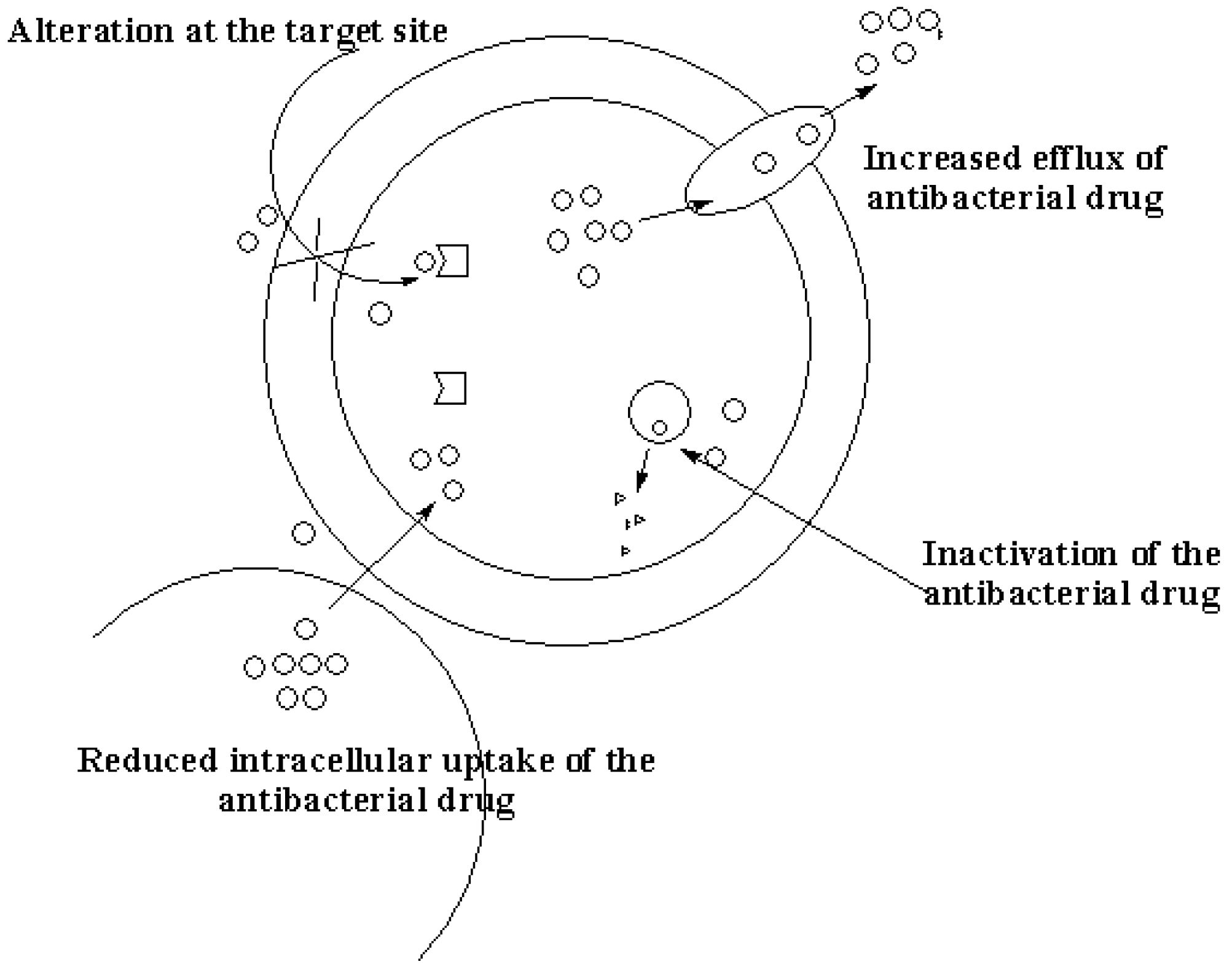
Figure 2.
Mode of action of nanoparticles on bacteria.
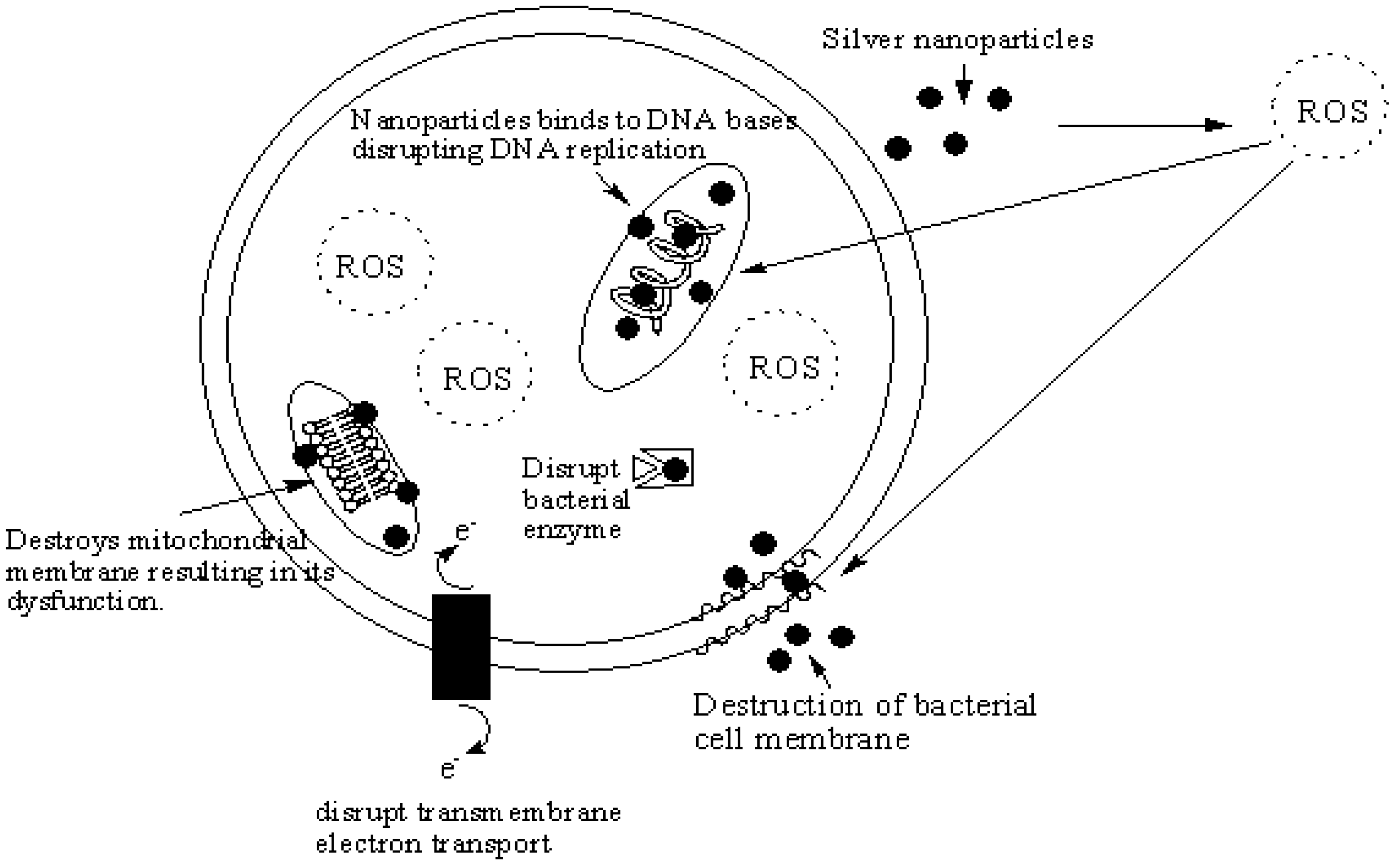
Figure 3.
Nanoshapes of silver nanoparticles.
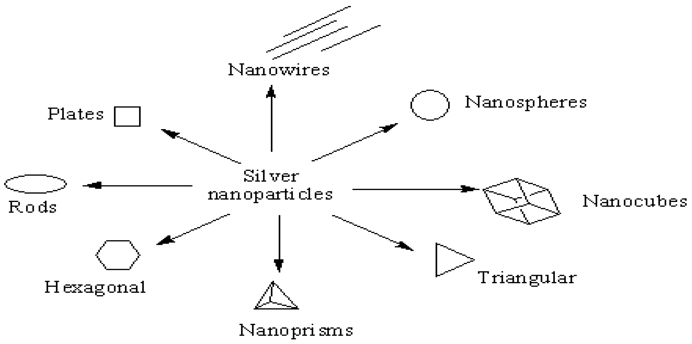
Figure 4.
Mode of action of nanoparticles on microphages.

Figure 5.
Mode of action of nanoparticles on viruses.


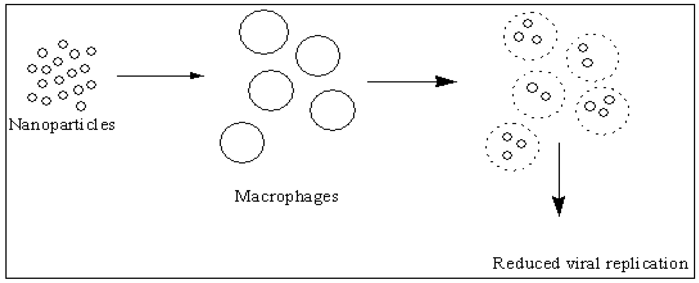
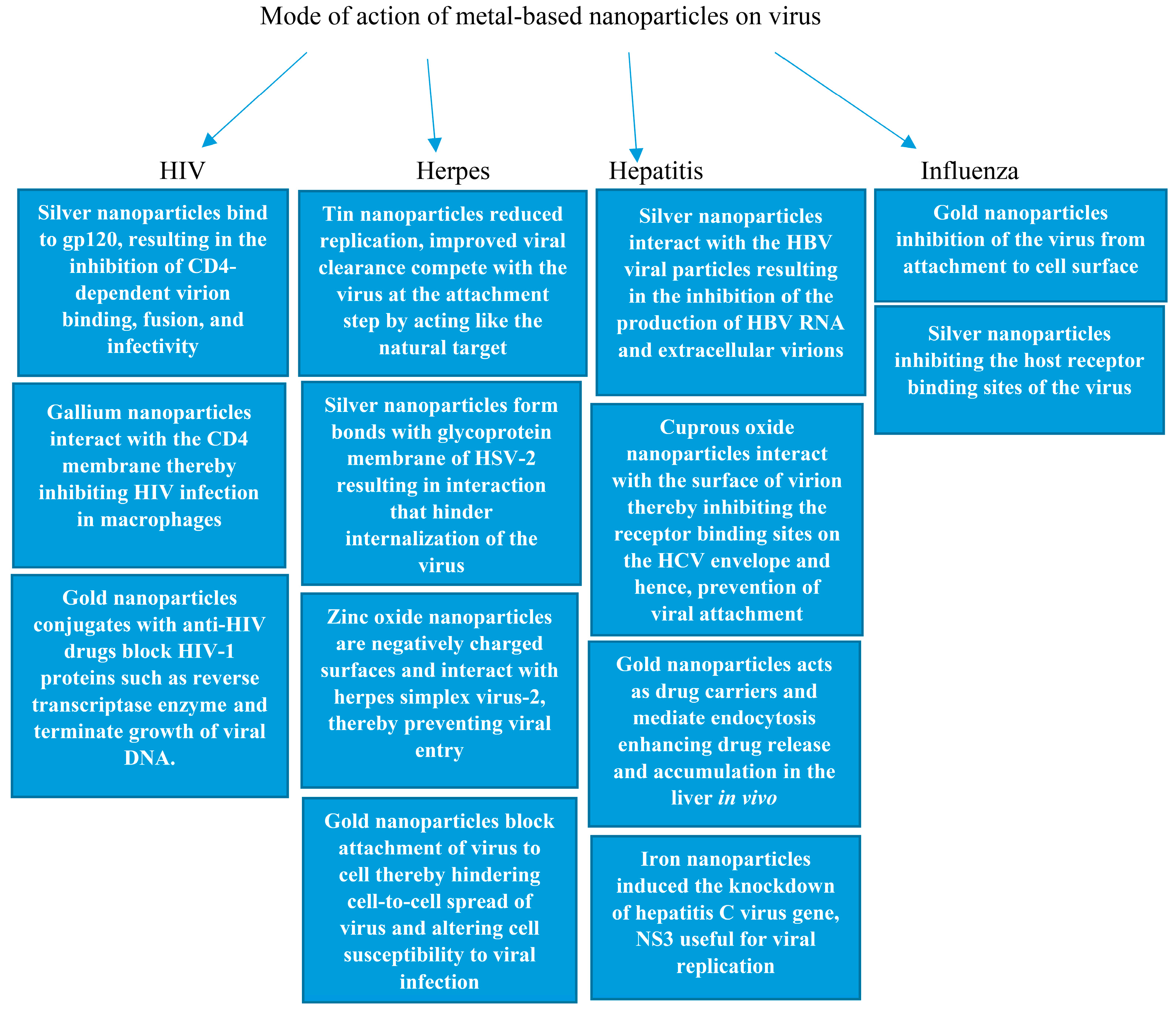
Table 1.
Silver nanoparticles with antibacterial activity.
| Metal Nanoparticles | Therapeutic Outcome | References |
|---|---|---|
| Silver nanoparticles | Effective against Escherichia coli and Staphylococcus aureus. | [24,25,26,27,28,29,30] |
| Sphere-shaped, and triangle shaped silver nanoparticles | The antibacterial activity of the nanoparticles against P. aeruginosa bacteria was enhanced for spherically shaped nanoparticles. The triangle-shaped silver nanoparticles exhibited enhanced antibacterial effect which is attributed to high-atom-density facets and interaction of the facets with the surface of the bacteria. | [31,32,33,34] |
| Rod-shaped silver nanoparticles | Triangular shaped nanoparticles exhibited high antibacterial activity against Escherichia coli than the spherical and rod shaped silver nanoparticles. | [32] |
| Hexagonal and nanoplates silver nanoparticles | Hexagonal-shaped silver nanoparticles were effective against S. aureus and E. coli when compared to the nanorod- and nanoplate-shaped silver nanoparticles. | [35,36] |
| Nanocube and nanowire-shaped silver nanoparticles | Nanocube-shaped silver nanoparticles exhibited the highest antibacterial activity because of their surface area, effective contact area, and facet reactivity. | [37] |
| Silver nanoparticles | Inhibited the growth of Mycobacterium tuberculosis | [38,39,40] |
| Silver nanoparticles | Effective against bacteria causing sexually transmitted disease e.g., Chlamydia trachomatis. | [41,42] |
| Silver nanoparticles in urinary catheter | Effective against bacteria that are responsible for urinary tract infections. | [43,44,45,46,47,48] |
| silver nanoparticles combined with polymixin B and rifampicin | Good synergistic effects in the treatment of Acinetobacter baumannii infection. | [49] |
| Silver nanoparticles combination with amoxicillin | Good synergistic effects against Escherichia coli resulting from chelation between active groups such as hydroxy and amido groups on amoxicillin with the nanosilver. | [50] |
| Silver nanoparticles combination with β-lactam; quinolone; aminoglycoside and polykeptide | Effective against drug-resistant bacteria Salmonella typhimurium. β-lactam class of antibiotics did not show synergistic effects because of its inability to form a complex with the nanoparticles. | [51] |
| Silver nanoparticles combination with gentamicin and penicillin | Excellent antibacterial effects against animal bacterial infections, Actinobacillus pleuropneumoniae, A. pleuropneumoniae and Pasteurella multocida. | [52] |
| Silver nanoparticles combination with visible blue light and either amoxicillin, azithromycin, clarithromycin, linezolid or vancomycin | Good synergistic antibacterial effects against methicillin-resistant Staphylococcus aureus. | [53] |
| Silver nanoparticles combined with either cefazolin, mupirocin, gentamycin, neomycin, tetracycline or vancomycin | Combination of nanoparticles with antibiotics was effective against Staphylococcus aureus, Pseudomonas aeruginosa and Escherichia coli. | [54] |
| Conjugation of cephalexin onto silver nanoparticles | Effective against E. coli and S. aureus by binding to the cell wall resulting in the destruction of the cell outer membrane. | [55] |
| Silver nanoparticles prepared using plants extracts | Stable nanoparticles with good antibacterial activity. | [56,57,58,59,60,61,62,63,64,65,66] |
| Silver nanoparticles prepared by biological methods using virus, bacteria and fungi | Good antibacterial activity. | [67,68,69,70,71,72,73] |
Table 2.
Iron oxide and copper oxide nanoparticles with antibacterial activity.
| Metal Nanoparticles | Therapeutic Outcome | References |
|---|---|---|
| Iron oxide nanoparticles | Good antibacterial activity on E. coli, P. vulgaris. Staphylococcus aureus | [74,75,76,77,78] |
| Iron oxide nanoparticles combination with erythromycin | Good synergistic antibacterial effects against S. pneumonia | [79] |
| Iron oxide nanoparticles | Inhibition of growth of Staphylococcus aureus, Escherichia coli, Pseudomonas aeruginosa and Serratia marcescens | [80,81] |
| Iron oxide nanoparticles coated with chitosan biomolecules | Good inhibition of growth of Bacillus subtilis and Escherichia coli bacteria | [82] |
| Iron oxide nanoparticles using Punica granatum peel extract | Strong antibacterial activity against Pseudomonas aeruginosa | [83] |
| Iron oxide nanoparticles | Good antibacterial effects against Staphylococcus aureus which is dependent on concentration | [85] |
| Chitosan coated iron nanoparticles | Inhibited the growth of Escherichia coli and Salmonella enteritidis | [86] |
| Iron oxide nanoparticles combined with ciprofloxacin | Poor antibacterial activity | [87] |
| Iron oxide nanoparticles prepared using leaf extract | Good antibacterial with varied shapes | [88,89,90,91,92,93,94,95,96,97] |
| Copper oxide nanoparticles | Very sensitive to E. coli and E. faecalis and less selective to K. pneumonia | [98] |
| copper oxide nanorods and multi-armed nanoparticles | Multi-armed nanoparticles exhibited higher antibacterial activity against E. coli than the nanorods | [99] |
| Copper oxide nanoparticles | The antibacterial activity of copper oxides is attributed to lipid peroxidation, generation of reactive oxygen species, protein oxidation and DNA degradation in bacteria cells | [100] |
| Copper oxide nanoparticles | antibacterial of the nanoparticles was dependent on the particle sizes | [101,102] |
| Copper oxide nanoparticles | The nanoparticles exhibited spherical shapes with high antibacterial activities against Bacillus subtilis and Salmonella choleraesuis | [103] |
| Copper oxide nanoparticles | The antibacterial activity of the nanoparticles was effective against K. pneumoniae, S. typhimurium, and E. aerogenes | [104] |
| Copper oxide nanoparticles | Effective against different strains of Staphylococcus aureus | [105] |
| Copper oxide nanoparticles | Good antibacterial activity of copper oxide nanoparticles against Gram-positive (B. subtilis and S. aureus) and Gram-negative (E. coli and P. aeruginosa) bacteria | [106] |
| Copper oxide nanoparticles | Good antibacterial activity against Escherichia coli and Pseudomonas aeruginosa | [107] |
| Copper oxide nanoparticles | Effective against Escherichia coli and Lactobacillus brevis | [108,109,110,111,112,113,114] |
Table 3.
Zinc oxide and aluminium oxide nanoparticles antibacterial activity.
| Metal Nanoparticles | Therapeutic Outcome | References |
|---|---|---|
| Zinc oxide nanoparticles | Good antibacterial activity against Klebsiella pneumonia that causes respiratory infection | [115] |
| Zinc oxide nanoparticles | The inhibition effect on the growth of B. subtilis was dependent on the concentration of the nanoparticles | [116,117] |
| Zinc oxide nanoparticles | The antibacterial effect against clinical isolate of Staphylococcus aureus was excellent | [118] |
| Zinc oxide nanoparticles | Effective against Campylobacter jejuni | [119] |
| Zinc oxide nanoparticles | Good antibacterial activity by ROS mediated membrane lipid oxidation of Escherichia coli, S. aureus, P. aeruginosa and V. anguillarum | [120,121,122,123] |
| Zinc oxide nanoparticles | Effective against E. coli | [124,125,126] |
| Zinc oxide nanoparticles | Effective against Gram-positive bacteria. The antibacterial effect was high on B. subtilis cells when compared to S. aureus | [127] |
| Zinc oxide nanoparticles coated with gentamicin | The antibacterial effects against Escherichia coli, Pseudomonas aeruginosa, Staphylococcus aureus, Bacillus cereus and Listeria monocytogenes was significant | [128] |
| Zinc oxide nanoparticles using aqueous extracts of P. crispum | Excellent antibacterial activity | [129] |
| Zinc oxide nanoparticles prepared from plants extract | Enhanced antibacterial activity | [130,131] |
| Aluminium oxide nanoparticles | Nanoparticles penetrated Candida cells disrupting the morphological and physiological activity of the cells. | [132,133] |
| aluminium oxide nanoparticles prepared from leaf extracts of lemongrass | Good antibacterial, activity against clinical isolates of P. aeruginosa was significant | [134] |
| Aluminium oxide nanoparticles | Effective against gram-positive and gram-negative bacteria | [135,136] |
Table 4.
Gold, titanium dioxide and gallium nanoparticles with antibacterial activity.
| Metal Nanoparticles | Therapeutic Outcome | References |
|---|---|---|
| Gold nanoparticles | Effective against bacterial infection in animal, Corynebacterium pseudotuberculosis | [137] |
| Gold nanoparticles | Effective against E. Coli | [138,139,140,141,142] |
| Gold nanoparticles | The nanoparticles were active against Gram-negative, Gram-positive uropathogens and multi-drug resistant pathogens | [139] |
| Gold nanoparticles | Active against enteric bacteria e.g., Escherichia coli, Staphylococcus aureus, Bacillus subtilis and Klebsiella pneumonia | [140] |
| Gold nanoparticles | Effective against E. coli, S. typhimurium DT104, and S. aureus | [141] |
| Gold nanoparticles | Inhibited growth of Salmonella typhi | [142] |
| Gold nanoparticles | Effective against Staphylococcus aureus and Escherichia coli | [143] |
| Gold nanoparticles combined with gentamicin | Effective against Escherichia coli | [144,145] |
| Gold nanoparticles capped with cefaclor | Potent antimicrobial activity against both Gram-positive (Staphylococcus aureus) and Gram-negative (Escherichia coli) bacteria | [145] |
| Gold nanoparticles | Potent antibacterial effect against multidrug-resistant Gram-negative bacteria | [146] |
| Gold nanoparticles prepared using banana peel extract | Good antibacterial activity | [147] |
| Gold nanoparticles combined with ofloxacin | Superior bactericidal property | [148] |
| Gold nanoparticles prepared using Stoechospermum marginatum | Enhanced antibacterial activity | [149] |
| Gold nanoparticle prepared from A. comosus extract | Useful purification processes for inhibiting the growth of bacteria | [150] |
| Gold nanoparticles prepared using aqueous leaves extract of Moringa oleifera | Effective against Staphylococcus aureus, Candida tropicalis, Candida krusei, Klebsiella pneumonia | [151] |
| Gold nanoparticles | Active against Escherichia coli | [152] |
| Gold nanoparticles combined with gentamicin | Good antibacterial activity | [153] |
| light-absorbing gold nanoparticles conjugated with specific antibodies | selective killing of the Gram-positive bacterium Staphylococcus aureus | [154,155,156,157] |
| Gold nanoparticles combined with vancomycin | Selective binding to the cell of Gram-positive bacteria, Gram-negative bacteria and antibiotic-resistant bacteria | [156,157] |
| Gold nanoparticles combined with ampicillin | Effective against E. coli, Micrococcus luteus and Staphylococcus aureus | [158] |
| Gold nanoparticles combined with streptomycin | Effective against E. coli, Micrococcus luteus and Staphylococcus aureus | [158] |
| Gold nanoparticles combined with kanamycin | Effective against E. coli, Micrococcus luteus and Staphylococcus aureus | [158] |
| Gold nanoparticles combined with levofloxacin | Inhibited growth of S. aureus and E. coli | [159] |
| Titanium dioxide nanoparticles | Effective against biofilm producing methicillin-resistant S. aureus | [160] |
| Titanium dioxide nanoparticles | Inhibited growth of bacteria that causes dental plaques | [161] |
| Titanium dioxide nanoparticles | Effective against Streptococcus mutans | [163] |
| Titanium dioxide nanoparticles | Effective against E. coli | [164,165,166,167,168] |
| Gallium nanoparticles | Inhibited the growth of mycobacteria | [169] |
| Gallium nanoparticles | Good antibacterial properties against Pseudomonas aeruginosa | [170] |
| Gallium nanoparticles | Inhibited growth of Mycobacterium tuberculosis significantly | [171] |
| Gallium nanoparticles | Disrupted F. Tularensis Fe metabolism | [172] |
Table 5.
Metal-based nanoparticles with antiviral activity.
| Nanoparticles | Infection | Therapeutic Outcome | References |
|---|---|---|---|
| Silver nanoparticles | HIV | Inhibition of CD4-dependent virion binding, fusion, and infectivity | [173,174,175,176,177] |
| Gallium nanoparticles | HIV | Suppressed co-infection of HIV and tuberculosis. Inhibition of viral protease | [178,179,180] |
| Gold nanoparticles combined with Azidothymidine | HIV | Inhibition of early stages of viral replication | [181,182] |
| Gold nanoparticles conjugated with raltegravir | HIV | Good anti-HIV activity | [183] |
| Gold nanoparticles | HIV | Inhibition of viral entry | [184] |
| Carbohydrate-coated gold nanoparticles conjugated with abacavir and lamivudine | HIV | The nanoparticles inhibited HIV viral replication | [185,186] |
| Peptide triazoles conjugated onto gold nanoparticle | HIV | Potent antiviral effects against HIV-1 | [187] |
| Tin nanoparticles | Herpes | Trapped HSV-1 before entry into the host cell | [188,189,190] |
| Silver nanoparticles | Herpes | Virus replication was inhibited | [191] |
| Tannic acid modified with silver nanoparticles | Herpes | reduced HSV-2 infection | [192] |
| Silver nanoparticles | Herpes | Inhibition of viral entry into the cell and prevention of subsequent infection | [193] |
| Polyurethane condom coated with silver nanoparticles | Herpes | Inhibition of HSV-1 and 2 infections | [194,195] |
| Zinc oxide | Herpes | Prevented viral entry and infection | [196,197] |
| Gold nanoparticle | Herpes | Inhibited viral attachment and penetration into the cells thereby preventing infections | [198,199] |
| Silver nanoparticles | Hepatitis | Interaction with the HBV viral particles resulting in the inhibition of the production of HBV RNA and extracellular virions | [200,201] |
| Iron oxide nanoparticles | Hepatitis | Induced the knockdown of hepatitis C virus gene, NS3. HCV NS3 gene encodes helicase and protease which are useful for viral replication | [202] |
| Cuprous nanoparticle | Hepatitis | Inhibited the entry of virus which included genotypes such as, 1a, 1b, and 2a thereby hindering viral replication | [203] |
| Gold nanoparticle loaded with interferon α | Hepatitis | Targeted delivery of interferon α | [204,205] |
| Gold and silver nanoparticles | Influenza | Effective against influenza A virus | [206,207] |
| Silver nanoparticles | Influenza | Effective against influenza viruses resulting in damage to their morphological structure. Inhibiting the host receptor binding sites of the virus | [208,209,210,211,212,213] |
Table 6.
Metal-based nanoparticles for the treatment of parasitic infections.
| Nanoparticles | Infection | Therapeutic Outcome | References |
|---|---|---|---|
| Silver nanoparticles | Malaria | Inhibition of the growth of P. falciparum in vivo and in vitro | [216,217,218,219] |
| Metal oxide nanoparticles (Fe3O4, MgO, ZrO2, Al2O3 and CeO2) | Malaria | Good to moderate antiplasmodial activity against P. falciparum | [220] |
| Gold nanoparticles | Malaria | Moderate delayed parasitemia rise in vivo, moderate antiplasmodial activity against P. falciparum | [221,222] |
| Silver nanoparticles | Leishmaniasis | Inhibition of proliferation and metabolic activity of promastigotes. Good antileishmanial activity in vitro and in vivo | [223,224,225,226,227,228,229] |
| Kaempferol-stabilized gold nanoparticles | Leishmaniasis | Effective against both wild and drug resistant strains | [230,231] |
| Metal-oxide nanoparticles (titanium dioxide nanoparticles, zinc oxide nanoparticles and magnesium oxide nanoparticles) | Leishmaniasis | Enhanced cytotoxic effects on promastigotes of L. major via induction of apoptosis | [232,233] |
| Silver nanoparticles | Helminth infections | Enhanced anthelmintic activity against worm | [234,235,236,237] |
| Gold nanoparticles | Helminth infections | Affected the physiological functioning of the parasite causing paralysis and subsequent death | [238] |
| Zinc oxide nanoparticles | Helminth infection | Disruption of the electron transport system inhibiting ATP production and the contractile movement of the parasite | [239] |
| Zinc oxide and iron oxide nanoparticles | Helminth infection | The anthelmintic activity of the metal oxides nanoparticles was via induction of oxidative stress | [240] |
© 2017 by the author. Licensee MDPI, Basel, Switzerland. This article is an open access article distributed under the terms and conditions of the Creative Commons Attribution (CC BY) license (http://creativecommons.org/licenses/by/4.0/).
Share and Cite
MDPI and ACS Style
Aderibigbe, B.A. Metal-Based Nanoparticles for the Treatment of Infectious Diseases. Molecules 2017, 22, 1370. https://doi.org/10.3390/molecules22081370
AMA Style
Aderibigbe BA. Metal-Based Nanoparticles for the Treatment of Infectious Diseases. Molecules. 2017; 22(8):1370. https://doi.org/10.3390/molecules22081370
Chicago/Turabian StyleAderibigbe, Blessing Atim. 2017. "Metal-Based Nanoparticles for the Treatment of Infectious Diseases" Molecules 22, no. 8: 1370. https://doi.org/10.3390/molecules22081370