Structure Elucidation and Botanical Characterization of Diterpenes from a Specific Type of Bee Glue

Faculty of Pharmacy A15, The University of Sydney, Sydney, NSW 2006, Australia
*
Author to whom correspondence should be addressed.
Molecules 2017, 22(7), 1185; https://doi.org/10.3390/molecules22071185
Submission received: 4 July 2017
/
Revised: 12 July 2017
/
Accepted: 12 July 2017
/
Published: 14 July 2017
Abstract
:Investigation of the single plant source bee glue type originating from Southern Australia resulted in the isolation and structure elucidation of major serrulatane diterpenes, novel 7,8,18-trihydroxyserrulat-14-ene (1), along with its oxidized product, 5,18-epoxyserrulat-14-en-7,8-dione (3) and known (18RS)-5,18-epoxyserrulat-14-en-8,18-diol (2). Exploration into the botanical origin revealed Myoporum insulare R. Br, as the plant source of the bee glue materials. This discovery was made through comparative analysis of the myoporum bee glue samples collected from the beehives, analyses of plant resinous exudate, and resin carried on the hind legs of bees foraging for bee glue.
1. Introduction
Bee glue (Propolis) is a resinous substance collected by worker honey bees, Apis mellifera, from specific plants and transported to the hives where it is mixed by bees with beeswax to form strongly adhesive material with antiseptic properties to aid in defense and for maintenance of hygiene in the hive [1,2,3]. There are well-documented instances of its usage in history i.e., Egyptians have used propolis to mummify corpses based on its preservation properties. Abu Ali Sina (Avicenna) the Persian scientist has mentioned propolis as the black wax [4]. Similarly, in other parts of the world such as South Africa, Eastern Europe, France, and Russia traditional folk medicine use of it has been recognized [5,6,7,8]. The chemical composition of the propolis depends on its plant source which varies with geographical location. There is no information showing that bees do any significant chemical processing on the collected resins [9]. The chemical groups of compounds identified in propolis samples include: terpenes, lignans, flavonoids, chalcones, stilbenes, cinnamates, aromatic acids and esters, aliphatic acids and esters, benzophenones, benzofurans and sugars [10,11]. Due to its unique flora, the Southern part of Australia (Kangaroo Island) has been recognized as an area rich in bees’ propolis [12]; therefore, a phytochemical analysis was done to categorize propolis types collected from different regions of the island. Two serrulatane diterpenes along with one oxidized product were isolated from a specific propolis type designated as “purple spot” and later called myoporum propolis after identification of its botanical source as Myoporum insulare R. Br. from the plant family of Scrophulariaceae Juss. Chemical structure elucidation was determined from 1D/2D NMR and 1H-NMR decoupling experiments, aided by comparison with published X-ray crystallographic data of serrulatane diterpenes [13,14,15]. The confirmation of the myoporum propolis samples’ botanical origin was completed by the determination of their chemical components along with observation of bees’ behavior and by analysis of the island flora.
2. Results
2.1. Structure Elucidation of Serrulatanes from Myoporum Bee Glue
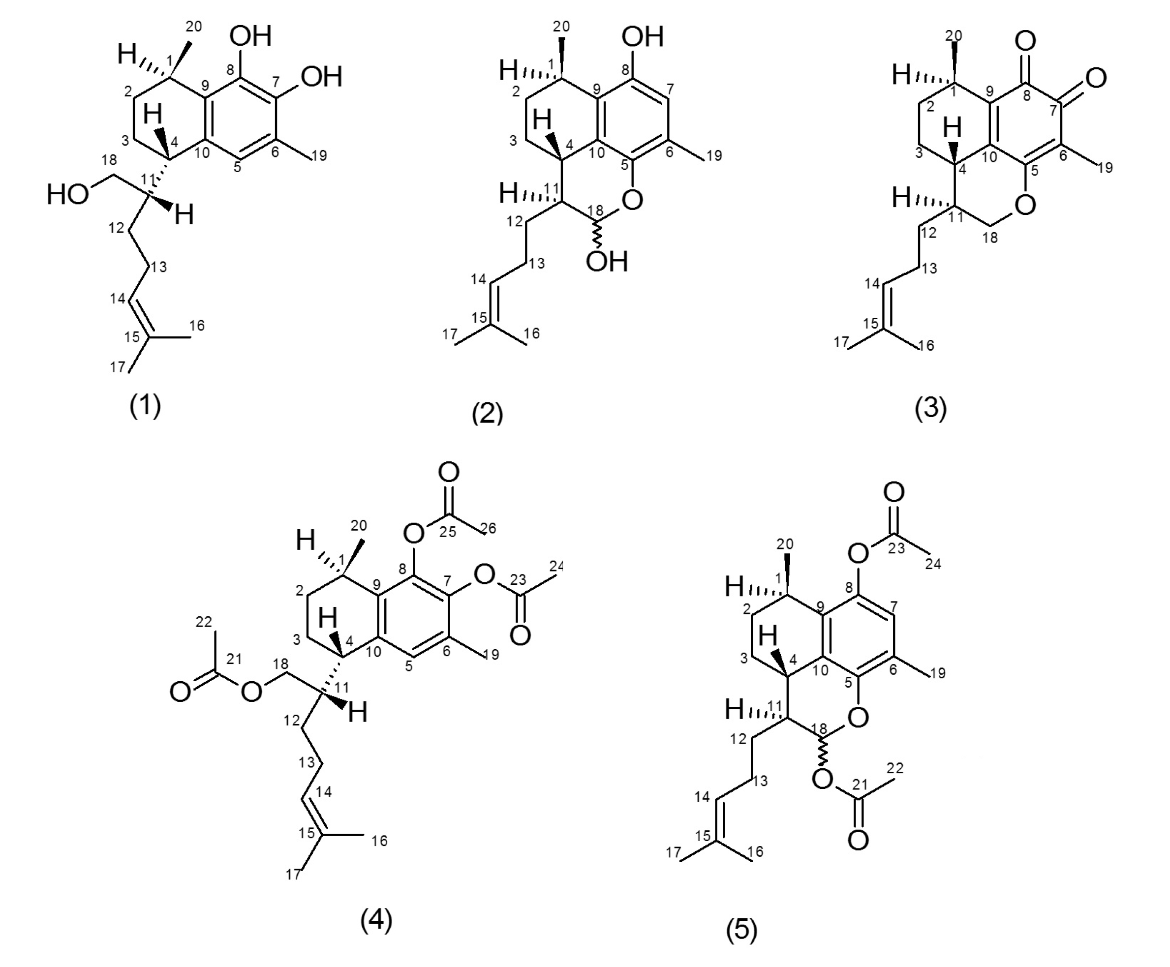
Propolis samples were collected broadly over the Island with beekeeping practice modified to minimize mixing of propolis from different plant sources. Propolis samples from different apiary sites were never mixed and collection from each site was done over a relatively short period of time i.e., approximately two weeks. To further minimize mixing from different plant sources, propolis collections were restricted to a maximum of 1 kg per sample bag and every bag was analyzed. More than 1000 samples were analyzed by thin-layer chromatography (TLC) and 1H-NMR studies and classified into six main propolis types; three types have been published [16,17] and the others are yet to be published [18]. Multiple propolis samples based on their relatively simple TLC and NMR pattern were identified as a likely single-source propolis of a type and initially named “purple spot” propolis (myoporum propolis) based on a characteristic purple spot observed by TLC and also purple coloration of the propolis and its ethanol solutions. This propolis type was extracted and fractionated by normal-phase short-column vacuum chromatography (NP-SCVC) to give two major components identified as novel 1 and known 2 diterpenes (Figure 1) from the interpretation of 1D/2D NMR spectroscopic and high-resolution electrospray ionization mass spectrometry (HRESIMS) data.
Compound 1 was a yellow liquid identified as 7,8,18-trihydroxyserrulat-14-ene. Its molecular mass was recorded as m/z 341.2089 [M + Na]+ (calcd. 341.2087) by HRESIMS analysis, hence the molecular formula was concluded as C20H30O3. With the use of 1H-NMR, 13C-NMR, HSQC, HMBC and 1H-1H COSY spectra, the chemical structure 1 was determined (Table 1 and Table 2).
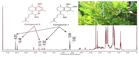
Analysis of 1H-1H COSY spectrum indicated the backbone relations from methyl group at position 20 to the di-methylated (C-16 and C-17) olefin moiety demonstrated by the bold lines. HMBC spectrum also confirmed the observed correlations (Figure 2). From the HMBC data strong correlation was noted for the methyl group at H-20 (δH 1.21) with C-1 (δc 26.9). Additionally, strong HMBC correlations were detected for δH 5.04 (H-14) to C-16 (δc 25.7) and C-17 (δc 17.7), which confirmed the position of the terminal di-methylated olefin moiety side chain. A one-proton singlet in the aromatic region at 6.56 ppm due to H-5 was noted in the 1H-NMR spectrum of compound 1. At 5.04 ppm, a broad triplet split integrating for one proton was recorded with a J value of 7.0 Hz which matched with (H-14) of the isoprene sidechain part of the serrulatane diterpene structure. At 5.42 and 5.23 ppm, two broad one-proton singlets were assigned to the catechol hydroxyl moieties situated on the aromatic ring (OH-8, OH-7), respectively. A two-proton doublet at 3.63 ppm with J value of 6.16 Hz was assigned to position H2-18. The hydrogen at position 1 (H-1) showed a one-proton signal at 3.07 ppm with a splitting pattern of a quintuplet of doublets and J values of J = 6.8, 1.6 Hz. Furthermore, H-4 resonated as a one-proton triplet of doublets at 2.76 ppm (J values 5.6 and 2.6 Hz) plus the three hydrogens at chemical shift of 2.20 ppm were assigned to H-19 corresponding to the methyl group on the aromatic ring. The other two methyl groups were noted at 1.68 and 1.58 ppm, matching to H-16 and H-17, respectively.
Similar procedures as for compound 1 were followed to elucidate the structures of other isolated chemical compounds. Compound 2 was defined as (18RS)-5,18-epoxyserrulat-14-en-8,18-diol, with its HRESIMS showing molecular mass of m/z 315.1970 [M − H]− (calcd. 315.1966) and a molecular formula of C20H28O3.
Structures 1 and 2 present a high degree of similarity in terms of their 1H-NMR and 13C-NMR chemical shift values for their non-aromatic ring and the olefinic side chain (Table 1 and Table 2). One significant difference is the presence of C-18 methine group in 2 when compared to compound 1, and the down-field shift of proton and carbon (δH 5.59 and δc 91.9) indicated the presence of C-18 hemiacetal methine group in 2. One more change between 1 and 2 takes place at aromatic proton position, C-7 (δc 115.9) in 2 compared with C-5 (δc 122.0) in 1.
Compound 3, the oxidized product of 1, with molecular mass of m/z 315.19573 [M + H]+ (calcd. 315.19547) based on HRESIMS, was assigned the molecular formula C20H26O3, and was identified as 5,18-epoxyserrulat-14-en-7,8-dione (Figure 1). It is proposed that compound 3 may be formed through a four-stage process with two stages of oxidation (Supporting information). The catechol moiety of 1 may be oxidized to an intermediate ortho-benzoquinone. In the second stage, the 18-hydroxyl group adds to the 5,6 double-bond to form the six-membered ring with the O-18 to C-5 bond. As a result of the addition, the intermediate has a 7,6-double-bond and a 7-hydroxy group. In the third stage, by keto to enol tautomersim, H-5 may be lost and a hydrogen gained on O-8 to give a catechol hydroxyl. The second catechol oxidation in the last stage gives compound 3. On account of oxidation reaction, catechol hydroxyl hydrogens are lost in the formation of the benzoquinone keto groups at position 7 and 8, which make C-7 and C-8 less shielded with δc 179.4 and δc 181.9, respectively. Compound 3 lacks the aromatic proton at position 5 compared to compound 1 and also there are changes to the splitting pattern for protons in position 18. In more detail, H-18a was detected at 3.86 ppm with the splitting pattern of a triplet and J value of 10.8 Hz and the other, H-18b, was noted at 4.49 ppm with dd splitting and J values of 10.8 and 3.6 Hz (Table 1). This pattern is consistent with the additional ring formed by oxidative cyclisation between position 5 and oxygen 18 to form an additional six-membered ring where H-18a is pseudoaxial and H-18b is pseudoequatorial.
2.2. Derivatization of Isolated Serrulatanes
To avoid quinonoid products formation of compound 1 and possible equilibrium of open and closed forms of compound 2 [19], for long duration NMR studies; rotating frame overhause effect spectroscopy (2D ROESY) and 1H-NMR decoupling, the hydroxyl groups were acetylated to provide respectively compounds 4 and 5 (Figure 1). The expanded and more specific C-NMR data from serrulatane derivatives enable a more definitive interpretation of the relative stereochemistry of the serrulatane core through closer comparison with the few available X-ray studies on serrulatane skeleton stereochemistry [13,14,15].
The acetylated compound 4, isolated as a pale yellow oil, was determined to have a molecular mass of m/z 467.2405 [M + Na]+ (calcd. 467.2404) in the HRESIMS and a molecular formula established as C26H36O6. The experimental HMBC connection between H-11 (δH 2.1) and C-4 (δc 36.9) showed that the C-11 (side chain) is directly attached to the non-aromatic ring, this was also supported by a substantial ion at m/z 275.06 in the mass spectrum caused by the cleavage of C-4/C-11 which is frequently reported among serrulatane compounds [20]. HMBC correlations from δH 1.15 (H-20) to C-1 (δc 27.6) and C-9 (δc 134.1) suggested the attachment of the methyl group to the C-1 of the bicyclic system and this was further established by decoupling experiments. The H-4 neighboring to an aromatic ring was specified by HMBC correlations from δH 2.88 (H-4) to C-5 (δc 128.1), C-9 (δc 134.1) and C-10 (δc 137.1). The cyclohexene ring foundation was complete by showing H-1 (δH 2.92) HMBC correlations to C-9 and C-10. The strong HMBC correlation from δH 4.11 proton to a carbonyl moiety confirmed the attachment of this acetoxy group to this methylene moiety, and the distinct HMBC correlations to C-4, C-11 and C-12, positioned it at H-18. High level of uniformity was achieved when comparisons were made among the 1H and 13C-NMR data (Table 1 and Table 2) of the aromatic ring plus the side-chain assignment, with other similar previous literature data [13,21].
Searching similar substructures accompanied by consideration of diterpene biosynthetic pathways indicated that compound 4 possessed a serrulatane skeleton. Allocating the relative configuration of the serrulatanes using 1H-NMR data only is very challenging [13]. The relative configurations for the chiral centers (C-1, C-4 and C-11) were clarified on the basis of 1H-NMR decoupling and ROESY data. When irradiation of the H-1 proton was examined, the doublet (J = 6.9 Hz) protons of the methyl group at δH 1.15 collapsed to a broad singlet. The similarity of H-1 splitting pattern with reported serrulatanes [22] suggested a distorted chair conformation for the cyclohexene ring with H-1 in pseudoequatorial orientation. According to the ROESY correlations of H-1 and H-2a, these protons are located on the same face of the molecule. The pseudoequatorial configuration of H-4 was understood from the intensive ROESY correlation between H-4 and H-5, consistent with the reported X-ray structure (CCDC 999272) [14]. Subsequently, ROESY correlations between H-1/H-2a and H-4/H-3a positioned H-1 and H-4 on the opposed faces of the molecule. In relation to the negative specific rotation for the published serrulatanes in literature [15,22], 1R*, 4S* configuration was assigned, consistent with obtained optical rotation results for the isolated serrulatanes presented in this study. The flexibility of the isoprene side chain complicated the configuration determination at C-11, however, from the spatial ROESY correlation between H-4/H-11, the configuration of H-11 was established as 11S. Therefore, the concluding relative configuration of the compound 4 best fits as 1R*, 4S*, 11S*, in agreement with reported similar configurations [14,22,23,24,25,26,27,28] for the serrulatane skeleton.
After isolating compound 2, elucidation difficulties were faced due to the hemiacetal cyclization and equilibrium of open and closed forms, therefore the acetylation of compound 2 was carried out, resulting in compound 5 with a molecular formula of C24H32O5 and molecular mass m/z 423.2143 [M + Na]+ (calcd. 423.2142) in the HRESIMS. Compound 5 1H-NMR spectrum presented a similar pattern to 4, with a significant doublet signal (J = 2.1 Hz) at δH 6.53 corresponding to the proton H-18 shifted to the lower field on forming the acetate, owing to the electron-withdrawing influence and anisotropy of the acetoxy group attached to position 18. 1H and 13C-NMR data (Table 1 and Table 2) were consistent with compound 5 being a serrulatane diterpene. The 1H and 13C-NMR spectra of compound 5 were almost identical with that of the published serrulatane [19], with the exception that in the side chain, the bond between C-14/C-15 was unsaturated. The relative configuration of compound 5 was determined as 1R*, 4S* and 11S* from the observed similarity with reported serrulatanes and X-ray crystal studies [13,14,15].
Molecular geometry affects 13C chemical shifts; hence, a possible underlying explanation for the downfield C-2 and C-3 chemical shifts noticed for structures 2, 3 and 5 could be addressed as the steric effects caused by cyclization and rigidity among these three mentioned structures; whereas in case of compound 1 and its derivative (4), 13C chemical shifts at these positions are noted more upfield, same pattern has been reported for similar serrulatane diterpenes as published in literature [22,25].
2.3. Identification of Botanical Origin
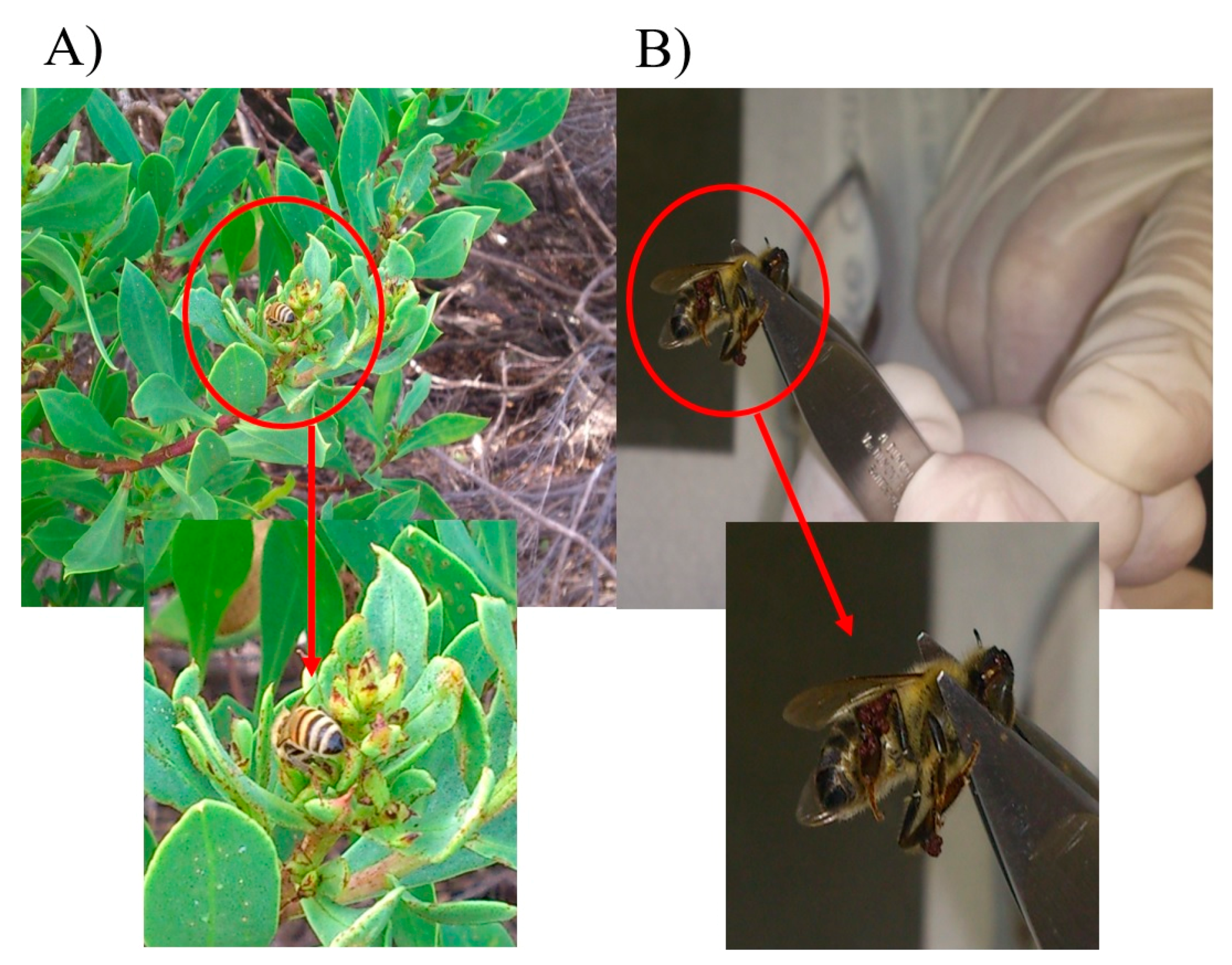
Focusing on the isolated chemical structures 1 and 2, our research group concentrated on searching to find the botanical origin of the myoporum propolis type. From the main skeleton of the compounds, serrulatane, plants of the genus, Eremophila, Scrophulariaceae family, were considered as possible sources. Since Eremophila species are rare on the Island, plants of the closely related genus Myoporum, of widespread occurrence on the Island, were also taken into consideration. By means of the flora distribution map of the Island for these categories [29,30,31,32], and having the location of apiary sites in mind, the best regions were recognized. Subsequently, bees were detected collecting resin from the sticky buds and leaves to produce propolis from a shrub or tree, called Myoporum insulare, near coastal zones (Figure 3, Appendix B video supplementary data).
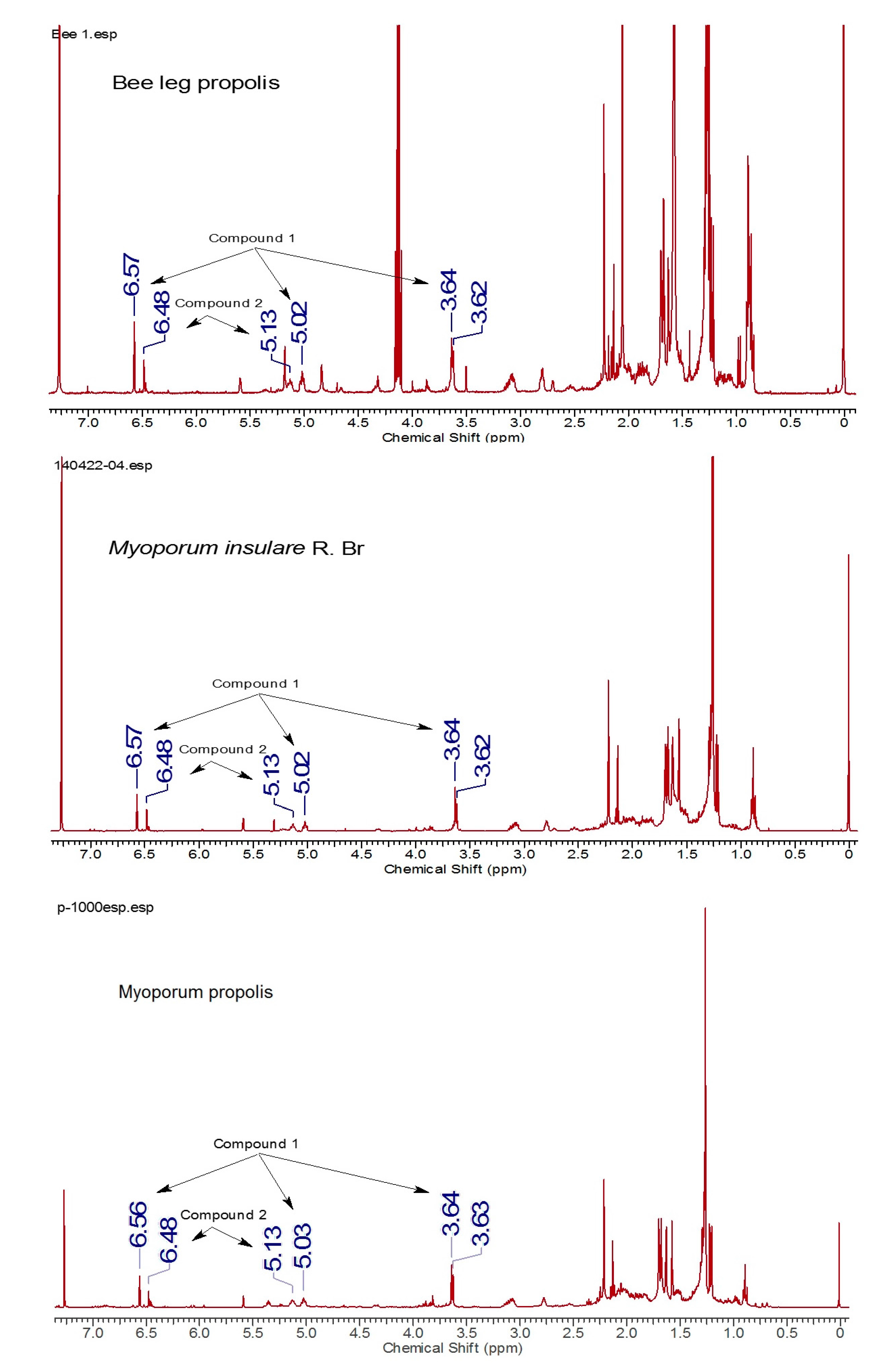
Plant samples were found in the vicinity of the beehives across the Island in sites where the myoporum propolis samples had been collected. Comparative NMR analysis of propolis on bee legs, myoporum propolis collected from the hives mats, plus the resin extracted from the shiny sticky sprout tips of the leaves of Myoporum insulare revealed identical chemical constituents’ profiles in comparable proportions (Figure 4).
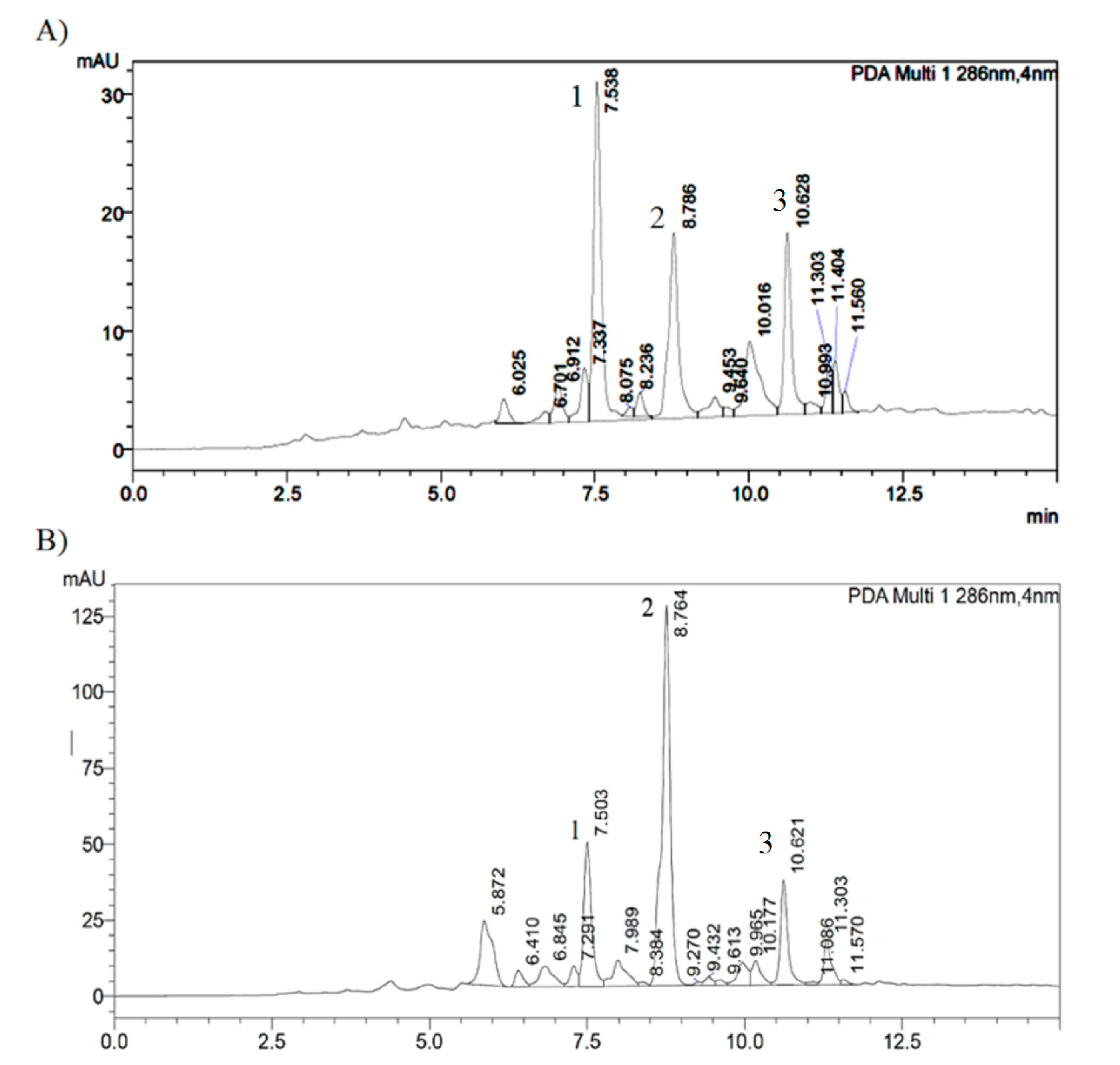
Moreover, Figure 5 presents comparative HPLC profiling of the sticky leaf surface resin extract of Myoporum insulare and bee legs propolis extract.
3. Discussion
Considering the unique flora in Australia, only a small number of studies have been done on the Australian bee glue compared with the numerous studies done in other parts of the world. Although there are a number of reports regarding diterpenes obtained from propolis around the world, there is no report of propolis comprising diterpenoids from Australia. Propolis is defined as one of the natural rich sources of biologically active diterpenes [12]. Compounds isolated from propolis may be assessed for their medical uses since knowledge of a degree of safety and possible efficacy exists through their traditional consumption as blends in propolis [33,34,35,36,37,38]. With attention to promising biological activities of diterpenes and considering the fact that no reports have been made about diterpenes from Australian propolis, Kangaroo Island propolis samples were documented to possess interesting composition, uncovering novel diterpenes. Chromatography isolation and NMR studies demonstrated that the main components were new serrulatanes, classifying a discrete propolis type, myoporum “purple spot” propolis, with potential for useful medicinal properties. The distinctive “purple spot” was found to originate from compound 1 by its oxidation reaction when exposed to oxygen and light. Based on its 1H-NMR spectrum, the “purple spot” has been identified as compound 3, formed by processes involving oxidation and cyclisation. The purple colour is representative of an ortho-benzoquinone chromophore [39,40]. Multiple propolis samples were analyzed, either collected by research team members or by local beekeepers from different sites over the Island in all seasons. To categorize the samples having the isolated serrulatanes, 1H-NMR methods and, to detect the plant source of this propolis type, the bee observation process along with flora distribution mapping were exploited. Bees foraging for propolis were captured from a shrub or tree, of the family Scrophulariaceae, dispersed along the coastal areas, identified as Myoporum insulare.
Matching profiles were confirmed among samples of shiny resinous plant leaves, propolis from bee legs and collected hive propolis using NMR and HPLC analysis. In the case of myoporum propolis samples, diterpenes 1 and 2 were noted as the major diterpenoid components. This is the first report about isolated diterpenes from this Island propolis.
4. Materials and Methods
4.1. General Experimental Procedures
Thin layer chromatography (TLC) sheets preloaded with silica gel 60-F254 were supplied from Merck (Darmstadt, Germany). Normal-phase short-column vacuum chromatography (NP-SCVC) was done using silica gel 60H (Merck, Darmstadt, Germany) packed into a sintered glass funnel with vacuum applied to give evenly packed stationary phase. A multiband UV-254/366 UV GL-58 mineral light lamp (Analytik Jena, Upland, CA, USA) was used for visualizing the TLC plates. All solvents used were purchased from Chem-Supply Pty Ltd., Port Adelaide, South Australia. The NMR solvents were purchased from Novachem Pty Ltd., Collingwood, Victoria, Australia. To evaporate the solvent fractions, a rotavapor model R-114 rotary evaporator (BÜCHI Labortechnik, Flawil, Switzerland). with a water bath temperature ranging from 40 °C to 60 °C was used. A vacuum pump V700 (Vacuubrand GMBH, Wertheim, Germany)., or Vacuubrand MD 4C NT diaphragm pump (Vacuubrand GMBH, Wertheim, Germany) with vacuum controller V-800 or V-850 were used along with the rotary evaporator and finally a NAPCO 5831 vacuum oven (NAPCO, Salt Lake City, UT, USA) with a Direct Torr vacuum pump (Sargent-Welch, Buffalo, NY, USA) was utilized to remove the last traces of solvents from samples. Analytical HPLC was performed on a Shimadzu UFLC, LC20AD pump, SIL-20A HT auto sampler (Shimadzu, Kyoto, Japan), with a Hewlett Packard Column, NUCLEOSIL 100C18, 5 μm, 4 mm × 125 mm (Shimadzu, Kyoto, Japan). Optical rotation measurements were performed by a Perkin-Elmer Model 341 LC Polarimeter (Perkin-Elmer, Waltham, MA, USA) with a sodium lamp (λ = 589 nm) using a 1 mL cell (length 2.5 cm). UV-vis spectra were measured on a Shimadzu UVmini-1240 spectrophotometer (Shimadzu, Kyoto, Japan). IR spectra were determined with a Shimadzu IRTracer-100 FT-IR spectrometer (Shimadzu, Kyoto, Japan). 1D/2D NMR (Nuclear magnetic resonance) analyses were done with Varian 400 MHz and Bruker 600 MHz systems. NMR spectra were internally referenced to tetramethylsilane (TMS) or 7.27 ppm for the peak of the residual H of CDCl3. Low resolution ESI-MS spectra were obtained from a Thermo Finnigan Polarisq ION trap MS/MS system (Thermo Fisher, Waltham, MA, USA). High resolution ESI-MS were measured on a Bruker Apex Qe 7T, Fourier Transform Ion Cyclotron Resonance mass spectrometer (Bruker, Bremen, Germany) equipped with an Apollo MTP ESI/MALDI Duel source (Bruker, Bremen, Germany).
4.2. Materials
A large number of standard 10-frame per box hives, each with a propolis mat underneath the hive cover lid, were the source of the propolis samples prepared by a number of beekeepers from colonies of European honey bee (Apis mellifera ligustica) in ten apiary sites situated in different locations of Kangaroo Island, South Australia. These sites were in the neighborhood of Hanson Bay (35°58’4” S; 136°49’1” E), Island Beach (35°47’49” S; 137°4745” E), Penneshaw (35°43’6” S; 137°56’25” E), Flour Cask Bay (35°51’35” S; 137°41’44” E), Vivonne Bay (35°59’1” S; 137°10’30” E), Rainy Creek (35°57’8” S; 137°12’55” E), Kingscote (35°39’11” S; 137°38’2” E), Brownlow (35°39’54” S; 137°37’8” E), and Eleanor River (35°57’59” S; 137°12’18” E). These locations are recognized to be a preferred environment for the plant known as Myoporum insulare on the Island. These sites delivered 23 propolis samples which were chemically studied, profiled and categorized by TLC analysis and 1H-NMR techniques and assessed to be at least 90% sourced from Myoporum insulare. Samples were cleaned, packed and deposited at −20 °C until advance analysis. The leaves from Myoporum insulare R. Br. were collected from the areas close to the apiary sites located in the Island from December 2013 to June 2016. A specimen was authenticated by the botanist, A/Prof Murray Henwood, John Ray Herbarium, as Myoporum insulare. Honey bees observed collecting resin from the leaves of Myoporum insulare were captured, capped and frozen in suitable plastic sample tubes. Parts of bee hind legs carrying propolis were cut and stored at −20 °C until analysis.
4.3. Extraction and Isolation of Serrulatane Diterpenes from Propolis
Propolis (2 g) was dissolved at room temperature in ethanol (40 mL) using an ultrasound sonicator, then the extract was filtered, and dried using a reduced pressure rotary evaporator followed by a vacuum oven. The dried extract (1.65 g) was taken onto a NP-SCVC column with 5 cm of bed diameter and 4 cm of height. The column was eluted using a stepwise gradient mobile-phase system with 50 mL fractions: hexane (Fractions A1 and A2); hexane:EtOAc, 4:1 (A3 and A4); hexane:EtOAc, 1:1 (A5 and A6); EtOAc (A7 and A8); EtOAc:EtOH, 4:1 (A9 and A10). Based on TLC analysis and NMR studies results, fractions A4 and A5 were combined and designated as fraction B. Exploiting the same technique, fractions A6 and A7 were combined together to give fraction B’. Fraction B (692 mg) was eluted using the stepwise gradient mobile-phase system hexane reaching hexane:EtOAc 4:1 and finishing with hexane: EtOAc 1:1. Totally 10 fractions (B1–10) were collected and fractions B3–5 were grouped together and chosen based on TLC and NMR analysis for further purification using NP-SCVC with CH2Cl2 and stepwise increasing the proportion of EtOAc to 1%, 2%, 4% and finally to 8% as eluent, ten fractions, C1–10, were collected of which fraction C5 provided compound 2 (153 mg). Further fractionation of fraction B’ (758 mg) was completed by NP-SCVC with CH2Cl2 and stepwise increasing the proportion of EtOAc to 2%, 4%, 8% and finally to 20% as mobile phase that afforded to 10 fractions (B’1–10). Through TLC and NMR analysis fraction B’7 was kept for one more purification step and fractions B’6 and B’8 were mixed together and called fraction D’. Fraction B’7 (86 mg) was loaded on NP-SCVC with CH2Cl2 and stepwise increasing the proportion of EtOAc to 1%, 2%, 4%, 8%, 12% and finally to 16% as eluent which resulted in 13 fractions (C’1–13) and fractions (C’11 and C’12) gave compound 1 (41 mg). Fraction D’ (249 mg) was further fractionated using NP-SCVC with CH2Cl2 and stepwise increasing the proportion of EtOAc to 1%, 2%, 4%, 8%, 12% and finally to 16% as mobile phase which gave 13 fractions (D’1–13) and fractions D’8–11 yielded compound 3 (62 mg).
Compound 1, C20H30O3, 7,8,18-trihydroxyserrulat-14-ene. Yellow oily material. [α] −27.3° (C = 1.0, CHCl3). UV (CH3OH) λmax nm (log ε) 279 (3.37) and 320 (2.49). IR (ν): 3381, 2924, 2864, 1625, 1581, 1448 cm−1. For 1H- & 13C-NMR, see Table 1 and Table 2. HRESIMS: m/z 341.2089 [M + Na]+ (calcd. 341.2087).
Compound 2, C20H28O3, (18RS)-5,18-epoxyserrulat-14-en-8,18-diol. Dark red oily material. [α] −21.5° (C = 1.0, CHCl3). UV (CH3OH) λmax nm (log ε) 293 (3.36) and 334 (2.91). IR (ν): 3327, 2970, 2926, 2860, 1647, 1558, 1448, 1043 cm−1. For 1H- & 13C-NMR, see Table 1 and Table 2. HRESIMS: m/z 315.1970 [M − H]− (calcd. 315.1966).
4.4. Acetylation of Serrulatane Diterpenes
The serrulatane 1 (400 mg) was dissolved in acetic anhydride (1 mL) and dry pyridine (1 mL) and stirred at room temperature overnight [41]. The mixture was quenched with distilled water (30 mL), and was extracted with CH2Cl2. The CH2Cl2 solution was extracted with 0.1 M hydrochloric acid (30 mL) to remove the residual trace of pyridine, and the aqueous phase was extracted using CH2Cl2 (3 × 40 mL). The CH2Cl2 solutions were dried over Na2SO4, filtered and concerted on a rotary evaporator to give a yellow oily residue. Finally, using short-column vacuum chromatography with CH2Cl2 and EtOAc (100:0 to 90:10) the compound 4 (434 mg, 77%) was purified and isolated as a yellow viscous liquid. The serrulatane 2 (80 mg) was dissolved in acetic anhydride (1 mL) and in dry pyridine (1 mL) and stirred at room temperature overnight, and with the same workup procedure as for preparation of 4 and using short-column vacuum chromatography with hexane and EtOAc (100:0 to 80:20) compound 5 (57.8 mg, 58%) was purified and isolated.
Supplementary Materials
Supplementary materials are available online. S1: NMR and HRESIMS spectra of diterpenes, Video S2: bees collecting resin from the leaves of Myoporum insulare.
Acknowledgments
Authors acknowledge the University of Sydney support, Postgraduate Research Awards (N.A. and A.N.) and Faculty of Pharmacy support to conduct this study. Thanks to C. Duke Faculty of Pharmacy, University of Sydney and N. Proschogo, Mass Spectroscopy Facility, School of Chemistry, University of Sydney for their advice and helpful discussions.
Author Contributions
Noushin Aminimoghadamfarouj did literature bibliography and wrote the paper. Noushin Aminimoghadamfarouj designed the experimental protocols. Noushin Aminimoghadamfarouj conducted the extraction and isolation of natural products from propolis and their derivatizations. Noushin Aminimoghadamfarouj and Alireza Nematollahi found the botanical source. Noushin Aminimoghadamfarouj, and Alireza Nematollahi analyzed the spectroscopic data and contributed in structures determination. All the authors read and approved the final version.
Conflicts of Interest
The authors declare no conflict of interest.
References
- Sforcin, J.M.; Bankova, V. Propolis: Is there a potential for the development of new drugs? J. Ethnopharmacol. 2011, 133, 253–260. [Google Scholar] [CrossRef] [PubMed]
- Lopes, A.A.; Ferreira, T.S.; Nesi, R.T.; Lanzetti, M.; Pires, K.M.; Silva, A.M.; Borges, R.M.; Silva, A.J.; Valenca, S.S.; Porto, L.C. Antioxidant action of propolis on mouse lungs exposed to short-term cigarette smoke. Bioorgan. Med. Chem. 2013, 21, 7570–7577. [Google Scholar] [CrossRef] [PubMed]
- Athikomkulchai, S.; Awale, S.; Ruangrungsi, N.; Ruchirawat, S.; Kadota, S. Chemical constituents of Thai propolis. Fitoterapia 2013, 88, 96–100. [Google Scholar] [CrossRef] [PubMed]
- Khalil, M.L. Biological activity of bee propolis in health and disease. Asian Pac. J. Cancer Preve. 2006, 7, 22–31. [Google Scholar]
- Castaldo, S.; Capasso, F. Propolis, an old remedy used in modern medicine. Fitoterapia 2002, 73, S1–S6. [Google Scholar] [CrossRef]
- Choudhari, M.K.; Punekar, S.A.; Ranade, R.V.; Paknikar, K.M. Antimicrobial activity of stingless bee (Trigona sp.) propolis used in the folk medicine of Western Maharashtra, India. J. Ethnopharmacol. 2012, 141, 363–367. [Google Scholar] [CrossRef] [PubMed]
- Kuropatnicki, A.K.; Szliszka, E.; Krol, W. Historical aspects of propolis research in modern times. Evid. Based Complement. Altern. Med. 2013, 2013, 964149–964160. [Google Scholar] [CrossRef] [PubMed]
- Da Cunha, M.G.; Franchin, M.; de Carvalho Galvao, L.C.; de Ruiz, A.L.; de Carvalho, J.E.; Ikegaki, M.; de Alencar, S.M.; Koo, H.; Rosalen, P.L. Antimicrobial and antiproliferative activities of stingless bee Melipona scutellaris geopropolis. BMC Complement. Altern. Med. 2013, 13, 23–32. [Google Scholar] [CrossRef] [PubMed]
- Trusheva, B.; Popova, M.; Bankova, V.; Simova, S.; Marcucci, M.C.; Miorin, P.L.; da Rocha Pasin, F.; Tsvetkova, I. Bioactive constituents of brazilian red propolis. Evid. Based Complement. Altern. Med. 2006, 3, 249–254. [Google Scholar] [CrossRef] [PubMed]
- Marcucci, M.C. Propolis: Chemical composition, biological properties and therapeutic activity. Apidologie 1995, 26, 83–99. [Google Scholar] [CrossRef]
- Bankova, V.S.; Castro, S.L.D.; Marcucci, M.C. Propolis: Recent advances in chemistry and plant origin. Apidologie 2000, 31, 3–15. [Google Scholar] [CrossRef]
- Aminimoghadamfarouj, N.; Nematollahi, A. Propolis Diterpenes as a Remarkable Bio-Source for Drug Discovery Development: A Review. Int. J. Mol. Sci. 2017, 18. [Google Scholar] [CrossRef] [PubMed]
- Tippett, L.M.; Massywestropp, R.A. Serrulatane Diterpenes from Eremophila-Duttonii. Phytochemistry 1993, 33, 417–421. [Google Scholar] [CrossRef]
- O’Hora, P.S.; Incerti-Pradillos, C.A.; Kabeshov, M.A.; Shipilovskikh, S.A.; Rubtsov, A.E.; Elsegood, M.R.J.; Malkov, A.V. Catalytic Asymmetric Crotylation of Aldehydes: Application in Total Synthesis of (−)-Elisabethadione. Chemistry 2015, 21, 4551–4555. [Google Scholar] [CrossRef] [PubMed]
- Abell, A.D.; Massy-Westropp, R.A. Eremophilane and Serrulatane Terpenoids from Eremophila rotundifolia. Aust. J. Chem. 1985, 38, 1263–1269. [Google Scholar] [CrossRef]
- Duke, C.C.; Tran, V.H.; Duke, R.K.; Abu-Mellal, A.; Plunkett, G.T.; King, D.I.; Hamid, K.; Wilson, K.L.; Barrett, R.L.; Bruhl, J.J. A sedge plant as the source of Kangaroo Island propolis rich in prenylated p-coumarate ester and stilbenes. Phytochemistry 2017, 134, 87–97. [Google Scholar] [CrossRef] [PubMed]
- Tran, V.H.; Duke, R.K.; Abu-Mellal, A.; Duke, C.C. Propolis with high flavonoid content collected by honey bees from Acacia paradoxa. Phytochemistry 2012, 81, 126–132. [Google Scholar] [CrossRef] [PubMed]
- Duke, C.; Duke, R.; Tran, V.H.; Abu-Mellal, A.; King, D. Kangaroo island propolis types and their distribution. In Proceedings of the 42th APIMONDIA International Apicultural Congress, Ukrain, Istanbul, Turkey, 29 September–4 October 2017. [Google Scholar]
- Abell, A.; Horn, E.; Jones, G.; Snow, M.; Massywestropp, R.; Riccio, R. The Structure of a Stable Serrulatane Diterpenoid Acetal From Eremophila-Rotundifolia. Aust. J. Chem. 1985, 38, 1837–1845. [Google Scholar] [CrossRef]
- Forster, P.G.; Ghisalberti, E.L.; Jefferies, P.R.; Poletti, V.M.; Whiteside, N.J. Serrulatane diterpenes from Eremophila spp. Phytochemistry 1986, 25, 1377–1383. [Google Scholar] [CrossRef]
- Syah, Y.M.; Ghisalberti, E.L. Serrulatane diterpenes from a new Eremophila species. Phytochemistry 1997, 45, 1479–1482. [Google Scholar] [CrossRef]
- Molina-Salinas, G.M.; Rivas-Galindo, V.M.; Said-Fernandez, S.; Lankin, D.C.; Munoz, M.A.; Joseph-Nathan, P.; Pauli, G.F.; Waksman, N. Stereochemical analysis of leubethanol, an anti-TB-active serrulatane, from Leucophyllum frutescens. J. Nat. Prod. 2011, 74, 1842–1850. [Google Scholar] [CrossRef] [PubMed]
- Hall, S.R.; Raston, C.L.; Skelton, B.W.; White, A.H. Structural studies of some serrulatane diterpenes. J. Chem. Soc. Perkin Trans. 1981. [Google Scholar] [CrossRef]
- Ndi, C.P.; Semple, S.J.; Griesser, H.J. Instability of Antibacterial Serrulatane Compounds from the Australian Plant Species Eremophila duttonii. Aust. J. Chem. 2012, 65, 20–27. [Google Scholar] [CrossRef]
- Ndi, C.P.; Semple, S.J.; Griesser, H.J.; Pyke, S.M.; Barton, M.D. Antimicrobial compounds from the Australian desert plant Eremophila neglecta. J. Nat. Prod. 2007, 70, 1439–1443. [Google Scholar] [CrossRef] [PubMed]
- Barnes, E.C.; Kavanagh, A.M.; Ramu, S.; Blaskovich, M.A.; Cooper, M.A.; Davis, R.A. Antibacterial serrulatane diterpenes from the Australian native plant Eremophila microtheca. Phytochemistry 2013, 93, 162–169. [Google Scholar] [CrossRef] [PubMed]
- Liu, Q.; Harrington, D.; Kohen, J.L.; Vemulpad, S.; Jamie, J.F. Bactericidal and cyclooxygenase inhibitory diterpenes from Eremophila sturtii. Phytochemistry 2006, 67, 1256–1261. [Google Scholar] [CrossRef] [PubMed]
- Croft, K.D.; Ghisalberti, E.L.; Jefferies, P.R.; Raston, C.L.; White, A.H.; Hall, S.R. Chemistry of Eremophila spp. 6. Stereochemistry and Crystal-Structure of Dihydroxyserrulatic Acid. Tetrahedron 1977, 33, 1475–1480. [Google Scholar] [CrossRef]
- Chinnock, R.J. Eremophila and Allied Genera: A Monograph of the Plant Family Myoporaceae; Rosenberg Publishing: Kenthurst, Australia, 2007. [Google Scholar]
- Overton, D.S.; Overton, B.M. Discover Kangaroo Island’s Native Plants; Environmental Realist: Kingscote, Australia, 2012. [Google Scholar]
- Ball, D.M.; Carruthers, S. Kangaroo Island Vegetation Mapping; Information and Data Analysis Branch, Planning SA, Department for Transport, Urban Planning and the Arts: Adelaide, Australia, 1998. [Google Scholar]
- Jackson, I.; Parks, S.A.N.; Service, W. The Flora of Kangaroo Island: From the Sketch Books of Ida Jackson; National Parks and Wildlife Service, Department of Environment and Planning: Adelaide, Australia, 1988. [Google Scholar]
- Righi, A.A.; Negri, G.; Salatino, A. Comparative chemistry of propolis from eight brazilian localities. Evid. Based Complement. Altern. Med. 2013, 2013, 267878–267892. [Google Scholar] [CrossRef] [PubMed]
- Burdock, G.A. Review of the biological properties and toxicity of bee propolis (propolis). Food Chem. Toxicol. 1998, 36, 347–363. [Google Scholar] [CrossRef]
- Araujo, M.J.; Mattar, N.S.; Reis, A.S.; Serra, I.C.; Fialho, E.M.; Assuncao, A.K.; Dutra, R.P.; Nogueira, A.M.; Liberio, S.A.; Guerra, R.N.; et al. Pharmacognostic and acute toxicological evaluation of Scaptotrigona aff. postica propolis extract in pre-clinical assays. Nat. Prod. Res. 2011, 25, 1037–1046. [Google Scholar] [CrossRef] [PubMed]
- Popova, M.P.; Graikou, K.; Chinou, I.; Bankova, V.S. GC-MS Profiling of Diterpene Compounds in Mediterranean Propolis from Greece. J. Agric. Food Chem. 2010, 58, 3167–3176. [Google Scholar] [CrossRef] [PubMed]
- Popova, M.; Trusheva, B.; Antonova, D.; Cutajar, S.; Mifsud, D.; Farrugia, C.; Tsvetkova, I.; Najdenski, H.; Bankova, V. The specific chemical profile of Mediterranean propolis from Malta. Food Chem. 2011, 126, 1431–1435. [Google Scholar] [CrossRef]
- Popova, M.; Bankova, V.; Tsvetkova, I.; Naydenski, C.; Silva, M.V. The first glycosides isolated from propolis: Diterpene rhamnosides. Z. Naturforsch. C 2001, 56, 1108–1111. [Google Scholar] [CrossRef] [PubMed]
- Jackson, H.; Kendal, L.P. The oxidation of catechol and homocatechol by tyrosinase in the presence of amino-acids. Biochem. J. 1949, 44, 477–487. [Google Scholar] [CrossRef] [PubMed]
- Busch, J.M. Enzymic browning in potatoes: A simple assay for a polyphenol oxidase catalysed reaction. Biochem. Educ. 1999, 27, 171–173. [Google Scholar] [CrossRef]
- Davis, R.A.; Carroll, A.R.; Pierens, G.K.; Quinn, R.J. New lamellarin alkaloids from the australian ascidian, didemnum chartaceum. J. Nat. Prod. 1999, 62, 419–424. [Google Scholar] [CrossRef] [PubMed]
Sample Availability: Samples of the compounds are not available from the authors. |
Figure 1.
Chemical structures of the diterpenes, compounds 1–5 from the myoporum bee glue.

Figure 2.
1H-1H COSY and HMBC correlations for compound 1.
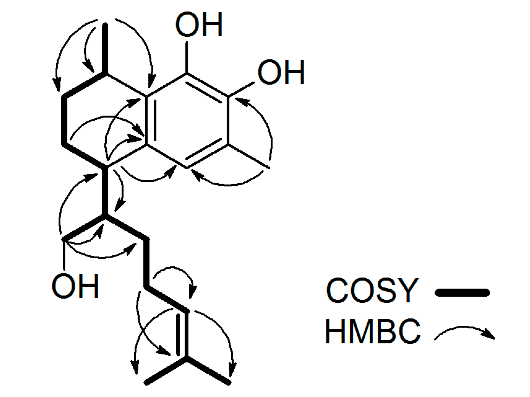
Figure 3.
(A) Propolis collection by bees from resinous leaves of Myoporum insulare; (B) Laboratory analysis of the bee legs with propolis collected from Myoporum insulare leaves surface.
Figure 3.
(A) Propolis collection by bees from resinous leaves of Myoporum insulare; (B) Laboratory analysis of the bee legs with propolis collected from Myoporum insulare leaves surface.

Figure 4.
1H-NMR spectra for bee leg propolis, plant resin and myoporum propolis.

Figure 5.
C-18 Reversed-phase HPLC chromatograms of (A) Myoporum insulare resinous leaf extract and (B) bee leg propolis extract. Compounds 1–3 are labeled. The gradient HPLC analysis was carried out using Shimadzu UFLC with a Hewlett Packard Column, NUCLEOSIL 100C18, 5 μm, 4 mm × 125 mm, eluted with mobile phase, consisted of methanol (phase A), and methanol: water: acetic acid (40:59.8:0.2) (phase B), detected at 286 nm with a UV-Vis detector Shimadzu SPD-20A.


Table 1.
1H-NMR data for Compounds 1–5 (400 MHz, δ in ppm J values (Hz) in parentheses, measured in CDCl3 relative to tetramethylsilane (TMS)).
Table 1.
1H-NMR data for Compounds 1–5 (400 MHz, δ in ppm J values (Hz) in parentheses, measured in CDCl3 relative to tetramethylsilane (TMS)).
| #H | 1 | 2 | 3 | 4 | 5 |
|---|---|---|---|---|---|
| 1 | 3.07 pd (6.8, 1.6) | 3.11 sextet (7.6) | 2.82 m | 2.92 m | 2.96 sextet (7.4) |
| 2 | A: 1.51 m B: 1.99 m | A: 2.25 m B: 1.42 m | A: 2.15 m B: 1.23 m | A: 1.87 m B: 1.50 m | A: 2.19 m B: 1.42 m |
| 3 | A: 1.66 m B: 1.86 m | A: 2.08 m B: 1.05 m | A: 2.13 m B: 1.11 m | A: 1.87 m B:1.60 m | A: 2.12 m B: 1.10 m |
| 4 | 2.76 td (5.6, 2.6) | 2.54 td (11.4, 3.6) | 2.14 m | 2.88 m | 2.54 td (11.6, 3.8) |
| 5 | 6.56 s | - | - | 6.92 s | - |
| 6 | - | - | - | - | - |
| 7 | - | 6.48 s | - | - | 6.70 s |
| 8 | - | - | - | - | - |
| 9 | - | - | - | - | - |
| 10 | - | - | - | - | - |
| 11 | 1.83 m | 1.52 m | 1.74 m | 2.12 m | 1.65 m |
| 12 | A: 1.36 m B: 1.25 m | A: 1.69 m B: 1.27 m | A: 1.19 m B: 1.76 m | A: 1.33 m B: 1.23 m | A: 1.73 m B: 1.30 m |
| 13 | A: 1.86 m B: 1.90 m | A: 2.20 m B: 2.09 m | A: 2.09 m B: 1.97 m | A: 1.99 m B: 1.87 m | A: 2.19 m B: 2.02 m |
| 14 | 5.04 bt (7.0) | 5.14 bt (7.0) | 5.07 bt (6.8) | 4.97 bt (7.1) | 5.08 bt (6.5) |
| 15 | - | - | - | - | - |
| 16 | 1.68 s | 1.70 s | 1.71 s | 1.66 s | 1.69 s |
| 17 | 1. 58 s | 1. 63 s | 1.63 s | 1. 54 s | 1.62 s |
| 18 | 3.63 d (6.2) | 5.59 bs | A: 3.86 t (10.8) B: 4.49 dd (10.8, 3.6) | 4.11 d (6.3) | 6.53 d (1.7) |
| 19 | 2.2 s | 2.13 s | 1.81 s | 2.14 s | 2.14 s |
| 20 | 1.21 d (7.0) | 1.28 d (6.9) | 1.13 d (6.9) | 1.15 d (7.0) | 1.23 d (6.8) |
| 21 | - | - | - | - | - |
| 22 | - | - | - | 2.06 s | 2.09 s |
| 23 | - | - | - | - | - |
| 24 | - | - | - | 2.29 s | 2.30 s |
| 25 | - | - | - | - | - |
| 26 | - | - | - | 2.31 s | - |
Table 2.
13C-NMR data for Compounds 1–5 (100 MHz, δ in ppm, measured in CDCl3 relative to TMS).
| #C | 1 | 2 | 3 | 4 | 5 |
|---|---|---|---|---|---|
| 1 | 26.9 | 27.8 | 28.7 | 27.6 | 28.3 |
| 2 | 26.5 | 31.7 | 30.8 | 26.9 | 31.4 |
| 3 | 20.5 | 26.6 | 25.3 | 19.2 | 26.2 |
| 4 | 37.9 | 31.9 | 40.1 | 36.9 | 32.3 |
| 5 | 122.0 | 141.7 | 162.8 | 128.1 | 145.6 |
| 6 | 121.3 | 122.9 | 114.4 | 128.3 | 123.9 |
| 7 | 139.7 | 115.9 | 179.4 | 139.3 | 122.5 |
| 8 | 141.6 | 146.8 | 181.9 | 140.6 | 142.4 |
| 9 | 127.2 | 125.2 | 139.4 | 134.1 | 130.9 |
| 10 | 130.7 | 124.3 | 149.7 | 137.1 | 123.8 |
| 11 | 45.4 | 40.5 | 37.1 | 41.9 | 38.9 |
| 12 | 29.8 | 28.9 | 29.1 | 28.3 | 28.3 |
| 13 | 26.2 | 25.4 | 24.8 | 26.1 | 25.1 |
| 14 | 124.4 | 124.1 | 123.1 | 123.9 | 123.5 |
| 15 | 131.7 | 131.9 | 132.9 | 132.0 | 132.6 |
| 16 | 25.7 | 25.7 | 25.7 | 25.7 | 25.7 |
| 17 | 17.7 | 17.7 | 17.8 | 17.6 | 17.7 |
| 18 | 64.4 | 91.9 | 71.8 | 65.4 | 90.2 |
| 19 | 15.5 | 15.9 | 7.6 | 16.1 | 15.9 |
| 20 | 21.1 | 22.9 | 21.7 | 21.4 | 22.8 |
| 21 | - | - | - | 171.2 | 170.2 |
| 22 | - | - | - | 20.9 | 21.1 |
| 23 | - | - | - | 168.1 | 170.0 |
| 24 | - | - | - | 20.4 | 21.2 |
| 25 | - | - | - | 168.3 | - |
| 26 | - | - | - | 20.5 | - |
© 2017 by the authors. Licensee MDPI, Basel, Switzerland. This article is an open access article distributed under the terms and conditions of the Creative Commons Attribution (CC BY) license (http://creativecommons.org/licenses/by/4.0/).
Share and Cite
MDPI and ACS Style
Aminimoghadamfarouj, N.; Nematollahi, A. Structure Elucidation and Botanical Characterization of Diterpenes from a Specific Type of Bee Glue. Molecules 2017, 22, 1185. https://doi.org/10.3390/molecules22071185
AMA Style
Aminimoghadamfarouj N, Nematollahi A. Structure Elucidation and Botanical Characterization of Diterpenes from a Specific Type of Bee Glue. Molecules. 2017; 22(7):1185. https://doi.org/10.3390/molecules22071185
Chicago/Turabian StyleAminimoghadamfarouj, Noushin, and Alireza Nematollahi. 2017. "Structure Elucidation and Botanical Characterization of Diterpenes from a Specific Type of Bee Glue" Molecules 22, no. 7: 1185. https://doi.org/10.3390/molecules22071185