Article

Investigation of the Anti-Leishmania (Leishmania) infantum Activity of Some Natural Sesquiterpene Lactones

by
Imke F. Wulsten
1,
Thais A. Costa-Silva
2,
Juliana T. Mesquita
2,
Marta L. Lima
2,3,
Mariana K. Galuppo
2,
Noemi N. Taniwaki
2,
Samanta E. T. Borborema
2,
Fernando B. Da Costa
4,
Thomas J. Schmidt
5,* and
Andre G. Tempone
2,*
1
Department of Biology, Chemistry, Pharmacy, Freie Universität Berlin, 14195 Berlin, Germany
2
Centre for Parasitology and Mycology, Instituto Adolfo Lutz (IAL), Av. Dr. Arnaldo, 351, CEP 01246-900 São Paulo, SP, Brazil
3
Instituto de Medicina Tropical, Universidade de São Paulo, Av. Dr. Enéas de Carvalho Aguiar, 470, CEP 05403-000 São Paulo, SP, Brazil
4
AsterBioChem Research Team, Laboratory of Pharmacognosy, School of Pharmaceutical Sciences of Ribeirão Preto, USP, Av. do Café s/n, 14040-903 Ribeirão Preto, SP, Brazil
5
Institute of Pharmaceutical Biology and Phytochemistry (IPBP), University of Münster, PharmaCampus, Corrensstraße 48, 48149 Münster, Germany
*
Authors to whom correspondence should be addressed.
Molecules 2017, 22(5), 685; https://doi.org/10.3390/molecules22050685
Submission received: 28 March 2017 / Revised: 19 April 2017 / Accepted: 19 April 2017 / Published: 25 April 2017

Abstract

:
Leishmaniases are neglected infectious diseases caused by parasites of the ‘protozoan’ genus Leishmania. Depending on the parasite species, different clinical forms are known as cutaneous, muco-cutaneous, and the visceral leishmaniasis (VL). VL is particularly fatal and the therapy presents limitations. In the search for new anti-leishmanial hit compounds, seven natural sesquiterpene lactones were evaluated against promastigotes and intracellular amastigotes of Leishmania (Leishmania) infantum, a pathogen causing VL. The pseudoguaianolides mexicanin I and helenalin acetate demonstrated the highest selectivity and potency against intracellular amastigotes. In addition, promastigotes treated with helenalin acetate were subject to an ultrastructural and biochemical investigation. The lethal action of the compound was investigated by fluorescence-activated cell sorting and related techniques to detect alterations in reactive oxygen species (ROS) content, plasma membrane permeability, and mitochondrial membrane potential. Helenalin acetate significantly reduced the mitochondrial membrane potential and the mitochondrial structural damage was also confirmed by transmission electron microscopy, displaying an intense organelle swelling. No alteration of plasma membrane permeability or ROS content could be detected. Additionally, helenalin acetate significantly increased the production of nitric oxide in peritoneal macrophages, probably potentiating the activity against the intracellular amastigotes. Helenalin acetate could hence be a useful anti-leishmanial scaffold for further optimization studies.

Graphical Abstract

1. Introduction

Leishmaniases are listed as neglected tropical diseases by the World Health Organization [1]. Nowadays, these diseases affect 98 countries. In Brazil, an increased number of cases has been observed in recent years, accompanied by a geographical spread [1]. Worldwide, an estimated 1.3 million new cases and 20,000–30,000 deaths are reported annually; around 310 million people are at risk of an infection [1].
Depending on the parasite species and determinants of susceptibility of the vertebrate hosts, Leishmania infections present different clinical forms: cutaneous, mucocutaneous, and visceral leishmaniasis (VL) [2,3]. Untreated VL accounts for the majority of deaths reported for the leishmaniases [1]. In VL, the parasites affect mainly the spleen, liver, and bone marrow, where they multiply inside macrophages, causing organ hypertrophy (hepatosplenomegaly) and dysfunction [4]. Besides the high prevalence, impact, and risk among the world population, existing treatments for VL exhibit strong limitations, including high toxicity leading to severe adverse effects, high costs, parasite resistance, and hospitalization due to parenteral administration [5,6]. Therefore, new therapies for this disease are desirable.
Natural products traditionally play an important role in search for new therapeutics. Their large structural diversity of secondary metabolites is a source of novel chemical structures as starting points for drug development [7,8,9,10,11]. Natural sesquiterpene lactones (STLs) are widespread secondary metabolites in plants of the family Asteraceae [12]. This class of compounds are known for a wide range of biological activities such as anticancer [13,14], gastric cytoprotective [15] but also cytotoxic [16] effects, and in vitro anti-trypanosomatid activities [17,18,19,20,21,22,23].
In this work, we evaluated the in vitro activity of the pseudoguaianolides mexicanin I and helenalin acetate; the guaianolides arglabin and cynaropicrin; eudesmanolide alantolactone; germacranolide parthenolide; and furanoheliangolide budlein A (Figure 1) against promastigotes and intracellular amastigotes of Leishmania (Leishmania) infantum, the etiologic agent of VL in wide parts of South America and the Mediterranean region. Using different techniques, we studied the mechanism of action of the most active and selective STL in L. (L.) infantum, evaluating different parameters of the parasite such as plasma membrane permeability, reactive oxygen species (ROS) content, and mitochondrial membrane potential (Δψm) as well as the production of nitric oxide (NO) by host cells.

2. Results and Discussion

Anti-L. (L.) infantum Activity of STL

Seven representative STLs (Figure 1) were chosen for this study since they had demonstrated different levels of in vitro anti-trypanosomatid activity against Trypanosoma brucei rhodesiense and Leishmania donovani (axenic amastigotes) in previous studies [22,23]. Their activity against L. (L.) infantum as well as their cytotoxicity and selectivity is demonstrated in Table 1. All compounds displayed in vitro activity against L. (L.) infantum promastigotes, with IC50 values in a concentration range between 3 and 59 μM. The pseudoguaianolides helenalin acetate (1) and mexicanin I (2) showed the most potent activity against the intracellular amastigotes and highest selectivity indices (S.I. = IC50(cytotox.)/IC50(amastigote)), with IC50 values of 1.15 μM (S.I. 7) and 1.73 μM (S.I. 5.3), respectively. Argablin (3) and cynaropicrin (4) also demonstrated selectivity against the intracellular amastigotes, but were about 6-fold less effective than the compounds 1 and 2, demonstrating IC50 values of 7.33 (S.I. 5.3) and 6.88 μM (S.I. 4.8), respectively. Compounds 1 and 2 were about 15-fold more effective than miltefosine, the standard drug used to treat VL. Effects of 1 on intracellular amastigotes as observed by light microscopy are shown in Figure S1, Supplementary Material.
The unesterified parent compound of 1, helenalin, and 2 were previously shown to have activity against L. (L.) mexicana at low concentrations, one of the species responsible for cutaneous leishmaniasis [20]. Considering the selectivity index and the high potency against promastigotes and intracellular amastigotes, compound 1 was selected for further studies aiming at biochemical and ultrastructural investigations in L. (L.) infantum parasites after treatment.
The parasite’s plasma membrane regulates the transport of nutrients, pH homeostasis, and homeostasis of other ions [24]. Thus, we investigated the effect of 1 on the parasite plasma membrane using the probe SYTOX Green. This probe shows fluorescence when it gets in contact with nucleic acids, reaching the intracellular milieu via a severely damaged plasma membrane [25]. No significant increase of SYTOX Green fluorescence could be detected after treatment with compound 1 (3 and 6 μM) during 60 min (Figure 2), in comparison with untreated parasites. Thus, it can be concluded that 1 has no influence on the plasma membrane permeability of the parasite.
It is known that vital processes of Leishmania promastigotes are irreversibly affected by helenalin at short periods of exposure (3 h) [20]. Having ruled out the plasma membrane as target, the next step was the investigation of a possible influence of helenalin acetate (1) on the mitochondria of L. (L.) infantum promastigotes, a recognized target for a number of antileishmanial compounds [26]. Leishmania is known to have a single mitochondrion as major supplier of cellular energy—i.e., ATP—through the metabolism of the respiratory chain [27]. The mitochondrial membrane potential (Δψm) is a crucial component in ATP production and its maintenance is essential for the parasite survival. Therefore, disturbances in the Δψm of L. (L.) infantum promastigotes treated with 1 were investigated using the fluorescent probe rhodamine 123. Rhodamine 123 (Rd123) is a fluorescent cationic dye that accumulates into polarized mitochondria. Depolarization, i.e., a decrease in Δψm, results in a lower accumulation of rhodamine and, consequently, in a reduced fluorescence [28]. Parasites treated for 1 h with compound 1 at a concentration representing the previously determined IC99 value (6 μM), decreased the fluorescence of Rd123 by 60% in comparison to untreated parasites (Figure 3a). This effect on the Δψm was similar to that obtained with 10 μM carbonyl cyanide 4-(trifluoromethoxy) phenylhydrazone (FCCP), a known mitochondrial uncoupler. A previous study with parthenolide (6) against axenic amastigotes (extracellular) of L. (L.) amazonensis, also demonstrated a mitochondrial depolarization with this sesquiterpene lactone [21]. The exact mechanism by which 1 or 6 affect the Δψm of Leishmania remains to be elucidated.
Derangements in Δψm are usually linked to overproduction of ROS, resulting in mitochondrial dysfunction and ultimately cell death [29,30]. Accordingly, the production of ROS in promastigotes treated with compound 1 was determined using the dye 2′,7′-dichlorodihydrofluorescein diacetate (H2DCFDA) and flow cytometry. The fluorescence intensity of this dye has been assumed to be proportional to the redox state of the parasite. In contrast to oligomycin (20 μM), a mitochondrial respiratory chain inhibitor of the ATP synthase (complex V), which increased the ROS production over time, promastigotes treated with 1 showed no alteration of ROS after 1 h of treatment (Figure 3b).
The impairment in Δψm accompanied by ROS generation is commonly described for STLs in cancer cell lines [31,32]. It was suggested that, in these cells, STLs cause a knockdown of crucial enzymes that participate in the redox defense metabolism, resulting in ROS accumulation [33]. Similarly, some STLs have been described to induce ROS generation concomitant with reduction of glutathione (GSH) content in Leishmania promastigotes. GSH is an important metabolite of the redox defense metabolism of the parasite as a precursor for the synthesis of trypanothione. It was proposed that STLs react with sulfhydryl groups by the Michael-type addition; thus, impairment of redox defense metabolism of parasites should not be ruled out in Leishmania [34]. However, the absence of an increase in ROS generation in promastigotes treated with 1, even in disturbed mitochondria as indicated by the affected Δψm, suggests that the redox defense metabolism was probably not affected by this STL throughout the incubation period. Likewise, psilostachyin C—another STL—also demonstrated a leishmanicidal effect through a lethal mechanism other than oxidative stress or reduction of GSH content in the parasite [34]. In summary, the mitochondrial dysfunction provoked by STLs in Leishmania may have distinct mechanisms.
In addition to these biochemical investigations, an ultrastructural study using Transmission Electron Microscopy (TEM) was used to investigate the alterations in L. (L.) infantum promastigotes treated with compound 1 at 1 μM over different incubation times. 1 induced early morphological and ultrastructural alterations in promastigotes after 30 min of incubation, with increasing damage throughout the time interval studied, 4 h (Figure 4). The integrity of plasma membrane and pericellular microtubules was preserved over 4 h, corroborating the aforementioned SYTOX Green assay (Figure 4). Similarly, 1 induced no alteration of the plasma membrane of L. (L.) mexicana promastigotes after a long-term treatment [20]. The previously observed mitochondrial dysfunction could now be confirmed by a visible swelling of the organelle (Figure 4b–f, red arrows) during the treatment with 1. The strong vacuolization of the cytoplasm was associated with autophagic processes as judged by presence of multivesicular structures (Figure 4f). Autophagy can be triggered by the cell for energy production, cell recycling, or death signalling [35]. Detachment of the nuclear membrane (Figure 4e, blue arrow) was initially observed after 3 h followed by chromatin decondensation after 4 h of incubation (Figure 4g, red arrow). Usually, this event precedes the DNA fragmentation which has also been reported to occur in L. (L.) mexicana promastigotes after long treatment with unesterified helenalin [20]. In summary, the ultrastructural analysis showed that compound 1 induced a rounded amastigote-like morphology (compare Figure 4a with Figure 4g) with intense cytoplasmatic vacuolization, formation of autophagosomes, swelling of the mitochondrion, and chromatin decondensation (Figure 4g).
Besides direct effects on the parasite, antileishmanial compounds can also trigger a lethal effect via macrophage activation mechanisms. Macrophages play an important role in the vertebrate immune system and represent the primary cells infected by Leishmania. Their activation profile is decisive for the resolution of the infection [36,37]. It has been demonstrated that macrophage-mediated killing of Leishmania could be an event associated with the up-regulation of NO. After stimulation with lipopolysaccharide (LPS), macrophages are activated to produce NO from arginine, controlling the proliferation of intracellular parasites such as L. (L.) major [38]. Considering the higher activity of 1 against the intracellular amastigotes of L. (L.) infantum as compared to extracellular promastigotes (Table 1), we investigated the possibility of the host cell participating in the elimination of the parasites. The NO levels of peritoneal macrophages treated with 1 at 3 μM were determined after 24 h incubation using the colorimetric Griess assay [39]. Our data demonstrated that 1 significantly increased the production of NO at similar levels as cells treated with LPS (positive control) (Figure 5). This result suggests that 1 elicited an immunomodulatory effect that may have contributed to reduce the infection by L. (L.) infantum via an NO-mediated mechanism.

3. Materials and Methods
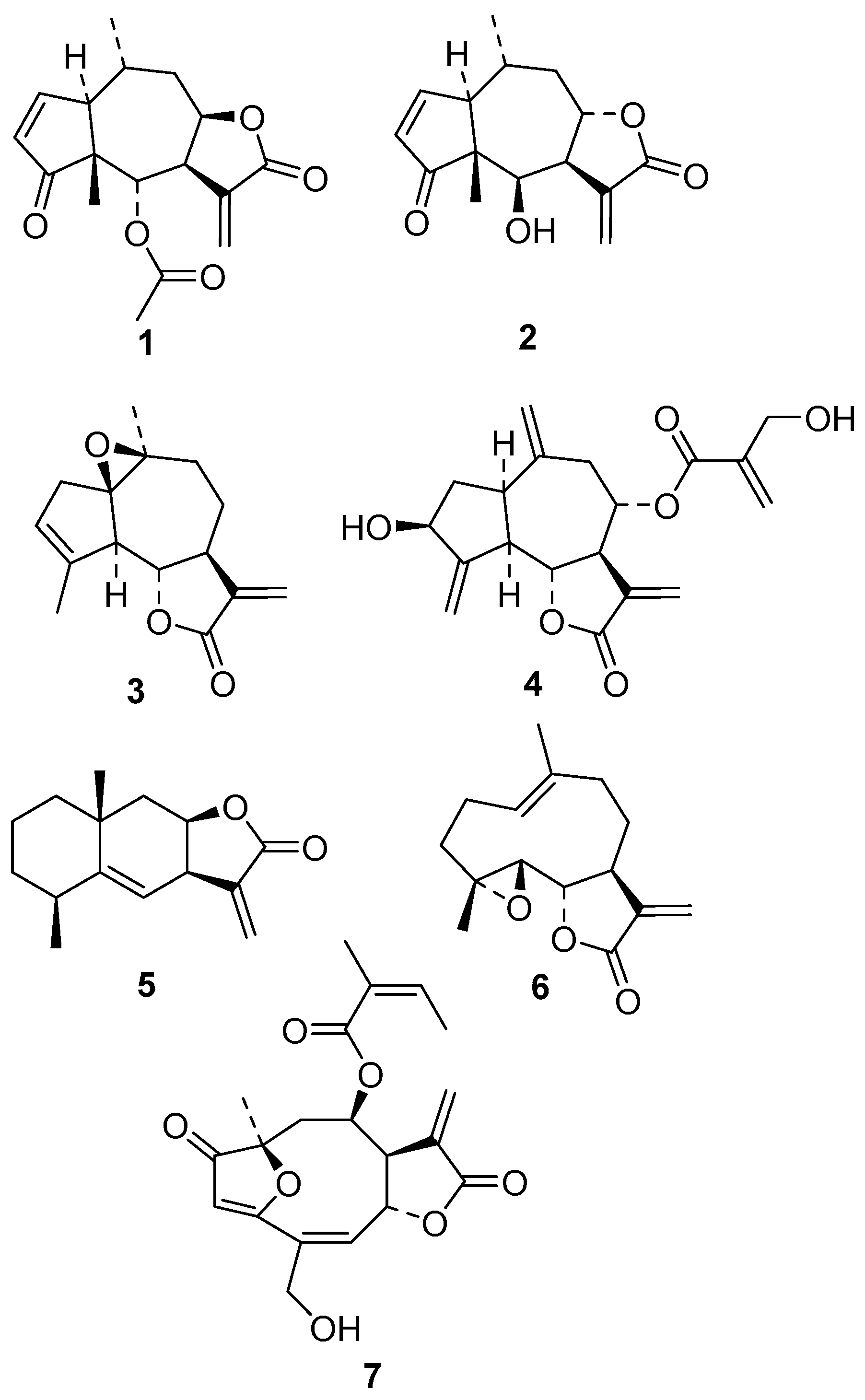
3.1. Compounds

The STLs investigated in this study were previously isolated in our laboratories (T.J.S., F.B.C.) from plants of the family Asteraceae. Helenalin acetate (1) and mexicanin I (2) were isolated from A. montana L. and Arnica acaulis (Walter) Britton, Sterns and Poggenb., respectively. The guaianolides arglabin (3) and cynaropicrin (4) originate from Artemisia glabella Kar. Et Kir. Fl. Alt. and Cynara cardunculus L., respectively. Alantolactone (5) is an eudesmanolide isolated from Inula helenium L. The germacranolide parthenolide (6) and the furanoheliangolide budlein A (7) were isolated from Tanacetum parthenium (L.) Sch. Bip. and Aldama robusta (Gardner) E.E.Schill and Panero (formerly: Viguiera robusta). Further details on the origin of these compounds and their purity were published previously [18,22,23]. In all cases, stock solutions were prepared with dimethyl sulfoxide (DMSO) which were subsequently diluted with the respective medium to the specific concentration used in the various bioassays.

3.2. Animals

Golden hamsters (Mesocricetus auratus Waterhouse) were used for the maintenance of the L. (L.) infantum (MHOM/BR/1972/LD) culture. Young female BALB/c mice (Mus musculus L.) with a weight of 18–24 g were used as a source of peritoneal macrophages.
Animals were provided by the animal facility of the Adolfo Lutz Institute, São Paulo, Brazil. The animals received water and food ad libitum and were kept in sterilized cages. The procedures involving animals were realized in agreement with the Guidelines for the Care and Use of Laboratory Animals from the National Academy of Sciences, USA. The project received ethical approval by the Research Ethics Commission of the Adolfo Lutz Institute/ Pasteur Institute (CEUA-IAL/Pasteur 02/2011), São Paulo, Brazil.

3.3. Leishmania (Leishmania) infantum Promastigote, Peritoneal Macrophages, and NCTC Cells Culture

L. (L.) infantum (MHOM/BR/1972/LD) promastigotes were maintained in M-199 medium supplemented with 10% heat-inactivated fetal bovine serum (FBS) (Gibco, Thermo Fisher Scientific, São Paulo, Brazil), 0.25% hemin (Sigma-Aldrich), and 5% human urine at 24 °C. Amastigotes were obtained from the spleen of previously infected hamsters by differential centrifugation. Macrophages were collected from the peritoneal cavity of BALB/c mice by washing with RPMI-1640 medium supplemented with 10% FBS, and were maintained at 37 °C in a 5% CO2-humidified incubator. Murine fibroblasts NCTC (clone L929 ATCC) were used to evaluate the cytotoxicity of the tested compounds. NCTC cells were maintained in M-199 medium supplemented with 10% FBS and 20 μg/mL gentamicin at 37 °C in a 5% CO2-humidified incubator.

3.4. Determination of 50% Inhibitory Concentration (IC50) against Leishmania and 50% Cytotoxicity Concentration (CC50) against NCTC

3.4.1. Promastigotes

Compounds were dissolved in DMSO and diluted in M-199 medium in 96-well microplates, with the highest concentration of 150 μM. Promastigotes of L. (L.) infantum in the late growth-phase were seeded at 1 × 10⁶/well and incubated with the compounds for 48 h. The viability of cells was determined using the MTT assay [40]. An internal control group was used with 0.5% DMSO (maximal concentration). Miltefosine was used as standard drug.

3.4.2. Intracellular Amastigotes of L. (L.) infantum

Murine peritoneal macrophages were collected from the peritoneal cavity of BALB/c mice, and the macrophages were seeded at 1 × 10⁵/well for 24 h in a 16-well slide. Amastigotes were prepared (as described previously), seeded at a ratio 1:10 (macrophages:amastigotes) and kept at 37 °C in a 5% CO2-humidified incubator for 24 h. Test compounds were incubated to the highest concentration of 100 μM with infected macrophages for 120 h. Miltefosine was used as a standard drug. Subsequently, the cells were fixed with methanol, stained with Giemsa (Merck KGaA, Darmstadt, Germany), and observed using a light microscope. The parasite burden was determined by the number of infected macrophages out of 400 cells.

3.4.3. Cytotoxicity

NCTC cells were seeded at 6 × 10⁴ in 96-well microplates and incubated with the compounds for 48 h. An internal control group was used with 0.5% DMSO (maximal concentration). The quantification of viable cells was assessed by the MTT assay [40]. The selectivity index (SI) was determined using the following equation: SI = CC50 in NCTC cells / IC50 against amastigotes.

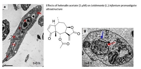
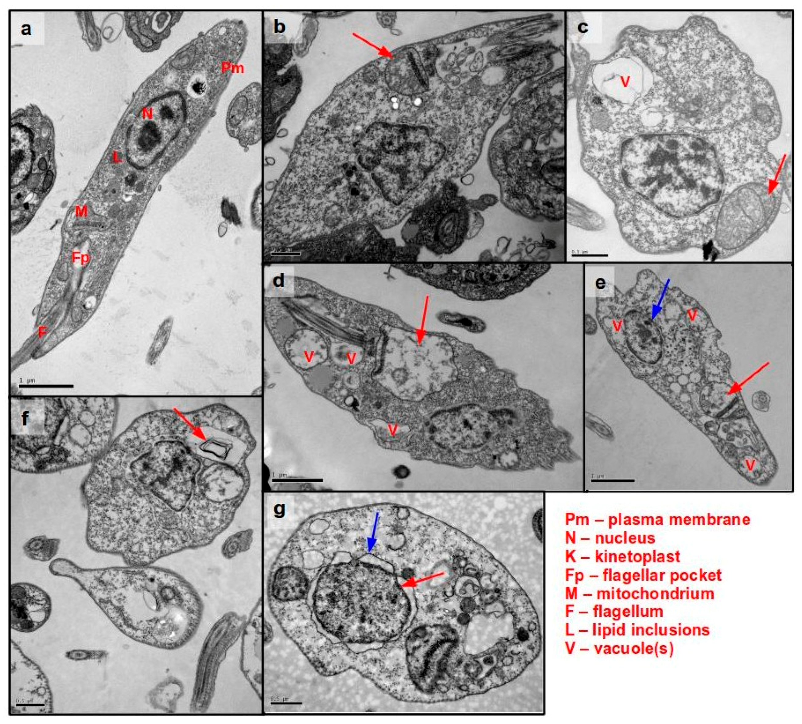
3.5. Ultrastructural Analysis of Cellular Damage in Promastigotes Treated with Helenalin Acetate, Using TEM

Ultrastructural studies were performed with promastigotes treated with compound 1 by TEM. L. (L.) infantum promastigotes in the late growth phase were washed and incubated with 120 μM of 1 (corresponding to the IC99 value obtained with 1 × 10⁶ parasites/well) at 2 × 10⁷/well in 200 μL M-199 medium. Compound incubation was performed for 0.5, 1, 2, 3, and 4 h under the previously described growth conditions. After centrifugation at 2800 rpm for 10 min, the parasites were fixed with 1 mL glutaraldehyde and the sample processing was performed according to Duarte et al. [41]. The TEM imaging was performed on a JEOL 1011 transmission electron microscope (Peabody, MA, USA).

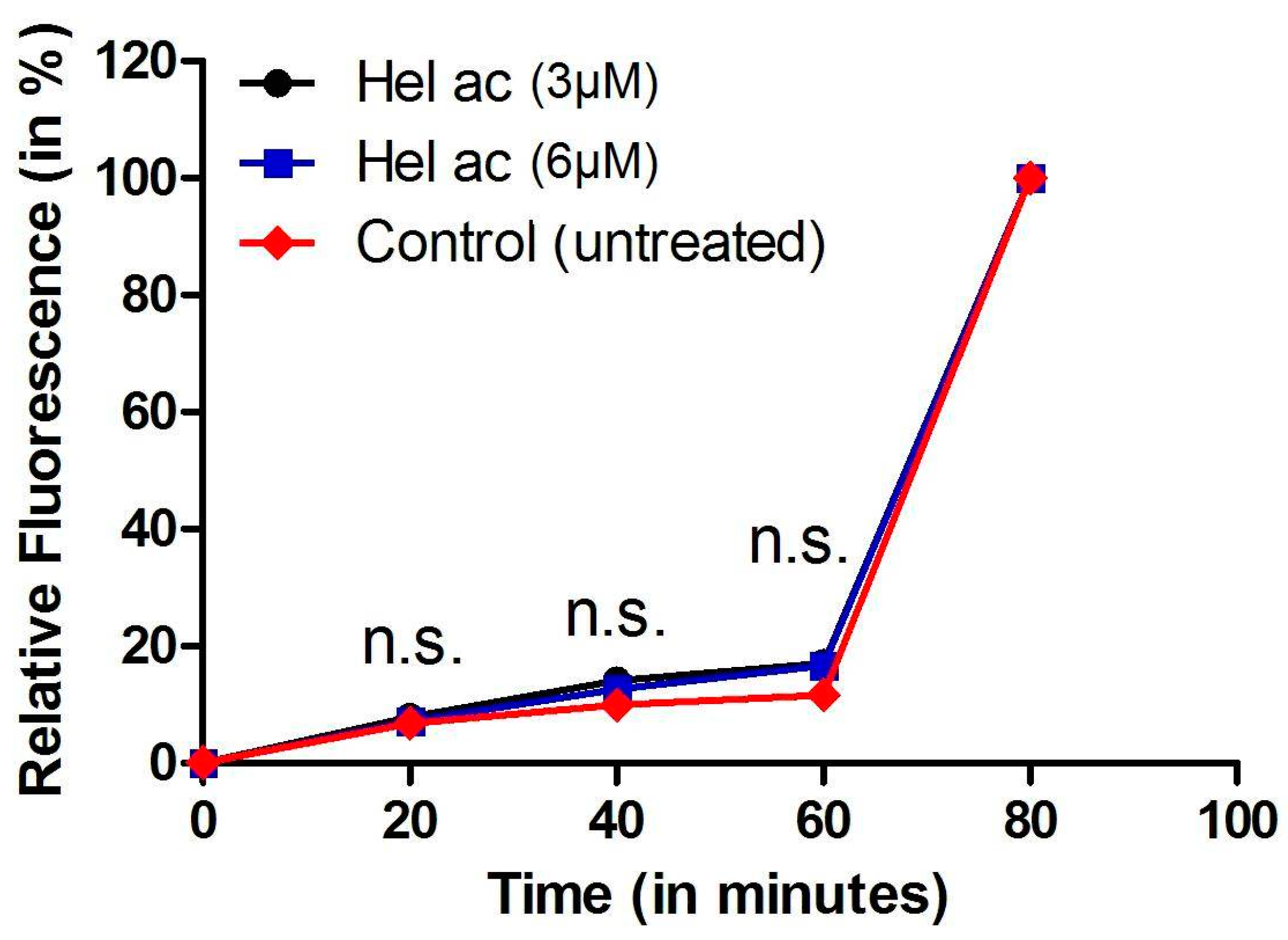
3.6. Determination of Plasma Membrane Permeability in Helenalin Acetate-Treated Promastigotes

Late-growth-phase (non-stationary) promastigotes were incubated with the probe SYTOX Green (Molecular Probes Inc., Eugene, OR, USA), as previously described [42]. Concentrations of 1 were chosen in accordance with the respective IC50 and IC99 values. 1 was added at 3 and 6 μM to 1 × 10⁶ cells/well, at 24 °C. The fluorescence was measured every 20 min (0–80 min). At the end of the assay (80 min), 0.1% Triton X-100 was added to all wells to obtain the maximum permeabilization of parasites. Fluorescence intensity was determined using a fluorimetric microplate reader (FilterMax F5 Multi-Mode Microplate Reader-Molecular Devices, Sunnyvale, CA, USA) with excitation and emission wavelengths of 485 and 520 nm, respectively. The following internal controls were used during the evaluation: (i) the background fluorescence of the compound at the respective wavelengths; (ii) the possible interference of DMSO; (iii) untreated promastigotes; and (iv) medium without any cells. Samples were tested in duplicate.

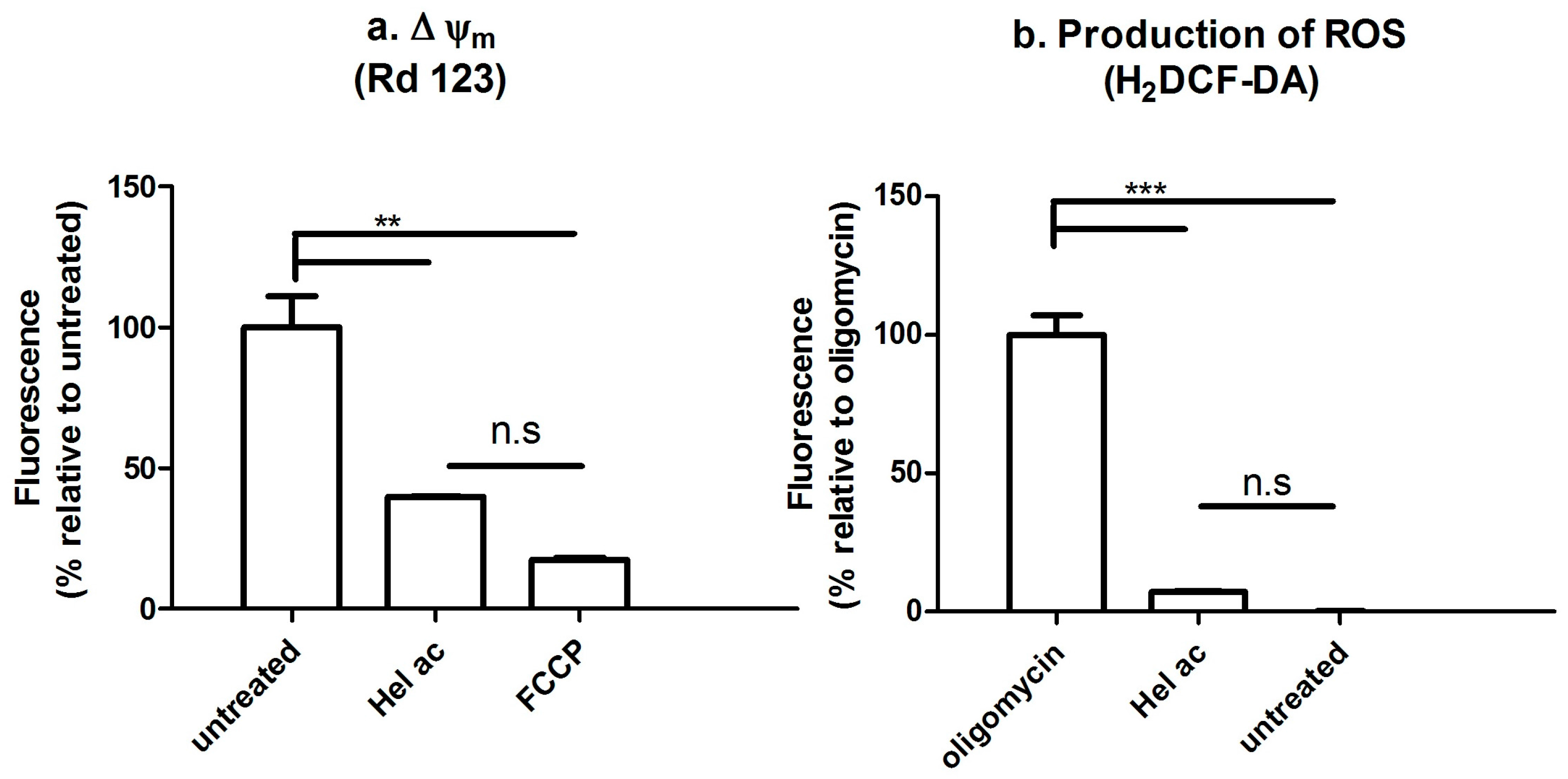
3.7. Determination of the Mitochondrial Membrane Potential (Δψm) and ROS by Fluorescence-Activated Cell Sorting

The Δψm and ROS content of promastigotes treated with compound 1 were determined using the fluorescence probes rhodamine 123 and H2DCFDA (Molecular Probes Inc., Eugene, OR, USA), respectively, and further evaluated by flow cytometry [25,43]. Briefly, 2 × 10⁶ promastigotes/well cultured until the late growth-phase were treated with 12 μM of 1 (corresponding to the IC99 value obtained with 1 × 10⁶ parasites/well). The parasites were incubated in HANKS’ balanced salts solution (HBSS; Sigma-Aldrich, Carlsbad, CA, USA), supplemented with 10 mM d-Glucose, at 26 °C for 1 h. Afterwards the parasites were washed and stained with rhodamine 123 (0.3 μg/mL) or H2DCFDA (5 μM) for 15 min under absence of light. The washed parasites were resuspended in PBS and the fluorescence was measured in a Flow Cytometer Attune Nxt (ThermoFisher Scientific, Waltham, MA, USA) using the forward scatter (FSC, relative cell size) and the side scatter detectors (SSC, cell granulometry or internal complexity), FL1-A (detecting fluorescence emission 530 ± 15 nm), and Attune Nxt software. As control, untreated promastigotes (not stained with Rd123 or H2DCFDA) served to monitor the basal fluorescence. FCCP (10 μM) and oligomycin (20 μM) provided information on the maximal mitochondrial depolarization and maximal ROS production, respectively. Samples were tested in duplicate. Two independent assays were performed.

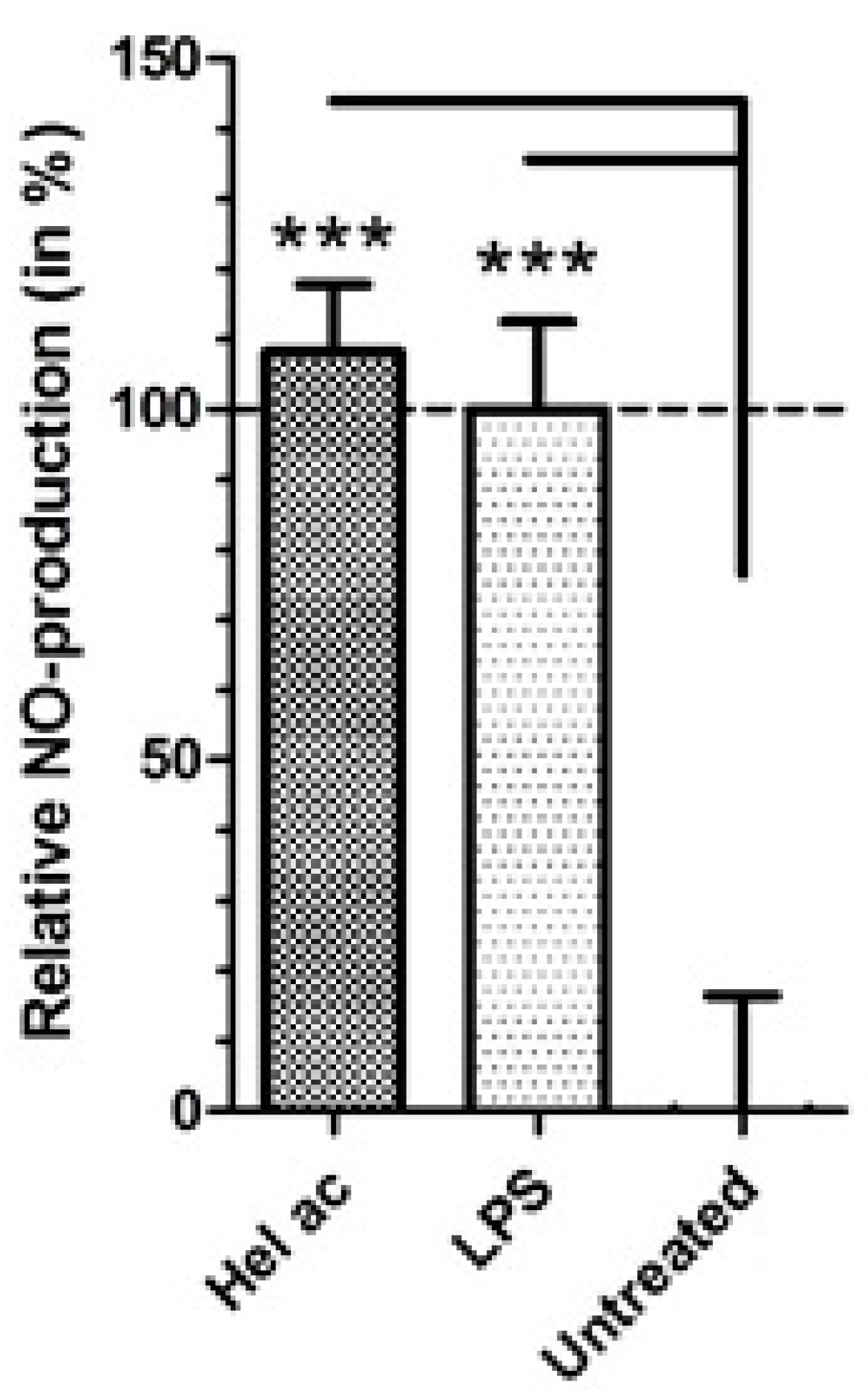
3.8. Production of Nitric Oxide (NO) in Macrophages Treated with Helenalin Acetate

The NO production of macrophages treated with 1 was performed using the Griess assay [39]. Briefly, 1 × 10⁵/well peritoneal macrophages of BALB/c mice were seeded overnight in a 96-well plate. Fresh medium containing compound 1 at 3 µM (corresponding to the CC25 value obtained with NCTC cells) or 5 µg/mL LPS (positive control) were added and incubated for 24 h at 37 °C. Afterwards the NO content of each of the cultures’ supernatant was analysed using the Griess assay. The samples’ NO content was determined by means of a standard curve prepared with NaNO2 at concentrations from 0 to 400 μM.

3.9. Statistical Analysis

Data processing and statistical analyses was done with GraphPad Prism® 5.0 (GraphPad Software, Inc. La Jolla, CA, USA). For determination of antileishmanial activity and NCTC cytotoxicity, the IC50 and CC50 values were calculated after normalization using sigmoidal dose-response curves. The evaluation of statistical significance of multiple pairs was done with a Tukey's range test, which corrects for the increased probability of a type I error during multiple comparison.

4. Conclusions

The present study revealed the in vitro anti-Leishmania (L.) infantum efficacy of seven STLs isolated from plants of the Asteraceae family. Helenalin acetate (1) and mexicanin I (2) showed the highest potency of all tested compounds and a certain selectivity against L. (L.) infantum intracellular amastigotes. Helenalin acetate (1) affected mitochondria at an early phase during incubation, causing depolarization of the mitochondrial membrane potential. These early events could be related to altered ATP production, since the mitochondrion is a pivotal organelle for bioenergetic homeostasis. Additionally, it was demonstrated that the activity of 1 against intracellular amastigotes could be, at least in part, related to an increased NO-production of the host cells since an elevated release of NO was induced by the compound in peritoneal macrophages. Helenalin acetate and, possibly, other STLs such as mexicanin I, may thus represent interesting scaffolds for the synthesis of derivatives with potent and selective activity against L. (L.) infantum.

Supplementary Materials

Supplementary materials are available online. Figure S1: Helenalin acetate activity against intracellular L. (L.) infantum amastigotes (GIEMSA-stained light microscopy images).

Acknowledgments

This work has received financial support from the São Paulo Research Foundation (FAPESP 2012/18756-1 and 2013/50228-8) and is part of the activities of the Research Network Natural Products against Neglected Diseases (ResNet NPND; http://www.ResNetNPND.org).

Author Contributions

T.J.S. and A.G.T. devised the study. T.J.S. provided the studied compounds. A.G.T. provided the laboratory facilities for all biological tests and supervised the study. TEM analysis was realized by N.N.T and I.F.W. I.F.W., S.E.T.B., T.A.C.-S. and J.T.M. performed the study and I.F.W. prepared a draft manuscript. M.K.G. and M.L.L. performed flow cytometry analysis. A.G.T. and T.J.S. edited the manuscript and prepared, together with I.F.W., the final submitted version.

Conflicts of Interest

The authors declare no conflict of interest. The founding sponsors had no role in the design of the study; in the collection, analyses, or interpretation of data; in the writing of the manuscript, or in the decision to publish the results.

References

  1. World Health Organization (WHO). The 17 Neglected Tropical Diseases. World Health Organization. Available online: http://www.who.int/neglected_diseases/diseases/en/ (accessed on 1 July 2016).
  2. Grimaldi, G.; Tesh, R.B. Leishmaniasis of the New World: Current concepts and implications for future research. Clin. Microbiol. Rev. 1993, 6, 230–250. [Google Scholar] [CrossRef] [PubMed]
  3. Lainson, R.; Shaw, J.J. Epidemiology and ecology of leishmaniasis in Latin-America. Nature 1978, 5664, 595–600. [Google Scholar] [CrossRef]
  4. De Freitas, E.O.; Leoratti, F.M.; Freire-de-Lima, C.G.; Morrot, A.; Feijó, D.F. The Contribution of Immune Evasive Mechanisms to Parasite Persistence in Visceral Leishmaniasis. Front. Immunol. 2016, 7, 153. [Google Scholar] [CrossRef] [PubMed]
  5. Moore, E.M.; Lockwood, D.N. Treatment of visceral leishmaniasis. J. Glob. Infect. Dis. 2010, 2, 151–158. [Google Scholar] [PubMed]
  6. Croft, S.L.; Olliaro, P. Leishmaniasis chemotherapy-challenges and opportunities. Clin. Microbiol. Infect. 2011, 10, 1478–1483. [Google Scholar] [CrossRef] [PubMed]
  7. Tempone, A.G.; de Oliveira, C.M.; Berlinck, R.G.S. Current approaches to discover marine antileishmanial natural products. Planta Med. 2011, 6, 572–585. [Google Scholar] [CrossRef] [PubMed]
  8. Sen, R.; Chatterjee, M. Plant derived therapeutics for the treatment of Leishmaniasis. Phytomedicine 2011, 12, 1056–1069. [Google Scholar] [CrossRef] [PubMed]
  9. Schmidt, T.J.; Khalid, S.A.; Romanha, A.J.; Alves, T.M.A.; Biavatti, M.W.; Brun, R.; Ferreira, V.F.; Lago, J.H.G.; Leon, L.L.; Lopes, N.P.; et al. The Potential of Secondary Metabolites from Plants as Drugs or Leads against Protozoan Neglected Diseases—Part I. Curr. Med. Chem. 2012, 14, 2128–2175. [Google Scholar]
  10. Schmidt, T.J.; Khalid, S.A.; Romanha, A.J.; Alves, T.M.A.; Biavatti, M.W.; Brun, R.; Ferreira, V.F.; Lago, J.H.G.; Leon, L.L.; Lopes, N.P.; et al. The Potential of Secondary Metabolites from Plants as Drugs or Leads against Protozoan Neglected Diseases—Part II. Curr. Med. Chem. 2012, 14, 2176–2228. [Google Scholar] [CrossRef]
  11. Newman, D.J.; Cragg, G.M. Natural products as sources of new drugs over the 30 years from 1981 to 2010. J. Nat. Prod. 2012, 3, 311–335. [Google Scholar] [CrossRef] [PubMed]
  12. Rodriguez, E.; Towers, G.N.H.; Mitchell, J.C. Biological activities of sesquiterpene lactones. Phytochemistry 1976, 15, 1573–1580. [Google Scholar] [CrossRef]
  13. Kupchan, S.M.; Eakin, M.A.; Thomas, A.M. Tumor inhibitors. Structure-cytotoxicity relationships among the sesquiterpene lactones. J. Med. Chem. 1971, 12, 1147–1152. [Google Scholar] [CrossRef]
  14. Robles, M.; Aregullin, M.; West, J.; Rodriguez, E. Recent studies on the zoopharmacognosy, pharmacology and neurotoxicology of sesquiterpene lactones. Planta Med. 1995, 3, 199–203. [Google Scholar] [CrossRef] [PubMed]
  15. Giordano, O.S.; Guerreiro, E.; Pestchanker, M.J.; Guzman, J.; Pastor, D.; Guardia, T. The gastric cytoprotective effect of several sesquiterpene lactones. J. Nat. Prod. 1990, 4, 803–809. [Google Scholar] [CrossRef]
  16. Ordóñez, P.E.; Sharma, K.K.; Bystrom, L.M.; Alas, M.A.; Enriquez, R.G.; Malagón, O.; Jones, D.E.; Guzman, M.L.; Compadre, C.M. Dehydroleucodine, a Sesquiterpene Lactone from Gynoxys verrucosa, Demonstrates Cytotoxic Activity against Human Leukemia Cells. J. Nat. Prod. 2016, 4, 691–696. [Google Scholar] [CrossRef] [PubMed]
  17. Brengio, S.D.; Belmonte, S.A.; Guerreiro, E.; Giordano, O.S.; Pietrobon, E.O.; Sosa, M.A. The sesquiterpene lactone dehydroleucodine (DhL) affects the growth of cultured epimastigotes of Trypanosoma cruzi. J. Parasitol. 2000, 2, 407–412. [Google Scholar] [CrossRef]
  18. Schmidt, T.J.; Brun, R.; Willuhn, G.; Khalid, S.A. Anti-trypanosomal activity of helenalin and some structurally related sesquiterpene lactones. Planta Med. 2002, 8, 750–751. [Google Scholar] [CrossRef] [PubMed]
  19. Jimenez-Ortiz, V.; Brengio, S.D.; Giordano, O.; Tonn, C.; Sánchez, M.; Burgos, M.H.; Sosa, M.A. The trypanocidal effect of sesquiterpene lactones helenalin and mexicanin on cultured epimastigotes. J. Parasitol. 2005, 1, 170–174. [Google Scholar] [CrossRef] [PubMed]
  20. Barrera, P.A.; Jimenez-Ortiz, V.; Tonn, C.; Giordano, O.; Galanti, N.; Sosa, M.A. Natural sesquiterpene lactones are active against Leishmania mexicana. J. Parasitol. 2008, 5, 1143–1149. [Google Scholar] [CrossRef] [PubMed]
  21. Tiuman, T.S.; Ueda-Nakamura, T.; Alonso, A.; Nakamura, C.V. Cell death in amastigote forms of Leishmania amazonensis induced by parthenolide. BMC Microbiol. 2014, 10, 14–152. [Google Scholar] [CrossRef] [PubMed]
  22. Schmidt, T.J.; Nour, A.M.M.; Khalid, S.A.; Kaiser, M.; Brun, R. Quantitative structure--antiprotozoal activity relationships of sesquiterpene lactones. Molecules 2009, 6, 2062–2076. [Google Scholar] [CrossRef] [PubMed]
  23. Schmidt, T.J.; Da Costa, F.B.; Lopes, N.P.; Kaiser, M.; Brun, R. In Silico prediction and experimental evaluation of furanoheliangolide sesquiterpene lactones as potent agents against Trypanosoma brucei rhodesiense. Antimicrob. Agents Chemother. 2014, 1, 325–332. [Google Scholar] [CrossRef] [PubMed]
  24. Zilberstein, D.; Dwyer, D.M. Protonmotive force-driven active transport of d-glucose and l-proline in the protozoan parasite Leishmania donovani. Proc. Natl. Acad. Sci. USA 1985, 6, 1716–1720. [Google Scholar] [CrossRef]
  25. Luque-Ortega, J.R.; Rivas, L. Characterization of the leishmanicidal activity of antimicrobial peptides. In Antimicrobial Peptides; Methods in Molecular Biology; Humana Press: London, UK, 2010; Volume 618, pp. 393–420. [Google Scholar]
  26. Carvalho, L.; Luque-Ortega, J.R.; Manzano, J.I.; Castanys, S.; Rivas, L.; Gamarro, F. Tafenoquine, an antiplasmodial 8-aminoquinoline, targets leishmania respiratory complex III and induces apoptosis. Antimicrob. Agents Chemother. 2010, 12, 5344–5351. [Google Scholar] [CrossRef] [PubMed]
  27. Van Hellemond, J.J.; Van Der Meer, P.; Tielens, A.G.M. Leishmania infantum promastigotes have a poor capacity for anaerobic functioning and depend mainly on respiration for their energy generation. Parasitology 1997, 114, 351–360. [Google Scholar] [CrossRef]
  28. Perry, S.W.; Norman, J.P.; Barbieri, J.; Brown, E.B.; Gelbard, H.A. Mitochondrial membrane potential probes and the proton gradient: A practical usage guide. Biotechniques 2011, 2, 98–115. [Google Scholar] [CrossRef] [PubMed]
  29. Kowaltowski, A.J.; Vercesi, A.E. Mitochondrial damage induced by conditions of oxidative stress. Free Radic. Biol. Med. 1999, 3–4, 463–471. [Google Scholar] [CrossRef]
  30. Mehta, A.; Shaha, C. Apoptotic death in Leishmania donovani promastigotes in response to respiratory chain inhibition: Complex II inhibition results in increased pentamidine cytotoxicity. J. Biol. Chem. 2004, 12, 11798–11813. [Google Scholar] [CrossRef] [PubMed]
  31. Martino, R.; Beer, M.F.; Elso, O.; Donadel, O.; Sülsen, V.; Anesini, C. Sesquiterpene lactones from Ambrosia spp. are active against a murine lymphoma cell line by inducing apoptosis and cell cycle arrest. Toxicol. In Vitro 2015, 7, 1529–1536. [Google Scholar] [CrossRef] [PubMed]
  32. Hamzeloo-Moghadam, M.; Aghaei, M.; Fallahian, F.; Jafari, S.M.; Dolati, M.; Abdolmohammadi, M.H.; Hajiahmadi, S.; Esmaeili, S. Britannin, a sesquiterpene lactone, inhibits proliferation and induces apoptosis through the mitochondrial signaling pathway in human breast cancer cells. Tumour Biol. 2015, 2, 1191–1198. [Google Scholar] [CrossRef] [PubMed]
  33. Shao, F.Y.; Wang, S.; Li, H.Y.; Chen, W.B.; Wang, G.C.; Ma, D.L.; Wong, N.S.; Xiao, H.; Liu, Q.Y.; Zhou, G.X.; et al. EM23, a natural sesquiterpene lactone, targets thioredoxin reductase to activate JNK and cell death pathways in human cervical cancer cells. Oncotarget 2016, 6, 6790–6808. [Google Scholar]
  34. Barrera, P.; Sülsen, V.P.; Lozano, E.; Rivera, M.; Beer, M.F.; Tonn, C.; Martino, V.S.; Sosa, M.A. Natural Sesquiterpene Lactones Induce Oxidative Stress in Leishmania mexicana. Evid. Based Complement. Altern. Med. 2013, 2013, 163404. [Google Scholar] [CrossRef] [PubMed]
  35. Edinger, A.L.; Thompson, C.B. Death by design: Apoptosis, necrosis and autophagy. Curr. Opin. Cell Biol. 2004, 6, 663–669. [Google Scholar] [CrossRef] [PubMed]
  36. Iniesta, V.; Carcelén, J.; Molano, I.; Peixoto, P.M.V.; Redondo, E.; Parra, P.; Mangas, M.; Monroy, I.; Campo, M.L.; Nieto, C.G.; et al. Arginase I Induction during Leishmania major Infection Mediates the Development of Disease. Infect. Immun. 2005, 9, 6085–6090. [Google Scholar] [CrossRef] [PubMed]
  37. Iniesta, V.; Gómez-Nieto, L.C.; Molano, I.; Mohedano, A.; Carcelén, J.; Mirón, C.; Alonso, C.; Corraliza, I. Arginase I induction in macrophages, triggered by Th2-type cytokines, supports the growth of intracellular Leishmania parasites. Parasite Immunol. 2002, 3, 113–118. [Google Scholar] [CrossRef]
  38. Mills, C.D.; Kincaid, K.; Alt, J.M.; Heilman, M.J.; Hill, A.M. M-1/M-2 macrophages and the Th1/Th2 paradigm. J. Immunol. 2000, 12, 6166–6173. [Google Scholar] [CrossRef]
  39. Lezama-Dávila, C.M.; Isaac-Márquez, A.P.; Barbi, J.; Cummings, H.E.; Lu, B.; Satoskar, A.R. Role of phosphatidylinositol-3-kinase-γ (PI3Kγ)-mediated pathway in 17β-estradiol-induced killing of L. mexicana in macrophages from C57BL/6 mice. Immunol. Cell Biol. 2008, 6, 539–543. [Google Scholar] [CrossRef] [PubMed]
  40. Tada, H.; Shiho, O.; Kuroshima, K.; Koyama, M.; Tsukamoto, K. An improved colorimetric assay for interleukin 2. J. Immunol. Methods 1986, 2, 157–165. [Google Scholar] [CrossRef]
  41. Duarte, M.I.; Mariano, O.N.; Takakura, C.F.; Everson, D.; Corbett, C.E. A fast method for processing biologic material for electron microscopic diagnosis in infectious disease. Ultrastruct. Pathol. 1992, 4, 475–482. [Google Scholar] [CrossRef]
  42. Pinto, E.G.; Pimenta, D.C.; Antoniazzi, M.M.; Jared, C.; Tempone, A.G. Antimicrobial peptides isolated from Phyllomedusa nordestina (Amphibia) alter the permeability of plasma membrane of Leishmania and Trypanosoma cruzi. Exp. Parasitol. 2013, 135, 655–660. [Google Scholar] [CrossRef] [PubMed]
  43. Mesquita, J.T.; Da Costa-Silva, T.A.; Borborema, S.E.; Tempone, A.G. Activity of imidazole compounds on Leishmania (L.) infantum chagasi: Reactive oxygen species induced by econazole. Mol. Cell. Biochem. 2014, 1–2, 293–300. [Google Scholar] [CrossRef] [PubMed]
Sample Availability: Samples of the compounds 17 are available from the authors (T.J.S.).
Figure 1. Structures of the tested sesquiterpene lactones (STLs). (1) Helenalin acetate; (2) Mexicanin I; (3) Arglabin; (4) Cynaropicrin; (5) Alantolactone; (6) Parthenolide; (7) Budlein A.
Molecules 22 00685 g001
Figure 2. Permeability of L. (L.) infantum plasma membrane incubated with compound 1 assessed spectrofluorimetrically by the fluorescence of the SYTOX Green. Promastigotes were treated with 1 (3 and 6 μM) for 60 min. At the end of this treatment, Triton X-100 was used for 100% permeabilization. Dimethyl sulfoxide (DMSO) was used as internal control and did not cause any alteration in membrane permeability (data not shown).
Molecules 22 00685 g002
Figure 3. (a) Depolarization of the mitochondrial membrane potential (Δψm) and (b) production of reactive oxygen species (ROS) of L. (L.) infantum promastigotes treated with compound 1 for 1 h. (a) Promastigotes were treated with 1 (6 μM) and then stained with rhodamine 123 (0.3 μg/mL) or (b) H2DCFDA (5 μM). Data were normalized to the intensity of Rd123 fluorescence achieved in non-depolarized untreated promastigotes (a) or H2DCFDA fluorescence achieved in promastigotes treated with oligomycin (b). In both cases, fluorescence was measured in 10,000 cells using a FL1 at 530 ± 15 nm on a Flow Cytometer Attune Nxt. Error bars depict the mean and standard deviation (SD) of duplicate samples. For each assay, one representative of two independently performed experiments is shown. n.s, not significant; *** indicates significant differences with the control at p < 0.001; ** indicates p < 0.01.
Molecules 22 00685 g003
Figure 4. Transmission electron microscopy imaging of L. (L.) infantum promastigotes treated with helenalin acetate (1). 2 × 10⁷ promastigotes/well were incubated with 1 for different periods of time. (a) Untreated control. (b) 0.5 h; (c) 1 h; (d) 2 h; (e,f) 3 h; (g) 4 h. The figure shows representative images taken from one out of two independent experiment. The observed ultrastructural observations appeared consistently in both independent Transmission Electron Microscopy (TEM) experiments.
Molecules 22 00685 g004
Figure 5. Production of nitric oxide (NO) by peritoneal macrophages of BALB/c mice treated with helenalin acetate (1). The production of NO was determined colorimetrically by the Griess assay. The data were blanked and normalized to the value of control cells (untreated macrophages); 1 × 10⁵ macrophages/well were incubated with (1) at 3 μM for 24 h. Untreated = Untreated macrophages; lipopolysaccharide (LPS) = 5 μg/mL lipopolysaccharide (positive control); Hel ac = helenalin acetate (1). Error bars depict the mean and SD of duplicate samples. *** α = 0.001. One representative of two independently performed experiments is shown.
Molecules 22 00685 g005
Table 1. In vitro antileishmanial activity and mammalian cytotoxicity of sesquiterpene lactones (STLs).
CompoundsL. (L.) infantum PromastigotesL. (L.) infantum AmastigotesCytotoxicitySI (NCTC/Amastigote)
IC50 (μM) (SD)IC50 (μM) (SD)CC50 (μM) (SD)
Helenalin acetate (1)3.53 (0.2)1.15 (0.22)8.12 (1.47)7.0
Mexicanin I (2)4.89 (1.2)1.73 (0.7)9.20 (2.74)5.3
Arglabin (3)29.98 (2.4)7.33 (4.0)39.35 (6.38)5.3
Cynaropicrin (4)30.59 (0.61)6.88 (3.5)33.54 (7.39)4.8
Alantolactone (5)9.94 (0.14)n.a.11.04 (1.61)-
Parthenolide (6)59.13 (4.00)89.20 (-)58.18 (9.14)<1
Budlein A (7)28.00 (7.87)n.a.9.05 (1.86)-
Miltefosine16.69 (3.49)17.80 (1.39)116.70 (5.30)6.5
IC50: 50% inhibitory concentration; CC50: 50% cytotoxic concentration; n.a.: not active to the highest concentration of 100 μM; SI: selectivity index based on CC50 in murine fibroblasts (NCTC)/IC50 in amastigotes.

