Article

LC-ESI-MS/MS Identification of Biologically Active Phenolic Compounds in Mistletoe Berry Extracts from Different Host Trees

by
Wioleta Pietrzak
1,
Renata Nowak
1,*,
Urszula Gawlik-Dziki
2,
Marta Kinga Lemieszek
3 and
Wojciech Rzeski
3,4
1
Department of Pharmaceutical Botany, Medical University of Lublin, Chodźki 1 Str., 20-093 Lublin, Poland
2
Department of Biochemistry and Food Chemistry, University of Life Sciences, Skromna 8 Str., 20-704 Lublin, Poland
3
Department of Medical Biology, Institute of Rural Health in Lublin, Jaczewskiego 2, 20-090 Lublin, Poland
4
Department of Virology and Immunology, Maria Curie Skłodowska University, Akademicka 19, 20-033 Lublin, Poland
*
Author to whom correspondence should be addressed.
Molecules 2017, 22(4), 624; https://doi.org/10.3390/molecules22040624
Submission received: 22 March 2017 / Revised: 5 April 2017 / Accepted: 6 April 2017 / Published: 12 April 2017
(This article belongs to the Collection Bioactive Compounds)

Abstract

:
A new, rapid, sensitive and selective liquid chromatography-electrospray ionization-tandem mass spectrometry (LC-ESI-MS/MS) method was developed to determine the content of flavonoid aglycones and phenolic acids in mistletoe berries (Viscum album L.) harvested from six different Polish host trees. Additionally, the total phenolic content (TPC) and total flavonoid content (TFC) as well as an antioxidant and antiproliferative activity were evaluated for the first time. The plant material was selectively extracted using ultrasound assisted maceration with methanol/water (8:2) solution. The obtained TPC and TFC results varied from 7.146 to 9.345 mg GA g−1 and from 1.888 to 2.888 mg Q g−1 of dry extracts, respectively. The LC-ESI-MS/MS analysis demonstrated the highest content of phenolic acids in mistletoe berries from Populus nigra ‘Italica’ L. and flavonoid aglycones in mistletoe berries from Tilia cordata Mill. (354.45 µg and 5.955 µg per g dry extract, respectively). The moderate antioxidant activity of investigated extracts was obtained. The studies revealed that the examined extracts decreased the proliferation of human colon adenocarcinoma cells line LS180 in a dose-dependent manner without cytotoxicity in the human colon epithelial cell line CCD 841 CoTr. Moreover, the obtained results suggest considerable impact of polyphenols on the anticancer activity of these extracts.

1. Introduction

Mistletoe (Viscum album) is a perennial evergreen shrub that grows as a hemiparasite on woody plant species. The plant contains large amounts of active components such as lectins, viscotoxins, phenylpropanes, lignans, flavonoids, amines and polysaccharides. Literature reports have demonstrated its antioxidant, anticancer, anti-inflammatory, and antibacterial properties as well as antidiabetic, antiepileptic, immunostimulatory and antiviral activity [1,2,3,4,5,6]. Mistletoe has been evaluated as a treatment for people with numerous types of cancers in several clinical studies [7,8,9], and the findings have suggested that adjuvant treatment of cancer patients with mistletoe extracts is associated with a better survival rate, alleviated conventional therapy side effects and improved quality of life. Among the different types of cancer, human colon cancer is the third most commonly diagnosed malignant disease [10]. Over the last decade there has been a great deal of evidence from genetic, pharmacological, and epidemiological data supporting the notion of a connection between antioxidant, and anti-inflammatory activity and tumorigenesis. High concentrations of free radicals are an important risk factor for the development of colon cancer. Anti-oxidant activity affects every facet of tumor development and might influence the efficacy of cancer therapies [11].
A variety of bioactive compounds, especially phenolic compounds, have been reported to possess important biological properties such as anticancer, antiviral, antioxidant and anti-inflammatory activities [12,13]. Some natural products are now being used for cancer prevention and/or therapy, and as adjuvants for conventional therapies.
The literature reports the beneficial effects of supportive care mistletoe extract therapy within adjuvant therapy (AT) protocols and long-term treatment with mistletoe extracts in stage I-III colorectal carcinoma patients, particularly improvement in AT-ADRs and symptoms and possible extension of disease-free survival [14]. The above information and sparse data regarding mistletoe berries in the literature [15,16,17,18] suggest that research into this subject area could provide valuable data about the chemical composition of mistletoe berries and their use.
The aim of the present work was to determine the chemical composition of mistletoe berries from different host trees using liquid chromatography-electrospray ionization-tandem mass spectrometry (LC-ESI-MS/MS). For the first time phenolic acids and flavonoid aglycones were analyzed in the extracts of mistletoe berries; the latter were assayed with a newly developed method. Moreover, the total phenolic and flavonoid contents of Viscum L. extracts were measured. The antioxidant activity of extracts was measured by four methods: reducing power (RED), metal chelating power (CHEL), inhibition of linoleic acid peroxidation (LPO) and free radical scavenging ability (ABTS).
Additionally, the antiproliferative and cytotoxic activities of the extracts were determined in human colon cancer cells LS180 and human colon epithelial cells CCD 841 CoTr, respectively. The antiproliferative activity of extracts was tested using MTT and BrdU assays while cytotoxicity was verified using the LDH test. Such trials are valuable to determine whether ethanolic extracts from mistletoe berries (without toxic proteins and lectins) could be useful in the treatment of some kinds of diseases, especially cancers.

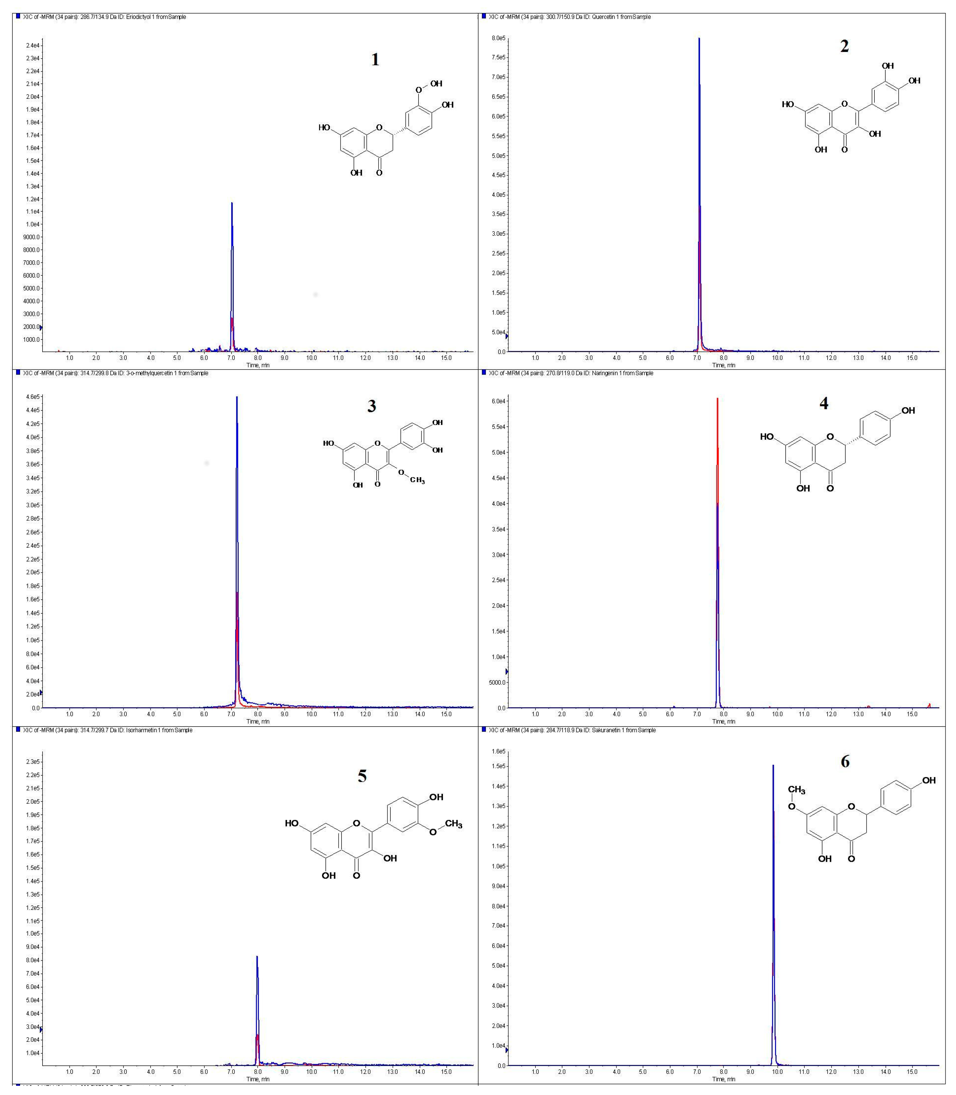
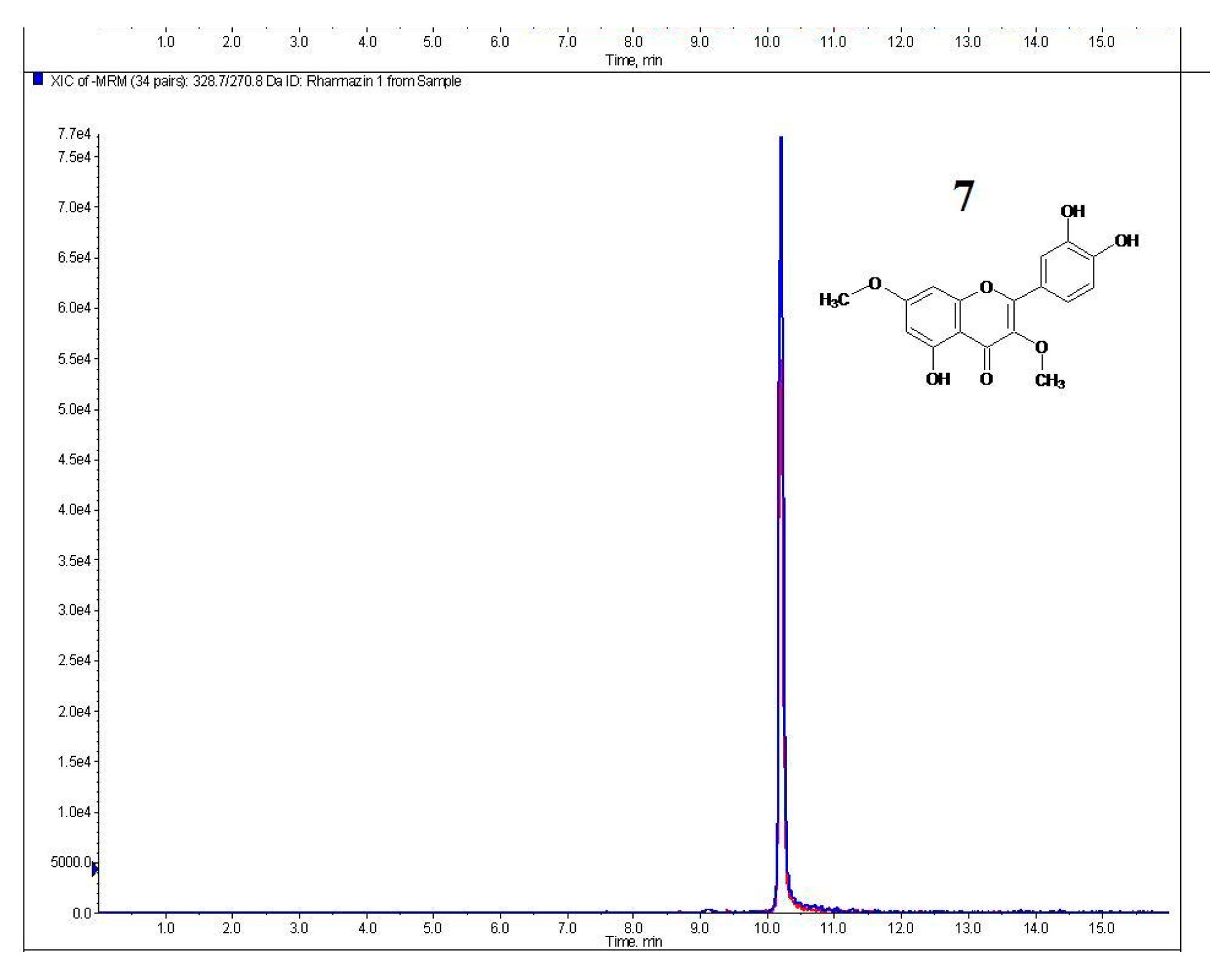
2. Results and Discussion

The purpose of the present study was to determine the phenolic acid and flavonoid aglycone contents and antioxidant activity of extracts from mistletoe berries harvested from different host trees. Phenolic compounds are widely distributed in the plant kingdom and are the most abundant secondary metabolites found in plants. The biological activity of phenolics is related, at least in part, to their antioxidant properties, due to both their free-radical scavenging activity and metal chelating properties [19,20,21]. There is growing scientific interest in the biological properties of polyphenols and flavonoids beneficial for prevention of age-related diseases, including cardiovascular diseases and cancer [22,23,24]. As previously mentioned, there are only a few scientific studies regarding mistletoe berries. Therefore, our findings aim to yield new valuable data on the chemical composition of Viscum album L.
The total phenolic and flavonoid contents of mistletoe extracts are shown in Table 1. The total phenolic content varied from 4.78 to 10.43 mg GA g−1 of dry extract and the total flavonoid content–from 0.27 to 0.428 mg Q g−1 of dry extract. The results of the present study demonstrated the highest amount of polyphenols and favonoids in extracts of mistletoe berries collected from Populus nigra “Italica” L. and Sorbus aucuparia L.
Liquid chromatography coupled with mass spectrometry (LC-MS) is one of the most effective analytical tools for organic compound analysis and has many advantages. It provides easy identification of eluted analytes as well as structural and molecular weight information, reduces sample preparation time; quantitative and qualitative data can be obtained easily from limited optimization of distributions [24,25,26].
A new method for the determination of flavonoid aglycones by LC-ESI-MS/MS was developed and applied for the first time. Chromatograms in MRM mode of standard solutions of flavonoid aglycones occurring in mistletoe berries are shown in Figure 1.
The majority of the tested compound were obtained from Tilia cordata Mill. and Populus nigra ‘Italica’ L. mistletoe berries (5.955 and 2.161 µg per g dry extract respectively) (Table 2).
Among free flavonoid aglycones, large amounts of naringenin (2.319 ± 0.046 µg per g dry extract), isorhamnetin (1.794 ± 0.09 µg per g dry extract), rhamnazin (1.223 ± 0.061 µg per g dry extract) and eriodictiol (0.201 ± 0.01 µg per g dry extract), were found in mistletoe berries from Tilia cordata Mill. A flavonoid aglycone characteristic of mistletoe berries from Populus nigra ‘Italica’ L. was sakuranetin (0.109 ± 0.0003 µg per g dry extract). These extracts also contained significant amounts of naringenin (0.427 ± 0.0001 µg per g dry extract), 3-O-methylquercetin (0.532 ± 0.004 µg per g dry extract), rhamnazin (0.472 ± 0.024 µg per g dry extract) and quercetin (0.056 ± 0.003 µg per g dry extract). Most of phenolic acids obtained by the LC-ESI-MS/MS method (Table 3) were determined for mistletoe berries from Sorbus aucuparia L. and Populus nigra ‘Italica’ L. (respectively 351.13 and 354.45 µg per g dry extract); the lowest amount was found in mistletoe berries from Pinus sylvestris L. (Viscum album subsp. austriacum).
The berries harvested from Fraxinus pensylvanica Marsh. contained large amounts of gallic acid (23.17 ± 0.58 µg per g dry extract), protocatechuic acid (80.79 ± 0.57 µg per g dry extract) and syringic acid (98.6 ± 0.69 µg per g dry extract). In other extracts, gallic acid was only present in trace amounts and the protocatechuic acid content ranged from trace amounts to a maximum value of 49.64 ± 0.50 µg per g dry extract for mistletoe berries from Malus domestica Borkh.
A characteristic compound for mistletoe berries from Malus domestica Borkh. and Pinus sylvestris L. (Viscum album subsp. austriacum) was ferulic acid (13.64 ± 0.67 and 11.81 ± 0.01 µg per g dry extract respectively). The extract of mistletoe from Populus nigra ‘Italica’ L. was characterized by the presence gentisic acid (3.26 ± 0.16 µg per g dry extract) and large amounts of sinapic and syringic acid (112.17 ± 1.79 and 137.99 ± 1.24 µg per g dry extract respectively).
The extracts from mistletoe berries coming from Sorbus aucuparia L. contained large amounts of caffeic acid (34.74 ± 0.52 µg per g dry extract), vanillic acid (43.9 ± 1.36 µg per g dry extract), sinapinic acid (107.08 ± 1.07 µg per g dry extract) and syringic acid (121.54 ± 1.58 µg per g dry extract).
The results of tests obtained for mistletoe berries from Tilia cordata Mill were noteworthy. In this case, synapinic acid was the characteristic phenolic acid present in large quantities (84.80 ± 1.36 µg per g dry extract).Moreover, biological research of mistletoe berries was carried out. The antioxidant activity of extracts was measured by four methods: reducing power (RED), metal chelating power (CHEL), inhibition of linoleic acid peroxidation (LPO) and free radical scavenging ability (ABTS)–Table 4.
The extract of mistletoe berries harvested from Tilia cordata Mill. showed the highest antioxidant activity measured by RED, LPO and CHEL methods. The highest antioxidant activity tested using ABTS was determined for the extract of mistletoe berries harvested from Sorbus aucuparia L. Fraxinus pensylvanica Marsh. and Populus nigra ‘Italica’ L. (EC50 7.72, 8.06 and 8.17 mg mL−1 of extract respectively).
The results of statistical analysis suggested that the phenolic and flavonoid content correlated with the results of antioxidant activity measured by the ABTS method (extracts which contained high amounts of phenolic and flavonoid compounds showed a low EC50 value, hence higher antioxidant activity—Table 5). Statistical analysis suggests that the antiradical activity is in 49% determined by the total phenolic content, in 65% by total flavonoid content and in 95% by total phenolic acids content (R2 × 100%).
The effects of extracts on human colon cancer cell proliferation were examined by MTT and BrdU assays, performed after 96 or 48 h, respectively, of LS180 cell treatment with the assessed compounds. The MTT assay results (upper panel of Figure 2) revealed the antiproliferative abilities of the studied extracts. The extracts isolated from mistletoe berries harvested from linden (Figure 2B) and poplar (Figure 2C) suppressed colon cancer cell proliferation in a dose-dependent manner, starting with the concentration of 50 μg/mL. At the highest concentration tested (100 μg/mL), a decrease in cell proliferation of approximately 30% versus control conditions was observed. The extracts obtained from mistletoe growing on Pinus sylvestris L. (Figure 2A) inhibited cancer cell proliferation only when the highest investigated concentration was used. The IC50 values calculated for all extracts were 1 mg/mL, 164 µg/mL and 202 µg/mL, respectively.
The antiproliferative properties of extracts were further verified by the more sensitive and specific BrdU assay, which enables one to determine the influence on DNA synthesis in proliferating cells. As presented in the bottom panel of Figure 2, 48 h of cancer cells exposure to the extracts did not affect DNA synthesis.
In vitro studies have revealed that ethanol extracts isolated from mistletoe berries decreased the proliferation of colon cancer cells in a dose-dependent manner. The effect was observed after 96 h of cells exposure and was attributed to altered mitochondrial activity. The changes were dependent on the tree species from which mistletoe was collected and used for extract preparation. The best results were obtained from the material harvested from Tilia cordata Mill. and Populus nigra ‘Italica’ L. Otherwise, the effects of extracts on the DNA synthesis in colon cancer cells were not observed.
Extract cytotoxicity was examined in human colon epithelial cells (CCD 841 CoTr) by means of an LDH test. Cells were exposed for 48 h to culture medium alone (control) or the extracts in concentrations ranging from 10 to 100 µg/mL. All tested extracts were nontoxic to colon epithelial cells in the whole range of investigated concentrations (Figure 3). Furthermore extracts isolated from mistletoe berries harvested from Tilia cordata Mill. in the highest concentration (Figure 3B) and Populus nigra ‘Italica’ L. in the concentration range 25–100 µg/mL (Figure 3C) significantly reduced the level of LDH release from treated cells.
Since anticancer agents should not be toxic for normal cells, the cytotoxic effects of extracts were evaluated in human intestinal epithelial cells. The findings revealed that the substances tested did not affect the integrity of colon epithelial cell membranes. Furthermore, the extracts isolated from mistletoe growing on Tilia cordata Mill. and Populus nigra ‘Italica’ L. were found to be able to reduce the level of LDH release from cells, which might indicate protective properties of the substances in question.
It has been reported that phenolic compounds possess important biological properties such as anticancer, antiviral, antioxidant and anti-inflammatory activities [13,27]. A substantial body of evidence supports the theory of anticancer properties of phenolic acids, and although the mechanisms are still not fully understood, they may include scavenging of free radicals, induction of enzymes involved in the metabolism of xenobiotics, regulation of gene expression, modulation of cellular signaling pathways including those involved in DNA damage repair, cell proliferation, apoptosis and invasion [12,28].
The large amounts of naringenin, isorhamnetin and rhamnazin were found in extracts from mistletoe berries from Tilia cordata Mill. and Populus nigra ‘Italica’ L. Moreover, in the extract from mistletoe berries from Populus nigra ‘Italica’ L. the largest content of phenolic acids were determined. In vitro studies showed that ethanolic extracts from mistletoe berries from Tilia cordata Mill. and Populus nigra ‘Italica’ L. decreased the proliferation of colon cancer cells to the greatest extent.
Many studies have shown various pharmacological effects including antioxidant, anti-inflammatory, antimutagenic and anticancer activity of naringenin (NAR) [29,30,31,32]. There is growing evidence that naringenin induces apoptosis in human colon cancer cells, showing no toxic effects on normal cells at a similar dose.
Activating transcription factor 3 (ATF3) is associated with apoptosis in human colon cancer. Song et al. [33] demonstrated that naringenin increases the transcriptional activation and increase of ATF3 protein level through p38 activation, and NAR-mediated ATF3 expression contributes, at least in part, to apoptosis in human colon cancer cells.
Moreover, Song et al. [34] demonstrated that NAR-induced proteasomal degradation of cyclin D1 might inhibit the cell proliferation in human colorectal cancer cells. Their study provides information on the apoptotic effect and the potential molecular mechanism of naringenin.
Likewise Leonardi et al. [35] indicated that naringnenin was promising as a naturally occuring chemopreventive agents against colon carciogenesis.
The beneficial effects of isorhamnetin, such as anti-inflammatory, antioxidant and anti-cancer have been reported in various tissues [36,37,38]. Seo et al. [39] determined that the antioxidant activity of isorhamnetin was correlated with its anti-cancer effects on colorectal cancer cells. The results demonstrated that isorhamnetin inhibits reactive oxygen species (ROS)-mediated hypoxia-induced hypoxia inducible factor-1α (HIF-1α) accumulation, which contributes to its anti-metastatic efficacy.
Furthermore, it has been found that isorhamnetin (IH) exerted a strong antiproliferative effect and increased cytotoxicity in combination with chemotherapeutic drugs. IH also inhibited the migratory/invasive properties of gastric cancer cells [38,40]. Previous studies and the preclinical evaluations suggest the antiproliferative and apoptogenic effects of rhamnazin [41,42].
To sum up, the literature findings suggest that naringenin, isorhamnetin and rhamnazin could be used as anti-cancer agents, particularly in colon cancer. Moreover, the presented mechanisms can explain the anti-cancer efficacy of various natural compounds with antioxidant effects.
As previously mentioned mistletoe has been evaluated as a treatment for people with numerous kinds of cancers including colon cancer [9]. The literature reports of the beneficial effect of supportive care mistletoe extract Iscador (ISC) therapy within adjuvant therapy (AT) protocols and long-term ISC treatment in stage I-III colorectal carcinoma patients, particularly improvement in AT-ADRs and symptoms and possible extension of disease-free survival [14].
It is worth mentioning that our studies relate to ethanolic extracts from mistletoe berries, without toxic proteins and lectins that are present in aqueous extract of mistletoe. Finally it should also be emphasized that the available literature lacks studies regarding contents of phenolic acids and flavonoid aglycones or antioxidant and biological activities of mistletoe berries.

3. Materials and Methods

3.1. Plant Material

Berries of V. album subsp. album were harvested from different host trees, i.e., Populus nigra L. ‘Italica’, Fraxinus pennsylvanica Marsh., Malus domestica Borkh., Sorbus aucuparia L., Tilia cordata Mill. and Pinus sylvestris L. growing close to the Lublin region (Poland) during the winter of 2013/14. The plant material was air-dried at room temperature and powdered.

3.2. Extraction Method

Portions of shredded mistletoe berries (10 g) were extracted using ultrasound assisted maceration at room temperature with 8:2 methanol/water solution (50 mL). The plant material was extracted thrice, 15 min each time. In all cases, the extracts were filtered, evaporated to dryness under vacuum and lyophilized in a Free Zone 1 apparatus (Labconco, Kansas City, KS, USA). The residue was weighted and placed in refrigerator (−30 °C). Immediately before analysis the extracts were redissolved in the same solvent as the one used for extraction to obtain a stock solutions. All samples were prepared in triplicate. Before LC-MS analysis, each test solution was filtered through a 0.45 μm membrane filter.

3.3. Total Phenolic and Flavonoid Content

The total phenolic content (TPC) was determined according to a modified Folin-Ciocalteu method described earlier [43]. The absorbance was measured at 680 nm after a 20-min incubation. Determinations were carried out using a standard curve prepared for gallic acid and the results were expressed as mg of gallic acid per 1 g of dry extract (GAE–gallic acid equivalent). The total flavonoid content (TFC) was determined according to the method described by Lamaison and Carret [44] with some modifications [45]. The absorbance was read at 430 nm after a 30-min incubation against a blank solution containing methanol instead of the test sample. The results were expressed as mg quercetin (Q) per 1 g of dry extract. All measurements were performed using an Infinite Pro 200F microplate reader (Tecan Group Ltd., Männedorf, Switzerland).

3.4. LC-ESI-MS/MS Analysis

3.4.1. Chromatographic Conditions and Apparatus

Phenolic acids and flavonoid aglycones were determined by reversed-phase high-performance liquid chromatography and electrospray ionization mass spectrometry (LC-ESI-MS/MS). An Agilent 1200 Series HPLC system (Agilent Technologies, Santa Clara, CA, USA) equipped with a binary gradient solvent pump, a degasser, an autosampler and a column oven connected to a 3200 QTRAP Mass spectrometer (AB Sciex, Framingham, MA, USA) was used.
The contents of free flavonoid aglycones were determined by a newly developed, simple and rapid method using liquid chromatography-electrospray ionization-tandem mass spectrometry. The compounds were separated at 25 °C on a Zorbax SB-C18 column (2.1 × 50 mm, 1.8-μm particle size; Agilent Technologies). The injection volume was 5 μL. Gradient elution was applied using water containing 0.1% HCOOH (A) and acetonitrile (B). The flow rate was 200 μL/min and the gradient was as follows: 0–0.5 min 5% B; 1–4 min 40% B; 5–9 min 50% B; 11–12 min 70% B; 12.5–13.5 min 90% B; 15–19 min 5% B. Contents of phenolic acids were determined by the LC-ESI-MS/MS method previously described by Nowacka et al. [46].

3.4.2. Optimisation of Quantitative Analysis Parameters

The 3200 QTRAP MS/MS system and an electrospray ion source in the negative mode were used. The optimal mass spectrometer parameters were determined experimentally and for analysis flavonoid aglycones were as follows: curtain gas 20 psi, capillary temperature 550 °C, nebulizer gas 30 psi, negative ionization mode source voltage-4500 V. Nitrogen was used as a curtain and collision gas. The data were acquired and processed using Analyst 1.5 software from AB Sciex (Framingham, MA, USA).
Multiple reaction monitoring (MRM) was used for quantitative analysis of compounds. The analytes were identified by comparing the retention times and m/z values obtained by MS and MS2 with the mass spectra of the corresponding standards tested under the same conditions. The calibration curves obtained in the MRM mode were used for quantification of all analytes. The identified flavonoid aglycones were quantified based on their peak areas and comparison with a calibration curve for the corresponding standards. Linearity ranges for calibration curves were specified.
The limits of detection (LOD) and quantification (LOQ) for phenolic acids and flavonoids aglycone were determined at a signal-to-noise ratio of 3:1 and 10:1, respectively, by injecting a series of dilute solutions of known concentrations. Table 6 and Table 7 summarize the optimized parameters for quantification of flavonoid aglycones.

3.5. Biological Activity

3.5.1. Antioxidant Activity

The antioxidant activity of extracts was measured by four methods. The antiradical activity was assayed using an improved ABTS decolorization assay [47,48]. The absorbance was measured at 734 nm. The extract ability to quench the ABTS free radical was determined using the following equation:
Scavenging% = ((AC − AA)/AA) × 100
where AC is the absorbance of control and AA is the absorbance of sample.
Reducing power (RED) was determined by the method of Oyaizu [49] The absorbance was measured at 700 nm with a spectrophotometer (Lambda 40, Perkin-Elmer, Waltham, MA, USA). Increased absorbance of the reaction mixture indicated increased reducing power.
Metal chelating power (CHEL) was determined by the method of Guo et al. [50]. Subsequently, the absorbance of the solution was measured spectrophotometrically at 562 nm. The percentage of inhibition of ferrozine-Fe2+ complex formation was calculated according to the following formula:
Inhibition% = (1 − (Ap/Ac)) × 100
where Ac is the-absorbance of control and Ap–the absorbance in the presence of sample.
The degree of inhibition of the hemoglobin-catalyzed linoleic acid peroxidation (LPO) was determined according to Kuo and Sun [51]. The absorbance of sample (As) was measured at 480 nm with a spectrophotometer for 5 min. The absorbance of blank (A0) was determined without hemoglobin added to the reaction mixture; the absorbance of control (A100) was measured with no sample added to the above mixture. Thus, the antioxidative activity of the sample was calculated as:
AA% = (1 − (As − A0)/(A100 − A0)) × 100
All the antioxidant activity results were expressed as EC50 values, the extract concentration providing 50% of the activity in a dose-dependent manner.

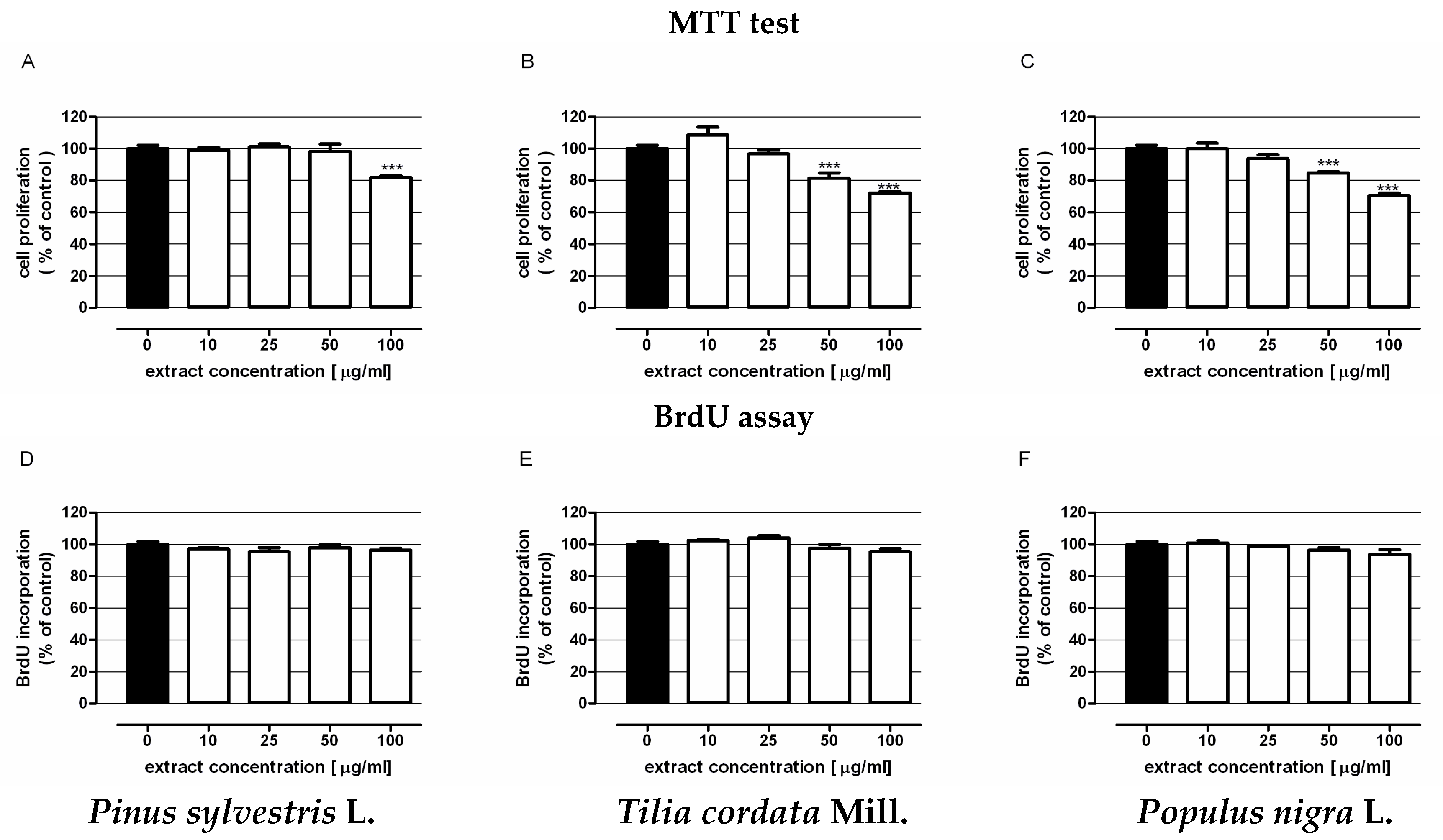
3.5.2. Examination of Extract Antiproliferative Activity

The human colon adenocarcinoma cell line LS180 was obtained from the European Collection of Cell Cultures (ECACC, Centre for Applied Microbiology and Research, Salisbury, UK). LS180 cells were grown in a 1:1 mixture of DMEM (Sigma-Aldrich, St. Louis, MO, USA) and Ham’s F-12 nutrient mixture (Sigma-Aldrich) supplemented with 10% foetal bovine serum (FBS) (Sigma-Aldrich), penicilin (100 U/mL) (Sigma-Aldrich) and streptomycin (100 μg/mL) (Sigma-Aldrich). Cells were maintained in a humidified atmosphere of 95% air and 5% CO2 at 37 °C.
The effects of extracts on cancer cells proliferation were determined by the MTT metabolism assay and BrdU incorporation immunoassay. The BrdU test was conducted using Cell Proliferation ELISA BrdU according to the manufacturer’s instructions (Roche Diagnostics GmbH, Penzberg, Germany). Both assays were performed in human adenocarcinoma cells (LS180). Details of the procedures are described elsewhere [52].

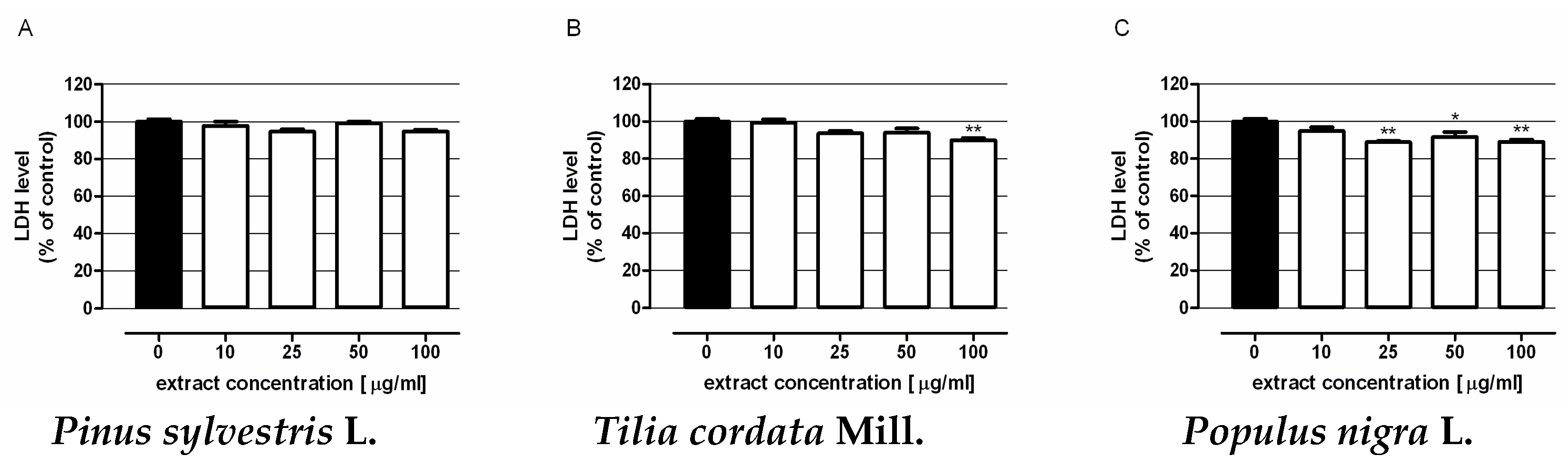
3.5.3. Assessment of Cytotoxicity of Extracts

The human colon epithelial cell line CCD 841 CoTr was purchased from American Type Culture Collection (ATCC, Manassas, VA, USA). CCD 841 CoTr cells were grown in DMEM (Sigma-Aldrich) supplemented with 10% foetal bovine serum (FBS) (Sigma-Aldrich), penicillin (100 U/mL) (Sigma-Aldrich) and streptomycin (100 μg/mL) (Sigma-Aldrich). Cells were maintained in a humidified atmosphere of 95% air and 5% CO2 at 33 °C. The cytotoxicity of extracts was evaluated using the colorimetric test based on the measurement of lactate dehydrogenase (LDH) release. Determinations were performed in human colon epithelial cells (CCD 841 CoTr) using a LDH-based in vitro toxicology assay kit (Sigma-Aldrich). Details of the procedure were described elsewhere [52].

3.6. Statistical Analysis

All results were expressed as a mean ± standard error of the mean (SEM) of three replications for each extract tested. Moreover the Pearson correlation coefficient and R2 coefficient were applied to show relationship between total phenolic content (TPC), total flavonoid content (TFC) total phenolic acids content (LC-ESI-MS/MS), total flavonoid aglycones (LC-ESI-MS/MS) and EC50 (ABTS) for mistletoes berries extracts from different host trees.
Calculations were performed in STATISTICA 10.0 (StatSoft PolandCracow, Poland). Statistical analysis was performed using the one-way ANOVA with the Tukey post-hoc test and column statistics used for comparisons. Significance was accepted at p < 0.05. The IC50 value of biological activity (concentration causing proliferation inhibition by 50% compared to control) was calculated according to the Litchfield-Wilcoxon method [53].

4. Conclusions

The chemical composition and biological activity of mistletoe berries (Viscum album L.) collected from different host plants were studied for the first time. Our findings demonstrated that the extracts of V. album contained large amounts of polyphenolic compounds. Phenolic acids and flavonoids may be the characteristic compounds indicating a subspecies of the host tree from which the mistletoe was collected. In vitro studies have revealed that ethanol extracts isolated from mistletoe berries decreased the proliferation of colon cancer cells in dose-dependent manner; the best results were obtained from Tilia cordata Mill. and Populus nigra ‘Italica’ L materials. Moreover, the substances assessed did not affect the integrity of colon epithelial cell membranes and extracts isolated from mistletoe growing on Tilia cordata Mill. and Populus nigra ‘Italica’ L. were able to reduce the level of LDH release from cells, which might prove the protective properties of the substances tested.
Moreover, this work presented newly developed method of phenolic compound using simple, rapid, and reliable liquid chromatography-electrospray ionization-tandem mass spectrometry. Tge present studies suggest that the phenolic acids and flavonoids might have potential as chemopreventive agents against colon tumor development.

Acknowledgments

The paper was developed using the equipment purchased within the Project “The equipment of innovative laboratories doing research on new medicines used in the therapy of civilization and neoplastic diseases” within the Operational Program Development of Eastern Poland 2007-2013, Priority Axis I Modern Economy, Operations I.3 Innovation Promotion.

Author Contributions

W.P. and R.N. completed the manuscript in collaboration with U.G.-D., M.K.L. and W.R. All authors read and approved the final manuscript.

Conflicts of Interest

The authors declare no conflict of interest.

References

  1. Onay-Uçar, E.; Karagöz, A.; Arda, N. Antioxidant activity of Viscum album spp. album. Fitoterapia 2006, 77, 556–560. [Google Scholar] [CrossRef] [PubMed]
  2. Orhan, D.D.; Aslan, M.; Sendogdu, N.; Ergun, F.; Yesilada, E. Evaluation of the hypoglycemic effect and antioxidant activity of three Viscum album subspecies (European mistletoe) in streptozotocin-diabetic rats. J. Ethnopharmacol. 2005, 98, 95–102. [Google Scholar] [CrossRef] [PubMed]
  3. Deeni, Y.Y.; Sadiq, N.M. Antimicrobial properties and phytochemical constituents of the leaves of African mistletoe (Tapinanthus dodoneifolius (DC) Danser) (Loranthaceae): An ethnomedicinal plant of Hausaland, Northern Nigeria. J. Ethnopharmacol. 2002, 83, 235–240. [Google Scholar] [CrossRef]
  4. Amabeoku, G.J.; Leng, M.J.; Syce, J.A. Antimicrobial and anticonvulsant activities of Viscum capense. J. Ethnopharmacol. 1998, 61, 237–241. [Google Scholar] [CrossRef]
  5. Gorter, R.; Khwaja, T.; Linder, M. Anti-HIV and immunomodulating activities of Viscum album (mistletoe). Int. Conf. AIDS 1992, 84, 19–24. [Google Scholar]
  6. Simirgiotis, M.J.; Quispe, C.; Areche, C.; Sepúlveda, B. Phenolic Compounds in Chilean Mistletoe (Quintral, Tristerix tetrandus) Analyzed by UHPLC–Q/Orbitrap/MS/MS and Its Antioxidant Properties. Molecules 2016, 21, 245. [Google Scholar] [CrossRef] [PubMed]
  7. Son, G.S.; Ryu, W.S.; Kim, H.Y.; Woo, S.U.; Park, K.H.; Bae, J.W. Immunologic response to mistletoe extract (Viscum album L.) after conventional treatment in patientswith operable breast cancer. J. Breast Cancer 2010, 13, 14–18. [Google Scholar] [CrossRef]
  8. Beuth, J.; Schneider, B.; Schierholz, J.M. Impact of complementary treatment of breast cancer patients with standardized mistletoe extract during aftercare: A controlled multicenter comparative epidemiological cohort study. Anticancer Res. 2008, 28, 523–527. [Google Scholar] [PubMed]
  9. Mistletoe Extracts (PDQ®)-Health Professional Version. Available online: https://www.cancer.gov/about-cancer/treatment/cam/hp/mistletoe-pdq#cit/section_5.28 (accessed on 20 January 2017 ).
  10. Siegel, R.; Desantis, C.; Jemal, A. Colorectal cancer statistics, 2014. CA Cancer J. Clin. 2014, 64, 104–117. [Google Scholar] [CrossRef] [PubMed]
  11. Li, W.-Y.; Chan, S.-W.; Guo, D.-J.; Yu, P.H.-F. Correlation between Antioxidative Power and Anticancer Activity in Herbs from Traditional Chinese Medicine Formulae with Anticancer Therapeutic Effect. Pharm. Biol. 2008, 45, 541–546. [Google Scholar] [CrossRef]
  12. Rosa, L.S.; Silva, N.J.A.; Soares, N.C.P.; Monteiro, M.C.; Teodoro, A.J. Anticancer Properties of Phenolic Acids in Colon Cancer A Review. J. Nutr. Food Sci. 2016, 6. [Google Scholar] [CrossRef]
  13. Batra, P.; Sharma, A.K. Anti-cancer potential of flavonoids: Recent trends and future perspectives. Biotech 2013, 3, 439–459. [Google Scholar] [CrossRef] [PubMed]
  14. Friedel, W.E.; Matthes, H.; Bock, P.R.; Zänker, K.S. Systematic evaluation of the clinical effects of supportive mistletoe treatment within chemo- and/or radiotherapy protocols and long-term mistletoe application in nonmetastatic colorectal carcinoma: Multicenter, controlled, observational cohort study. J. Soc. Integr. Oncol. 2009, 7, 29–43. [Google Scholar]
  15. Edlund, U.; Hensel, A.; Frӧse, D.; Pfűller, U.; Scheffler, A. Polysaccharides from fresh Viscum album L. Berry extract and their interaction with Viscum album Agglutinin I. Arzneimittel-Forschung/Drug Res. 2000, 7, 645–651. [Google Scholar] [CrossRef] [PubMed]
  16. Azuma, J.I.; Kim, N.H.; Heux, L.; Vuong, R.; Chanzy, H. Cellulose system in viscin from mistletoe berries. Cellulose 2000, 1, 3–19. [Google Scholar] [CrossRef]
  17. Wagner, H.; Jordan, E. An immunologically active arabinogalactan from Viscum album ‘berries’. Phytochemistry 1988, 8, 2511–2517. [Google Scholar] [CrossRef]
  18. Cammarata, P.L.; Cajelli, E. Free amino acid content of Viscum album L. berries parasitizing the Pinus silvestris L. and Pinus nigra Arnold var. austriaca. Boll. Chim. Farm. 1967, 8, 521–526. [Google Scholar]
  19. Carbonaro, M.; Virgili, F.; Carnovale, E. Evidence for protein-tannin interaction in legumes: Implications in the antioxidant properties of faba bean tannins. Lebens-Wiss Technol. 1996, 29, 743–750. [Google Scholar] [CrossRef]
  20. Rice-Evans, C.A.; Miller, N.J.; Paganga, G. Structure-antioxidant activity relationships of flavonoids and phenolic acids. Free Radic. Biol. Med. 1996, 20, 933–956. [Google Scholar] [CrossRef]
  21. Okuda, T.; Yoshida, T.; Hatano, T. Antioxidant effects of tannins and related polyphenols. In Phenolic Compounds in Food and their Effects on Health. II. Antioxidants and Cancer prevention; Ho, C.T., Lee, C.Y., Huang, M.T., Eds.; American Chemical Society: Washington DC, USA, 1992; pp. 87–97. [Google Scholar]
  22. Amzad Hossain, M.; Mizanur Rahman, S.M. Isolation and characterization of flavonoids from the leaves of medicinal plant Orthosiphon stamineus. Arabian J. Chem. 2015, 8, 218–221. [Google Scholar] [CrossRef]
  23. Williamson, G.; Manach, C. Bioavailability and bioefficacy of polyphenols in humans. II. Review of 93 intervention studies. Am. J. Clin. Nutr. 2005, 81, 243S–255S. [Google Scholar] [PubMed]
  24. Wilkins, C. Impressive development in mass spektrometry. Trends Anal. Chem. 2007, 26, 65–67. [Google Scholar] [CrossRef]
  25. Watson, J.T.; Sparkman, O.D. Introduction to Mass Spectrometry. Instrumentation, Applications and Strategies for Data Interpretation; John Wiley & Sons Ltd.: Anglia, UK, 2007. [Google Scholar]
  26. Jiang, Z.; Wang, Y.; Zheng, Y.; Yang, J.; Zhang, L. Ultra high performance liquid chromatography coupled with triple quadrupole mass spectrometry and chemometric analysis of licorice based on the simultaneous determination of saponins and flavonoids. J. Sep. Sci. 2016, 39, 2928–2940. [Google Scholar] [CrossRef] [PubMed]
  27. Saibabu, V.; Zeeshan, F.; Luqman, A.K.; Saif, H. Therapeutic Potential of Dietary Phenolic Acids. Adv. Pharm. Sci. 2015, 2015, 823539. [Google Scholar] [CrossRef] [PubMed]
  28. Pandey, K.B.; Rizvi, S.I. Plant polyphenols as dietary antioxidants in human health and disease. Oxid. Med. Cell. Longev. 2009, 2, 270–278. [Google Scholar] [CrossRef] [PubMed]
  29. Pietta, P.G. Flavonoids as antioxidants. J. Nat. Prod. 2000, 63, 1035–1042. [Google Scholar] [CrossRef] [PubMed]
  30. Esmaeili, M.A.; Alilou, M. Naringenin attenuates CCl4-induced hepatic inflammation by the activation of an Nrf2-mediated pathway in rats. Clin. Exp. Pharmacol. Physiol. 2014, 41, 416–422. [Google Scholar] [CrossRef] [PubMed]
  31. Choi, J.S.; Park, K.Y.; Moon, S.H.; Rhee, S.H.; Young, H.S. Antimutagenic effect of plant flavonoids in the Salmonella assay system. Arch. Pharm. Res. 1994, 17, 71–75. [Google Scholar] [CrossRef] [PubMed]
  32. Ekambaram, G.; Rajendran, P.; Magesh, V.; Sakthisekaran, D. Naringenin reduces tumor size and weight lost in N-methyl-N′-nitro-N-nitrosoguanidine-induced gastric carcinogenesis in rats. Nutr. Res. 2008, 28, 106–112. [Google Scholar] [CrossRef] [PubMed]
  33. Song, H.M.; Park, G.H.; Eo, H.J.; Jeong, J.B. Naringenin-Mediated ATF3 Expression Contributes to Apoptosis in Human Colon Cancer. Biomol. Ther. 2016, 2, 140–146. [Google Scholar]
  34. Song, H.M.; Park, G.H.; Eo, H.J.; Lee, J.W.; Kim, M.K.; Lee, J.R.; Lee, M.H.; Koo, J.S.; Jeong, J.B. Anti-Proliferative Effect of Naringenin through p38-Dependent Downregulation of Cyclin D1 in Human Colorectal Cancer Cells. Biomol. Ther. (Seoul) 2015, 23, 339–344. [Google Scholar] [CrossRef] [PubMed]
  35. Leonardi, T.; Vanamala, J.; Taddeo, S.S.; Davidson, L.A.; Murphy, M.E.; Patil, B.S.; Wang, N.; Carroll, R.J.; Chapkin, R.S.; Lupton, J.R.; et al. Apigenin and naringenin suppress colon carcinogenesis through the aberrant crypt stage in azoxymethane-treated rats. Exp. Biol. Med. 2010, 235, 710–717. [Google Scholar] [CrossRef] [PubMed]
  36. Yang, J.H.; Kim, S.C.; Shin, B.Y.; Jin, S.H.; Jo, M.J.; Jegal, K.H.; Kim, Y.W.; Lee, J.R.; Ku, S.K.; Cho, I.J.; et al. O-Methylated flavonol isorhamnetin prevents acute inflammation through blocking of NF-κB activation. Food Chem. Toxicol. 2013, 59, 362–372. [Google Scholar] [CrossRef] [PubMed]
  37. Yang, J.H.; Shin, B.Y.; Han, J.Y.; Kim, M.G.; Wi, J.E.; Kim, Y.W.; Cho, I.J.; Kim, S.C.; Shin, S.M.; Ki, S.H. Isorhamnetin protects against oxidative stress by activating Nrf2 and inducing the expression of its target genes. Toxicol. Appl. Pharmacol. 2014, 15, 293–301. [Google Scholar] [CrossRef] [PubMed]
  38. Saud, S.M.; Young, M.R.; Jones-Hall, Y.L.; Ileva, L.; Evbuomwan, M.O.; Wise, J.; Colburn, N.H.; Kim, Y.S.; Bobe, G. Chemopreventive activity of plant flavonoid isorhamnetin in colorectal cancer is mediated by oncogenic Src and β-catenin. Cancer Res. 2013, 73, 5473–5484. [Google Scholar] [CrossRef] [PubMed]
  39. Seo, S.; Seo, K.; Ki, S.H.; Shin, S.M. Isorhamnetin Inhibits Reactive Oxygen Species-Dependent Hypoxia Inducible Factor (HIF)-1α Accumulation. Biol. Pharm. Bull. 2016, 39, 1830–1838. [Google Scholar] [CrossRef] [PubMed]
  40. Ramachandran, L.; Manu, K.A.; Shanmugam, M.K.; Li, F.; Siveen, K.S.; Vali, S.; Kapoor, S.; Abbasi, T.; Surana, R.; Smoot, D.T.; et al. Isorhamnetin Inhibits Proliferation and Invasion and Induces Apoptosis through the Modulation of Peroxisome Proliferator-activated Receptor γ Activation Pathway in Gastric Cancer. J. Biol. Chem. 2012, 287, 38028–38040. [Google Scholar] [CrossRef] [PubMed]
  41. Philchenkov, A.A.; Zavelevych, M.P. Rhamnazin inhibits proliferation and induces apoptosis of human jurkat leukemia cells in vitro. Ukr. Biochem. J. 2015, 87, 122–128. [Google Scholar] [CrossRef] [PubMed]
  42. Yu, Y.; Cai, W.; Pei, C.G.; Shao, Y. Rhamnazin, a novel inhibitor of VEGFR2 signaling with potent antiangiogenic activity an antitumor efficacy. Biochem. Biophys. Res. Commun. 2015, 458, 913–919. [Google Scholar] [CrossRef] [PubMed]
  43. Olech, M.; Nowak, R.; Los, R.; Rzymowska, J.; Malm, A.; Chruściel, K. Biological activity and composition of teas and tinctures prepared from Rosa rugosa Thunb. Cent. Eur. J. Biol. 2012, 7, 172–182. [Google Scholar] [CrossRef]
  44. Lamaison, J.L.C.; Carret, A. Teneurs en principaux flavonoides des fleurs de Cratageus monogyna Jacq et de Cratageus Laevigata (Poiret D.C) en Fonction de la vegetation. Plant Méd. Phytothér. 1990, 25, 12–16. [Google Scholar]
  45. Pietrzak, W.; Nowak, R.; Olech, M. Effect of extraction method on phenolic content and antioxidant activity of mistletoe extracts from Viscum album subsp. abietis. Chem. Pap. 2014, 68, 976–982. [Google Scholar] [CrossRef]
  46. Nowacka, N.; Nowak, R.; Drozd, M.; Olech, M.; Los, R.; Malm, A. Analysis of phenolic constituents, antiradical and antimicrobial activity of edible mushrooms growing wild in Poland. LWT-Food Sci. Technol. 2014, 59, 689–694. [Google Scholar] [CrossRef]
  47. Gawlik-Dziki, U.; Świeca, M.; Dziki, D.; Sęczyk, Ł.; Złotek, U.; Różyło, R.; Kaszuba, K.; Ryszawy, D.; Czyż, J. Anticancer and Antioxidant Activity of Bread Enriched with Broccoli Sprouts. BioMed Res. Int. 2014. [Google Scholar] [CrossRef] [PubMed]
  48. Re, R.; Pellegrini, N.; Proteggente, A.; Pannala, A.; Yang, M.; Rice-Evans, C. Antioxidant activity applying an improved ABTS radical cation decolorization assay. Free Radic. Biol. Med. 1999, 26, 1231–1237. [Google Scholar] [CrossRef]
  49. Oyaizu, M. Studies on products of browning reaction–antioxidative activities of products of browning reaction prepared from glucosamine. Jpn. J. Nutr. 1984, 44, 307–315. [Google Scholar] [CrossRef]
  50. Guo, J.T.; Lee, H.L.; Chiang, S.H.; Lin, H.I.; Chang, C.Y. Antioxidant properties of the extracts from different parts of broccoli in Taiwan. J. Food Drug Anal. 2001, 9, 96–101. [Google Scholar]
  51. Kuo, J.-M.; Yeh, D.-B.; Pan, B. Rapid photometric assay evaluating antioxidative activity in edible part material. J. Agric. Food Chem. 1999, 47, 3206–3209. [Google Scholar] [CrossRef] [PubMed]
  52. Lemieszek, M.K.; Cardoso, C.; Nunes, F.H.F.M.; Barros, A.I.R.N.A.; Marques, G.; Pożarowski, P.; Rzeski, W. Boletus edulis biological active biopolymers induce cell cycle arrest in human colon adenocarcinoma cells. Food Funct. 2013, 4, 575–585. [Google Scholar] [CrossRef] [PubMed]
  53. Litchfield, J.T.; Wilcoxon, F.A. A simplified method of evaluating dose-effect experiments. J. Pharmacol. Exp. Ther. 1949, 96, 99–113. [Google Scholar] [PubMed]
Sample Availability: Samples of the compounds are available from the authors.
Figure 1. The chromatograms in MRM mode of standards solution of flavonoid aglycones occurring in mistletoe berries: 1–Eriodictiol; 2–Quercetin; 3–3-o-Methylquercetin; 4–Naringenin; 5–Isorhamnetin; 6–Sakuranetin; 7–Rhamnazin.
Molecules 22 00624 g001aMolecules 22 00624 g001b
Figure 2. The influence of mistletoe berries extracts on the proliferation of human colon adenocarcinoma cell line LS180. Cells were exposed for 96 h (upper panel) or 48 h (bottom panel) to extracts isolated from berries of mistletoe harvested from Pinus sylvestris L. (A,D); Tilia cordata Mill. (B,E); Populus nigra L. ‘Italica’ (C,F). Cell proliferation was quantified by MTT test (upper panel) or BrdU assay (bottom panel). Results are presented as the mean ± SEM of 4–5 measurements. *** p < 0.001 vs. control, one-way ANOVA test; post test: Tukey.
Molecules 22 00624 g002
Figure 3. Cytotoxicity of mistletoe berries extracts in human colon epithelial cell line CCD 841 CoTr. Cells were exposed for 48 h to extracts isolated from berries of mistletoe harvested from Pinus sylvestris L. (A); Tilia cordata Mill. (B); Populus nigra L. ‘Italica’ (C). Extract cytotoxicity was measured by means of the LDH assay. Results are presented as the mean ± SEM of 4 measurements. * p < 0.05 vs. control, ** p < 0.01 vs. control, one-way ANOVA test; post test: Tukey.
Molecules 22 00624 g003
Table 1. Total phenolic content (TPC) and flavonoid content (TFC) of methanol extracts from mistletoe berries harvested from various host trees.
SpeciesHost TreeTPC
(mg GA g−1 of dry extract)
TFC
(mg Q g−1 of dry extract)
V. albumFraxinus pensylvanica Marsh.8.82 ± 0.18 ns0.365 ± 0.005 ns
Malus domestica Borkh.6.71 ± 0.32 *0.356 ± 0.006 ns
Sorbus aucuparia L9.66 ± 0.28 ns0.428 ± 0.007 ns
Populus nigra ‘Italica’ L.10.43 ± 0.200.404 ± 0.017
Tilia cordata Mill.4.78 ± 0.09 *0.286 ± 0.007 ns
V. austriacumPinus sylvestris L.7.11 ± 0.20 ns0.270 ± 0.007 ns
Values are presented in mean ± SEM, n = 3 and evaluated by one-way ANOVA (post test: Tukey) to compare each extract to Populus nigra ‘Italica’ L. extract. Differences were considered to be statistically significant if p < 0.05; ns–no significant differences; * p < 0.05.
Table 2. Flavonoid aglycones contents in extracts from mistletoe berries collected from different trees host. Values are presented in mean ± SEM, n = 3.
CompoundV. albumV. austriacum
Fraxinus pensylvanica Marsh.Malus domestica Borkh.Sorbus aucuparia L.Populus nigra ‘Italica’ L.Tilia cordata Mill.Pinus sylvestris L.
Flavonoid aglycones (µg per g dry extracts)
Taxifolintrace a- btrace atrace atrace a- b
Myricetin- b- b- b- b- b- b
Morin- b- b- b- b- b- b
Eriodictiol0.011 ± 0.001trace a0.011 ± 0.0010.014 ± 0.0010.201 ± 0.010.001 ± 0.0001
Luteolin- b- b- btrace a- b- b
Quercetintrace a0.025 ± 0.00010.006 ± 0.00010.056 ± 0.003trace a0.026 ±0.0001
3-O-Methylquercetin0.255 ± 0.0020.163 ± 0.0080.510 ± 0.0200.532 ± 0.0040.409 ± 0.020.009 ± 0.0001
Apigenin- b- btrace atrace atrace atrace a
Naringenin0.111 ± 0.0020.102 ± 0.0020.427 ± 0.00010.681 ± 0.0142.319 ± 0.0460.251 ± 0.013
Kaempferol- b- b- btrace a- btrace a
Isorhamnetintrace a0.099 ± 0.0050.106 ± 0.0050.297 ± 0.0061.794 ± 0.090.013 ± 0.001
Rhamnetintrace atrace atrace atrace atrace atrace a
Chrysin- b- b- b- b- b- b
Sakuranetin0.014 ± 0.00010.001 ± 0.00020.054 ± 0.0030.109 ± 0.00030.009 ± 0.00010.004 ± 0.0001
Prunetin- b- b- b- b- b- b
Rhamnazin0.009 ± 0.00010.067 ± 0.0030.122 ± 0.0020.472 ± 0.0241.223 ± 0.0610.008 ± 0.0001
TOTAL0.40.4571.2362.1615.9550.312
a trace–trace amounts; - b not detected.
Table 3. Phenolic acid contents in extracts from mistletoe berries collected from different trees host. Values are presented in mean ± SEM, n = 3.
CompoundV. albumV. austriacum
Fraxinus pensylvanica Marsh.Malus domestica Borkh.Sorbus aucuparia L.Populus nigra ‘Italica’ L.Tilia cordata Mill.Pinus sylvestris L.
Phenolic Acids (µg per g dry Extracts)
Gallic acid23.17 ± 0.58trace atrace atrace atrace a- b
Protocatechuic acid80.78 ± 0.5740.88 ± 1.5929.03 ± 1.4531.07 ± 1.55trace atrace a
Gentisic acid- b- btrace a3.26 ± 0.16trace atrace a
4-OH-benzoic acid11.23 ± 0.115.20 ± 0.142.72 ± 0.140.79 ± 0.043.51 ± 0.18trace a
Vanilic acid26.1 ± 1.02Trace a43.9 ± 1.3644.1 ± 2.21trace atrace a
Caffeic acid12.78 ± 0.591.57 ± 0.0834.74 ± 0.529.40 ± 0.12trace atrace a
Syringic acid98.60 ± 0.6924.90 ± 0.05121.54 ± 1.58137.99 ± 1.2449.35 ± 0.125.77 ± 0.05
p-Coumaric acid1.19 ± 0.03trace atrace atrace atrace a7.07 ± 0.05
Ferulic acid4.65 ± 0.00513.64 ± 0.676.94 ± 0.287.69 ± 0.086.66 ± 0.0711.81 ± 0.01
Salicylic acid4.32 ± 0.171.22 ± 0.015.18 ± 0.027.98 ± 0.070.12 ± 0.0052.06 ± 0.03
Veratric acid- b- b- b- b- b- b
Synapic acid78.50 ± 2.3671.82 ± 0.43107.08 ± 1.07112.17 ± 1.7984.80 ± 1.368.60 ± 0.09
3-OH-cinnamic acid- b- b- b- b- b- b
Rosmarinic acidtrace atrace atrace atrace atrace atrace a
TOTAL341.32159.23351.13354.45144.4455.31
a trace–trace amounts; - b not detected.
Table 4. Antioxidant activity–reducing power (RED), metal chelating power (CHEL), Inhibition of linoleic acid peroxidation (LPO) and free radicals scavenging ability (ABTS) in methanol extracts from mistletoe berries harvested from various host trees.
SpeciesHost TreesAntioxidant Activity (EC50 in mg mL−1 of extract)
REDABTSCHELLPO
V. albumFraxinus pensylvanica Marsh.1.04 ± 0.02 ns8.06 ± 0.12 ns0.88 ± 0.18 *5.30 ± 0.03 *
Malus domestica Borkh.1.21 ± 0.02 ns13.03 ± 0.06 **0.54 ± 0.03 ns7.55 ± 0.19 **
Sorbus aucuparia L.1.05 ± 0.01 ns7.72 ± 0.13 **0.59 ± 0.10 ns6.60 ± 0.33 *
Populus nigra ‘Italica’ L.1.08 ± 0.018.17 ± 0.020.54 ± 0.035.65 ± 0.44
Tilia cordata Mill.0.92 ± 0.03 ns11.31 ± 0.14 **0.45 ± 0.10 ns5.57 ± 0.43 ns
V. austriacumPinus sylvestris L.1.33 ± 0.02 ns14.77 ± 0.25 **0.60 ± 0.01 ns10.24 ± 0.25 **
Values are presented in mean ± SEM, n = 3 and evaluated by one-way ANOVA (post test: Tukey) to compare each extract to Populus nigra ‘Italica’ L. extract. Differences were considered to be statistically significant if p < 0.05; ns—no significant differences; * p < 0.05; ** p < 0.01.
Table 5. Pearson correlation coefficient and R2 coefficient showing relationship between total phenolic content (TPC), total flavonoid content (TFC) total phenolic acids content (LC-ESI-MS/MS), total flavonoid aglycones (LC-ESI-MS/MS) and EC50 (ABTS) for mistletoes berries extracts from different host trees.
Content/EC50R (X,Y)R2
TPC & EC50 (ABTS)−0.700.49
TFC & EC50 (ABTS)−0.810.65
Total phenolic acids content (LC-ESI-MS/MS) & EC50 (ABTS)−0.970.95
Total flavonoid aglycones content (LC-ESI-MS/MS) & EC50 (ABTS)−0.070.01
Table 6. Summary of optimized parameters for the quantitative analysis of flavonoid aglycones.
CompoundRetention Time (min)Q1
(m/z)
Q3
(m/z)
DP a
(V)
EP b
(V)
CEP c
(V)
CE d
(eV)
CXP e
(V)
Taxifolin6.17302.7124.9−45−3.5−18−260
284.8−45−3.5−18−14−4
Myricetin6.47316.7136.9−55−9−14−320
150.9−55−9−14−260
Morin6.82300.7124.9−50−3.5−20−240
106.9−50−3.5−20−300
Eriodictiol7.07286.7134.9−45−6−12−320
150.9−45−6−12−18−2
Luteolin7.08284.7132.9−75−9−18−380
150.9−75−9−18−260
Quercetin7.13300.7150.9−60−2.5−12−260
178.8−60−2.5−12−20−2
3-O-Methylquercetin7.32314.7299.8−55−9.5−22−18−4
270.8−55−9.5−22−26−4
Apigenin7.77268.8117−70−9.5−12−440
106.8−70−9.5−12−340
Naringenin7.81270.8119−50−11.5−12−340
150.9−50−11.5−12−220
Kaempferol7.94284.7116.8−70−5−12−460
93−70−5−12−520
Isorhamnetin8.09314.7299.7−65−2.5−26−20−4
150.9−65−2.5−26−300
Rhamnetin8.85314.7165−60−5.5−18−240
120.9−60−5.5−18−360
Chrysin9.83252.8208.9−80−10−14−22−2
142.9−80−10−14−260
Sakuranetin9.89284.7118.9−60−5.5−12−340
164.8-60-5.5−12−20−2
Prunetin10.18282.8267.7−55−12−18−20−4
238.7−55−12−18−26−2
Rhamnazin10.31328.7270.8−70−3−28−26−2
313.8−70−3−28−14−4
a DP–Declustering Potential; - b EP–Entrance Potential; c CEP–Cell Entrance Potential; d CE–Collision Energy; e CXP–Collision Cell Exit Potential.
Table 7. Analytical parameters of LC-MS/MS quantitative method.
CompoundLOD
(ng/µL)
LOQ
(ng/µL)
R2Linearity Range
(ng/µL)
Taxifolin0.0150.050.99720.05–5
Myricetin0.0020.0040.99590.011–3.6
Morin0.00150.0030.99510.01–5
Eriodictiol0.0050.0150.99760.015–5
Luteolin0.0240.040.99610.04–4
Quercetin0.00150.0030.99600.031–3.1
3-O-Methylquercetin0.0010.0020.99620.011–3.7
Apigenin0.0030.0040.99520.012–6.3
Naringenin0.0250.0330.99590.033–3.3
Kaempferol0.020.0330.99730.033–20
Isorhamnetin0.0120.0240.99650.04–60
Rhamnetin0.0020.0060.99550.006–0.625
Chrysin0.0250.0420.99540.042–2.5
Sakuranetin0.0340.0460.99550.072–7.2
Prunetin0.050.0750.99670.2–20
Rhamnazin0.0540.0720.99620.072–7.2

