Article

Quantitative Analysis of Psoralea corylifolia Linne and its Neuroprotective and Anti-Neuroinflammatory Effects in HT22 Hippocampal Cells and BV-2 Microglia

1
Herbal Medicine Research Division, Korea Institute of Oriental Medicine, Daejeon 34054, Korea
2
Korean Medicine Life Science, University of Science & Technology, Daejeon 34113, Korea
*
Author to whom correspondence should be addressed.
These authors contributed equally to this work.
Molecules 2016, 21(8), 1076; https://doi.org/10.3390/molecules21081076
Submission received: 12 July 2016 / Revised: 5 August 2016 / Accepted: 12 August 2016 / Published: 17 August 2016
(This article belongs to the Section Medicinal Chemistry)

Abstract

:
The seeds of Psoralea corylifolia L. (P. corylifolia), also known as “Bo-Gol-Zhee” in Korea, are used in a traditional herbal medicine for treating various skin diseases. In the present study, we performed quantitative analyses of the seven standard components of P. corylifolia: psoralen, angelicin, neobavaisoflavone, psoralidin, isobavachalcone, bavachinin, and bakuchiol, using high-performance liquid chromatography. We also investigated the neuroprotective and anti-neuroinflammation effects of P. corylifolia and its standard components in the hippocampal cell line HT22 and microglia cell line BV-2. A 70% ethanol extract of P. corylifolia was prepared and the seven standard components were separated using C-18 analytical columns by gradient solvents with acetonitrile and water, and ultraviolet detection at 215, 225 and 275 nm. The analytical method showed high linearity, with a correlation coefficient of ≥0.9999. The amounts of the standard components ranged from 0.74 to 11.71 mg/g. Among the components, bakuchiol (11.71 mg/g) was the most potent phytochemical component of P. corylifolia. Furthermore, we analyzed the inhibitory effects of the components from P. corylifolia to determine the bioactive compound needed to regulate neuronal cell changes. Angelicin, isobavachalcone, and bakuchiol suppressed lipopolysaccharide (LPS)-stimulated nitric oxide production in LPS-treated BV-2 microglia more significantly than did the other components. In HT22 hippocampal cells, neobavaisoflavone and bakuchiol had more potent inhibitory activity against hydrogen peroxide-induced cell death. Taken together of the quantification and efficacy analyses, bakuchiol appeared to be the most potent bioactive phytochemical component of P. corylifolia for the potential treatment of neurodegenerative diseases.

Graphical Abstract

1. Introduction

The seeds of Psoralea corylifolia L. (P. corylifolia), also called “Bo-Gol-Zhee” in Korean and Buguzhi in Chinese, are used in a traditional herbal medicine. P. corylifolia belongs to the genus Psoralea and the seeds have been used widely for the treatment of various types of skin diseases such as vitiligo, alopecia areata, leukoderma, and psoriasis [1]. To date, over 90 compounds have been identified from P. corylifolia. Among them, the seven major compounds are psoralen, angelicin, neobavaisoflavone, psoralidin, isobavachalcone, bavachinin, and bakuchiol (Figure 1), which were reported as biologically active standard components [2]. P. corylifolia and its standard components are known to have medicinal properties in combating diabetes [3,4], obesity [5], tumorigenesis [6,7], oxidative stress [4,8], and inflammation [9,10], and to have estrogen-like effects [11,12]. Several groups have reported the possibility of using P. corylifolia or its major components as drug(s) for treating neurodegenerative diseases or depression [13,14,15]. However, no report has determined the active components of P. corylifolia against crucial cellular changes such as neuroinflammation and neuronal cell damage. Our present study investigated the biological activity of seven components of P. corylifolia in the prevention of neuroinflammation in BV-2 microglia and the neuroprotection of HT22 hippocampal cells.

2. Results

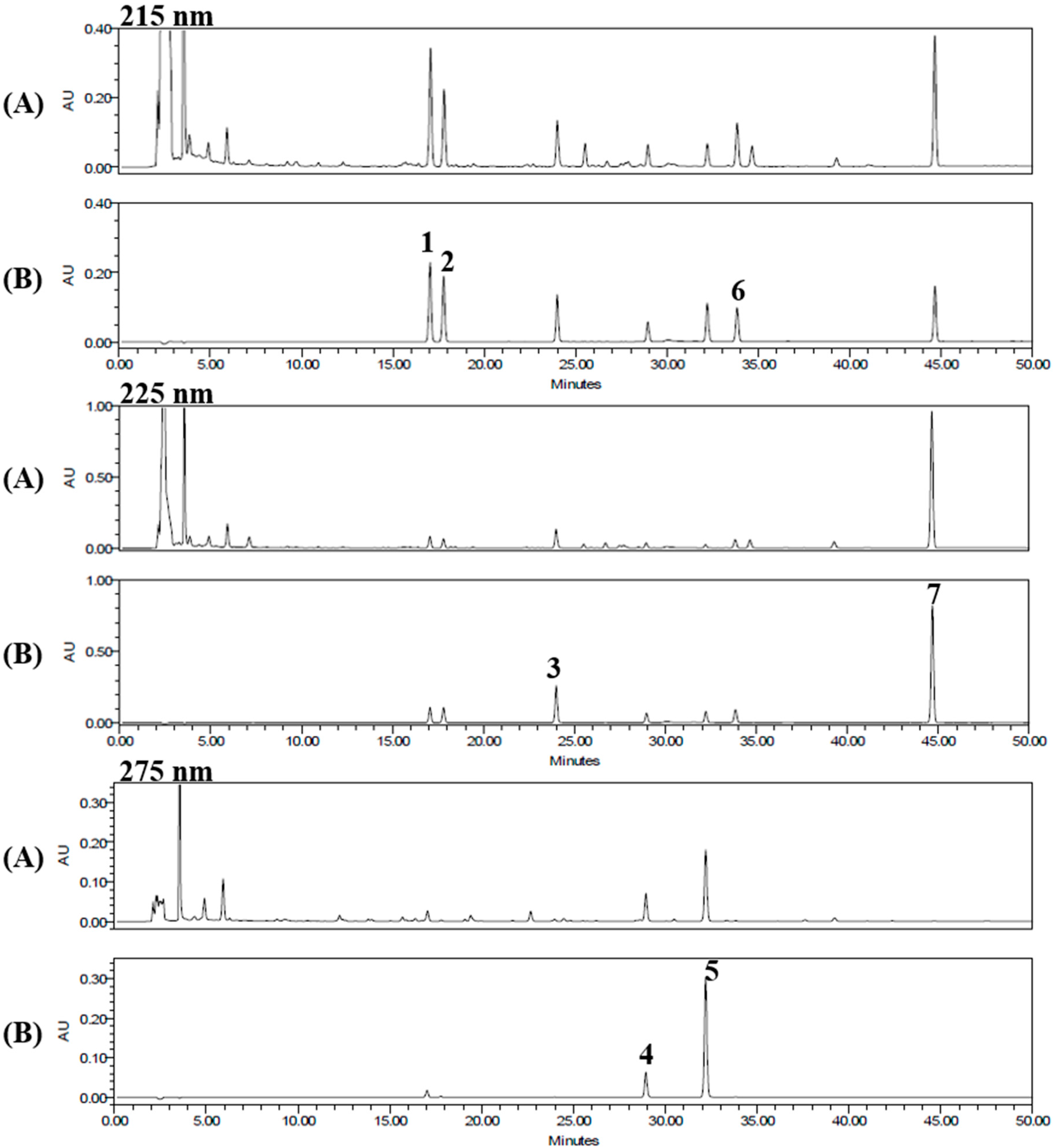
2.1. Optimization of High-Performance Liquid Chromatography (HPLC) Separation

We used HPLC for separation of the seven standard components from the 70% ethanol extract of the seeds of P. corylifolia. The established conditions of the mobile phase was shown in Table 1. Under these established HPLC methods, the seven standard components were resolved within 45 min. The retention times of the psoralen, angelicin, neobavaisoflavone, psoralidin, isobavachalcone, bavachinin, and bakuchiol were 17.05, 17.79, 23.99, 28.95, 32.21, 33.84, and 44.65 min, respectively. HPLC chromatograms of the 70% ethanol extract of the seeds of P. corylifolia and the standard mixture are shown in Figure 2.

2.2. Linearity, Limits of Detection (LOD), and Limits of Quantification (LOQ)

The linear relationships between the peak areas (y) and concentrations (x, μg/mL) of the components were expressed by the regression equations (y = ax + b) given in Table 2. The established analytical method showed high linearity with a correlation coefficient (r2) of ≥0.9999. The calibration curves showed good linearity over the concentration range 3.125–100 μg/mL, except for bakuchiol (12.5–400 μg/mL). The LODs and LOQs for the seven standard components were in the range 0.102–0.988 μg/mL and 0.309–2.995 μg/mL, respectively.

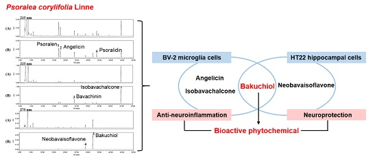
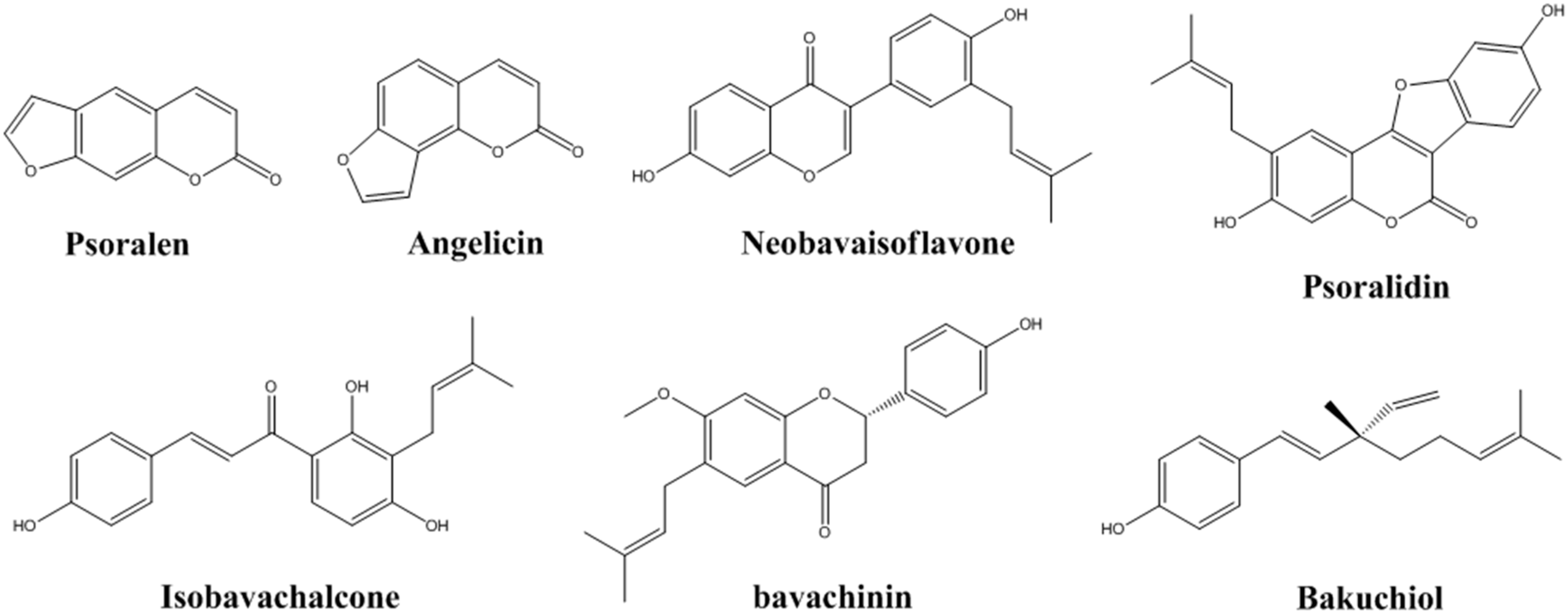
2.3. Determination of the Seven Standard Components in P. corylifolia

The established HPLC analytical method was applied to the simultaneous quantification of the seven components in the seed of P. corylifolia. The amounts of the seven standard components ranged from 0.74 mg/g to 11.71 mg/g. Among the components, bakuchiol was the most abundant compound in the seed of P. corylifolia. The results for the content of each component are shown in Table 3.

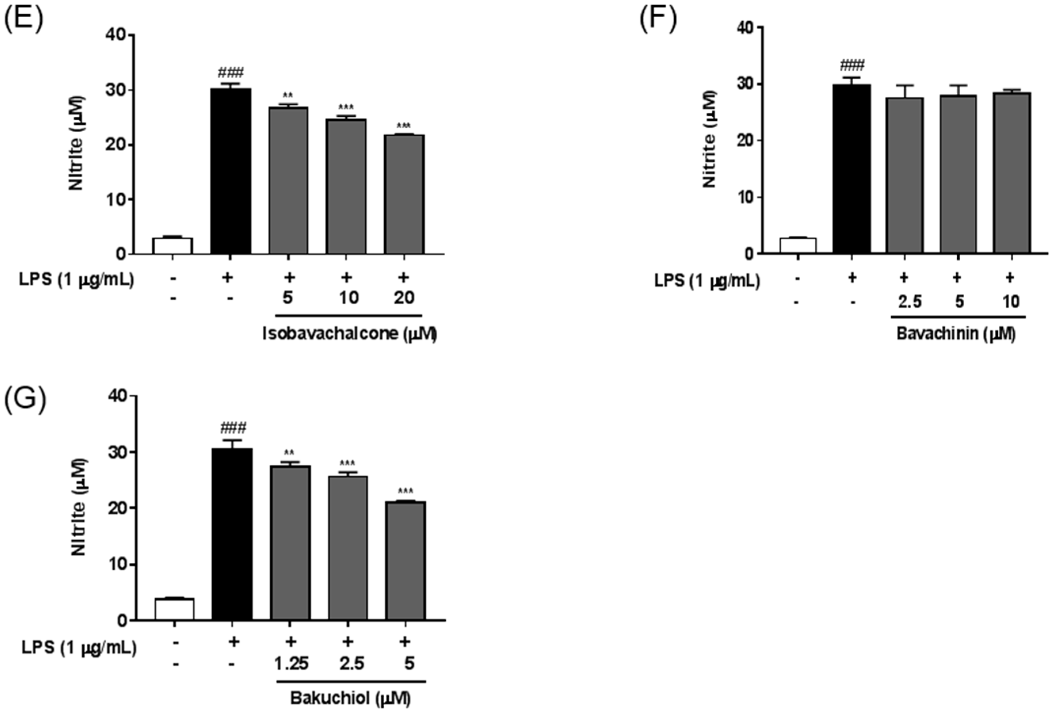
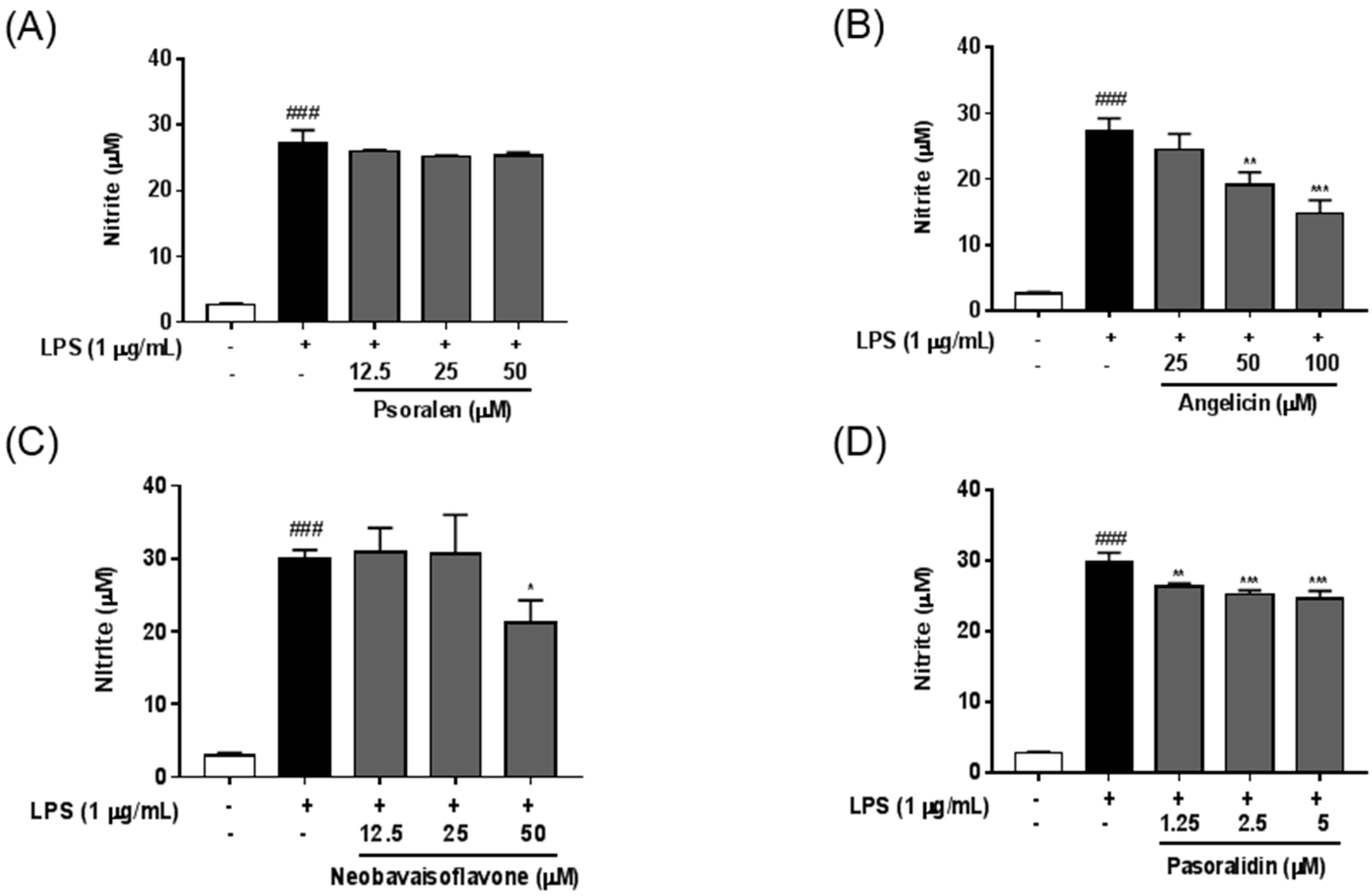
2.4. Anti-Neuroinflammatory Effects of Seven Standard Components in P. corylifolia

We evaluated the effect of the seven standard components of P. corylifolia on LPS-induced nitric oxide (NO) production in BV-2 microglia cells. As shown in Figure 3, LPS stimulation strongly increased NO production in BV-2 cells compared with untreated controls and, treatment with LPS (1 μg/mL) did not induce obvious decrease of cell viability, suggesting that LPS were non-toxic to microglia cell during this concentration range. Among these components, angelicin, isobavachalcone, and bakuchiol had the most significant inhibitory effect on the LPS-induced NO production in dose-dependent manners. Psoralidin also significantly suppressed LPS-induced NO production in BV-2 cells. Neobavaisoflavone had an inhibitory effect only at 50 μM. These effects were not caused by cytotoxicity because components at these concentrations did not show any significant reductions in cell viability.

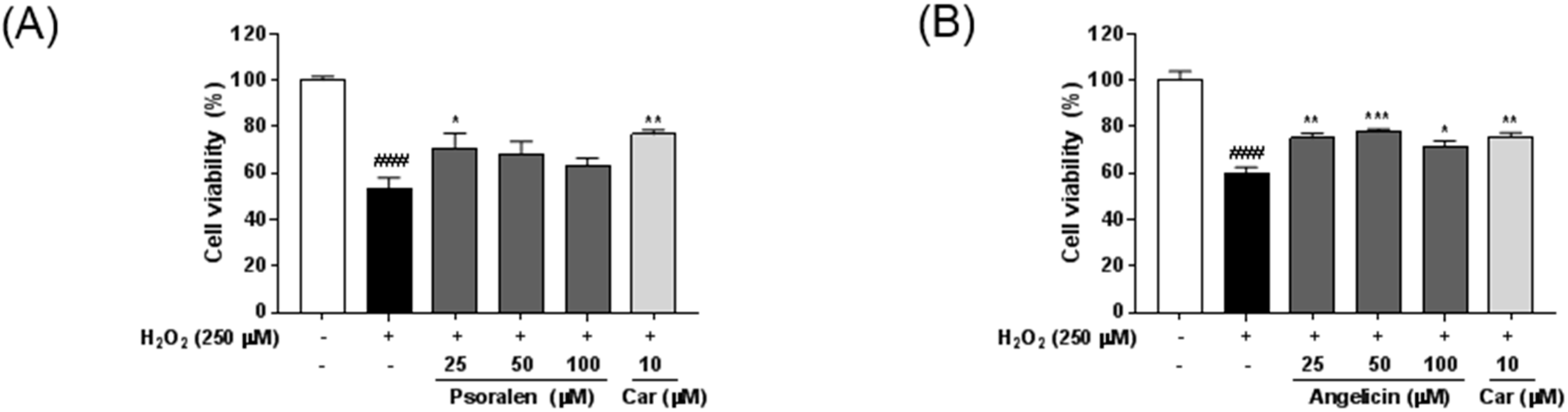
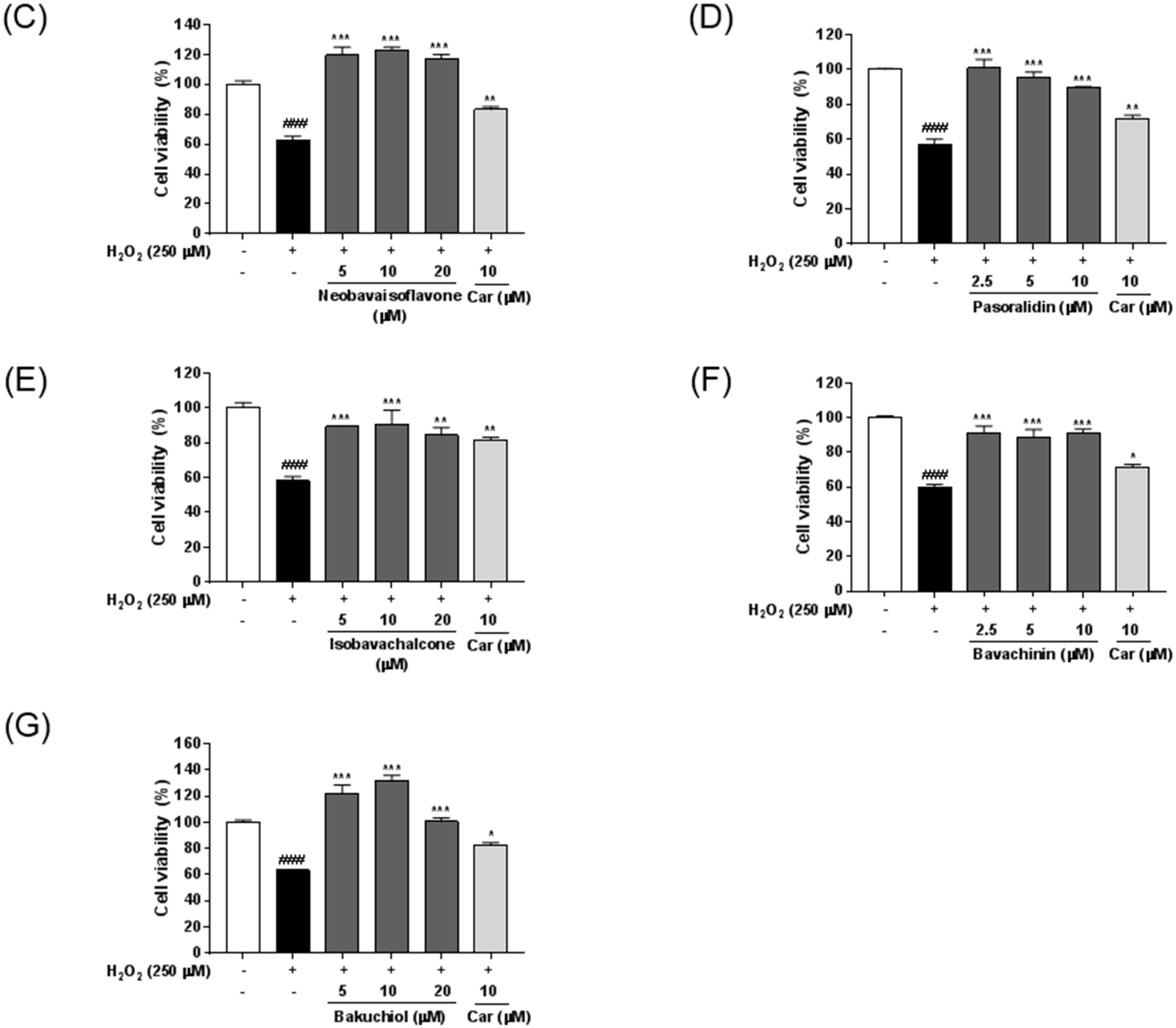
2.5. Neuroprotective Effects of Seven Standard Compounds in P. corylifolia

To investigate whether the seven standard components of P. corylifolia acted against neuronal cell damage, HT22 mouse hippocampal cells were treated with hydrogen peroxide (H2O2) in the presence or absence of various concentrations of each component. Cytotoxicity of seven standard components was determined using CCK assay (Supplementary Figure S1), and nontoxic concentrations of each component were used for the following experiments. As shown in Figure 4, H2O2 treatment significantly reduced the viability of HT22 cells compared with untreated controls. All the standard components exerted protective effects against H2O2-induced neuronal cell damage. Psoralen weakly reversed H2O2-induced neuronal cell death, but only at 25 μM. Neobavaisoflavone and bakuchiol had the most significant inhibitory effects against H2O2-induced damage. Carvedilol was used as a positive control [16].

3. Discussion

Neuroinflammation plays an important role in the pathogenesis of various neurodegenerative diseases such as Alzheimer’s disease and other dementias, Parkinson’s disease, and Huntington’s disease [17]. Microglia act as key mediators of neuroinflammation although they represent only about 10% of the total cell population in the central nervous system [18,19]. Activated microglia generate proinflammatory cytokines such as tumor necrosis factor-alpha, interleukin (IL)-1, and IL-6 and trigger the production of NO [20]. Excessive NO production stimulates the generation of reactive nitrogen species and mediates neuronal cell death [21,22]. Thus, the targeting NO production is thought to be a valuable clinical approach for the treatment of neurodegenerative diseases [23].
In neurodegenerative diseases, H2O2 is one of the most important mediators of oxidative stress detected under pathological conditions. H2O2 generation is required to mediate the complete sequence of events occurring in oxidative stress-induced neuronal cell death [24]. In previous studies, researchers employed H2O2 overload as a neurotoxic challenge paradigm to evaluate aspects of neuroprotection in murine hippocampal HT22 cell [25,26].
In the present study, we examined whether the seven components influenced the production of NO in LPS-stimulated BV-2 microglia. Among the components, angelicin, psoralidin, iso-bavachalcone, neobavaisoflavone and bakuchiol significantly decreased the LPS-induced NO production in BV-2 cells. Neobavaisoflavone inhibited the NO production only at higher concentration (50 μM) and psoralidin showed mild effects on the NO inhibition compared with the other components. Angelicin, isobavachalcone, and bakuchiol reduced the NO levels in dose-dependent manners in LPS-treated BV-2 cells. We also investigated the inhibitory effects of P. corylifolia and its seven components using HT22 hippocampal cells damaged by H2O2. All tested compounds revealed their potential as neuroprotective agents. However, psoralen and angelicin had weaker inhibitory activities than the others. Neobavaisoflavone and bakuchiol most significantly inhibited the H2O2-induced death of HT22 cells. In quantification of the seven components from P. corylifolia, bakuchiol was the most prevalent (11.71 mg/g) compared with the other six components. Overall, considering the quantification and bio-efficacy analyses, bakuchiol proved the most potent bioactive phytochemical of P. corylifolia for the potential treatment of neurodegenerative diseases.
Bakuchiol from P. corylifolia has a variety of biological activities such as inhibiting tumorigenesis [27,28], fungal activity [29], viral infections [30], bone loss [31], and hepatotoxicity [32], and has estrogenic-like effects [33]. Of note, Chaudhuri et al. reported that bakuchiol can act as an anti-aging compound via regulation of retinol-like gene expression [34]. Taken together, we consider that bakuchiol might be more useful for treating age-associated neurodegenerative diseases such as Alzheimer’s disease. Further studies will be necessary to understand the molecular mechanisms responsible for the regulation of neuronal cell responses using in vitro and in vivo experimental models of Alzheimer’s disease. In addition, the safety of bakuchiol should be elucidated by toxicology testing.

4. Materials and Methods

4.1. Plant Material

Seeds of Psoralea corylifolia were purchased from the Kwangmyungdang herbal market (Ulsan, Korea). A voucher specimen has been deposited at the Herbal Medicine Research Division, Korea Institute of Oriental Medicine.

4.2. Chemicals and Reagents

The standard components, psoralen, angelicin, neobavaisoflavone, psoralidin, isobavachalcone, bavachinin, and bakuchiol were purchased from Shanghai Sunny Biotech Co., Ltd. (Shanghai, China). The chemical structures of the standard components were shown in Figure 1. The purities of these standard components were ≥98.0% by high-performance liquid chromatography (HPLC) analysis. The HPLC-grade solvents, acetonitrile and water, were obtained from J. T. Baker Chemical Co. (Phillipsburg, NJ, USA).

4.3. Apparatus and Chromatographic Conditions

Quantitative analysis was conducted using a Waters Alliance e2695 system (Waters Corp., Milford, MA, USA) equipped with a pump, degasser, column oven, autosampler, and photodiode array detector (Waters Corp., #2998). The data were acquired and processed using Empower software (version 3; Waters Corp). Chromatographic separation for the seven standard components was carried out at room temperature using Luna C18 analytical columns (250 mm × 4.6 mm, 5 μm) supplied by Phenomenex (Torrance, CA, USA) with a gradient solvent system of acetonitrile and water. The ultraviolet (UV) wavelengths for detecting components were 215 nm for psoralen, angelicin and bavachinin; 225 nm for neobavaisoflavone and bakuchiol; and 275 nm for psoralidin and isobavachalcone. The flow rate was 1.0 mL/min and the injection volume was 10 μL.

4.4. Preparation of Standard Solutions

The seven components were weighed accurately, dissolved in methanol at 1.0 mg/mL and stored at below 4 °C. The stock solutions were diluted to yield a series of standard solutions with different concentrations for quantitative analysis.

4.5. Preparation of Sample Solutions

Dried seeds of P. corylifolia (50 g) were extracted twice with 70% ethanol (300 mL) by refluxing for 2 h. The extracted solution was filtered through a filter paper (5 μm), and evaporated using a rotary evaporator under a vacuum to dryness (8.279 g). The 70% ethanol extract of the seeds of P. corylifolia was weighed accurately and dissolved in methanol at 20 mg/mL. The sample solution was filtered through a syringe filter (0.45 μm) for HPLC analysis.

4.6. Calibration Curve and Determination of the Limit of Detection (LOD) and Limit of Quantification (LOQ)

The calibration curves of components were obtained by assessment of the peak areas of the standard solutions at six different concentrations. The tested concentration ranges were 3.125–100 μg/mL for psoralen, angelicin, neobavaisoflavone, psoralidin, isobavachalcone, and bavachinin, and 12.5–400 μg/mL for bakuchiol. The LOD and LOQ for the seven standard components were calculated using the slope of the calibration curve and the standard deviation (SD) of the intercept as follows: LOD = 3.3 × (SD of the response/slope of the calibration curve); and LOQ = 10 × (SD of the response/slope of the calibration curve).

4.7. Cell Lines and Culture

Mouse microglia BV-2 is a cell lines generated by infecting with a v-raf/v-myc oncogene carrying retrovirus in primary microglial cell [35]. Mouse hippocampal HT22 is a cell line immortalized a subclone of the original clone HT4 [36]. The murine microglial cell line BV2 and hippocampal HT22 cells were purchased from the American Type Culture Collection (Manassas, VA, USA) and cultured in Dulbecco’s Modified Eagle’s medium (Hyclone/Thermo Fisher Scientific, Rockford, IL, USA), supplemented with 10% fetal bovine serum (Hyclone/Thermo Fisher) and penicillin/streptomycin under 5% CO2 in air at 37 °C.

4.8. Nitric Oxide (NO) Assay

NO synthesis was analyzed by determining the accumulation of nitrite (NO2) in culture supernatants using the Griess Reagent System (Promega, Madison, WI, USA). BV-2 cells were pretreated with seven standard components for 2 h and treated with lipopolysaccharide (LPS; 1 μg/mL, Sigma-Aldrich, St. Louis, MO, USA) for an additional 22 h. After collecting the culture supernatants, equal volumes of supernatant and sulfanilamide solution were mixed, incubated for 10 min at room temperature, and then added to naphthylethylenediamine dihydrochloride solution for an additional 5 min. The absorbance was measured at 540 nm by using an Epoch microplate spectrophotometer. The nitrite concentration was determined from a standard curve (100, 50, 25, 12.5, 6.25, 3.13, 1.56 μM) generated using sodium nitrite (NaNO2) solutions. Determine average absorbance value of each experimental sample. Determine its concentration by comparison to the standard curve.

4.9. Measurement of Neuroprotective Activity

HT22 cells were plated on 96-well microplates at a density of 5 × 10³/well and co-treated with hydrogen peroxide (H2O2, 250 μM, Sigma-Aldrich) and various concentrations of each marker compound for 6 h. Cell counting Kit-8 (CCK-8) solution (Dojindo, Kumamoto, Japan) was added, and the cells were incubated for 4 h. The absorbance was read at 450 nm on an Epoch Microplate Spectrophotometer (BioTek Instruments, Inc., Winooski, VT, USA). The cell viability was calculated using the following equation:
Cell viability ( % ) = Mean OD in drug treated cells Mean OD in untreated cells × 100

4.10. Statistical Analysis

The data are expressed as the mean ± standard error of the mean (SEM). Data were analyzed using one-way analysis of variance and Dunnett’s multiple comparisons test; p < 0.05 was considered statistically significant.

Supplementary Materials

Supplementary materials can be accessed at: https://www.mdpi.com/1420-3049/21/8/1076/s1.

Acknowledgments

This study was supported by a research grant (K16293) from the Korea Institute of Oriental Medicine (KIOM).

Author Contributions

Y.J.K., H.-S.L., and S.-J.J. participated in the design of the study data analyses and manuscript preparation. Y.J.K. and J.L. conducted P. corylifolia extract and analysis of seven constituents in P. corylifolia extract using HPLC. H.-S.L. and S.-J.J. conducted the biological examination of in vitro. All authors read and approved the final manuscript.

Conflicts of Interest

The authors declare no conflict of interest.

References

  1. Uikey, S.K.; Yadav, A.; Sharma, A.K.; Rai, A.K.; Raghuwanshi, D.; Badkhane, Y. The botany, chemistry, pharmacological and therapeutic application of Psoralea corylifolia L.: A review. Int. J. Phytomed. 2011, 2, 100–107. [Google Scholar] [CrossRef]
  2. Zhang, X.; Zhao, W.; Wang, Y.; Lu, J.; Chen, X. The Chemical Constituents and Bioactivities of Psoralea corylifolia Linn.: A Review. Am. J. Chin. Med. 2016, 44, 35–60. [Google Scholar] [CrossRef] [PubMed]
  3. Lee, H.; Li, H.; Noh, M.; Ryu, J.H. Bavachin from Psoralea corylifolia Improves Insulin-Dependent Glucose Uptake through Insulin Signaling and AMPK Activation in 3T3-L1 Adipocytes. Int. J. Mol. Sci. 2016, 17. [Google Scholar] [CrossRef] [PubMed]
  4. Seo, E.; Lee, E.K.; Lee, C.S.; Chun, K.H.; Lee, M.Y.; Jun, H.S. Psoralea corylifolia L. seed extract ameliorates streptozotocin-induced diabetes in mice by inhibition of oxidative stress. Oxid. Med. Cell. Longev. 2014. [Google Scholar] [CrossRef] [PubMed]
  5. Seo, E.; Oh, Y.S.; Jun, H.S. Psoralea corylifolia L. Seed Extract Attenuates Nonalcoholic Fatty Liver Disease in High-Fat Diet-Induced Obese Mice. Nutrients 2016, 8, 83. [Google Scholar] [CrossRef] [PubMed]
  6. Jung, B.; Jang, E.H.; Hong, D.; Cho, I.H.; Park, M.J.; Kim, J.H. Aqueous extract of Psoralea corylifolia L. inhibits lipopolysaccharide-induced endothelial-mesenchymal transition via downregulation of the NF-kappaB-SNAIL signaling pathway. Oncol. Rep. 2015, 34, 2040–2046. [Google Scholar] [PubMed]
  7. Rajan, V.; Tripathi, J.; Variyar, P.; Pandey, B.N. Mechanism of cytotoxicity by Psoralea corylifolia extract in human breast carcinoma cells. J. Environ. Pathol. Toxicol. Oncol. 2014, 33, 265–277. [Google Scholar] [CrossRef] [PubMed]
  8. Dang, Y.; Ling, S.; Duan, J.; Ma, J.; Ni, R.; Xu, J.W. Bavachalcone-induced manganese superoxide dismutase expression through the AMP-activated protein kinase pathway in human endothelial cells. Pharmacology 2015, 95, 105–110. [Google Scholar] [CrossRef] [PubMed]
  9. Lee, K.M.; Kim, J.M.; Baik, E.J.; Ryu, J.H.; Lee, S.H. Isobavachalcone attenuates lipopolysaccharide-induced ICAM-1 expression in brain endothelial cells through blockade of toll-like receptor 4 signaling pathways. Eur. J. Pharmacol. 2015, 754, 11–18. [Google Scholar] [CrossRef] [PubMed]
  10. Yang, H.J.; Youn, H.; Seong, K.M.; Yun, Y.J.; Kim, W.; Kim, Y.H.; Lee, J.Y.; Kim, C.S.; Jin, Y.W.; Youn, B. Psoralidin, a dual inhibitor of COX-2 and 5-LOX, regulates ionizing radiation (IR)-induced pulmonary inflammation. Biochem. Pharmacol. 2011, 82, 524–534. [Google Scholar] [CrossRef] [PubMed]
  11. Liu, X.; Nam, J.W.; Song, Y.S.; Viswanath, A.N.; Pae, A.N.; Kil, Y.S.; Kim, H.D.; Park, J.H.; Seo, E.K.; Chang, M. Psoralidin, a coumestan analogue, as a novel potent estrogen receptor signaling molecule isolated from Psoralea corylifolia. Bioorg. Med. Chem. Lett. 2014, 24, 1403–1406. [Google Scholar] [CrossRef] [PubMed]
  12. Park, J.; do Kim, H.; Ahn, H.N.; Song, Y.S.; Lee, Y.J.; Ryu, J.H. Activation of Estrogen Receptor by Bavachin from Psoralea corylifolia. Biomol. Ther. 2012, 20, 183–188. [Google Scholar] [CrossRef] [PubMed]
  13. Chen, Y.; Wang, H.D.; Xia, X.; Kung, H.F.; Pan, Y.; Kong, L.D. Behavioral and biochemical studies of total furocoumarins from seeds of Psoralea corylifolia in the chronic mild stress model of depression in mice. Phytomedicine 2007, 14, 523–529. [Google Scholar] [CrossRef] [PubMed]
  14. Yi, L.T.; Li, Y.C.; Pan, Y.; Li, J.M.; Xu, Q.; Mo, S.F.; Qiao, C.F.; Jiang, F.X.; Xu, H.; Lu, X.B.; et al. Antidepressant-like effects of psoralidin isolated from the seeds of Psoralea corylifolia in the forced swimming test in mice. Prog Neuropsychopharmacol. Biol. Psychiatry 2008, 32, 510–519. [Google Scholar] [CrossRef] [PubMed]
  15. Im, A.R.; Chae, S.W.; Zhang, G.J.; Lee, M. Neuroprotective effects of Psoralea corylifolia Linn seed extracts on mitochondrial dysfunction induced by 3-nitropropionic acid. BMC Complement. Altern. Med. 2014, 14. [Google Scholar] [CrossRef] [PubMed]
  16. Ouyang, Y.; Chen, Z.; Tan, M.; Liu, A.; Chen, M.; Liu, J.; Pi, R.; Fang, J. Carvedilol, a third-generation beta-blocker prevents oxidative stress-induced neuronal death and activates Nrf2/ARE pathway in HT22 cells. Biochem. Biophys. Res. Commun. 2013, 441, 917–922. [Google Scholar] [CrossRef] [PubMed]
  17. Kielian, T. Neuroinflammation: Good, bad, or indifferent? J. Neurochem. 2014, 130, 1–3. [Google Scholar] [CrossRef] [PubMed]
  18. Kraft, A.D.; Harry, G.J. Features of microglia and neuroinflammation relevant to environmental exposure and neurotoxicity. Int. J. Environ. Res. Public Health 2011, 8, 2980–3018. [Google Scholar] [CrossRef] [PubMed]
  19. Streit, W.J.; Mrak, R.E.; Griffin, W.S. Microglia and neuroinflammation: A pathological perspective. J. Neuroinflamm. 2004, 1, 14. [Google Scholar] [CrossRef] [PubMed]
  20. Goodwin, J.L.; Uemura, E.; Cunnick, J.E. Microglial release of nitric oxide by the synergistic action of beta-amyloid and IFN-gamma. Brain Res. 1995, 692, 207–214. [Google Scholar] [CrossRef]
  21. Vincent, V.A.; Tilders, F.J.; van Dam, A.M. Production, regulation and role of nitric oxide in glial cells. Mediators Inflamm. 1998, 7, 239–255. [Google Scholar] [CrossRef] [PubMed]
  22. Stefano, G.B.; Kim, E.; Liu, Y.; Zhu, W.; Casares, F.; Mantione, K.; Jones, D.A.; Cadet, P. Nitric oxide modulates microglial activation. Med. Sci. Monit. 2004, 10, 11594. [Google Scholar]
  23. Yuste, J.E.; Tarragon, E.; Campuzano, C.M.; Ros-Bernal, F. Implications of glial nitric oxide in neurodegenerative diseases. Front. Cell. Neurosci. 2015, 9, 322. [Google Scholar] [CrossRef] [PubMed]
  24. Zhao, Z.Y.; Luan, P.; Huang, S.X.; Xiao, S.H.; Zhao, J.; Zhang, B.; Gu, B.B.; Pi, R.B.; Liu, J. Edaravone protects HT22 neurons from H2O2-induced apoptosis by inhibiting the MAPK signaling pathway. CNS Neurosci. Ther. 2013, 19, 163–169. [Google Scholar] [CrossRef] [PubMed]
  25. Vedder, H.; Teepker, M.; Fischer, S.; Krieg, J.C. Characterization of the neuroprotective effects of estrogens on hydrogen peroxide-induced cell death in hippocampal HT22 cells: Time and dose-dependency. Exp. Clin. Endocrinol. Diabetes 2008, 108, 120–127. [Google Scholar] [CrossRef] [PubMed]
  26. Teepker, M.; Anthes, N.; Krieg, J.C.; Vedder, H. 2-OH-estradiol, an endogenous hormone with neuroprotective functions. J. Psychiatr. Res. 2003, 37, 517–523. [Google Scholar] [CrossRef]
  27. Kim, J.E.; Kim, J.H.; Lee, Y.; Yang, H.; Heo, Y.S.; Bode, A.M.; Lee, K.W.; Dong, Z. Bakuchiol suppresses proliferation of skin cancer cells by directly targeting Hck, Blk, and p38 MAP kinase. Oncotarget 2016, 7, 14616–14627. [Google Scholar] [PubMed]
  28. Li, L.; Chen, X.; Liu, C.C.; Lee, L.S.; Man, C.; Cheng, S.H. Phytoestrogen Bakuchiol Exhibits in Vitro and in Vivo Anti-breast Cancer Effects by Inducing S Phase Arrest and Apoptosis. Front. Pharmacol. 2016, 7, 128. [Google Scholar] [CrossRef] [PubMed]
  29. Nordin, M.A.; Abdul Razak, F.; Himratul-Aznita, W.H. Assessment of Antifungal Activity of Bakuchiol on Oral-Associated Candida spp. Evid. Based Complement. Altern. Med. 2015, 2015. [Google Scholar] [CrossRef] [PubMed]
  30. Shoji, M.; Arakaki, Y.; Esumi, T.; Kohnomi, S.; Yamamoto, C.; Suzuki, Y.; Takahashi, E.; Konishi, S.; Kido, H.; Kuzuhara, T. Bakuchiol Is a Phenolic Isoprenoid with Novel Enantiomer-selective Anti-influenza A Virus Activity Involving Nrf2 Activation. J. Biol. Chem. 2015, 290, 28001–28017. [Google Scholar] [CrossRef] [PubMed]
  31. Lim, S.H.; Ha, T.Y.; Kim, S.R.; Ahn, J.; Park, H.J.; Kim, S. Ethanol extract of Psoralea corylifolia L. and its main constituent, bakuchiol, reduce bone loss in ovariectomised Sprague-Dawley rats. Br. J. Nutr. 2009, 101, 1031–1039. [Google Scholar] [CrossRef] [PubMed]
  32. Park, E.J.; Zhao, Y.Z.; Kim, Y.C.; Sohn, D.H. Protective effect of (S)-bakuchiol from Psoralea corylifolia on rat liver injury in vitro and in vivo. Planta Med. 2005, 71, 508–513. [Google Scholar] [CrossRef] [PubMed]
  33. Mao, H.; Wang, H.; Ma, S.; Xu, Y.; Zhang, H.; Wang, Y.; Niu, Z.; Fan, G.; Zhu, Y.; Gao, X.M. Bidirectional regulation of bakuchiol, an estrogenic-like compound, on catecholamine secretion. Toxicol. Appl. Pharmacol. 2014, 274, 180–189. [Google Scholar] [CrossRef] [PubMed]
  34. Chaudhuri, R.K.; Bojanowski, K. Bakuchiol: A retinol-like functional compound revealed by gene expression profiling and clinically proven to have anti-aging effects. Int. J. Cosmet. Sci. 2014, 36, 221–230. [Google Scholar] [CrossRef] [PubMed]
  35. Blasi, E.; Barluzzi, R.; Bocchini, V.; Mazzolla, R.; Bistoni, F. Immortalization of microflial cells by a v-raf/v-myc carrying retrovirus. J. Neuroimmunol. 1990, 27, 229–237. [Google Scholar] [CrossRef]
  36. Morimoto, B.H.; Koshland, D.E., Jr. Induction and expressio of long- and short-term neurosecretory potentiation in a neural cell line. Neuron 1990, 5, 875–880. [Google Scholar] [CrossRef]
  • Sample Availability: Not available.
Figure 1. Chemical structures of the seven marker compounds of P. corylifolia.
Molecules 21 01076 g001
Figure 2. HPLC chromatograms of the 70% ethanol extract of P. corylifolia seeds (A); and its standard mixture (B) at 215 nm, 225 nm, and 275 nm. Psoralen (1), angelicin (2), neobavaisoflavone (3), psoralidin (4), isobavachalcone (5), bavachinin (6), and bakuchiol (7).
Molecules 21 01076 g002
Figure 3. Anti-neuroinflammatory effects of the seven components from P. corylifolia in LPS-stimulated BV-2 cells. Cells were pretreated with various concentrations of each components for 2 h and then stimulated with LPS (1 μg/mL) for an additional 22 h. Results are shown for psoralen (A); angelicin (B); neobavaisoflavone (C); psoralidin (D); isobavachalcone (E); bavachinin (F); and bakuchiol (G). The production of NO was determined using Griess reagents. The results are expressed as the mean ± SEM of three independent experiments. ### p < 0.01 versus vehicle control cells; * p < 0.05, ** p < 0.01, and *** p < 0.001 versus LPS-treated cells.
Molecules 21 01076 g003aMolecules 21 01076 g003b
Figure 4. Neuroprotective effects of the seven components from P. corylifolia in H2O2-treated HT22 cells. Cells were cotreated with various concentrations of each components and H2O2 (250 μM) for 6 h. Psoralen (A); angelicin (B); neobavaisoflavone (C); psoralidin (D); isobavachalcone (E); bavachinin (F); and bakuchiol (G). Cell viability was assessed using CCK-8 assays. Carvedilol (Car) was used as a positive control. The results are expressed as mean ± SEM of three independent experiments. ### p < 0.01 versus vehicle control cells; * p < 0.05, ** p < 0.01, and *** p < 0.001 versus H2O2-treated cells.
Molecules 21 01076 g004aMolecules 21 01076 g004b
Table 1. Condition of mobile phase for HPLC analysis.
Time (min)Flow Rate (mL/min)Mobile Phase
Water (%)Acetonitrile (%)
01.07525
401.02080
461.00100
521.00100
Table 2. Linear range, regression equation, correlation coefficients, LODs, and LOQs for compounds.
CompoundLinear Range (μg/mL)Regression Equation (y = ax + b) aCorrelation Coefficient (r2)LOD b (μg/mL)LOQ c (μg/mL)
Slope (a)Intercept (b)
Psoralen3.125–10081466736970.99990.1020.309
Angelicin3.125–10068433619420.99990.1030.313
Neobavaisoflavone3.125–10046488255751.00000.2390.725
Psoralidin3.125–100242131833.91.00000.1340.407
Isobavachalcone3.125–100113946605941.00000.1750.529
Bavachinin3.125–10039030335500.99990.1900.576
Bakuchiol12.5–40038481−298881.00000.9882.995
a y = ax + b, y means peak area and x means concentration (μg/mL); b LOD (Limit of detection): 3.3 × (SD of the response/slope of the calibration curve); c LOQ (Limit of quantitation): 10 × (SD of the response/slope of the calibration curve).
Table 3. The content of maker compounds in the seed of Psoralea corylifolia L.
CompoundContent (mg/g)
Psoralen1.902 ± 0.003
Angelicin1.506 ± 0.003
Neobavaisoflavone1.321 ± 0.011
Psoralidin1.310 ± 0.010
Isobavachalcone0.736 ± 0.006
Bavachinin1.623 ± 0.011
Bakuchiol11.713 ± 0.088

