
Transdermal Permeation and Anti-Inflammation Activities of Novel Sinomenine Derivatives

Abstract
:1. Introduction
2. Results
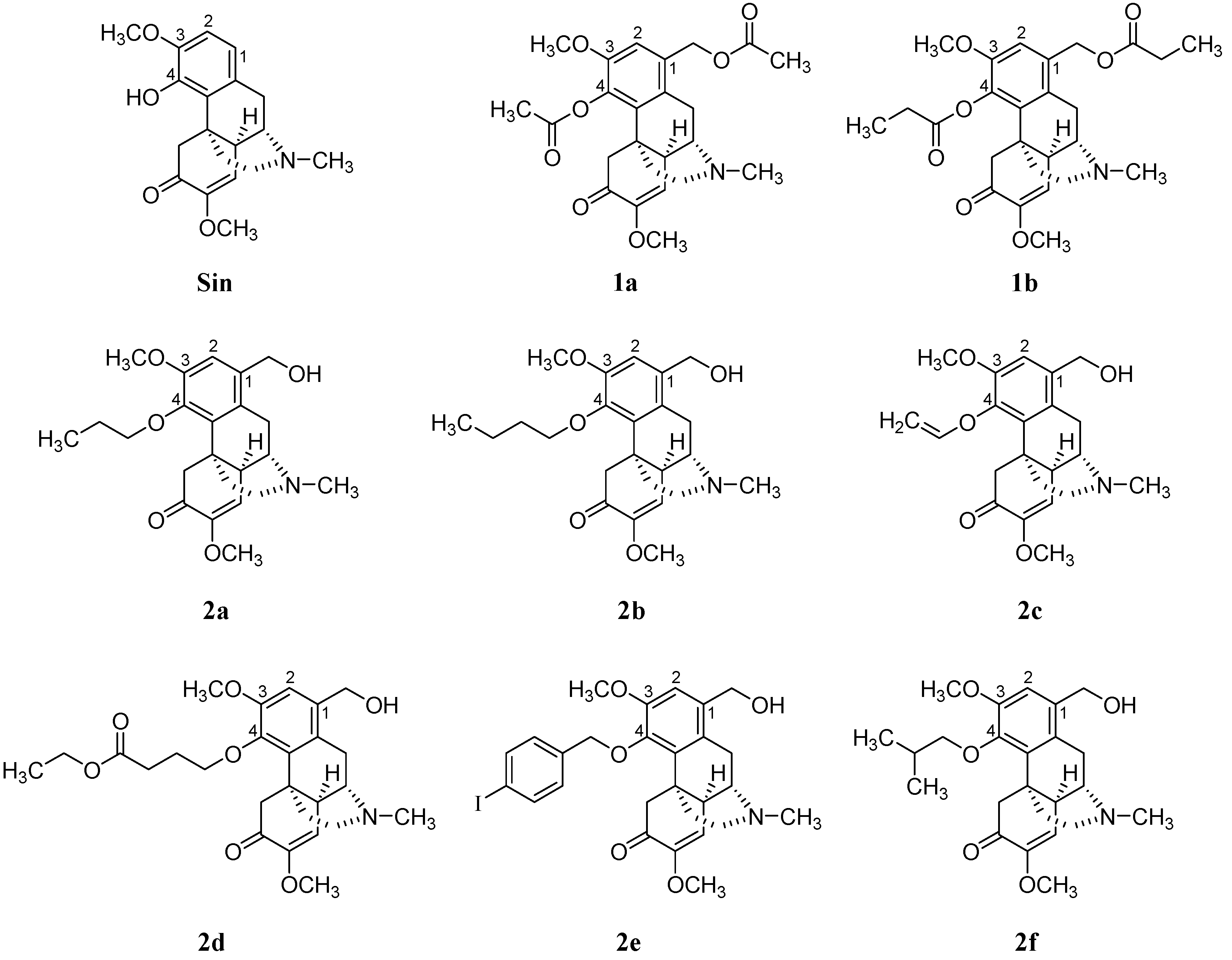
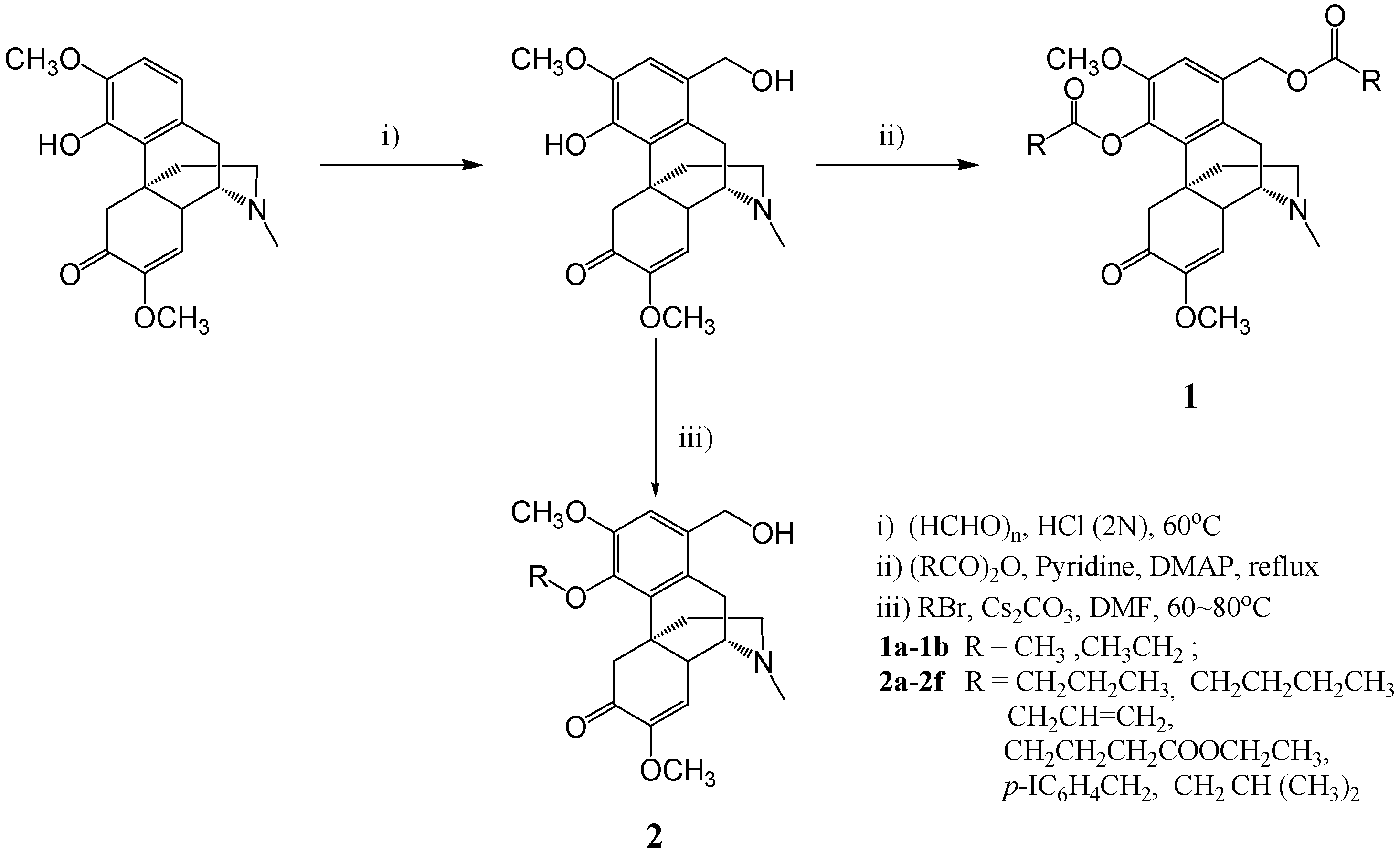
2.1. Compound Synthesis
2.2. Transdermal Permeation In Vitro
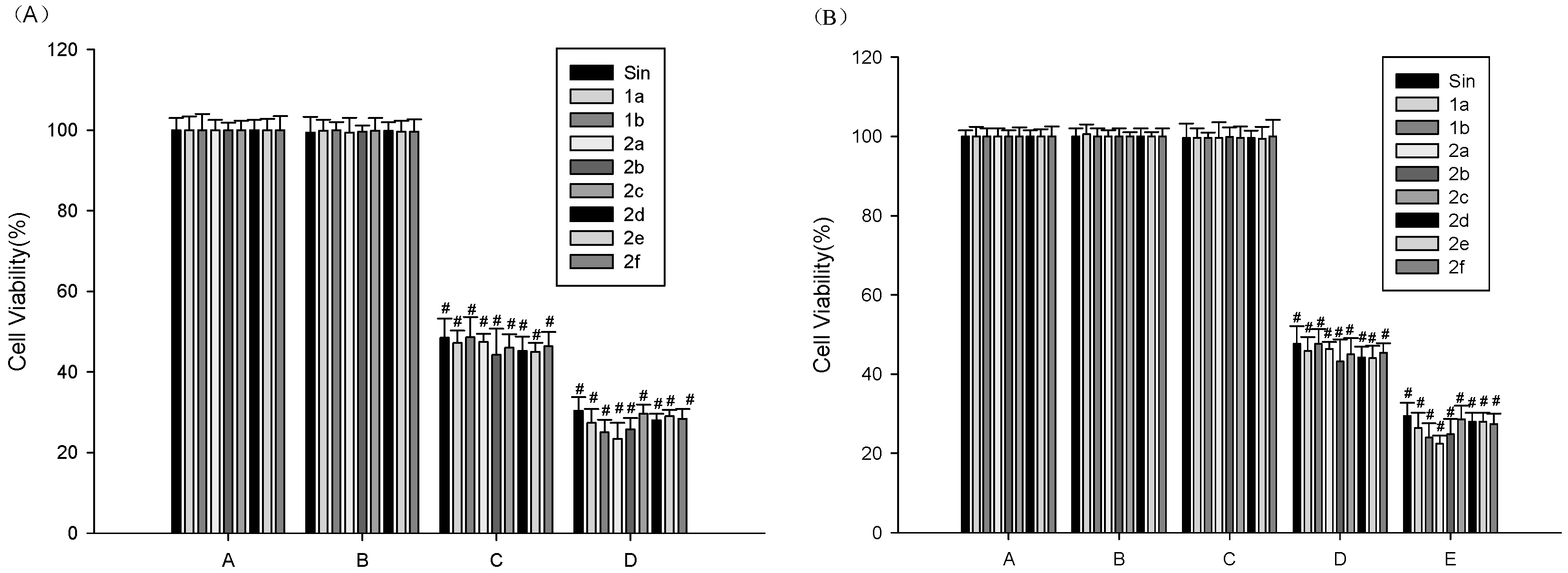
2.3. MTT Assay for Cell Viability
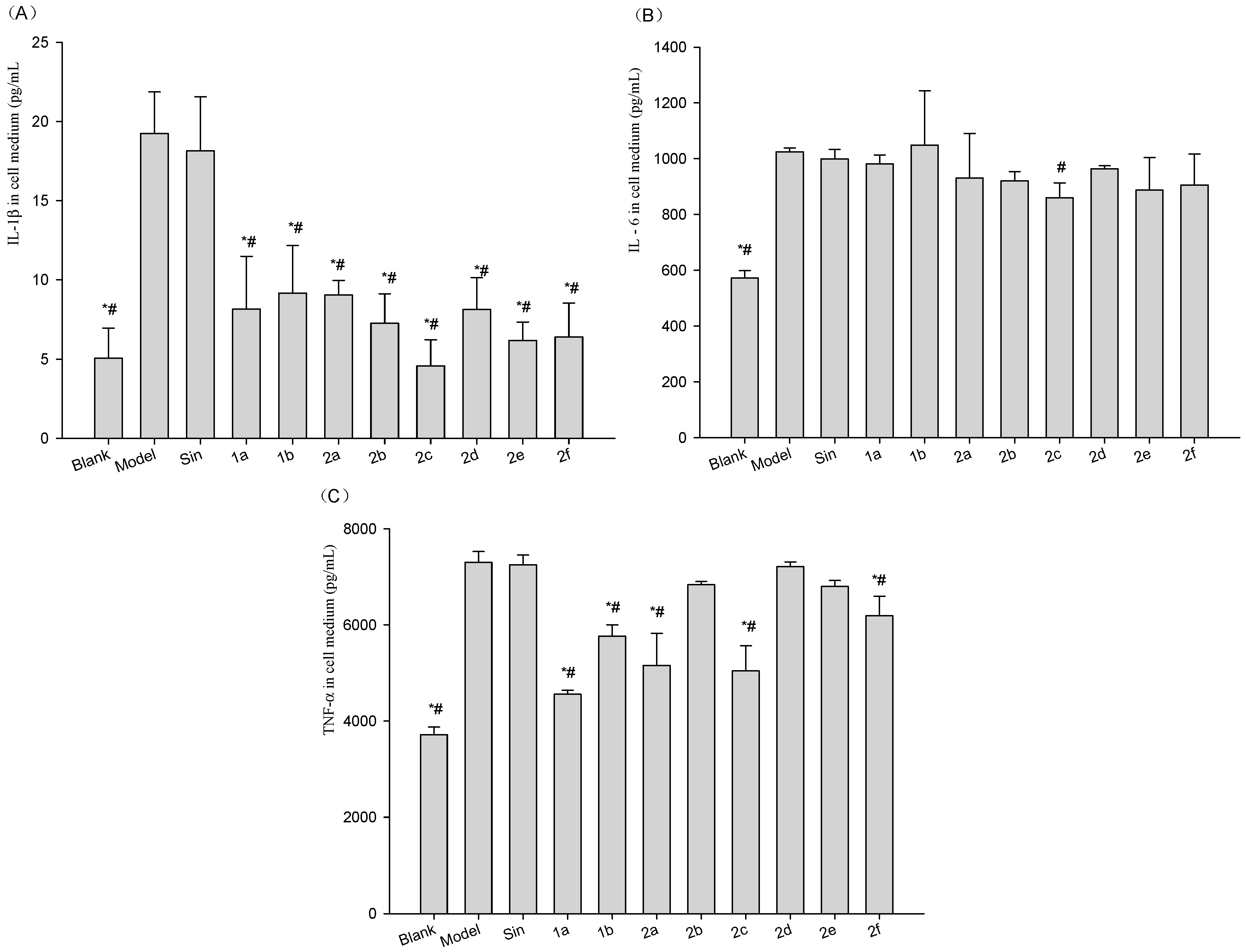
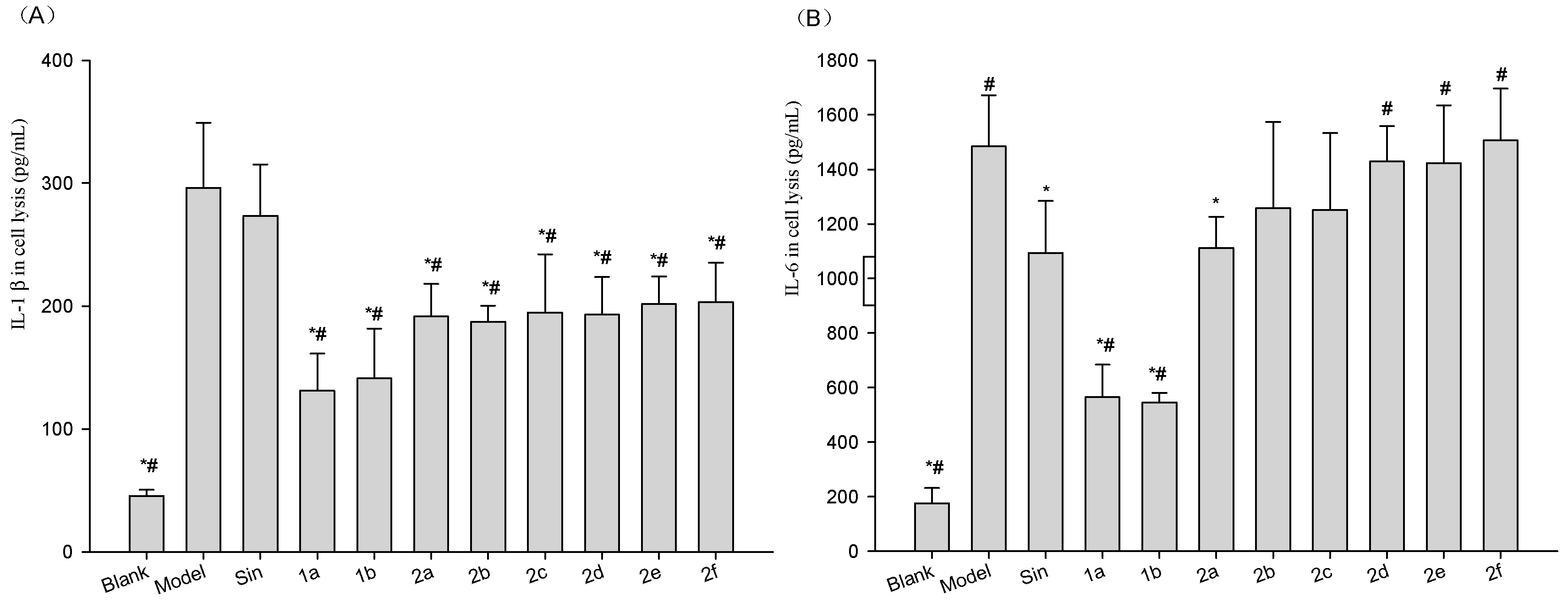
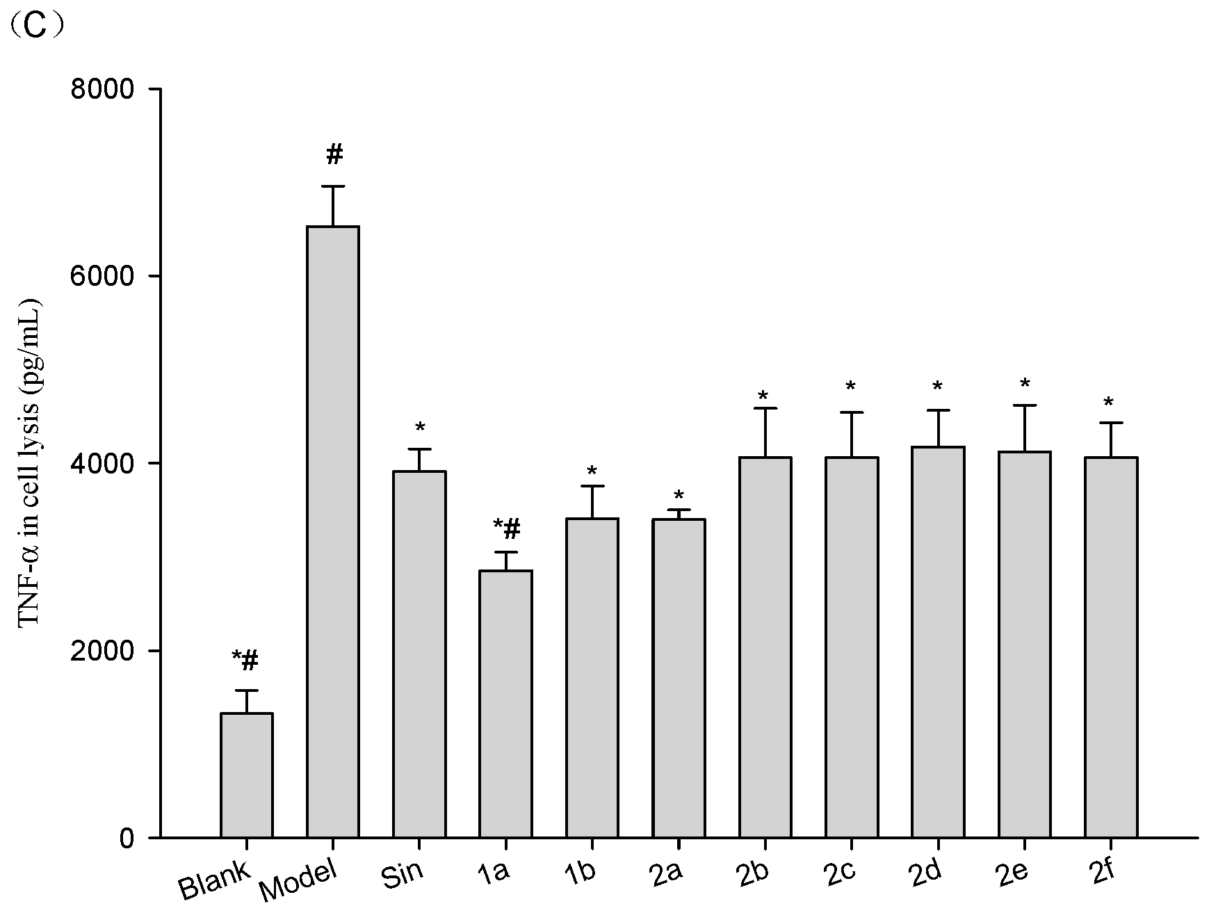
2.4. Effect of Compounds on the Levels of IL1β, IL6 and TNF-α in Cell Medium and Lysis of LPS-Stimulated Raw264.7 Cells
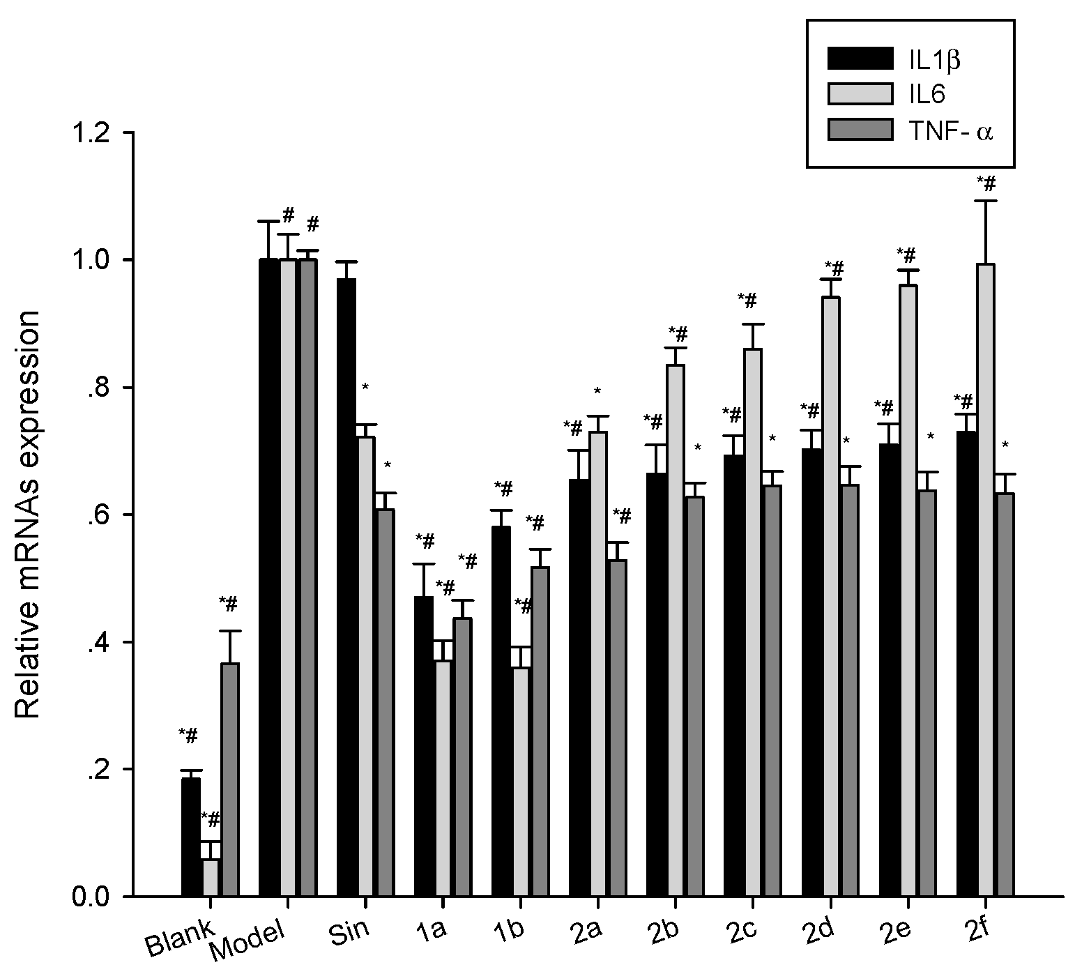
2.5. Effect of Compounds on the Expression of mRNA for IL1β, IL6 and TNF-α in LPS-Stimulated Mouse Raw264.7 Cells
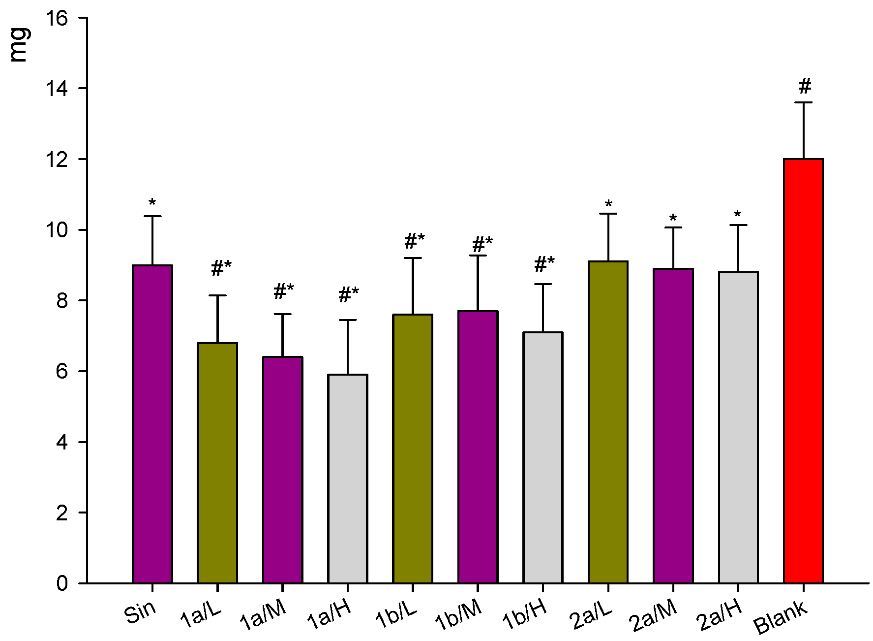
2.6. Inhibition of Dimethylbenzen-Induced Mouse Ear Edema by Sin Derivatives
2.7. Inhibition of Carrageenan-Induced Rat Paw Edema by Sin Derivatives
2.8. Inhibition of Acetic Acid-Induced Increase in Vascular Permeability
3. Discussion
4. Materials and Methods
4.1. Materials
4.1.1. Animals
4.1.2. Drugs and Reagents
4.2. Methods
4.2.1. Compound Synthesis
Synthesis of Sinomenine Benzyl Alcohol Intermediate
Target Compound 1a and 1b Synthesis
Target Compound 2a–2f Synthesis
4.2.2. Transdermal Permeation In Vitro
4.2.3. MTT Assay for the Measuring of Cell Viability
4.2.4. In Vitro Inhibition of the Inflammatory Factors, IL1β, IL6 and TNF-α
4.2.5. Dimethylbenzene-Induced Mouse Ear Edema
4.2.6. Carrageenan-Induced Rat Paw Edema
4.2.7. Induction of Increased Capillary Permeability with Acetic acid
4.3. Statistical Analysis
5. Conclusions
Supplementary Materials
Acknowledgments
Author Contributions
Conflicts of Interest
References
- Chung, I.M.; Ketharnathan, S.; Thiruvengadam, M.; Rajakumar, G. Rheumatoid arthritis: The stride from researchto clinical practice. Int. J. Mol. Sci. 2016, 17, 900. [Google Scholar] [CrossRef] [PubMed]
- Wong, C.K.; Chen, D.P.; Tam, L.S.; Li, E.K.; Yin, Y.B.; Lam, C.W. Effects of inflammatory cytokine IL27 on the activation of fibroblast-like synoviocytes in rheumatoid arthritis. Arthritis. Res. Ther. 2010, 12, R129. [Google Scholar] [CrossRef] [PubMed]
- Wong, R.; Davis, A.M.; Badley, E.; Grewal, R.; Mohammed, M. Prevalence of Arthritis and Rheumatic Disease around the World: A Growing Burden and Implications for Health Careneeds; ACREU and University Health Network: Toronto, TO, Canada, 2010. [Google Scholar]
- Chopra, A.; Abdel-Nasser, A. Epidemiology of rheumatic musculoskeletal disorders in the developing world. Best. Pract. Res. Clin. Rheumatol. 2008, 22, 583–604. [Google Scholar] [CrossRef] [PubMed]
- Zhao, X.X.; Peng, C.; Zhang, H.; Qin, L.P. Sinomenium acutum: A review of chemistry, pharmacology, pharmacokinetics, and clinical use. Pharm. Biol. 2012, 50, 1053–1061. [Google Scholar] [CrossRef] [PubMed]
- Xu, M.; Liu, L.; Qi, C.; Deng, B.; Cai, X. Sinomenine versus NSAIDs for the treatment of rheumatoid arthritis: A systematic review and meta-analysis. Planta Med. 2008, 74, 1423–1429. [Google Scholar] [CrossRef] [PubMed]
- Wang, J.P.; Ruan, J.L.; Zhang, C.G.; Ye, Y.J.; Cai, Y.L.; Wu, Y.X. Development and evaluation of the Sinomenine transdermal patch. Pak. J. Pharm. Sci. 2008, 4, 407–410. [Google Scholar]
- Wu, J.; Li, Y.H. The side effects and prevention of drug treatment by Zhengqingfengtongning. Chin. J. Med. Guide 2012, 14, 250–251. [Google Scholar]
- Ye, M.R.; Liu, L.; Zeng, Y.E.; Zhang, L.Q.; Tan, Y.H.; Deng, S.J.; Huang, G.Y. Studies on the relationship between Sinomenine distribution and its organic toxic ology. Chin. Pharmacol. Bull. 2001, 17, 65–69. [Google Scholar]
- Fu, S.X.; Chang, S.S.; Li, Y.S.; Wang, N.C. The toxicity and general pharmacological actions of sinomenine. Acta Pharm. Sin. 1963, 10, 673–676. [Google Scholar]
- Zhang, B.H.; Zhang, X.Z.; Li, J.; Cai, H.Y.; Xue, C.B. Studies on effect of azone on percutaneous absorption of Sinomenine gel in vitro. Chin. Hosp. Pharm. J. 2002, 22, 707–709. [Google Scholar]
- Li, F.; Hao, B.H.; Li, W.Z.; Liu, S.; Tang, B.B.; Du, S.J. Pharmacokinetics of transdermal absorption of Sinomenine in patch under electroporation condition. Acad. J. Second Mil. Med. Univ. 2008, 29, 1096–1098. [Google Scholar] [CrossRef]
- Jin, J.; Teng, P.; Liu, H.L.; Wu, J.; Liu, Y.M.; Xu, Q. Microfluidics assisted synthesis and bioevaluation of sinomenine derivatives as antiinflammatory agents. Eur. J. Med. Chem. 2013, 62, 280–288. [Google Scholar] [CrossRef] [PubMed]
- Wu, F.C.; Feng, X.Z.; Wu, K.M.; Chen, G.F.; Huang, Y.M.; Ye, X.R. Structure modified compound of sinomenine, and prepartion method. Chem. Abstr. 2006, 146, 100910. [Google Scholar]
- Lou, Y.T.; Zhou, H.B.; Zou, J.; Yan, L.C.; Bi, E.G.; Sun, B.; Yao, Z.J. Modification of poorly bioactive Sinomenine into more poten immuneosuppressive agents by embedding of drug-like fragments. Tetrahedron Lett. 2010, 51, 485–488. [Google Scholar] [CrossRef]
- Wang, M.; Ma, L.Y.; Lou, Y.T.; Bian, C.; Zhou, T.T.; Zhou, H.B.; Liao, H.Z.; Ma, Z.; Yin, D.S.; Chen, A.Z.; et al. Sinomenine derivatives with embedment of nitrogen-containing heterocycles exhibiting potent TNF-alpha inhibitory activity. Sci. China Chem. 2012, 55, 2537–2547. [Google Scholar] [CrossRef]
- Roy, S.D.; Fujiki, J.; Fleitman, J.S. Permeabilities of alkylp-aminobenzoates through living skin equivalent and cadaver skin. J. Pharm. Sci. 1993, 82, 1266–1268. [Google Scholar] [CrossRef] [PubMed]
- He, L.G.; Li, X.L.; Zeng, X.Z.; Duan, H.; Wang, S.; Lei, L.S.; Li, X.Z.; Liu, S.W. Sinomenine induces apoptosis in RAW264.7 cell-derived osteoclasts in vitro via caspase-3 activation. Acta Pharm. Sin. 2014, 35, 203–210. [Google Scholar] [CrossRef] [PubMed]
- Niu, X.F.; Yao, H.; Li, W.F.; Mu, Q.L.; Li, H.N.; Hu, H.; Li, Y.M.; Huang, H.M. δ-Amyrone, a specific inhibitor of cyclooxygenase-2, exhibits anti-inflammatory effects in vitro and in vivo of mice. Int. Immunopharmacol. 2014, 21, 112–118. [Google Scholar] [CrossRef] [PubMed]
- Dai, G.F.; Zhao, J.; Jiang, Z.W.; Zhu, L.P.; Xu, H.W.; Ma, W.Y.; Chen, X.R.; Dong, R.J.; Li, W.Y.; Liu, H.M. Anti-inflammatory effect of novel andrographolide derivatives through inhibition of NO and PGE2 production. Int. Immunopharmacol. 2011, 11, 2144–2149. [Google Scholar] [CrossRef] [PubMed]
- Hadgraft, J. Skin deep. Eur. J. Pharm. Biopharm. 2004, 58, 291–299. [Google Scholar] [CrossRef] [PubMed]
- Wang, Y.; Zhou, L.L.; Ling, J.J. Effect of penetration enhancers on the transdermal penetration of Sinomenine liposome patch. Chin. J. Chin. Mater. Med. 2005, 28, 567–570. [Google Scholar]
- Potts, R.O.; Guy, R.H. A predictive algorithm for skin permeability: The effects of molecular size and hydrogen bond activity. Pharm. Res. 1995, 12, 1628–1633. [Google Scholar] [CrossRef] [PubMed]
- Ding, C.; Cicuttini, F.; Li, J.; Jones, G. Targeting IL-6 in the treatment of inflammatory and autoimmune diseases. Expert Opin. Investig. Drug 2009, 18, 1457–1466. [Google Scholar] [CrossRef] [PubMed]
- Ohshima, S.; Saeki, Y.; Mima, T.; Sasai, M.; Nishioka, K.; Nomura, S.; Kopf, M.; Katada, Y.; Tanaka, T.; Suemura, M.; et al. Interleukin 6 plays a key role in the development of antigen-induced arthritis. Proc. Natl. Acad. Sci. USA 1998, 95, 8222–8226. [Google Scholar] [CrossRef] [PubMed]
- Feldmann, M.; Brennan, F.M.; Maini, R.N. Role of cytokines in rheumatoid arthritis. Annu. Rev. Immunol. 1996, 14, 397–440. [Google Scholar] [CrossRef] [PubMed]
- McInnes, I.B.; Schett, G. Cytokines in the pathogenesis of rheumatoid arthritis. Nat. Rev. Immunol. 2007, 7, 429–442. [Google Scholar] [CrossRef] [PubMed]
- Rubbert-Roth, A.; Finckh, A. Treatment options in patients with rheumatoid arthritis failing initial TNF inhibitor therapy: A critical review. Arthritis Res. Ther. 2009, 11, S1. [Google Scholar] [CrossRef] [PubMed]
- Brennan, F.M.; McInnes, I.B. Evidence that cytokines play a role in rheumatoid arthritis. J. Clin. Investig. 2008, 118, 3537–3545. [Google Scholar] [CrossRef] [PubMed]
- Zhou, H.; Wong, Y.F.; Wang, J.; Cai, X.; Liu, L. Sinomenine ameliorates arthritis via MMPs, TIMPs, and cytokines in rats. Biochem. Biophys. Res. Commun. 2008, 376, 352–357. [Google Scholar] [CrossRef] [PubMed]
- Abu-Ghefreh, A.A.; Canatan, H.; Ezeamuzie, C.I. In vitro and in vivo anti-inflammatory effects of Andrographolide. Int. Immunopharmacol. 2009, 9, 313–318. [Google Scholar] [CrossRef] [PubMed]
- Sample Availability: Not available.


| Compounds | Time (h) | ||||
|---|---|---|---|---|---|
| 2 | 4 | 6 | 8 | 10 | |
| Sinomenine | 60.35 ± 2.13 | 70.03 ± 2.43 | 72.54 ± 3.15 | 74.87 ± 2.08 | 74.48 ± 3.01 |
| 1a | 98.05 ± 1.79 * | 98.05 ± 2.01 * | 98.05 ± 1.87 * | 98.05 ± 1.97 * | 93.59 ± 2.53 * |
| 1b | 97.28 ± 1.59 * | 97.28 ± 1.78 * | 97.28 ± 2.03 * | 97.28 ± 2.05 * | 92.46 ± 2.42 * |
| 2a | 83.05 ± 1.95 * | 84.28 ± 3.01 * | 85.36 ± 2.12 * | 84.57 ± 3.03 * | 83.29 ± 2.89 * |
| 2b | 76.24 ± 2.49 * | 76.82 ± 2.23 * | 77.35 ± 2.48 | 76.28 ± 2.55 | 75.98 ± 2.64 |
| 2c | 78.05 ± 1.79 * | 82.05 ± 2.01 * | 82.05 ± 1.87 * | 81.05 ± 1.97 * | 79.59 ± 1.53 |
| 2d | 77.28 ± 2.59 * | 77.82 ± 2.03 * | 78.31 ± 1.78 | 77.28 ± 2.05 | 76.46 ± 2.32 |
| 2e | 79.05 ± 2.13 * | 69.25 ± 3.31 | 62.75 ± 2.51 * | 57.81 ± 2.65 * | 45.52 ± 2.05 * |
| 2f | 68.05 ± 2.31 * | 69.25 ± 3.31 | 72.75 ± 2.43 | 70.81 ± 2.56 | 68.52 ± 2.55 * |
| Compounds | Dose (mg/kg) | Ratio of Paw Volume Increase | |||||
|---|---|---|---|---|---|---|---|
| 1 h | 2 h | 3 h | 4 h | 5 h | 6 h | ||
| Sin | 3.250 | 0.1148 ± 0.0157 * | 0.1799 ± 0.0209 * | 0.1836 ± 0.0158 * | 0.1695 ± 0.0126 | 0.1841 ± 0.0199 * | 0.1726 ± 0.0244 * |
| 1a | 0.325 | 0.0968 ± 0.0116 *,# | 0.0711 ± 0.0139 *,# | 0.1288 ± 0.0236 *,# | 0.1171 ± 0.0168 *,# | 0.1334 ± 0.0211 *,# | 0.1449 ± 0.0251 *,# |
| 1a | 3.250 | 0.0459 ± 0.0154 *,# | 0.0710 ± 0.0163 *,# | 0.0904 ± 0.0157 *,# | 0.0931 ± 0.0226 *,# | 0.1129 ± 0.0186 *,# | 0.1086 ± 0.0181 *,# |
| 1a | 32.500 | 0.0415 ± 0.0085 *,# | 0.0583 ± 0.0118 *,# | 0.0867 ± 0.0154 *,# | 0.0963 ± 0.0206 *,# | 0.0945 ± 0.0154 *,# | 0.1148 ± 0.0277 *,# |
| 1b | 0.325 | 0.0555 ± 0.0163 *,# | 0.0803 ± 0.0180 *,# | 0.1359 ± 0.0273 *,# | 0.1287 ± 0.0289 *,# | 0.1432 ± 0.0251 *,# | 0.1497 ± 0.0219 *,# |
| 1b | 3.250 | 0.0503 ± 0.0144 *,# | 0.0728 ± 0.0143 *,# | 0.1012 ± 0.0197 *,# | 0.1119 ± 0.0209 *,# | 0.1250 ± 0.0164 *,# | 0.1287 ± 0.0262 *,# |
| 1b | 32.500 | 0.0480 ± 0.0130 *,# | 0.0702 ± 0.0248 *,# | 0.0968 ± 0.0242 *,# | 0.1070 ± 0.0292 *,# | 0.1098 ± 0.0271 *,# | 0.1160 ± 0.0314 *,# |
| Model group | Same volume of physiological saline | 0.1789 ± 0.0141 # | 0.1990 ± 0.0202 # | 0.2265 ± 0.0191 # | 0.2286 ± 0.0206 # | 0.2181 ± 0.0277 # | 0.2071 ± 0.0231 # |
| Group | Dose | OD590nm |
|---|---|---|
| Vehicle control | / | 0.2833 ± 0.0280 |
| Sin | 3.250 mg/kg | 0.2284 ± 0.0189 * |
| 1a | 3.250 mg/kg | 0.1819 ± 0.0168 *,# |
| 1b | 3.250 mg/kg | 0.2279 ± 0.0205 * |
| Gene Names | NCBI Accession No. | Primers (5′->3′) | Sizes (bp) |
|---|---|---|---|
| IL-1β | NM_008361 | Forward: GAAATGCCACCTTTTGACAGTG | 116 |
| Reverse: TGGATGCTCTCATCAGGACAG | |||
| IL-6 | NM_031168 | Forward: CTGCAAGAGACTTCCATCCAG | 131 |
| Reverse: AGTGGTATAGACAGGTCTGTTGG | |||
| TNF-α | NM_013693.2 | Forward: GGGCTTCCAGAACTCCA | 213 |
| Reverse: GCTACAGGCTTGTCACTCG | |||
| GAPDH | NM_008084 | Forward: AGGTCGGTGTGAACGGATTTG | 95 |
| Reverse: GGGGTCGTTGATGGCAACA |
© 2016 by the authors. Licensee MDPI, Basel, Switzerland. This article is an open access article distributed under the terms and conditions of the Creative Commons Attribution (CC-BY) license ( http://creativecommons.org/licenses/by/4.0/).
Share and Cite
Zhao, Z.-J.; Zhao, C.; Xiao, J.; Wang, J.-C. Transdermal Permeation and Anti-Inflammation Activities of Novel Sinomenine Derivatives. Molecules 2016, 21, 1520. https://doi.org/10.3390/molecules21111520
Zhao Z-J, Zhao C, Xiao J, Wang J-C. Transdermal Permeation and Anti-Inflammation Activities of Novel Sinomenine Derivatives. Molecules. 2016; 21(11):1520. https://doi.org/10.3390/molecules21111520
Chicago/Turabian StyleZhao, Zi-Jian, Chang Zhao, Jing Xiao, and Jian-Cheng Wang. 2016. "Transdermal Permeation and Anti-Inflammation Activities of Novel Sinomenine Derivatives" Molecules 21, no. 11: 1520. https://doi.org/10.3390/molecules21111520