Article

New Phragmalin-Type Limonoids from Chukrasia tabularis and Their α-Glucosidase Inhibitory Activity

1
Key Laboratory of Biology and Genetic Resources of Tropical Crops, Ministry of Agriculture, Institute of Tropical Bioscience and Biotechnology, Chinese Academy of Tropical Agricultural Sciences, Haikou 571101, China
2
College of Pharmacy and Chemistry, Dali University, Dali 671000, China
*
Authors to whom correspondence should be addressed.
These authors contributed equally to this work.
Molecules 2016, 21(1), 58; https://doi.org/10.3390/molecules21010058
Submission received: 12 November 2015 / Revised: 25 December 2015 / Accepted: 29 December 2015 / Published: 5 January 2016
(This article belongs to the Section Natural Products Chemistry)

Abstract

:
Phytochemical investigation on the stems of C. tabularis led to the isolation of five new phragmalin-type limonoids and six known ones. The structures of the new compounds 15, named chukbularisins A–E, were elucidated by spectroscopic techniques (IR, HRESIMS, 1D and 2D NMR) and comparisons with published data. All the compounds were evaluated for in vitro α-glucosidase inhibitory activity. Compounds 2, 3, 4, 5, and 8 exhibited inhibitory activity against α-glucosidase with IC50 values of 0.06 ± 0.008, 0.04 ± 0.002, 0.52 ± 0.039, 1.09 ± 0.040, and 0.20 ± 0.057 mM, respectively (using acarbose as positive control, IC50 0.95 ± 0.092 mM).

1. Introduction

The genus Chukrasia (Meliaceae) comprising only Chukrasia tabularis A. Juss and Chukrasia tabularis var. velutina, which are mainly distributed in the tropical areas of Asia, such as India, Malaysia, and southern China [1]. C. tabularis is a timber tree, which is widely cultivated in southern China for the use of urban afforestation and pot culture because it is an evergreen tree. Additionally, its root bark has been used for a long time as a traditional medicine for dispelling wind and heat from the body by the peoples in the tropical areas of Asia [2]. Previous phytochemical studies have reported a number of phragmalin-type limonoids from this plant [3], such as normal phragmalins and their orthoesters, 13/14/18-cyclopropanyl phragmalin-type orthoesters, C(15)-acyl phragmalins, 16-dinorphragmalins, C(15)-acyl 16-dinorphragmalins, 19-dinorphragmalins, and 16,19-dinorphragmalins [4,5,6,7,8,9,10,11,12,13,14,15], and their interesting biological properties including insecticidal, cytotoxic, anti-inflammatory, and delaying of rectifier (Ik) k+ current [16,17,18,19,20,21].
This study was focused on the isolation and identification of new bioactive limonoids from Chukrasia tabularis A. Juss. Bioactivity screening indicated that the EtOAc-soluble extract of the stems of C. tabularis showed significant α-glucosidase inhibitory activity. Subsequent chemical investigation led to the identification of five new phragmalin-type limonoids 15 that we have named chukbularisins A–E, along with six known analogues 611 (Figure 1). Compounds 2, 3, 4, 5, and 8 showed inhibitory activities against α-glucosidase. To the best of our knowledge, the α-glucosidase inhibitory activity in vitro of limonoids has not yet been reported before. We report herein the isolation, structural elucidation as well as the α-glucosidase inhibitory activity evaluation of eleven limonoids from C. tabularis.
Figure 1. Structures of compounds 111.
Molecules 21 00058 g001

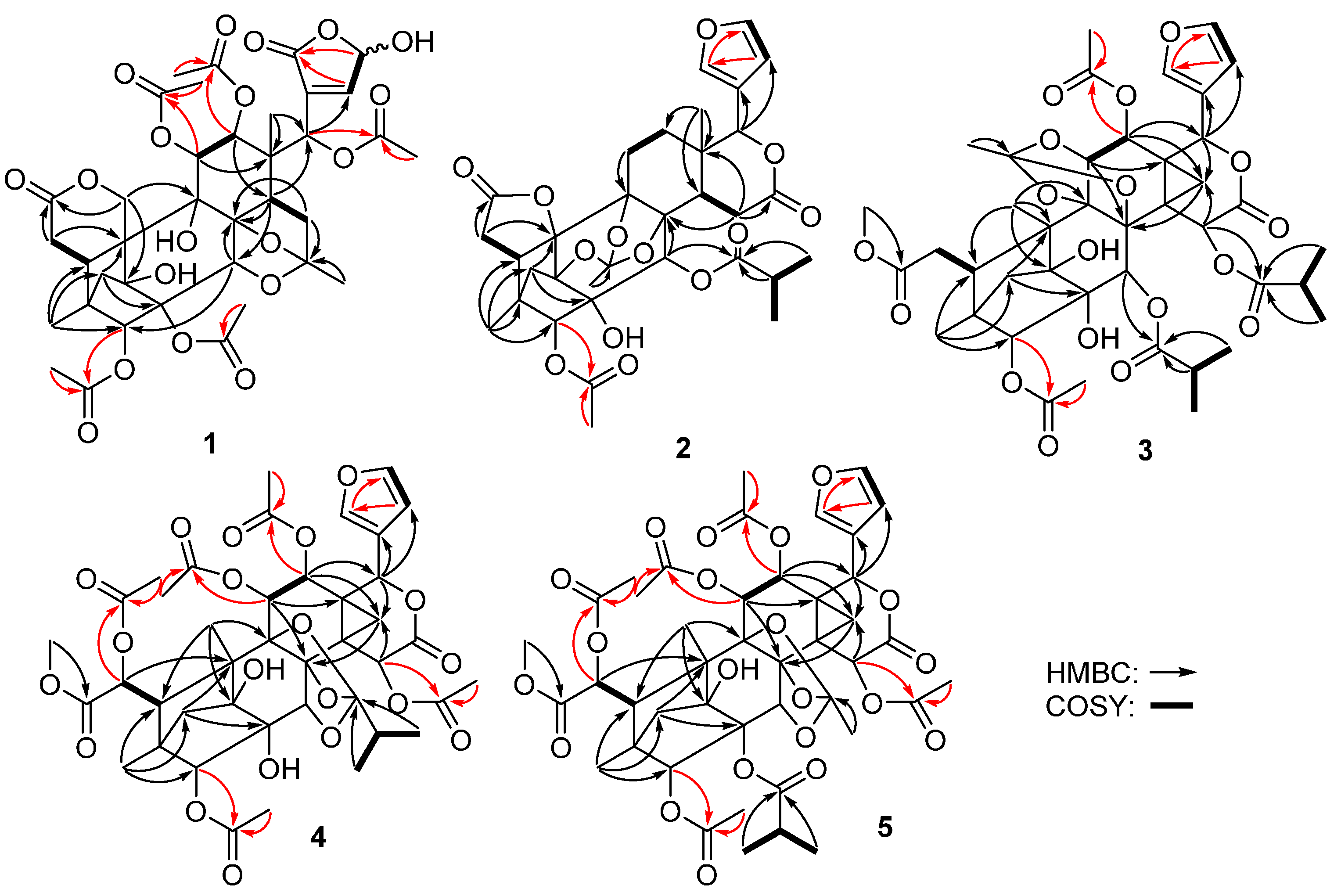
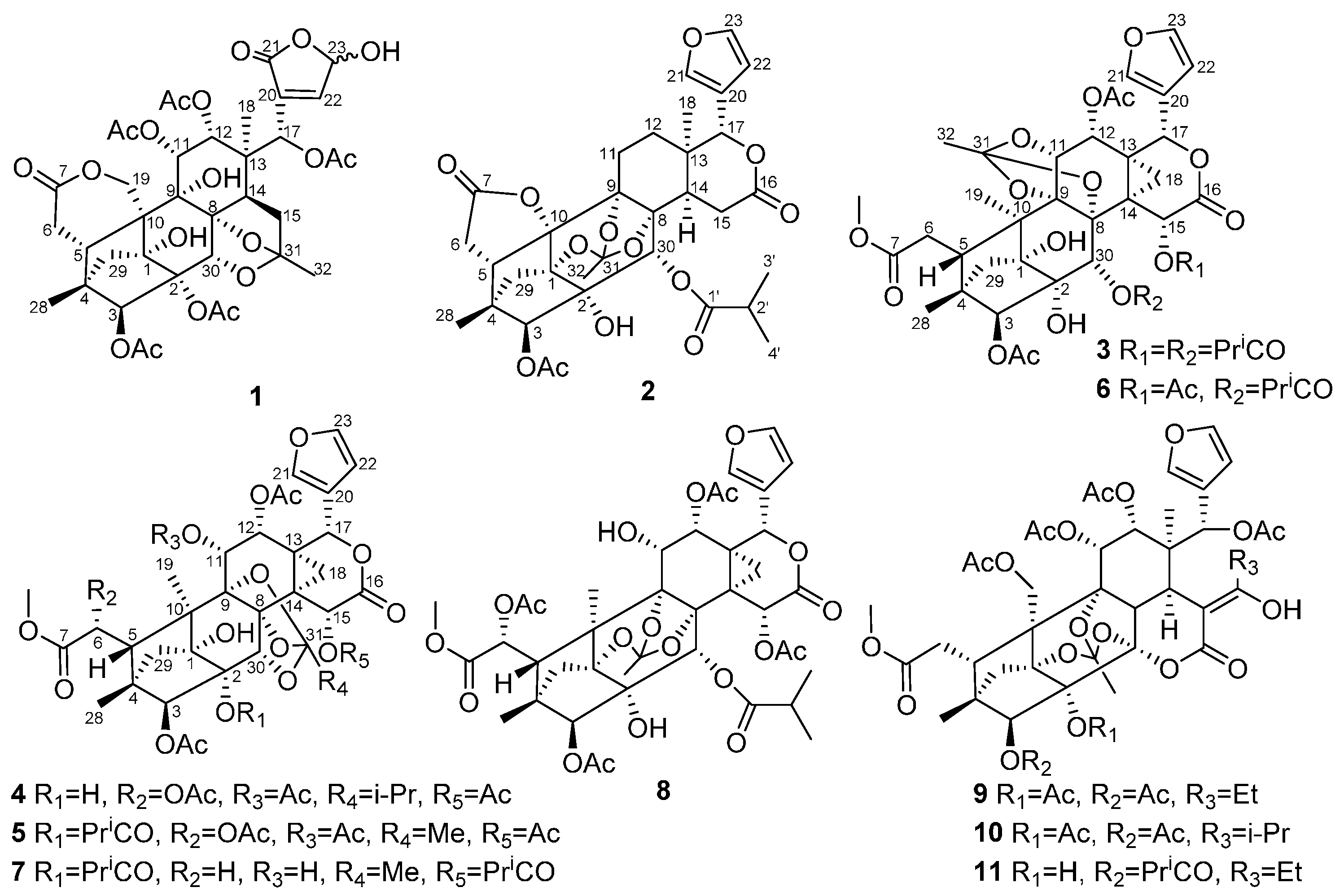
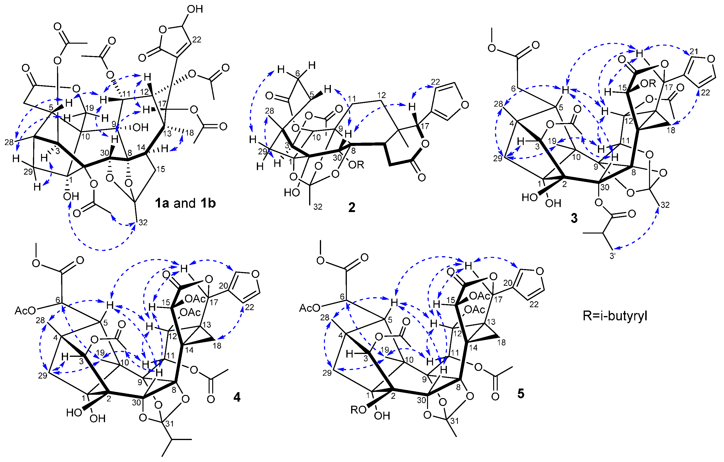
2. Results and Discussion

The EtOAc-soluble extract of the stems of C. tabularis was subjected to repeated column chromatography to afford five new phragmalin-type limonoids 15, and six known analogues 611 (Figure 1). Compound 1 was obtained as a white amorphous powder. Its molecular formula was established as C37H44O19 from a pseudomolecular ion peak at m/z 810.2817 ([M + NH4]+ calcd. 810.2815) in the HRESIMS, indicating 16 degrees of unsaturation. The IR spectrum showed hydroxyl group (3443 cm−1), carbonyl group (1746 cm−1), and olefinic bond (1636 cm−1) absorption bands. The 1H- and 13C-NMR spectra of 1 showed two sets of resonances with a ratio of 3:2 for isomers 1a and 1b. The 1H-NMR, 13C-NMR along with the HSQC data of the major isomer 1a revealed the presence of two angular methyls (δH 1.02, 0.90; δC 18.8, 15.2), five acetoxyls, and typical CH2-29 signals of a 4,29,1-ring-bridge [δH 2.09 and 2.02; δC 38.9]. Furthermore, the acetoxyls at C-3 (δC 83.4), C-11 (δC 70.6), C-12 (δC 71.2), and C-17 (δC 72.0) were revealed by the HMBC correlations from H-3 (δH 5.31), H-11 (δH 5.47), H-12 (δH 5.38), and H-17 (δH 5.72) to the corresponding carbonyls of the acetoxyl groups, respectively. The remaining acetoxyl was subsequently assigned to C-2 on the basis of its downfield shifted carbon resonance at δC 81.1 (for the case of 2-OH, the C-2 carbon resonance normally appeared at ca. δC 78.0). The HMBC correlations between C-7 (δC 172.7) and H-6 (δH 2.31) and one of the oxygenated C-19 methylene signals at δH 5.00 (H-19a) indicated the presence of the characteristic C-6–C-7 appendage of a phragmalin-type limonoid and the six-membered C-7/C-19 δ-lactone ring. A HMBC correlation between H-15a (δH 2.62) and the ketal carbon resonance at δC 113.7 (C-31), instead of the correlation between H-15 and the C-16 carbonyl in common phragmalins, indicated that 1a is a 16-decarboxylated phragmalin limonoid. The HMBC correlation between the ketal carbon and the methyl group signal H-32 (δH 1.65) suggested the linkage of the methyl to the ketal carbon, a biosynthetically extended C2 unit (C-31 and C-32) attached at C-15. The HMBC correlation between H-30 (δH 4.45) and the ketal carbon suggested the presence of an ether bridge between C-31 and C-30 (Figure 2). These data showed great similarity to those of chuktabularin B [10], except that a lactone carbonyl (δC 167.7) and a hemiacetal methine (δH 6.09; δC 95.7) signals replaced the corresponding two olefinic methine signals. HMBC correlations from H-17 to C-22, from H-22 and H-23 to C-21, and 1H-1H COSY correlation of H-22/H-23 indicated that a 23-hydroxy-20(22)-en-21,23-γ-lactone moiety instead of a β-furyl ring moiety located at C-17 in 1a. The relative configuration of 1a was elucidated using a ROESY experiment (Figure 3 and Table 1), in which the ROESY correlations of H-11/H-5, H-11/H-30, H-17/H-12, H-17/H-30 and 3-OAc/H-17, indicated that 3-OAc, H-5, H-11, H-12, H-17, and H-30 are co-facial and randomly assigned as β-oriented. ROESY correlations of Me-18/H-14, 9-OH/Me-18, 1-OH/Me-32, Me-32/2-OAc and H-29b/H-3 revealed that these protons adopt an α-orientation. The ROESY correlations of H-19a/1α-OH and H-19b/H-29a revealed that the six-membered 7,19-lactone ring was α-directed. Thus, the relative configuration of 1a in solution was established by a ROESY experiment as depicted. Comparison of the NMR data of 1a and 1b indicated that they had a same planar core structure. The only significant differences between 1a and 1b were the chemical shifts of carbons around C-23 (Table 1), suggesting that stereochemistry at hemiacetal C-23 was to be epimerized. This tautomerism has also been found in similar compounds, such as dysoxylumic acid B [22] and walsogyne A [23], and compound 1 was named as chukbularisin A.
Figure 2. Key HMBC and 1H-1H COSY correlations for compounds 15.
Molecules 21 00058 g002
Figure 3. Key ROESY correlations for compounds 15.
Molecules 21 00058 g003
Table 1. NMR spectroscopic data of compound 1 (isomers 1a and 1b) in CDCl3 (δ in ppm, J in Hz).
No.1a1b
δC aδH bROESY cδC aδH bROESY c
185.7 s 85.7 s
281.1 s 81.1 s
383.4 d5.31 (s)29b83.3 d5.31 (s)29b
445.7 s 45.8 s
540.8 d2.00 (m)11, 2840.7 d2.00 (m)11, 28
6a31.5 t2.31 (m, 2H) 31.5 t2.31 (m, 2H)
6b
7172.7 s 172.8 s
889.3 s 89.4 s
975.0 s 75.2 s
1052.6 s 52.6 s
1170.6 d5.47 (d, 3.8)5, 12, 3070.6 d5.54 (d, 3.8)5, 12, 30
1271.2 d5.38 (d, 3.8)11, 1771.4 d5.38 (d, 3.8)11, 17
1341.8 s 41.8 s
1444.6 d3.21 (dd, 12.2, 7.1)1844.5 d3.21 (dd, 12.2, 7.1)18
15a35.4 t2.62 (dd, 11.9, 7.1) 35.4 t2.63 (dd, 11.9, 7.1)
15b1.94 (dd, 12.2, 11.9)1.94 (dd, 12.2, 11.9)
1772.0 d5.72 (s)12, 30, 3-OAc72.1 d5.73 (s)12, 30, 3-OAc
1818.8 q1.02 (s)14, 9-OH18.8 q1.02 (s)14, 9-OH
19a69.5 t5.00 (d, 12.5)1-OH69.5 t4.97 (d, 12.5)1-OH
19b4.18 (dd, 12.5, 4.7)29a4.18 (dd, 12.5, 4.7)29a
20133.2 s 133.5 s
21167.7 s 167.7 s
22147.5 d7.38 (br s) 148.3 d7.34 (br s)
2395.7 d6.09 (t, 10.9) 96.2 d6.09 (t, 10.9)
2815.2 q0.90 (s)515.2 q0.90 (s)5
29a38.9 t2.09 (d, 11.8)19b38.9 t2.09 (d, 11.8)19b
29b2.02 (d, 11.8)32.02 (d, 11.8)3
3070.9 d4.45 (s)11, 1770.9 d4.42 (s)11, 17
31111.3 s 111.7 s
3218.8 q1.65 (s)1-OH, 2-OAc18.8 q1.65 (s)1-OH, 2-OAc
2-OAc170.2 s2.09 (s)32170.1 s2.09 (s)32
20.9 q20.9 q
3-OAc168.7 s2.45 (s)17168.9 s2.45 (s)17
21.0 q21.0 q
11-OAc171.5 s2.13 (s) 171.2 s2.12 (s)
20.8 q20.8 q
12-OAc170.5 s2.08 (s) 170.5 s2.08 (s)
20.3 q20.3 q
17-OAc170.7 s2.11 (s) 170.7 s2.11 (s)
20.2 q20.2 q
1-OH 4.86 (s)32, 19a 4.85 (s)32, 19a
9-OH 3.32 (s)18 3.30 (s)18
a Recorded at 125 MHz; b Recorded at 500 MHz; c Recorded at 500 MHz.
Compound 2 was isolated as a white amorphous powder, and the IR absorbance bands at 3455, and 1745 cm−1, suggested the presence of hydroxyl and carbonyl groups. The molecular formula C33H38O13 was determined by the pseudomolecular ion peak at m/z 643.2383 ([M + H]+ calcd. 643.2385) in the HRESIMS, indicating 15 degrees of unsaturation. The 13C and DEPT NMR showed presence of six methyls, five methylenes, nine methines (three oxygenated, and three olefinic ones) and thirteen quaternary carbons (six oxygenated, and four ester carbonyls). The 1H- and 13C-NMR spectroscopic data were similar to those of andirolide V isolated from Carapa guianensis [24], except for the downfield-shifted C-10 carbon signal and the absence of the oxygenated C-19 methylene signals. Detailed analysis of the NMR data of 2 further revealed that the A, B, C, D, and E rings of a phragmalin-type limonoid remained intact. The isobutyryloxyl was assigned to C-30 (δC 69.8) by the HMBC correlations from H-30 (δH 5.64) to C-1’ of the isobutyryloxyl, while the only acetoxyl was attached to C-3 according to the HMBC correlation from H-3 (δH 4.83) to the acetoxyl carbonyl. The HMBC correlations from H-6a and H2-29 to the oxygenated and remarkably deshielded C-10 (δC 86.4) revealed the loss of CH2-19 and the formation of the five-membered 7,10-γ-lactone ring. The degrees of unsaturation of 2 and the 14 mass units less in its molecular formula compared to that of andirolide V further confirmed this deduction. Planar structure of 2 was finally characterized by analysis of 1H-1H COSY and HMBC data as depicted in Figure 2. The relative configuration of 2 was assigned the same as that of andirolide V based on the explanation of ROESY NMR analysis (Figure 3 and Table 2). Thus, compound 2 (chukbularisin B) was determined as a 19-norphragmalin limonoid, a rare pentanortriterpenoid that only two limonoids of this type had been reported to the best of our knowledge [14,15].
Compound 3 was obtained as a white amorphous powder. The molecular formula C41H50O18 was determined by the pseudomolecular ion peak at m/z 869.2623 ([M + K]+ calcd. 869.2629) in the HRESIMS. The IR spectrum of 3 exhibited absorptions for OH groups at 3464 cm−1 and an ester carbonyl at 1727 cm−1. The 1H- and 13C-NMR data of 3 (Table 2) showed highly similarity to those of chubularinsin H [21], except for the absence of NMR signals for an acetoxy group at C-6. Moreover, the chemical shift of C-6 (δC 33.1) in 3 was upfield shifted (ca. ∆δC 37.5 ppm) compared with that of chubularinsin H, indicating the lack of a 6-OAc. This inference was further supported by the 58 mass units less in its molecular formula compared to that of chubularinsin H and 2D NMR data. Finally, the planar structure of 3 was characterized by analysis of 1H-1H COSY and HMBC data as depicted in Figure 2.
The relative configuration of 3 was assigned the same as that of chubularinsin H based on the explanation of ROESY correlations (Figure 3 and Table 2). Thus, the structure of 3 (chukbularisin C) was determined to be a 6-deacetoxy derivative of chubularinsin H.
Table 2. NMR spectroscopic data of compounds 2 and 3 in CDCl3 (δ in ppm, J in Hz).
No.23
δC aδH bROESY cδC aδH bROESY c
184.8 s 83.0 s
279.8 s 76.7 s
382.8 d4.83 (s)2885.9 d5.49 (s)29b
444.0 s 44.1 s
539.1 d2.94 (d, 8.4)3038.1 d2.58 (d, 11.9)12, 17, 28
6a30.2 t2.77 (d, 12.6)29a33.1 t2.66 (d, 12.3)
6b2.59 (dd, 12.6, 8.4)2.45 (d, 12.3
7174.3 s 173.9 s
886.5 s 78.5 s
984.5 s 90.6 s
1086.4 s 45.1 s
11a22.9 t1.64 (overlapped) 75.0 d4.17 (d, 3.6)12, 19
11b2.03 (m)
12a29.0 t1.53 (m) 66.7 d5.14 (br d, 3.6)5, 11, 17
12b1.41 (overlapped)
1334.9 s 31.3 s
1442.6 d2.10 (dd, 10.6, 2.1) 31.1 s
15a
15b
29.9 t3.15 (dd, 19.6, 2.1) 69.4 d7.16 (br d, 2.8)17, 30
2.72 (dd, 19.6, 10.6)
16169.8 s 167.1 s
1778.6 d5.32 (s)22, 3070.2 d6.42 (s)5, 12, 15, 21
18a20.3 q1.15 (s) 18.8 t2.64 (dd, 7.0, 3.1)
18b1.44 (d, 7.0)
19 14.4 q1.31 (s)11, 29a
20121.0 s 122.3 s
21141.0 d7.47 (br s) 142.2 d7.47 (br s)17
22109.7 d6.40 (br s)17109.9 d6.50 (br d, 1.6)
23143.6 d7.43 (br s) 143.4 d7.39 (br t, 1.6)
2814.3 q1.01 (s)314.8 q0.83 (s)5, 29b
29a39.5 t1.87 (s, 2H)6b39.0 t1.92 (s, 2H)19
29b3, 28
3069.8 d5.64 (s)5, 1769.4 d5.39 (s)15, 3-OAc
31119.8 s 119.9 s1.66 (s)3′
3221.0 q1.75 (s) 16.4 q
3-OAc170.1 s2.19 (s) 169.3 s2.22 s30
21.6 q21.2 q
12-OAc 170.9 s1.66 (s)
20.0 q
7-OCH3 52.6 q3.75 (s)
15-OCOCHMe2
1’ 177.9 s
2’ 34.2 d2.92 (m)
3’ 19.9 q1.32 (d, 7.0)
4’ 18.0 q1.25 (d, 7.0)
30-OCOCHMe2
1’175.4 s 173.9 s
2’34.6 d2.56-2.61 (m) 34.0 d2.51 (m)
3’18.2 q1.11 (d, 7.0) 19.5 q1.19 (d, 7.0)32
4’19.3 q1.19 (d, 7.0) 18.9 q1.17 (d, 7.0)
1-OH 2.85 (s)
2-OH 2.85 (s) 3.38 (s)
a Recorded at 125 MHz; b Recorded at 500 MHz; c Recorded at 500 MHz.
Compound 4 was isolated as a white amorphous powder and the IR absorbance bands at 3454 and 1735 cm−1 suggested the presence of hydroxyl and carbonyl groups. The molecular formula C41H48O20 was determined by the pseudomolecular ion peak at 883.2627 m/z ([M + Na]+ calcd. 883.2631) in the HRESIMS, indicating 18 degrees of unsaturation. The 13C and DEPT NMR showed the presence of ten methyls, two methylenes, twelve methines and seventeen quaternary carbons. The combined features of its 1H- and 13C-NMR spectra suggested that compound 4 was also a phragmalin-type limonoid with a β-substituted furanyl ring and typical CH2-29 proton signals of 4,29 1-ring-bridge in phragmalins. Furthermore, comparison of the 1H- and 13C-NMR data (Table 3) of 4 with those of tabularisin R [25] indicated that their structures showed high similarity. The only structural difference between them was in the presence of one additional acetoxyl group at C-3 in 4 replacing the 3-OH in tabularisin R, which was further confirmed by the downfield shifted H-3 (∆δH 1.55 ppm) signal of 4 owning to the acetylation effect, and the HMBC correlation from H-3 (δH 5.36) to the carbonyl (δC 169.0). The relative configuration of 4 was assigned the same as that of tabularisin R based on the explanation of ROESY correlations (Figure 3 and Table 3). Thus, the structure of 4 (chukbularisin D) was determined to be a 3-O-acetyl derivative of tabularisin R.
Compound 5 was isolated as a white amorphous powder. The molecular formula C43H50O21 was determined by the pseudomolecular ion peak at m/z 925.2737 ([M + Na]+ calcd. 925.2737) in the HRESIMS. IR data exhibited the presence of hydroxyls (3452 cm−1) and carbonyl groups (1736 cm−1). Comparison of the 1H- and 13C-NMR data (Table 3) of 5 with those of tabularisin C [7] indicated that their structures were closely related, and that they only differed in the nature of the oxygenated group at C-11. The corresponding HMBC correlation between the acetoxyl carbonyl and H-11 (δH 5.61) indicated that the 11-OH in tabularisin C was replaced by a 11-OAc group in 5. Finally, the planar structure of 5 was characterized by analysis of 1H-1H COSY and HMBC data as depicted in Figure 2. The relative configuration of 5 was established to be the same as tabularisin C by the ROESY data (Figure 3 and Table 3). Thus, the structure of 5 was elucidated and it was named chukbularisin E.
Table 3. NMR spectroscopic data of compounds 4 and 5 in CDCl3 (δ in ppm, J in Hz).
No.45
δC aδH bROESY cδC aδH bROESY c
184.6 s 83.9 s
276.0 s 83.1 s
385.5 d5.36 (s)29b85.8 d5.27 (s)
444.4 s 44.6 s
544.7 d2.81 (br s)12, 17, 28, 3043.9 d2.80 (s)12, 17, 28, 30
671.3 d6.26 (br s)1971.2 d6.22 (s)19
7172.1 s 172.1 s
886.6 s 86.5 s
984.2 s 84.7 s
1049.4 s 49.5 s
1167.1 d5.66 (d, 4.9)12, 15, 1967.0 d5.61 (d, 4.9)12, 15, 19
1266.7 d5.42 (d, 4.9)5, 11, 1766.5 d5.47 (d, 4.9)5, 11, 17
1329.6 s 29.8 s
1425.1 s 24.9 s
1569.7 d6.94 (br s)11, 17, 3070.5 d6.99 (d, 2.6)11, 17, 30
16166.0 s 165.7 s
1772.1 d6.50 (s)5, 12, 15, 2171.8 d6.44 (s)5, 12, 15, 21
18a16.2 t2.70 (dd, 7.2, 2.5)2217.8 t2.71 (dd, 7.2, 2.6)
18b1.51 (d, 7.2)1.43 (br d, 7.2)
1917.8 q1.37 (s)6, 11, 29a17.6 q1.36 (s)6, 11, 29a
20122.2 s 122.2 s
21142.1 d7.49 (br s)17142.1 d7.52 (br s)17
22109.7 d6.49 (br d, 1.4)18b109.9 d6.51 (br d, 1.3)
23143.4 d7.38 (br t, 1.6) 143.4 d7.38 (br t, 1.7)
2815.6 q0.96 (s)5, 29b15.6 q0.92 (s)5, 29b
29a40.2 t2.16 (d, 11.0)1940.8 t1.71 (br d, 11.4)19
29b1.83 (d, 11.0)3, 282.28 (br d, 11.4)28
3079.4 d4.09 (s)5, 15, 3-OAc76.0 d5.05 (s)5, 15
31119.4 s 116.2 s
3229.2 d2.15 (m) 15.8 q1.66 (s)
3317.1 q1.07 (d, 7.0)
3417.0 q1.05 (d, 7.0)
3-OAc169.0 s2.21 (s)30168.6 s2.33 (s)
21.1 q20.6 q
6-OAc169.3 s2.21 (s) 168.9 s2.20 (s)
21.3 q21.1 q
11-OAc169.2 s2.05 (s) 169.0 s2.07 (s)
20.9 q21.2 q
12-OAc170.1 s1.53 (s) 170.1 s1.54 (s)
19.3 q19.3 q
15-OAc169.0 s2.23 (s) 169.2 s2.22 (s)
21.1 q20.9 q
7-OCH353.7 q3.79 (s) 53.7 q3.79 (s)
2-OCOCHMe2
1’ 175.9 s
2’ 34.6 d2.50-2.55 (m)
3’ 18.9 q1.17 (d, 7.0)
4’ 18.9 q1.20 (d, 7.0)
1-OH 3.28 (s) 3.50 (s)
2-OH 3.47 (s)
a Recorded at 125 MHz; b Recorded at 500 MHz; c Recorded at 500 MHz.
The known compounds were identified as tabularisin E (6) [26], chubularisin E (7) [21], chubularisin K (8) [21], chukvelutilide B (9) [9], chukvelutilide D (10) [9], and chukvelutilide H (11) [25], respectively, by interpreting their data and making comparisons with literature values.
α-Glucosidase inhibitors are used in the treatment of non-insulin-dependent diabetes mellitus. In order to find in vitro α-glucosidase inhibitory agents among these compounds, some optimizations had been done to the reaction system, which was referred to Li [27]. The results showed that compounds 2, 3, 4, 5, and 8 exhibited α-glucosidase inhibitory activity with IC50 values of 0.06 ± 0.008, 0.04 ± 0.002, 0.52 ± 0.039, 1.09 ± 0.040, and 0.20 ± 0.057 mM, respectively (Table 4), among which compound 3 is 24 times more potent than the positive control (acarbose, IC50 0.95 ± 0.092 mM). Structure–activity relationship analysis revealed that the furanyl ring and the C-16/17 δ-lactone ring in these phragmalin limonoids are important for the α-glucosidase inhibitory activity. Thus, phragmalin limonoids might be promising agents for treatment and prevention of diabetes and need be further investigated for this purpose.
Table 4. In vitro α-glucosidase inhibitory activities of compounds 111.
CompoundIC50 Value (mM) aCompoundIC50 Value (mM) a
17
20.06 ± 0.00880.20 ± 0.057
30.04 ± 0.0029
40.52 ± 0.03910
51.09 ± 0.04011
6Acarbose b0.95 ± 0.092
a Values present mean ± SD of triplicate experiments; b Positive control; “–”inactive.

3. Experimental Section

3.1. General Procedures

Optical rotations were measured on an Autopol III polarimeter (Rudolph Research Analytical, Hackettstown, NJ, USA). Melting points were determined on a Beijing Taike X-5 stage apparatus (Beijing Taike Instrument Company, Beijing China) and are uncorrected. UV spectra were recorded on a DU800 spectrophotometer (Beckman, Brea, CA, USA). IR spectra were obtained on a 380 FT-IR spectrometer (Thermo, Pittsburgh, PA, USA). NMR experiments were recorded for 1H-NMR at 500 MHz and 13C-NMR at 125 MHz on an AV III spectrometer (Bruker, Bremen, Germany) using TMS as an internal standard. HRESIMS were acquired using an API QSTAR Pulsar mass spectrometer (Bruker). Column chromatographic separations were carried out by using silica gel (60–80 mesh and 200–300 mesh; Qingdao Haiyang Chemical Group Corporation, Qingdao, China), MCI gel CHP-20P (75–150 μm; Mitsubishi Chemical Industries Co. Ltd., Tokyo, Japan), Rp-18 (20–45 μm; Fuji Silysia Chemical Ltd., Durham, NC, USA) and Sephadex LH-20 (40–70 μm; Merck, Darmstadt, Germany). Silica gel (200–300 mesh), silica gel H (10–40 μm) and precoated silica GF254 plates for analytical TLC were produced by Qingdao Haiyang Chemical Company, Ltd. The spots on TLC were visualized by spraying with 5% H2SO4-ethanol solution.

3.2. Plant Material

The stems of Chukrasia tabularis were collected in Haikou, Hainan Province, P.R. China, in July 2014, which was identified by Dr. Jun Wang, Institute of Tropical Bioscience and Biotechnology, Chinese Academy of Tropical Agriculture Science, where a voucher specimen (No. 20140726) was deposited.

3.3. Extraction and Isolation

The air-dried stems of C. tabularis (110.0 kg) were pulverized and extracted with 95% ethanol (314 L) three times (7, 5, 3 days), at room temperature. The combined ethanol extract was then filtered through absorbent gauze, and the filtrate was concentrated under reduced pressure to remove the ethanol. Then, the residue (13.7 kg) was suspended in H2O and partitioned with petroleum ether, EtOAc, and n-BuOH successively. All the extracts were separately combined and evaporated to dryness under reduced pressure. These three fractions were designated as PEF (30.0 g), EAF (1700.0 g), and BUF (800.0 g), respectively. According to TLC analysis, the EtOAc fraction (1700.0 g) was separated into 18 fractions on a silica gel column (30 × 120 cm) using a step gradient elution of petroleum ether–EtOAc (20:1, 10:1, 5:1, 2:1, 1:1, and 0:1, v/v). Fr.17 (120.0 g) was subjected to silica gel (10 × 55 cm) vacuum liquid chromatography and eluted with CHCl3–MeOH (1:0, 100:1, 50:1, 25:1, 15:1, 10:1, 5:1, 2:1, 1:1, and 0:1, v/v) to provide 10 fractions (Fr.17-1–Fr.17-10). Fr.17-1 (3.5 g) was applied to ODS gel (3 × 40 cm) eluting with MeOH–H2O (from 3:7 to 1:0) to yield Fr.17-1-1–7. Fr.17-1-5 (350.0 mg) was chromatographed on Sephadex LH-20 gel (3 × 100 cm) with CHCl3–MeOH (v/v, 1:1), followed by silica gel (1.2 × 50 cm) eluting with petroleum ether–EtOAc (v/v, 6:4) to afford compound 1 (4.0 mg). Fr.15 (268.0 g) was subjected to silica gel (10 × 55 cm) vacuum liquid chromatography and eluted with CHCl3–EtOAc (1:0, 20:1, 10:1, 5:1, 1:1, and 0:1, v/v) to provide eight fractions (Fr.15-1–Fr.15-8). Fr.15-2 (36.8 g) was first subjected to a MCI gel column, eluted with MeOH–H2O (from 5:5 to 1:0) to yield Fr.15-2-1–15-2-4. Fr.15-2-1 (9.0 g) was applied to ODS gel (3 × 40 cm) eluting with MeOH–H2O (from 3:7 to 1:0) to yield Fr.15-2-1-1–20. Fr.15-2-1-5 (220.0 mg) was chromatographed on Sephadex LH-20 gel (3 × 100 cm) with CHCl3–MeOH (v/v, 1:1), followed by silica gel (1.2 × 50 cm) eluting with petroleum ether–EtOAc (v/v, 8:3) to afford 2 (8.0 mg) and 11 (8.0 mg). Fr.15-2-1-11 (850.0 mg) was chromatographed on Sephadex LH-20 gel (3 × 100 cm) with CHCl3–MeOH (v/v, 1:1), followed by silica gel (1.2 × 50 cm) eluting with petroleum ether–CHCl3–isopropanol (v/v/v, 5:5:0.07) to afford 3 (3.5 mg) and 8 (10.0 mg). Fr.15-2-1-13 (580.0 mg) was chromatographed on Sephadex LH-20 gel (3 × 100 cm) with CHCl3–MeOH (v/v, 1:1), followed by silica gel (1.2 × 50 cm) eluting with petroleum ether–EtOAc (v/v, 10:3) to afford compound 9 (15.0 mg) and 10 (8.0 mg). Fr.15-3 (26.8 g) was first subjected to a MCI gel column, eluted with MeOH–H2O (from 5:5 to 1:0) to yield Fr.15-3-1–15-3-8. Fr.15-3-3 (5.0 g) was applied to ODS gel (3 × 40 cm) eluting with MeOH–H2O (from 3:7 to 1:0) to yield Fr.15-3-3-1–18. Fr.15-3-3-5 (250.0 mg) was chromatographed on Sephadex LH-20 gel (3 × 100 cm) with CHCl3–MeOH (v/v, 1:1), followed by silica gel (1.2 × 50 cm) eluting with petroleum ether–CHCl3–isopropanol (v/v/v, 5:5:0.06) to afford 4 (7.0 mg) and 6 (10 mg). Fr.15-3-3-10 (300.0 mg) was chromatographed on Sephadex LH-20 gel (3 × 100 cm) with CHCl3–MeOH (v/v, 1:1), followed by silica gel (1.2 × 50 cm) eluting with petroleum ether–EtOAc (v/v, 8:3) to afford 5 (4.6 mg) and 7 (10 mg).
Chukbularisin A (1): White amorphous powder; mp 201–203 °C; [ α ] D 28 = +55° (c 0.30, CHCl3); UV (CHCl3): λmax (log ε) 240 (3.60) nm; IR (KBr) νmax 3443, 2923, 2853, 1746, 1636, 1217, 1043, 598 cm−1; 1H- and 13C-NMR data see Table 1; positive-mode HRESIMS m/z 810.2817 [M + NH4]+ (calcd. for C37H44O19NH4, 810.2815).
Chukbularisin B (2): White amorphous powder; mp 185–186 °C; [ α ] D 28 = +123° (c 0.20, CHCl3); UV (CHCl3) λmax (log ε): 240 (3.37) nm; IR (KBr) νmax 3455, 2923, 1745, 1640, 1215, 1072, 760 cm−1; 1H- and 13C-NMR data see Table 1; positive-mode HRESIMS m/z 643.2383 [M + H]+ (calcd. for C33H39O13, 643.2385).
Chukbularisin C (3): White amorphous powder; mp 198–199 °C; [ α ] D 28 = +68° (c 0.20, CHCl3); UV (CHCl3): λmax (log ε) 240 (3.61) nm; IR (KBr) νmax 3464, 2954, 1727, 1655, 1278, 1119, 1074 cm−1; 1H- and 13C-NMR data see Table 2; positive-mode HRESIMS m/z 869.2623 [M + K]+ (calcd. for C41H50O18K, 869.2629).
Chukbularisin D (4): White amorphous powder; mp 190–191 °C; [ α ] D 28 = +146° (c 0.05, CHCl3); UV (CHCl3): λmax (log ε) 248 (4.35) nm; IR (KBr) νmax 3454, 2926, 2088, 1735, 1634, 1383, 503 cm−1; 1H- and 13C-NMR data see Table 2; positive-mode HRESIMS m/z 883.2627 [M + Na]+ (calcd. for C41H48O20Na, 883.2631).
Chukbularisin E (5): White amorphous powder; mp 208–209 °C; [ α ] D 28 = +135° (c 0.10, CHCl3); UV (CHCl3): λmax (log ε) 246 (3.87) nm; IR (KBr) νmax 3452, 2923, 1736, 1638, 1383, 1099, 491 cm−1; 1H- and 13C-NMR data see Table 2; positive-mode HRESIMS m/z 925.2737 [M + Na]+ (calcd. for C43H50O21Na, 925.2737). (See Figures S1–S40 for more details about the original spectra of NMR and positive-mode HRESIMS data for the compounds 15).

3.4. α-Glucosidase Inhibitory Assays

The compounds tested in vitro for α-glucosidase activities were performed on the UV spectrophotometer, and the method used was that of Li [27]. The optimized procedure was as follows: 20 μL of 0.2 U/mL α-glucosidase has been added into 0.1mM potassium phosphate buffer (pH 6.8, 112 μL), then mixed with the testing sample (8 μL). After being preincubated at 37 °C for 15 min, 20 μL of 2.5 mmol/L 4-nitrophenyl-α-d-glucopyranoside was added and then mixed. The reaction was carried out at 37 °C for 15 min and stopped by adding 0.2 M solution of Na2CO3 (80 μL). The optical density values of the reaction mixture were the mean values of three measurements, which were performed at 405 nm wavelength. Acarbose (National Institutes for Food and Drug Control, Beijing, China, purity > 99.99%) was used as the positive control.

4. Conclusions

In conclusion, eleven limonoids including five new ones were isolated from the stems of C. tabularis based on its α-glucosidase inhibitory activity. Compounds 2, 3, 4, 5, and 8 displayed comparable or stronger α-glucosidase inhibition activity than acarbose (IC50 0.95 ± 0.092 mM) with IC50 values of 0.06 ± 0.008, 0.04 ± 0.002, 0.52 ± 0.039, 1.09 ± 0.040, and 0.20 ± 0.057 mM, respectively. It is worth noting that compound 3 is 24 times more potent than acarbose, and may serve as an attractive leading compound for the development of potent α-glucosidase inhibition agents.

Supplementary Materials

Supplementary materials can be accessed at: https://www.mdpi.com/1420-3049/21/1/58/s1.

Acknowledgments

This research was financially supported by Agro-scientific Research in the Public Interest (201303117), National Support Science and Technology Subject (2013BAI11B04), Major Technology Project of Hainan Province (ZDZX2013008-4) and National Nonprofit Institute Research Grant of CATAS-ITBB (ITBB2015RC03).

Author Contributions

Jun-Lin Peng and Jun Wang performed the isolation and structure elucidation of the constituents. Fan-Dong Kong contributed in the interpretation of the spectra and also part of the preparation of the manuscript. Zi-Qi Liu, Pei Wang and Bei Jiang partially contributed the structure elucidation, analyzed the data and together with Jun-Lin Peng prepared the manuscript. Cui-Juan Gai conducted the α-glucosidase inhibitory activity assay. Wen-Li Mei and Hao-Fu Dai planned, designed and organized the whole research of this study. All authors approved the final version of the manuscript.

Conflicts of Interest

The authors declare no conflict of interest.

References

  1. Chen, S.K.; Chen, B.Y.; Li, H. Flora Republicae Popularis Sinicae. In Chinese Flora (Zhongguo Zhiwu Zhi); Science Press: Beijing, China, 1997; Volume 43, pp. 47–49. [Google Scholar]
  2. Editorial Committee of the Administration Bureau of Traditional Chinese Medicine. Chemical composition and structure. In Chinese Material Medica (Zhonghua Bencao), 1st ed.; Shanghai Science and Technology Press: Shanghai, China, 1999; Volume 5, pp. 31–32. [Google Scholar]
  3. Tan, Q.G.; Luo, X.D. Meliaceous limonoids: Chemistry and Biological Activities. Chem. Rev. 2011, 111, 7437–7522. [Google Scholar] [CrossRef] [PubMed]
  4. Ragettli, T.; Tamm, C. Die Chukrasine A, B, C, D und E, fünf neue Tetranortriterpene aus Chukrasia tabularis A. Juss. Helv. Chim. Acta 1978, 61, 1814–1831. [Google Scholar] [CrossRef]
  5. Connolly, J.D.; Labbe, C.; Rycroft, D.S. Tetranortriterpenoids and related substances. Part 20. New tetranortriterpenoids from the seeds of Chukrasia tabularis (Meliaceae); simple esters of phragmalin and 12α-acetoxy phragmalin. J. Chem. Soc. Perkin Trans. 1978, 1, 285–288. [Google Scholar] [CrossRef]
  6. Luo, J.; Wang, J.S.; Cao, W.J.; Kong, L.Y. A new phragmalin-type limonoid from Chukrasia tabularis var. velutina. Chin. J. Nat. Med. 2011, 9, 98–100. [Google Scholar]
  7. Fan, C.Q.; Wang, X.N.; Yin, S.; Zhang, C.R.; Wang, F.D.; Yue, J.M. Tabularisins A–D, phragmalin ortho esters with new skeleton isolated from the seeds of Chukrasia tabularis. Tetrahedron 2007, 63, 6741–6747. [Google Scholar] [CrossRef]
  8. Luo, J.; Wang, J.S.; Luo, J.G.; Wang, X.B.; Kong, L.Y. Velutabularins A–J, phragmalin-type limonoids with novel cyclic moiety from Chukrasia tabularis var. velutina. Tetrahedron 2011, 67, 2942–2948. [Google Scholar] [CrossRef]
  9. Luo, J.; Wang, J.S.; Wang, X.B.; Huang, X.F.; Luo, J.G.; Kong, L.Y. Chukvelutilides A–F, phragmalin limonoids from the stem barks of Chukrasia tabularis var. velutina. Tetrahedron 2009, 65, 3425–3431. [Google Scholar] [CrossRef]
  10. Zhang, C.R.; Yang, S.P.; Liao, S.G.; Fan, C.Q.; Wu, Y.; Yue, J.M. Chuktabularins A–D, four new limonoids with unprecedented carbon skeletons from the stem bark of Chukrasia tabularis. Org. Lett. 2007, 9, 3383–3386. [Google Scholar] [CrossRef] [PubMed]
  11. Luo, J.; Wang, J.S.; Luo, J.G.; Wang, X.B.; Kong, L.Y. Chukvelutins A–C, 16-Norphragmalin Limonoids with Unprecedented Skeletons from Chukrasia tabularis var. velutina. Org. Lett. 2009, 11, 2281–2284. [Google Scholar] [CrossRef] [PubMed]
  12. Luo, J.; Wang, J.S.; Wang, X.B.; Luo, J.G.; Kong, L.Y. Chuktabularins E–T, 16-Norphragmalin Limonoids from Chukrasia tabularis var. velutina. J. Nat. Prod. 2010, 73, 835–843. [Google Scholar] [CrossRef] [PubMed]
  13. Zhang, C.R.; Fan, C.Q.; Zhang, L.; Yang, S.P.; Wu, Y.; Lu, Y.; Yue, J.M. Chuktabrins A and B, two novel limonoids fom the twigs and leaves of Chukrasia tabularis. Org. Lett. 2008, 10, 3183–3186. [Google Scholar] [CrossRef] [PubMed]
  14. Yin, J.L.; Di, Y.T.; Fang, X.; Liu, E.D.; Liu, H.Y.; He, H.P.; Li, S.L.; Li, S.F.; Hao, X.J. Tabulvelutin A, the first 19-nor limonoid with unprecedented ring system from Chukrasia tabularis var. velutina. Tetrahedron Lett. 2011, 52, 3083–3085. [Google Scholar] [CrossRef]
  15. Liu, H.B.; Zhang, H.; Li, P.; Gao, Z.B.; Yue, J.M. Chukrasones A and B: Potential Kv1.2 Potassium Channel Blockers with New Skeletons from Chukrasia tabularis. Org. Lett. 2012, 14, 4438–4441. [Google Scholar] [CrossRef] [PubMed]
  16. Nakatani, M.; Abdelgaleil, S.A.M.; Saad, M.M.G.; Huang, R.C.; Doe, M.; Iwagawa, T. Phragmalin limonoids from Chukrasia tabularis. Phytochemistry 2004, 65, 2833–2841. [Google Scholar] [CrossRef] [PubMed]
  17. Luo, J.; Wang, J.S.; Wang, X.B.; Luo, J.G.; Kong, L.Y. Phragmalin-type limonoid orthoesters from Chukrasia tabularis var. velutina. Chem. Pharm. Bull. 2011, 59, 225–230. [Google Scholar] [CrossRef] [PubMed]
  18. Luo, J.; Li, Y.; Wang, J.S.; Lu, J.; Wang, X.B.; Luo, J.G.; Kong, L.Y. Twelve Novel and Diverse 16-Norphragmalin-Type Limonoids from Chukrasia tabularis var. velutina. Chem. Pharm. Bull. 2012, 60, 195–204. [Google Scholar] [CrossRef] [PubMed]
  19. Luo, J.; Li, Y.; Wang, J.S.; Lu, J.; Kong, L.Y. Three new C-15-isobutyryl 16-norphragmalin-type limonoids from Chukrasia tabularis var. velutina. Phytochem. Lett. 2012, 5, 249–252. [Google Scholar] [CrossRef]
  20. Zhang, C.R.; Yang, S.P.; Chen, X.Q.; Wu, Y.; Zhen, X.C.; Yue, J.M. Limonoids from the twigs and leaves of Chukrasia tabularis. Helv. Chim. Acta 2008, 91, 2338–2350. [Google Scholar] [CrossRef]
  21. Liu, H.B.; Zhang, H.; Li, P.; Wu, Y.; Gao, Z.B.; Yue, J.M. Kv1.2 potassium channel inhibitors from Chukrasia tabularis. Org. Biomol. Chem. 2012, 10, 1448–1458. [Google Scholar] [CrossRef] [PubMed]
  22. Luo, X.D.; Wu, S.H.; Wu, D.G.; Ma, Y.B.; Qi, S.H. Novel antifeeding limonoids from Dysoxylum hainanense. Tetrahedron 2002, 58, 7797–7804. [Google Scholar] [CrossRef]
  23. Mohamad, K.; Hirasawa, Y.; Lim, C.S.; Awang, K.; Hadi, A.H.A.; Takeya, K.; Morita, H. Ceramicine A and walsogyne A, novel limonoids from two species of Meliaceae. Tetrahedron Lett. 2008, 49, 4276–4278. [Google Scholar] [CrossRef]
  24. Sakamoto, A.; Tanaka, Y.; Inoue, T.; Kikuchi, T.; Kajimoto, T.; Muraoka, O.; Yamada, T.; Tanaka, R. Andirolides Q–V from the flower of andiroba (Carapa guianensis, Meliaceae). Fitoterapia 2013, 90, 20–29. [Google Scholar] [CrossRef] [PubMed]
  25. Li, Y.; Luo, J.; Wang, Q.; Kong, L.Y. Two new limonoids from the stem barks of Chukrasia tabularis var. velutina. J. Asian Nat. Prod. Res. 2011, 13, 781–786. [Google Scholar] [CrossRef] [PubMed]
  26. Zhang, C.R.; Yang, S.P.; Zhu, Q.; Liao, S.G.; Wu, Y.; Yue, J.M. Nortriterpenoids from Chukrasia tabularis var. velutina. J. Nat. Prod. 2007, 70, 1616–1619. [Google Scholar] [CrossRef] [PubMed]
  27. Li, T.; Zhang, X.D.; Song, Y.W.; Liu, J.W. A microplate-based screening method for alpha-glucosidase inhibitors. Chin. J. Clin. Pharmacol. Ther. 2005, 10, 1128–1134. [Google Scholar]
  • Sample Availability: Samples of the compounds 111 are available from the authors.

