Next Article in Journal
Mini Review of Phytochemicals and Plant Taxa with Activity as Microbial Biofilm and Quorum Sensing Inhibitors
Next Article in Special Issue
Removal of Indoor Volatile Organic Compounds via Photocatalytic Oxidation: A Short Review and Prospect
Previous Article in Journal
Is Stevia rebaudiana Bertoni a Non Cariogenic Sweetener? A Review
Previous Article in Special Issue
Nanocasting of Periodic Mesoporous Materials as an Effective Strategy to Prepare Mixed Phases of Titania
 
 
Erratum published on 11 March 2016, see Molecules 2016, 21(3), 347.
Article

Mechanism of NO Photocatalytic Oxidation on g-C3N4 Was Changed by Pd-QDs Modification

1
Department of Science and Environmental Studies, The Hong Kong Institute of Education, Tai Po, New Territories, Hong Kong, China
2
Laboratory of Environmental Sciences and Technology, Xinjiang Technical Institute of Physics & Chemistry, and Key Laboratory of Functional Materials and Devices for Special Environments, Chinese Academy of Sciences, Urumqi 830011, China
*
Authors to whom correspondence should be addressed.
Molecules 2016, 21(1), 36; https://doi.org/10.3390/molecules21010036
Submission received: 1 December 2015 / Revised: 16 December 2015 / Accepted: 17 December 2015 / Published: 26 December 2015

Abstract

:
Quantum dot (QD) sensitization can increase the light absorption and electronic transmission of photocatalysts. However, limited studies have been conducted on the photocatalytic activity of photocatalysts after modification by noble metal QDs. In this study, we developed a simple method for fabricating Pd-QD-modified g-C3N4. Results showed that the modification of Pd-QDs can improve the NO photocatalytic oxidation activity of g-C3N4. Moreover, Pd-QD modification changed the NO oxidation mechanism from the synergistic action of h+ and O2 to the single action of ·OH. We found that the main reason for the mechanism change was that Pd-QD modification changed the molecular oxygen activation pathway from single-electron reduction to two-electron reduction. This study can not only develop a novel strategy for modifying Pd-QDs on the surface of photocatalysts, but also provides insight into the relationship between Pd-QD modification and the NO photocatalytic oxidation activity of semiconductor photocatalysts.

1. Introduction

Since Fujishima and Honda discovered oxygen and hydrogen evolution at a semiconductor electrode under light irradiation in 1972 [1], semiconductor photocatalysis have attracted worldwide attention in the fields of water or air purification [2,3,4,5], water splitting [6] and CO2 photoreduction [7,8]. Over the past four decades, various photocatalysts, such as oxides, sulfides and oxynitrides, have been developed to utilize solar energy for environmental purification and energy conversion [9,10,11,12]. However, most of these materials can only be excited by ultraviolet (UV) light, which occupies only 4% of solar light. Therefore, many researchers turn to develop new photocatalysts that are active under visible light. Among these endeavors, Wang et al. [13] reported that graphitic carbon nitride (g-C3N4) can produce oxygen or hydrogen by water splitting under visible light irradiation. More importantly, this metal-free polymer can withstand acid alkalis and high temperature because of the strong covalent bonds between carbon and nitride atoms. These advantages make g-C3N4 one of the most significant catalysts in the field of photocatalysis [14,15,16,17]. To increase the efficiency of this attractive material, many methods, such as surface area enhancement, anionic or cationic doping and coupling with other semiconductors, have been developed by researchers [18,19,20]. Despite all of these properties, the visible light photocatalytic activity of g-C3N4 is still low because of the fast recombination of the photogenerated carriers. Therefore, more efficient methods for improving the visible light photocatalytic activity of g-C3N4 should be designed and developed.
Quantum dots (QD) are nanoparticles with a unique feature, i.e., they can generate more than one electron for every absorbed photon. A recent report showed that QD sensitization could increase the light absorption and electronic transmission of photocatalysts. For example, C dots can enhance the performance of many photocatalysts, such as g-C3N4, TiO2 and Bi2MoO6, because of their ultrafast electron transferability [21,22,23]. The presence of CdS-QDs can favor the electron transfer and enhance the photoactivity of Zn1−xCdxS [24]. However, in all previous studies, the enhancement mechanism of photoactivity that is induced by the QDs sensitization is still in dispute. Some previous studies showed that the interfacial electron transfer from QDs to photocatalysts was due to the quantum confinement effect, whereas other previous studies consider that QDs can capture the photogenerated electrons and inhibit the recombination of electron-hole pairs [25,26]. Therefore, finding out how QDs will influence the photoreactivity of photocatalysts is considerably important, but challenging.
Some studies have demonstrated that the existence of noble metal on the surfaces of g-C3N4 particles could improve the photocatalytic activity of g-C3N4 by inhibiting the recombination of photoinduced electron-hole pairs [27,28,29]. However, few research works carefully studied the photocatalytic activity of g-C3N4 when it was modified by noble metal QDs. Although noble metals were often deposited on g-C3N4 surfaces, the sizes of these metals were larger than that of QDs. In view of the advantages of noble metals and QDs for g-C3N4 photocatalytic activity, we speculate that noble metal QD modification will be more effective than noble metal partials to improve the g-C3N4 photocatalytic activity. In this study, we have successfully prepared g-C3N4, which was modified with palladium QDs (PQDs), by a chemical reduction method for the first time. The resulting materials were carefully characterized and then used for the photocatalytic removal of organic NO under visible light irradiation. A series of experiments was designed to clarify the roles of PQDs on g-C3N4 photocatalysis under visible light. The reasons for the enhancement of the photocatalytic activity were analyzed in detail.

2. Experimental Section

2.1. Synthesis of Photocatalysts

Graphitic carbon nitride (g-C3N4) was prepared by calcining melamine in an alumina crucible with cover at 500 °C for 2 h and with an initial heating rate of 20 °C/min. This melamine was further calcined at 520 °C for 2 h. This procedure was similar to our previous paper [30].
PQDs-modified g-C3N4 was synthesized by an in situ chemical reduction method. In a typical synthetic procedure, the as-prepared g-C3N4 powder was added to 50 mL PdCl2 solution (0.5 g/dm3). After 20 min, the suspension was centrifuged and washed by distilled water many times. Then, the suspension was added to 50 mL of NaH2PO2·2H2O solution (20 g/dm3) under stirring. When it was stirred for 20 min, the suspension was centrifuged and washed thoroughly with distilled water. Finally, the sample was dried at 50 °C in a vacuum drying chamber. The final sample was denoted as PQDs-g-C3N4.

2.2. Characterization

The powder X-ray diffraction (XRD) patterns were recorded on a Bruker D8 Advance diffractometer (BRUKER, Berlin, Germany) with the DAVINCI design and monochromatized Cu Kα radiation (λ = 1.5418 Å). Transmission electron microscopy (TEM) images were obtained on a JEOL JSM-2010 microscope (JEOL, Tokyo, Japan) with an accelerating voltage of 200 kV. The TEM samples were prepared by dispersing the final powders in ethanol, and then, the dispersion was dropped on lacey support film grids. UV-VIS diffuse reflectance spectra were obtained by using a UV-VIS spectrometer (Shimadzu UV-2550, Tokyo, Japan) with BaSO4 as a reference, and these spectra were converted from reflection to absorbance by the Kubelka–Munk method. XPS measurements were performed in a VG scientific ESCALAB Mark II spectrometer (ESCALAB, London, UK), which was equipped with two ultra-high vacuum chambers. All of the binding energies were calibrated to the C1s peak at 284.6eV of the surface adventitious carbon.

2.3. Photocatalytic Activity Test

The photocatalytic activities of the resulting samples were tested for the photocatalytic removal of NO under visible light irradiation. In the experiments, NO removal at ppb levels was performed at ambient temperature in a continuous flow reactor. The rectangular reactor with a volume of 4.5 L (30 cm × 15 cm × 10 cm (L × W × H)) was made of stainless steel and covered with quartz glass. One sample dish, which contained the g-C3N4 or PQDs-g-C3N4 film, was placed in the middle of the reactor. A LED lamp (λ = 448 nm), which was vertically placed outside of the reactor above the sample dish, was used as the visible light source.
g-C3N4 and PQDs-g-C3N4 films were prepared by coating an aqueous suspension of g-C3N4 or PQDs-g-C3N4 onto a glass dish with a diameter of 12 cm. g-C3N4 or PQDs-g-C3N4 (0.15 g) was added to 15 mL of H2O and ultrasonicated for 20 min. Subsequently, the aqueous suspension was coated onto the glass dish, which was then dried at 60 °C until the water was completely removed.
NO gas was obtained from a compressed gas cylinder with traceable National Institute of Standards and Technology specifications. The NO concentration was diluted to about 600 ppb by the air stream that was supplied by a zero air generator. The flow rate was controlled at 1 L/min by a mass flow controller. After the adsorption–desorption equilibrium among gases and photocatalysts was achieved, the lamp was turned on. The NO concentration was continuously measured by using a Model T200 chemiluminescence NO analyzer (Teledyne, Thousand Oaks, CA, USA). NO removal efficiency (η) was calculated as follows:
η (%) = (1 − C/C0) × 100%
where C and C0 are the concentrations of NO in the outlet stream and the feeding stream, respectively.

2.4. Trapping Experiment

Active species trapping experiments were performed to investigate the NO removal mechanism. Potassium iodide (KI) and tert-butyl alcohol (TBA) were chosen as hole and ·OH scavengers, respectively. Argon was used to remove oxygen during the photocatalytic process. Typically, 0.15 g of photocatalyst with different trapping agents was added into 15 mL of H2O and ultrasonicated for 20 min. The aqueous suspensions were then coated onto the sample dish. Subsequently, the coated dish was dried at 60 °C until the water was completely removed. Finally, the coated dishes were used in NO removal experiments.
To eliminate the effect of H2O2, 1 mg·L−1 H2O2 was added into the sample dish under visible light in the absence of photocatalyst. Then, the NO concentration was measured by the NO analyzer.

3. Results and Discussion

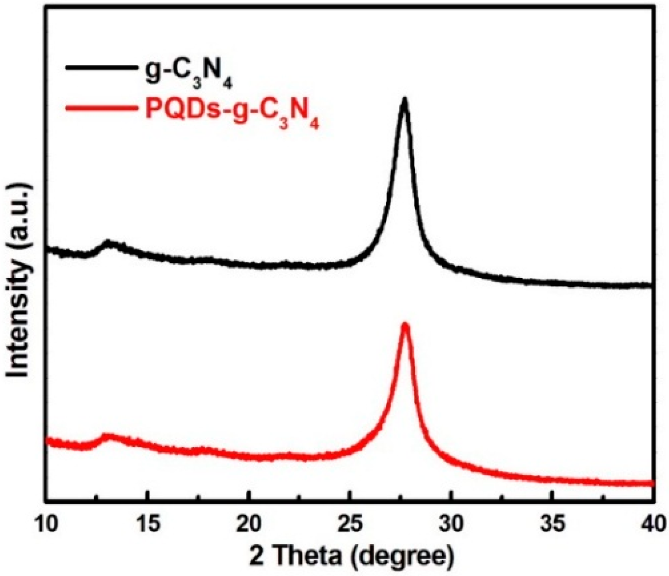
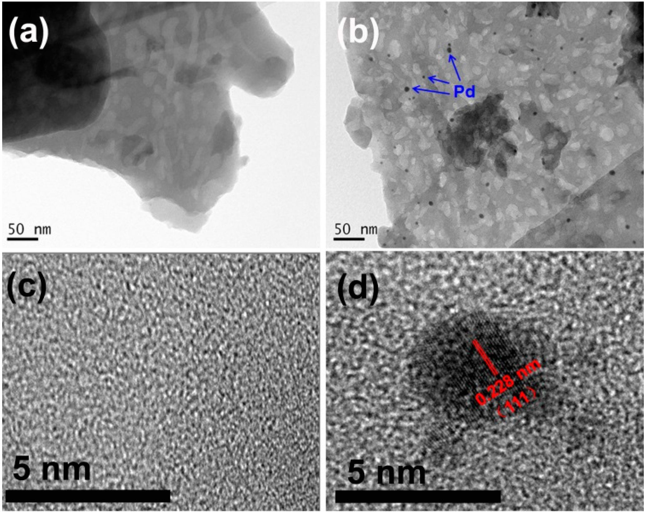
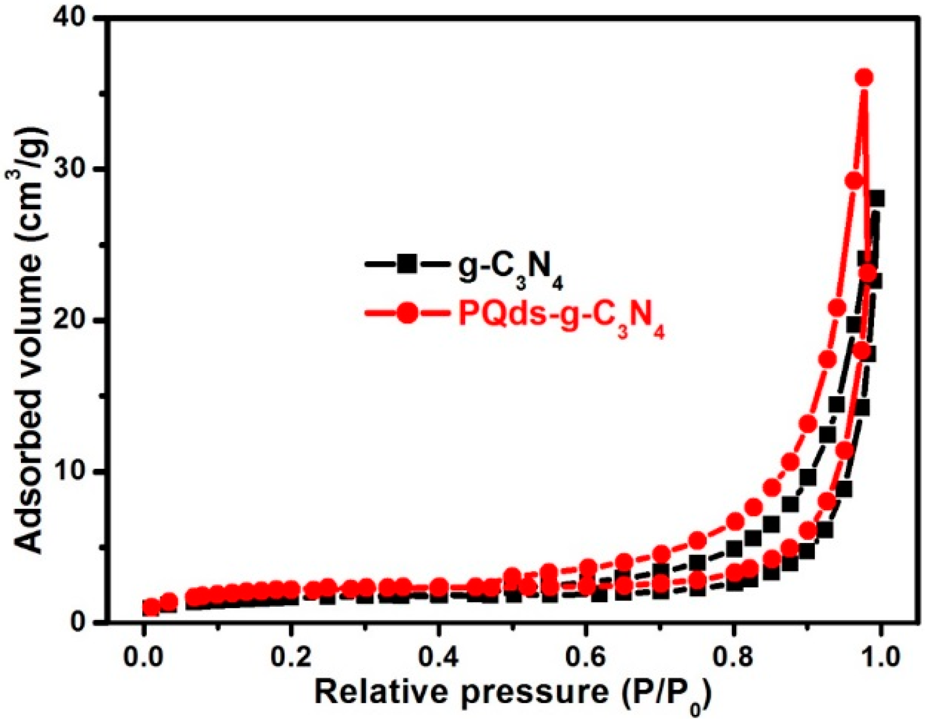
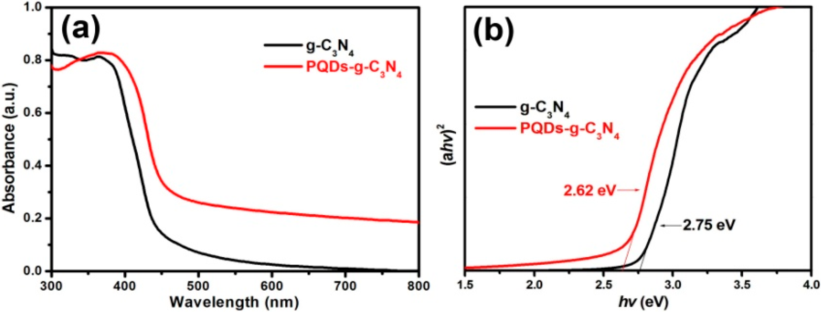
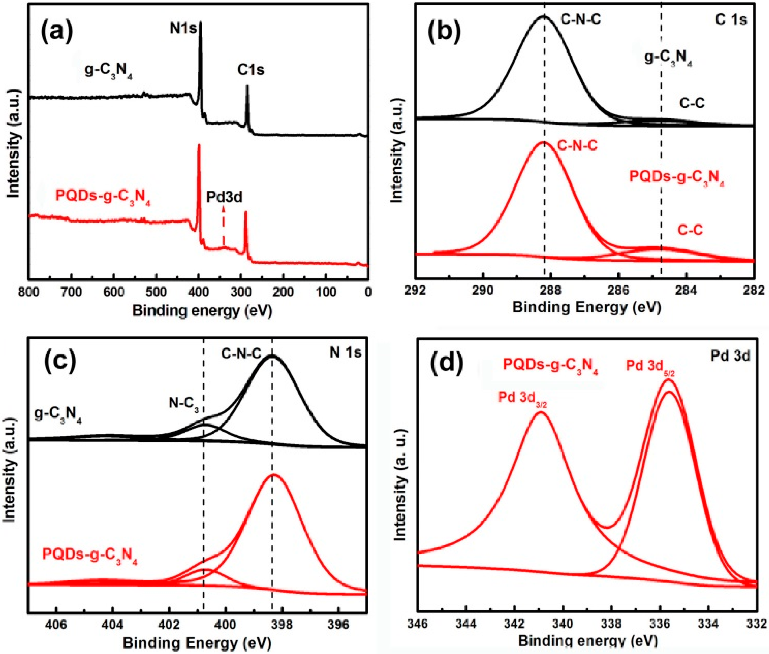
3.1. Structural Characterization of Result Samples

XRD was used to characterize the phase structure of the products. Figure 1 shows the powder XRD patterns of the as-prepared samples, where two peaks are found in all of the samples. The small angle peak at 13.08°, which corresponded to 0.676 nm, was due to the stacking of the interlayer. The strongest peak at 27.41°, which corresponded to 0.326 nm, was due to the stacking of the conjugated aromatic system that was indexed as the (002) peak for graphitic materials. No other peaks, such as Pd, could be detected in the XRD pattern of PQDs-g-C3N4, which indicates that the content of deposited Pd was considerably low to determine its existence and that Pd was dispersed uniformly onto the g-C3N4 surfaces.
Figure 1. The powder XRD patterns of the as-prepared samples. PQD, palladium QD.
Molecules 21 00036 g001
X-ray photoelectron spectroscopy (XPS) was used to investigate the chemical compositions of the photocatalyst structures. Figure 2a shows the survey spectra of the two resulting samples, in which g-C3N4 was composed of two elements, C and N, whereas PQDs-g-C3N4 was composed of three elements, C, N and Pd. As shown in Figure 2b, the C 1s spectrum could be fitted with two peaks at binding energies of 284.6 and 288.2 eV for both g-C3N4 and PQDs-g-C3N4, which is indicative of two different carbons in these two samples. The major peak at 288.2 eV was ascribed to the existence of sp2-hybridized carbon in C–N–C coordination, whereas the peak at 284.6 eV was assigned to the surface adventitious carbon. In the N 1s spectrum, the spectra of both samples could be separated into three binding energies (Figure 2c). The strongest peak at 398.7 eV could be assigned to sp2-hybridized nitrogen in the C–N–C groups. The peak at 400.2 eV was usually attributed to the tertiary nitrogen N-C3 groups. The weak additional signal at 401.3 eV could be attributed to the amino functional groups with hydrogen (C–N–H), which might be related to structural defects and incomplete condensation. Figure 2d showed the characteristicPd3d spectrum of PQDs-g-C3N4. The binding energies at 340.7 and 335.1 eV could be attributed to Pd 3d3/2 and 3d5/2, respectively, which correspond to the Pd°. These results confirmed the presence of Pd loads on g-C3N4surface.
Figure 2. XPS spectra of the resultants: (a) survey of the sample g-C3N4 and PQDs-g-C3N4; (b) C1s of the sample g-C3N4 and PQDs-g-C3N4; (c) N1s of the sample g-C3N4 and PQDs-g-C3N4; (d) Pd 3d of the sample p-g-C3N4.
Molecules 21 00036 g002
The microstructures of the resulting samples were investigated by TEM. Figure 3 shows the TEM image of g-C3N4 and PQD-g-C3N4. As shown in Figure 3a, the morphology of g-C3N4 was platelet-like. When g-C3N4 was treated in an in situ chemical reduction, its morphology was still platelet-like, but with many dark spherical spots on its surface. The dark spherical spots represented Pd-QDs with an average diameter of about 4.5 nm. Figure 3c shows the HRTEM image of PQD-g-C3N4. The lattice spacing of Pd nanoparticles was estimated at 0.228 nm, which corresponds to the (111) plane of Pd.
Figure 3. (a) TEM images of g-C3N4 and (b) PQDs-g-C3N4 (Inset: the blue arrows represent Pd QDs) samples; (c) HRTEM images of g-C3N4 and (d) PQDs-g-C3N4 (Inset: the red lines represent the lattice fringes of Pd QDs with d spacing of ca. 0.228 nm) samples.
Molecules 21 00036 g003

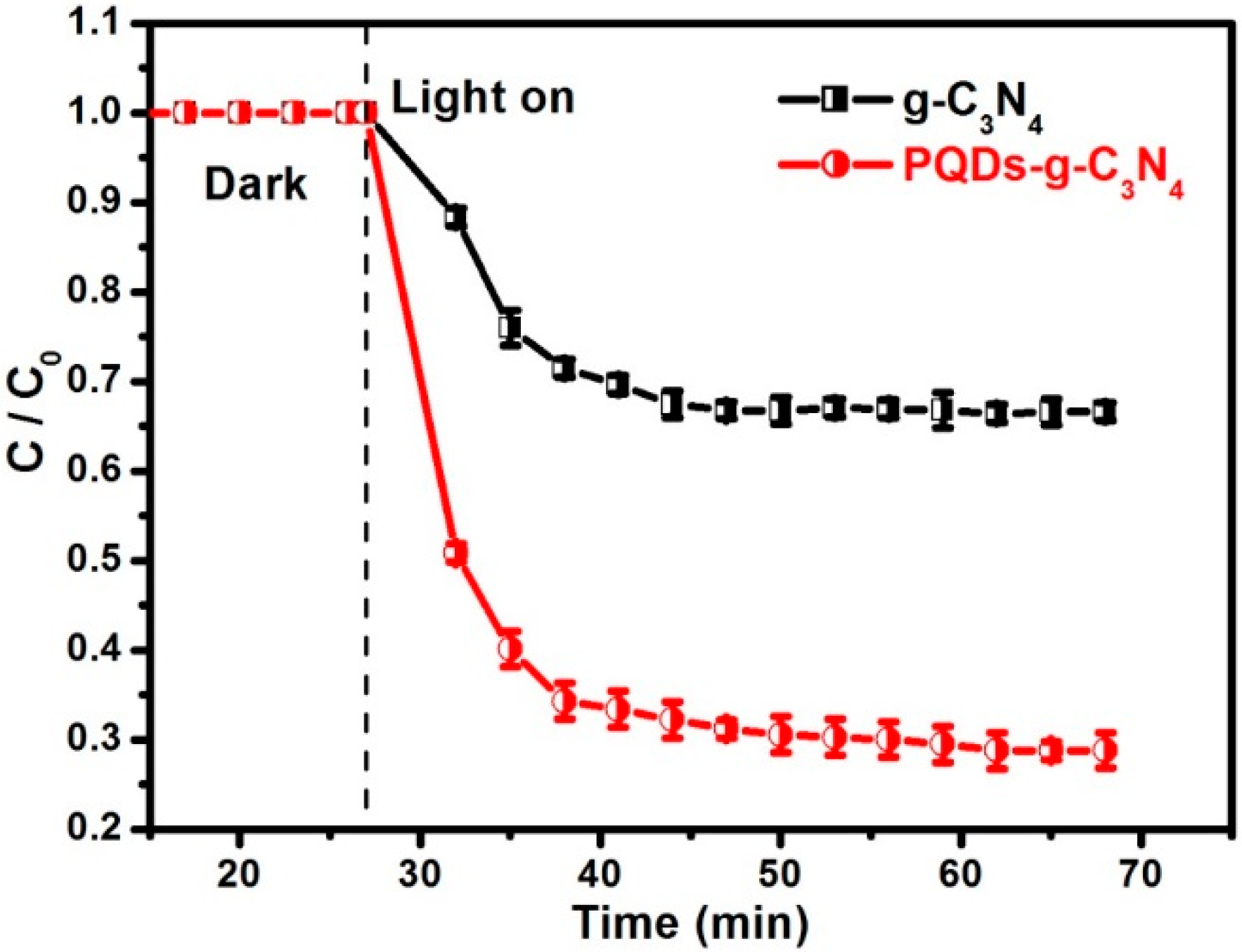
3.2. Photocatalytic Activities

The photocatalytic activities of the as-prepared g-C3N4 and PQD-g-C3N4 samples were tested in the photocatalytic oxidation of NO under visible light irradiation (λ > 420 nm). Figure 4 shows the photocatalytic activities of different photocatalysts and NO self-removal in the absence of any photocatalyst. The removal of NO was negligible under visible light irradiation (λ > 420 nm) without photocatalyst for 40 min, which indicates NO stabilization under visible light irradiation. However, in the presence of the g-C3N4, 34% oxidation efficiency of NO was observed after irradiation for 40 min. Interestingly, the introduction of PQDs remarkably enhanced the removal efficiency of NO, asPQDs-g-C3N4 could remove 72% of NO within 40 min. Obviously, PQD modification could significantly improve the photocatalytic activity of g-C3N4.
Figure 4. The photooxidation of NO over the samples g-C3N4 and PQDs-g-C3N4 under visible light irradiation (λ > 420 nm).
Molecules 21 00036 g004

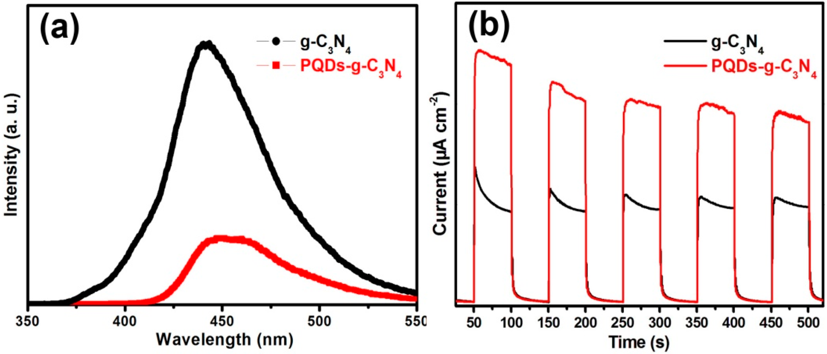
3.3. Mechanism of Activity Enhancement

As the photocatalytic activity is often related to the specific surface area, nitrogen sorption was used to measure the surface areas of the resulting samples. However, the measurement results showed that the surface areas of g-C3N4 and PQD-g-C3N4 were 7.9 and 8.2 m2/g, respectively (Figure 5). Therefore, the enhanced photocatalytic activity of PQDs-g-C3N4 could not be related to the surface area.
Figure 5. N2 adsorption-desorption isotherms and Barret–Joyner–Halenda (BJH) pore size distribution plots (inset) of g-C3N4 and PQDs-g-C3N4 samples.
Molecules 21 00036 g005
Aside from surface areas, photoabsorption and photoexcitation strongly affect the photocatalytic activity of photocatalysts. We therefore measured UV-VIS absorption spectra of g-C3N4 and PQD-g-C3N4 and found varying absorption edges of the two samples (Figure 6). The intrinsic absorption edge of PQDs-g-C3N4 showed a slight red shift compared to g-C3N4. Meanwhile, the absorption spectra of PQD-g-C3N4 extended to the whole visible light region, even in the infrared region, thereby enhancing light absorbance. This is not surprising, because QD sensitization could increase the light absorption and electronic transmission of photocatalysts. Assuming g-C3N4 is a direct semiconductor, plots of the (ahv)2 vs. the energy of absorbed light afford the band gaps of samples, as shown in Figure 6b. The calculated band gaps were approximately 2.75 and 2.62 eV for g-C3N4 and PQDs-g-C3N4, respectively. In such a case, the enhanced light harvesting and narrowed band gap of PQDs-g-C3N4 may result in the formation of more photogenerated electrons.
Figure 6. UV-VIS absorption spectra (a) and plots of (αhν)2 vs. energy () (b) of the sample g-C3N4 and PQDs-g-C3N4.
Molecules 21 00036 g006
After photoexcitation, the photogenerated electrons may undergo two fates: to migrate to the surface of the photocatalyst for subsequent chemical reactions and to recombine with photogenerated holes. Photoluminescence (PL) spectra were then used to investigate the recombination and separation of photogenerated electrons and holes in the two samples. Figure 7a displays the PL spectra of g-C3N4 and PQDs-g-C3N4 under the 320-nm excitation. The strong emission peak around 455 nm was derived from the direct electron and hole recombination of the band transition. The weaker PL peak intensity of PQDs-g-C3N4 undoubtedly confirmed that the modification of Pd-QDs could suppress the recombination of photogenerated charge carriers. Generally, photocatalyst with more photogenerated electrons and a lower electron-hole recombination rate would produce a higher photocurrent. Reasonably, the photocurrent that was generated on the PQD-g-C3N4 electrode would be higher than that generated on g-C3N4. This hypothesis could be confirmed by photocurrent measurement (Figure 7b). Therefore, the modification of Pd-QDs would favor the visible light absorption and separation of photogenerated carriers, which would eventually produce more carriers to remove NO.
Figure 7. Photoluminescence (PL) spectra of g-C3N4 and PQDs-g-C3N4 (a); current-time curves of g-C3N4 and PQDs-g-C3N4 (b).
Molecules 21 00036 g007

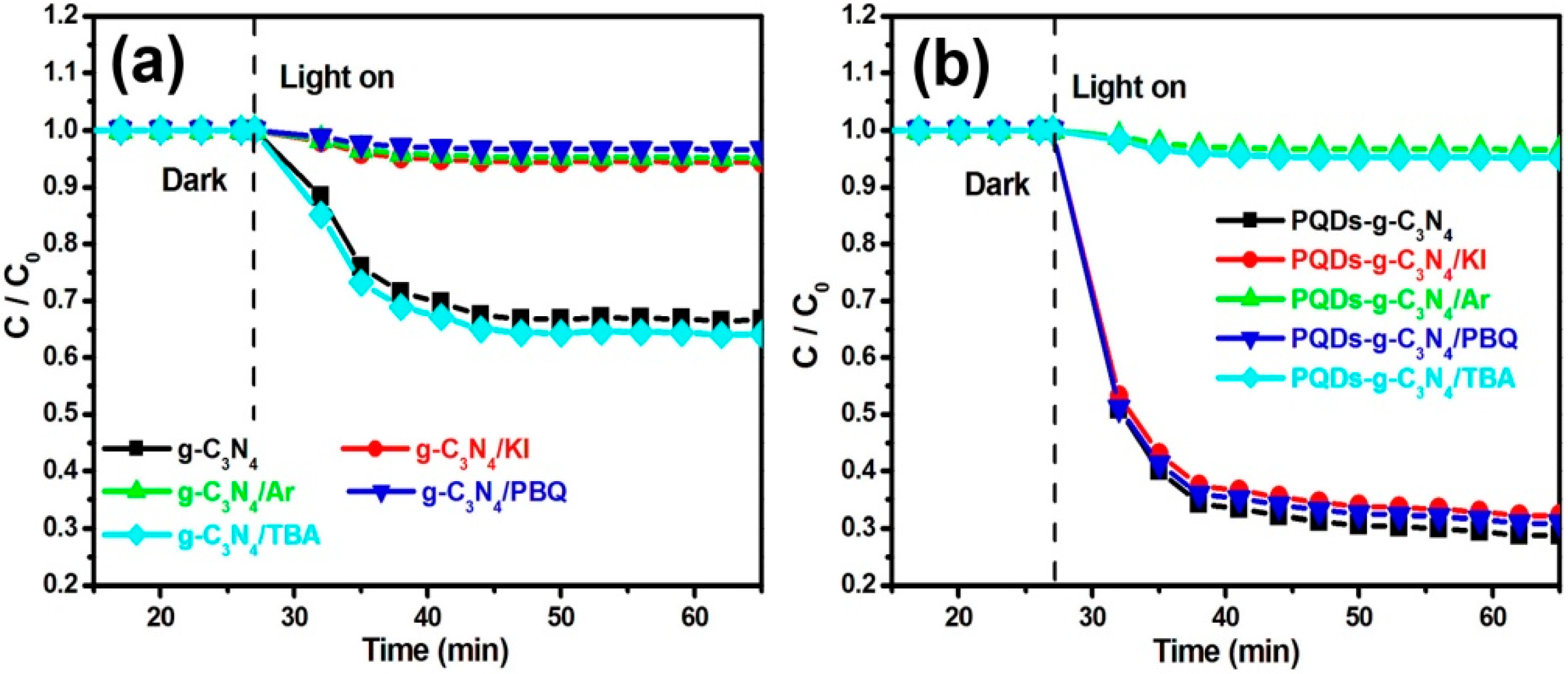
3.4. The Mechanism of NO Removal

In general, the photocatalytic removal of NO is attributable to several active species, such as the hydroxyl radicals (·OH), the superoxide (·O2), hydrogen peroxide (H2O2) and holes. These active species are formed in the following reactions:
Photocatalyst + solar light → h+ + e
e + O2 → ·O2
·O2 + 2H+ + e→ H2O2
H2O2 + e → 2·OH
h+ + H2O → ·OH + H+
To investigate the possible photocatalytic removal mechanism of NO over g-C3N4 and PQDs-g-C3N4, several experiments were performed to explore this mechanism. Potassium iodide (KI) was used to trap photogenerated holes. As shown in Figure 8, the rate of NO removal over g-C3N4 was suppressed completely when KI was added (5.5% removal efficiency of NO). This result suggests that the hole (h+) was significant in the photocatalytic removal process of NO on the g-C3N4. Aside from the photogenerated holes, O2 is an important factor in the photocatalytic process, because it can produce the superoxide (·O2), hydrogen peroxide (H2O2) and hydroxyl radicals (·OH). To test the role of dissolved O2 in degradation, high-purity argon was poured into the reaction to ensure that the reaction was operated without O2. As shown in Figure 8, the removal rate of NO on both g-C3N4 (4.7% removal efficiency of NO) and PQDs-g-C3N4 (3.3% removal efficiency of NO) was suppressed completely, which indicates that O2 is a necessary factor for NO photocatalytic removal. This result also implies that one or more of three active species (·O2, H2O2 and ·OH) are the major contributing factors for the pollutant’s degradation. To test this hypothesis, scavengers, such as p-benzoquinone (PBQ) for ·O2 and tert-butyl alcohol (TBA) for ·OH, were utilized in the photocatalytic process of the two samples. As shown in Figure 8a,b, the presence of PBQs could completely inhibit the photocatalytic activity of g-C3N4 (3.2% removal efficiency of NO); however, it had no effect on NO removal on PQDs-g-C3N4 (69.1% removal efficiency of NO). Interestingly, the presence of TBA did not influence the NO removal rate of g-C3N4 (35.8% removal efficiency of NO), but could significantly suppress the NO removal on PQDs-g-C3N4 (4.5% removal efficiency of NO). As a result, h+ and ·O2 have major functions in the removal process of NO on g-C3N4, whereas ·OH plays a major role in the NO removal process over PQDs-g-C3N4.
Figure 8. Comparison of NO photoremoval in different photo-catalysis systems under visible light irradiation (λ > 420 nm): (a) g-C3N4; (b) PQDs-g-C3N4. PBQ, p-benzoquinone; TBA, tert-butyl alcohol.
Molecules 21 00036 g008
To find out the reasons for the mechanism change of NO removal induced by the PQDs’ modification, we further employed the 5,5-dimethyl-pyrroline N-oxide (DMPO) spin-trapping electron spin resonance (ESR) technique to measure the reactive oxygen species, which were generated during photocatalysis. Four characteristic peaks of DMPO-·O2 were obviously observed in methanolic suspensions of g-C3N4 (Figure 9a), which reflects that ·O2 could be produced via the photocatalysis of g-C3N4. However, no peaks could be found in the aqueous dispersion of g-C3N4, which suggests that ·OH was not generated in the g-C3N4 system (Figure 9b).
Figure 9. (a) ESR spectra of g-C3N4 in a methanol aqueous dispersion for DMPO-·O2 and in an aqueous dispersion for DMPO-·OH; (b) ESR spectra of PQDs-g-C3N4 in a methanol aqueous dispersion for DMPO-·O2 and in an aqueous dispersion for DMPO-·OH; (c) ·OH has major functions in the removal process of NO on PQD-g-C3N4.
Molecules 21 00036 g009
We further measured the DMPO spin-trapping ESR spectra of PQDs-g-C3N4 in a methanol aqueous dispersion for DMPO-·O2 (Figure 9a) and in an aqueous dispersion for DMPO-·OH (Figure 9b). We found that the only ·OH was generated on PQDs-g-C3N4. According to previous reports, molecular oxygen can be activated through a single-electron reduction pathway (e → ·O2 → H2O2 → ·OH) or a two-electron reduction pathway (e → H2O2 → ·OH) [31]. Because ·O2 had not been detected in the PQD-g-C3N4 system, we therefore believe that PQD modification changed the molecular oxygen activation pathway from a single-electron reduction to a two-electron reduction. This change was related to the fact that Pd could enhance the oxygen adsorption ability and electron transport performance of PQDs-g-C3N4. Therefore, ·O2 has major functions in the removal process of NO on g-C3N4, whereas.OH has major functions in the removal process of NO on PQD-g-C3N4 (Figure 9c).

4. Conclusions

In this study, we developed a practical method to fabricate Pd-QD-modified g-C3N4. The modification of Pd-QDs not only favored the visible light absorption of g-C3N4, but also improved the separation efficiency of photogenerated carriers and, thus, enhanced the NO photocatalytic oxidation activity. More interestingly, Pd-QD modification changed the NO removal mechanism from the synergistic action of h+ and ·O2 to the single action of ·OH. We found that the main reason for the mechanism change was that Pd-QDs modification changed the molecular oxygen activation pathway from single-electron reduction to two-electron reduction. This study could not only develop a novel strategy to modify the Pd-QDs on the surface of photocatalysts, but also shed light on the deep understanding of the relationship between Pd-QD modification and the NO photocatalytic removal activity of semiconductor photocatalysts.

Acknowledgments

This research was financially supported by the research grant of the Early Career Scheme (ECS 809813) from the Research Grant Council, Hong Kong Special Administrative Region (SAR) Government, and Internal Research Grants (R3588) and (R3633) from The Hong Kong Institute of Education.

Author Contributions

Guohui Dong and Wingkei Ho conceived and designed the experiments; Yuhan Li and Liping Yang performed the experiments; Yuhan Li analyzed the data; Guohui Dong and Wingkei Ho contributed reagents/materials/analysis tools; Yuhan Li wrote the paper.

Conflicts of Interest

The authors declare no conflict of interest.

References

  1. Fujishima, A.; Honda, K. Electrochemical photolysis of water at a semiconductor electrode. Nature 1972, 238, 37–38. [Google Scholar] [CrossRef] [PubMed]
  2. Hoffmann, M.R.; Martin, S.T.; Choi, W.; Bahnemann, D.W. Environmental applications of semiconductor photocatalysis. Chem. Rev. 1995, 95, 69–96. [Google Scholar] [CrossRef]
  3. Carraway, E.R.; Hoffman, A.J.; Hoffmann, M.R. Photocatalytic oxidation of organic acids on quantum-sized semiconductor colloids. Environ. Sci. Technol. 1994, 28, 786–793. [Google Scholar] [CrossRef] [PubMed]
  4. Marta, I.L. Heterogeneous photocatalysis transition metal ions in photocatalytic systems. Appl. Catal. B 1999, 23, 89–114. [Google Scholar]
  5. Fujishima, A.; Rao, T.N.; Tryk, D.A. Titanium dioxide photocatalysis. J. Photochem. Photobiol. C 2000, 1, 1–21. [Google Scholar] [CrossRef]
  6. Yamasita, D.; Takata, T.; Hara, M.; Kondo, J.N.; Domen, K. Recent progress of visible-light-driven heterogeneous photocatalysts for overall water splitting. Solid State Ion. 2004, 172, 591–595. [Google Scholar] [CrossRef]
  7. Ettedgui, J.; Diskin-Posner, Y.; Weiner, L.; Neumann, R. Photoreduction of carbon dioxide to carbon monoxide with hydrogen catalyzed by a Rhenium(I) phenanthroline-polyoxometalate hybrid complex. J. Am. Chem. Soc. 2011, 133, 188–190. [Google Scholar] [CrossRef] [PubMed]
  8. Zhang, L.; Wang, W.; Jiang, D.; Gao, E.; Sun, S. Photoreduction of CO on BiOCl nanoplates with the assistance of photoinduced oxygen vacancies. Nano Res. 2015, 3, 821–831. [Google Scholar] [CrossRef]
  9. Kim, Y.I.; Salim, S.; Huq, M.J.; Mallouk, T.E. Visible light photolysis of hydrogen iodide using sensitized layered semiconductor particles. J. Am. Chem. Soc. 1991, 113, 9561–9563. [Google Scholar] [CrossRef]
  10. Lee, Y.; Terashima, H.; Shimodaira, Y.; Teramura, K.; Hara, M.; Kobayashi, H.; Domen, K.; Yashima, M. Zinc germanium oxynitride as a photocatalyst for overall water splitting under visible light. J. Phys. Chem. C 2007, 111, 1042–1048. [Google Scholar] [CrossRef]
  11. Maeda, K.; Takata, T.; Hara, M.; Saito, N.; Inoue, Y.; Kobayashi, H.; Domen, K. GaN:ZnO solid solution as a photocatalyst for visible-light-driven overall water splitting. J. Am. Chem. Soc. 2005, 127, 8286–8287. [Google Scholar] [CrossRef] [PubMed]
  12. Hitoki, G.; Takata, T.; Kondo, J.N.; Hara, M.; Kobayashi, H.; Domen, K. An oxynitride, TaON, as an efficient water oxidation photocatalyst under visible light irradiation(λ <500 nm). Chem. Commun. 2002, 16, 1698–1699. [Google Scholar]
  13. Wang, X.W.; Maeda, K.; Thomas, A.; Takanabe, K.; Xin, G.; Carlsson, J.M.; Domen, K.; Antonietti, M. A metal-free polymeric photocatalyst for hydrogen production from water under visible light. Nat. Mater. 2009, 8, 76–80. [Google Scholar] [CrossRef] [PubMed]
  14. Chen, X.F.; Zhang, J.S.; Fu, X.Z.; Antonietti, M.; Wang, X.C. Fe-g-C3N4-Catalyzed oxidation of benzene to phenol using hydrogen peroxide and visible light. J. Phys. Chem. A 2009, 131, 11658–11659. [Google Scholar] [CrossRef] [PubMed]
  15. Ding, Z.X.; Chen, X.F.; Antonietti, M.; Wang, X.C. Synthesis of transition metal-modified carbon nitride polymers for selective hydrocarbon oxidation. ChemSusChem 2010, 4, 274–281. [Google Scholar] [CrossRef] [PubMed]
  16. Yan, S.C.; Li, Z.S.; Zou, Z.G. Photodegradation of rhodamine b and methyl orange over boron-doped g-C3N4 under visible light irradiation. Langmuir 2010, 26, 3894–3910. [Google Scholar] [CrossRef] [PubMed]
  17. Dong, G.H.; Ai, Z.H.; Zhang, L.Z. Efficient anoxic pollutant removal with oxygen functionalized graphitic carbon nitride under visible light. RSC Adv. 2014, 4, 5553–5560. [Google Scholar] [CrossRef]
  18. Dong, G.H.; Ho, W.K.; Li, Y.H.; Zhang, L.Z. Facile synthesis of porous graphene-like carbon nitride (C6N9H3) with excellent photocatalytic activity for NO removal. Appl. Catal. B Environ. 2015, 174, 477–485. [Google Scholar] [CrossRef]
  19. Liu, G.; Niu, P.; Sun, C. Unique electronic structure induced high photoreactivity of sulfur-doped graphitic C3N4. J. Am. Chem. Soc. 2010, 132, 11642–11648. [Google Scholar] [CrossRef] [PubMed]
  20. Dong, G.H.; Zhang, L.Z. Synthesis and enhanced Cr(VI) photoreduction property of formate anion containing graphitic carbon nitride. J. Phys. Chem. C 2013, 117, 4062–4068. [Google Scholar] [CrossRef]
  21. Liu, J.; Liu, Y.; Liu, N.; Kang, Z. Metal-free efficient photocatalyst for stable visible water splitting via a two-electron pathway. Science 2015, 347, 970–974. [Google Scholar] [CrossRef] [PubMed]
  22. Lin, Z.; Xue, W.; Chen, H.; Lin, J.M. Peroxynitrous-acid-induced chemiluminescence of fluorescent carbon dots for nitrite sensing. Anal. Chem. 2011, 83, 8245–8251. [Google Scholar] [CrossRef] [PubMed]
  23. Di, J.; Xia, J.; Ji, M.; Li, H.; Hui, X.; Chen, R. The synergistic role of carbon quantum dots for the improved photocatalytic performances of Bi2MoO6. Nanoscale 2015, 7, 11433–11443. [Google Scholar] [CrossRef] [PubMed]
  24. Li, G.S.; Zhang, D.Q.; Yu, J.C. A new visible-light photocatalyst: CdS quantum dots embedded mesoporous TiO2. Environ. Sci. Technol. 2009, 43, 7079–7085. [Google Scholar] [CrossRef] [PubMed]
  25. Leutwyler, W.K.; Bürgi, S.L.; Burgl, H. Semiconductor clusters, nanocrystals, and quantum dots. Science 1996, 271, 933–937. [Google Scholar]
  26. Shen, J.; Zhu, Y.; Yang, X.; Li, C. Graphene quantum dots: emergent nanolights for bioimaging, sensors, catalysis and photovoltaic devices. Chem. Commun. 2012, 48, 3686–3699. [Google Scholar] [CrossRef] [PubMed]
  27. Liu, Q.; Zhang, J. Graphene supported Co-g-C3N4 as a novel metal–macrocyclic electrocatalyst for the oxygen reduction reaction in fuel cells. Langmuir 2013, 29, 3821–3828. [Google Scholar] [CrossRef] [PubMed]
  28. Samanta, S.; Martha, S.; Parida, K. Facile synthesis of Au/g-C3N4 nanocomposites: an inorganic/organic hybrid plasmonic photocatalyst with enhanced hydrogen gas evolution under visible-light irradiation. ChemCatChem 2014, 6, 1453–1462. [Google Scholar] [CrossRef]
  29. Shalom, M.; Guttentag, M.; Fettkenhauer, C.; Inal, S.; Neher, D.; Llobet, A.; Antonietti, M. In situ formation of heterojunctions in modified graphitic carbon nitride: Synthesis and noble metal free photocatalysis. Chem. Mater. 2014, 26, 5812–5818. [Google Scholar] [CrossRef]
  30. Dong, G.H.; Zhang, L.Z. Porous structure dependent photoreactivity of graphitic carbon nitride under visible light. J. Mater. Chem. 2012, 22, 1160–1166. [Google Scholar] [CrossRef]
  31. Dong, G.H.; Ai, Z.H.; Zhang, L.Z. Total aerobic destruction of azo contaminants with nanoscale zero-valent copper at neutral pH: Promotion effect of in-situ generated carbon center radicals. Water Res. 2014, 66, 22–30. [Google Scholar] [CrossRef] [PubMed]
  • Sample Availability: Samples are available from the authors.

