Article

Blumea balsamifera Oil for the Acceleration of Healing of Burn Injuries

1
Tropical Crops Genetic Resources Institute, Chinese Academy of Tropical Agricultural Sciences, Danzhou 571737, China
2
School of Traditional Chinese Medicine, Guangdong Pharmaceutical University, Guangzhou 510006, China
3
Key Laboratory of Crop Gene Resources and Germplasm Enhancement in Southern China, Danzhou 571737, China
4
Hainan Provincial Engineering Research Center for Blumea balsamifera, Danzhou 571737, China
*
Authors to whom correspondence should be addressed.
Molecules 2015, 20(9), 17166-17179; https://doi.org/10.3390/molecules200917166
Submission received: 12 June 2015 / Revised: 2 September 2015 / Accepted: 14 September 2015 / Published: 17 September 2015
(This article belongs to the Section Medicinal Chemistry)

Abstract

:
Blumea balsamifera oil (BBO) is a main extract obtained from Blumea balsamifera (L.) DC (Ainaxiang) leaves, which are widely used as a traditional medicine by the Miao and Li Nations to promote skin trauma or burn injury healing. This study was initiated to investigate the healing efficacy in deep second-degree burn model in rats. The rats were treated by BBO for 21 consecutive days. The rate of healing, scabs dropped time and re-epithelialization time were observed every three days for 21 days after burn injury. The samples were collected from different treated rats by sacrificing the animals on the 1st, 2nd, 5th, 9th, 14th, and 21st day post-burn creation. Then, the water content of burn tissue was measured. Plasma interleukin-1 (IL-1) and tumor necrosis factor-alpha (TNF-α) levels were evaluated, and the tissue expressions of basic fibroblast growth factor (bFGF), vascular endothelial growth factor (VEGF), and transforming growth factor-beta (TGF-β) were determined along with skin histopathology. The results showed that the water content of tissue was significantly reduced, the scabs dropped time shortened, and healing accelerated after treatment with BBO in the burn injury rats. Furthermore, the expressions of growth factors were significantly increased in the tissue; however, the levels of inflammatory factors on plasma decreased. This study confirms the efficacy of BBO consumption on burn injuries.

1. Introduction

Burn injuries, caused by heat, light, electricity, radiation and chemicals, are one of the most common and devastating forms of trauma, particularly partial- and full-thickness burns [1]. According to the report of Institute of Burn Third Military Medical University, about 5000–10,000 in one million people suffered from burn in China every year [2]. Depending on the severity, burn injuries are classified into three degrees, whereby the second degree is divided into shade and deep degrees [3]. Burn-wound could result in the increase of free radical-mediated damage and reactive oxygen species. It can delay granulation tissue formation, reduce angiogenesis and decrease collagen reorganization. Furthermore, due to its significantly prolonged period of rehabilitation, the healing of burn injuries is more difficult than that of ordinary wounds and is also associated with higher economic costs [4,5].
Current methods to treat burn injuries include antibiotics, antiphlogistics, and silver salt, which possesses major drawbacks and unwanted side effects [6,7,8,9,10]. Nowadays, the alternative and complementary medicines, such as traditional Chinese medicine and aromatherapy, are being used, because they are moderately beneficial to effective with little toxicity and are less expensive than synthetic drugs. Many plants and plant-derived products have been found to possess potent wound-healing or burn-wound-healing activity [11,12,13,14].
Blumea balsamifera (L.) DC., (B. balsamifera) an important medicinal herb the has been used as a traditional ethnic medicine for thousands of years in China, preferentially grows in the wild throughout Southeast Asia, such as Hainan, Guizhou, Yunnan, Guangdong, and Taiwan provinces [15,16,17]. In some ancient Chinese minorities such as Li, Miao and Zhuang, the whole plant or its leaves were used to treat eczema, dermatitis, beriberi, lumbago, menorrhagia, rheumatism, skin injury, and as an insecticide [18,19]. Previous studies suggested that leaf extracts display antibacterial [20], plasmin-inhibitory [21], free radical-scavenging [22], NO inhibitory [23] and anticancer [24,25] activities. Phytochemical analysis revealed that B. balsamifera leaves contained considerable amounts of volatile oil and flavonoids. In addition, the volatile oil analysis by GC-MS showed that its dominant constituents were l-borneol, d-camphor, caryophyllene and ledol [26,27,28]. As an important plant source of l-borneol, B. balsamifera was appointed as the only plant source for Aipian by Pharmacopoeia of the People’s Republic of China (2010) [29,30]. BBO was produced from industrialized Aipian, and used to treat burn-wound healing by the local ethnic minorities in Guizhou, in the southwest of China [19,31].
However, several questions still remain unanswered: Is the essential oil from B. balsamifera really effective for burn-wound healing? How does it generate the salutary effects? What are the mechanisms of action? With these queries in mind, a series of experiments were designed to investigate the effect of BBO on burn injuries.

2. Results and Discussion

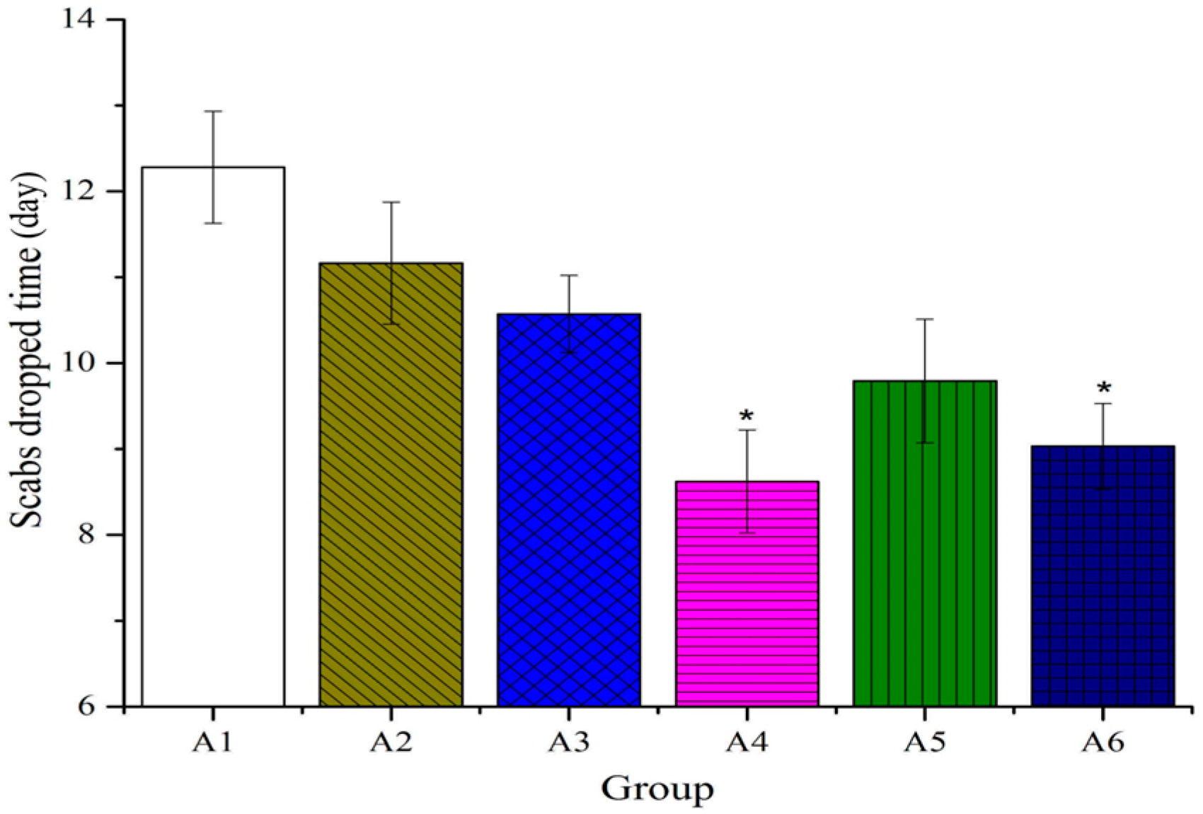
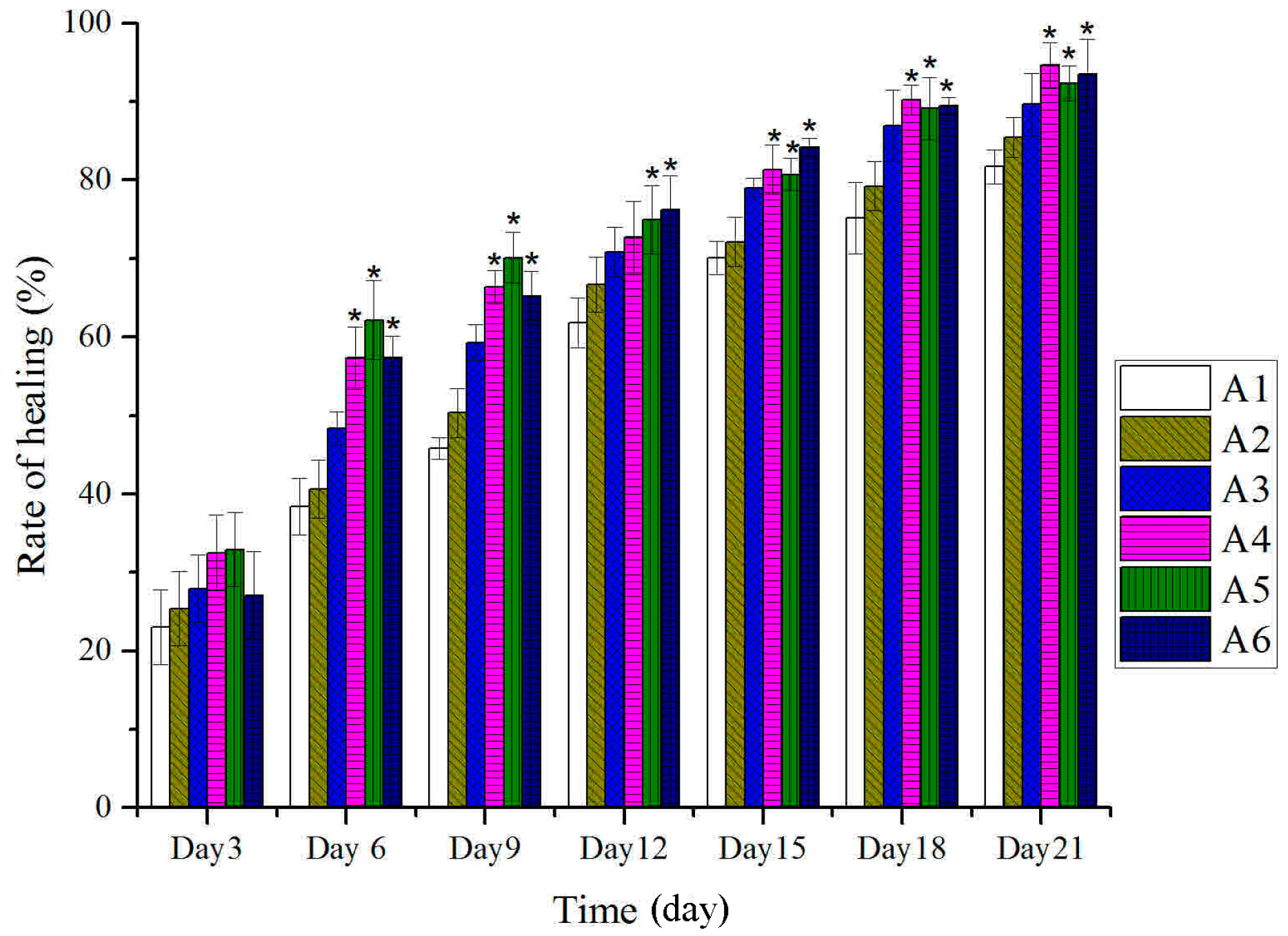
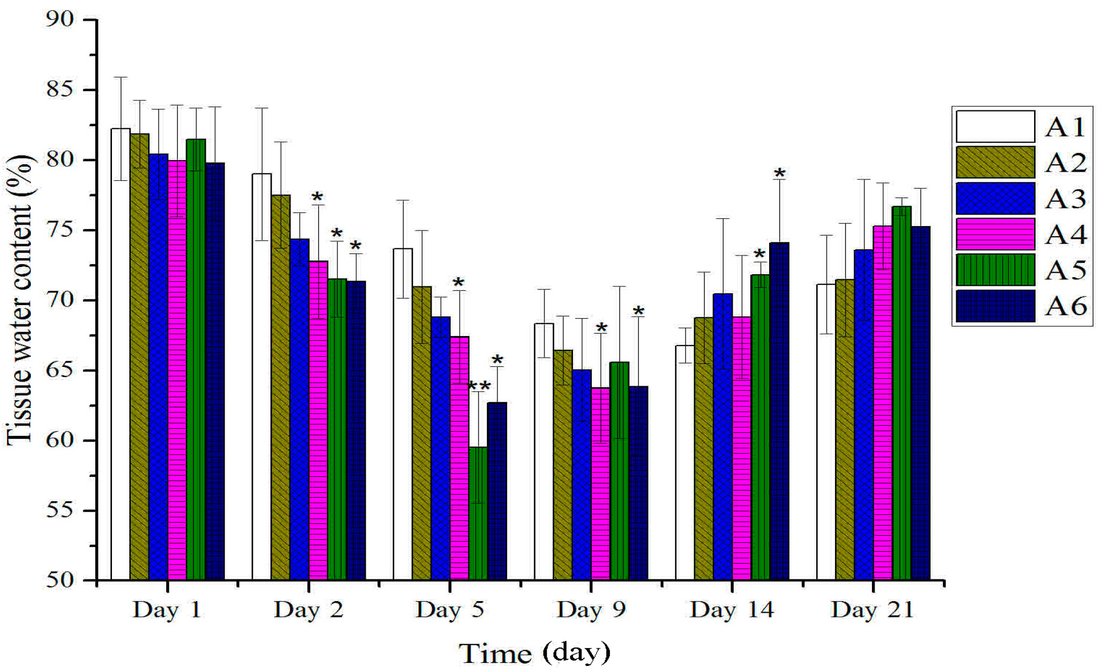
2.1. Effect of Different Treated Groups of BBO on General Observation in Rats

As shown in Figure 1, no remarkable differences were observed between treated and untreated groups in the tissue water content on the first day after burn. However, on the second day after burn, the percentage of water content significantly decreased compared to the control group. The scabs dropped time was 12.28 days (range, 11–13 days), control group; 11.16 days (range, 10–12 days), 80% ethanol (vehicle) group; 10.57 days (range 9–11 days), burn ointment (standard group); 9.15 days (range 8–10 days), BBO groups (Figure 2). On the fourth day after burn, the rate of wound healing treated with BBO was significantly greater than the control group. The burn wound was almost completely healed (>90%) on the 21st day (Figure 3), the re-epithelialization time correspondingly shortened.
Figure 1. Effect of different concentration or fractions of BBO on the tissue water content in rats. Values are expressed as mean ± S.D. (n = 4 animals). *: p < 0.05 vs. control; **: p < 0.01 vs. control.
Molecules 20 17166 g001
Figure 2. Effect of different concentration or fractions of BBO on the scabs dropped time in rats. Values are expressed as mean ± S.D. (n = 4 animals). *: p < 0.05 vs. control.
Molecules 20 17166 g002
Figure 3. Effect of different concentration or fractions of BBO on the rate of wound healing in rats. Values are expressed as mean ± S.D. (n = 4 animals). *: p < 0.05 vs. control.
Molecules 20 17166 g003

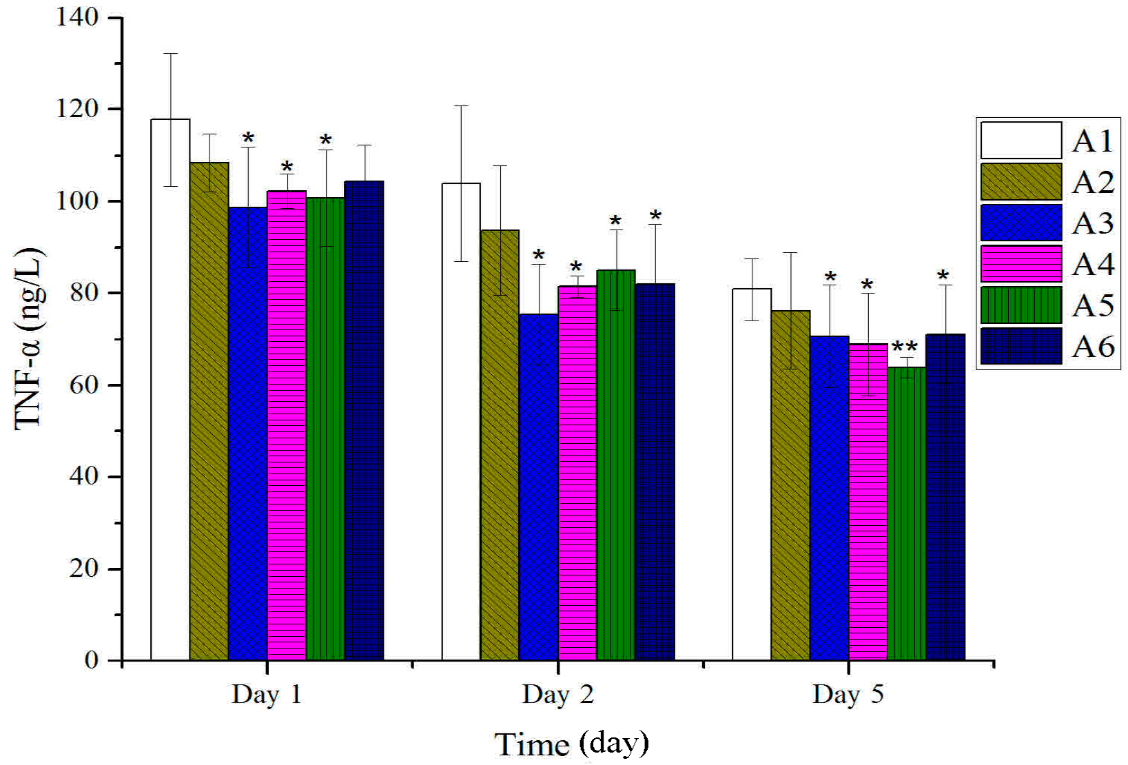
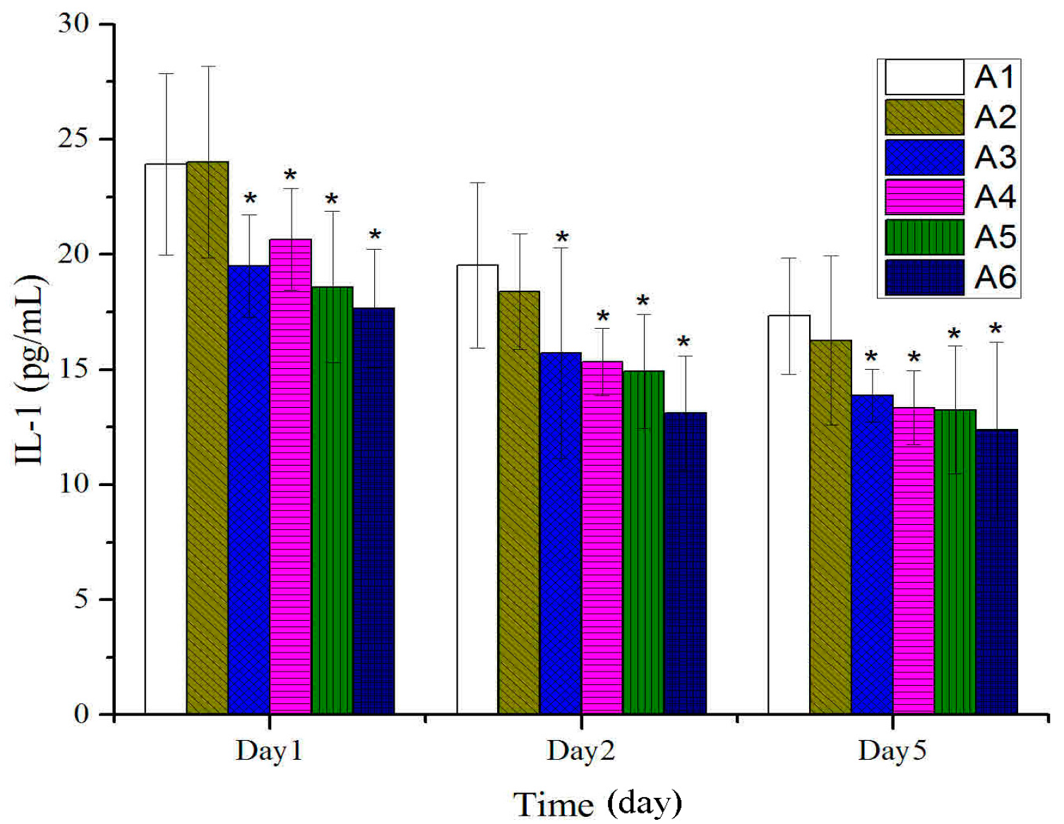
2.2. Effect of Different Treated Groups of BBO on Plasma IL-1 and TNF-α Level in Rats

As shown in Figure 4, a prominent decrease in IL-1 was observed with each BBO treatment five days after burn. As shown in Figure 5, the levels of TNF-α on plasma rapidly declined after being treated with different concentration or fractions of BBO five days after burn. No remarkable differences were observed between treated and untreated groups on the fifth day after burn.
Figure 4. Effect of different concentration or fractions of BBO on plasma IL-1 in rats. Values are expressed as mean ± S.D. (n = 4 animals). *: p < 0.05 vs. control.
Molecules 20 17166 g004
Figure 5. Effect of different concentrations or fractions of BBO on plasma TNF-α in rats. Values are expressed as mean ± S.D. (n = 4 animals). *: p < 0.05 vs. control; **: p < 0.01 vs. control.
Molecules 20 17166 g005

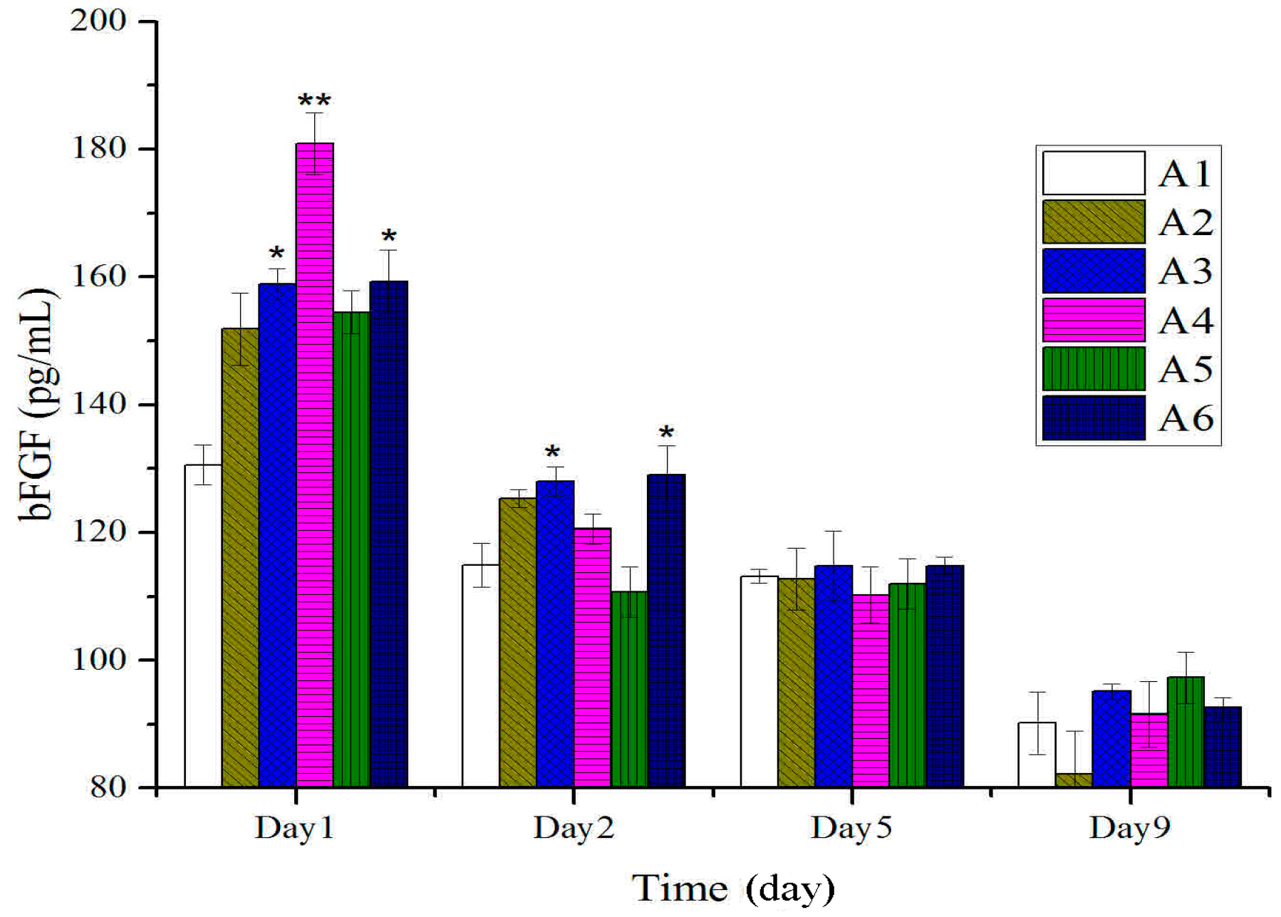
2.3. Expressions of bFGF in Tissues after Treatment with BBO in Rats

As shown in Figure 6, at the early stage after burn (first day), expressions of bFGF of treated groups with different concentration of BBO were significantly higher than those of the control group and vehicle group. With prolonging of the treatment time, there were no significant shifts in the bFGF levels at different groups.
Figure 6. Expressions of bFGF in tissues after treatment with BBO in rats. Values are expressed as mean ± S.D. (n = 4 animals). *: p < 0.05 vs. control; **: p < 0.01 vs. control.
Molecules 20 17166 g006

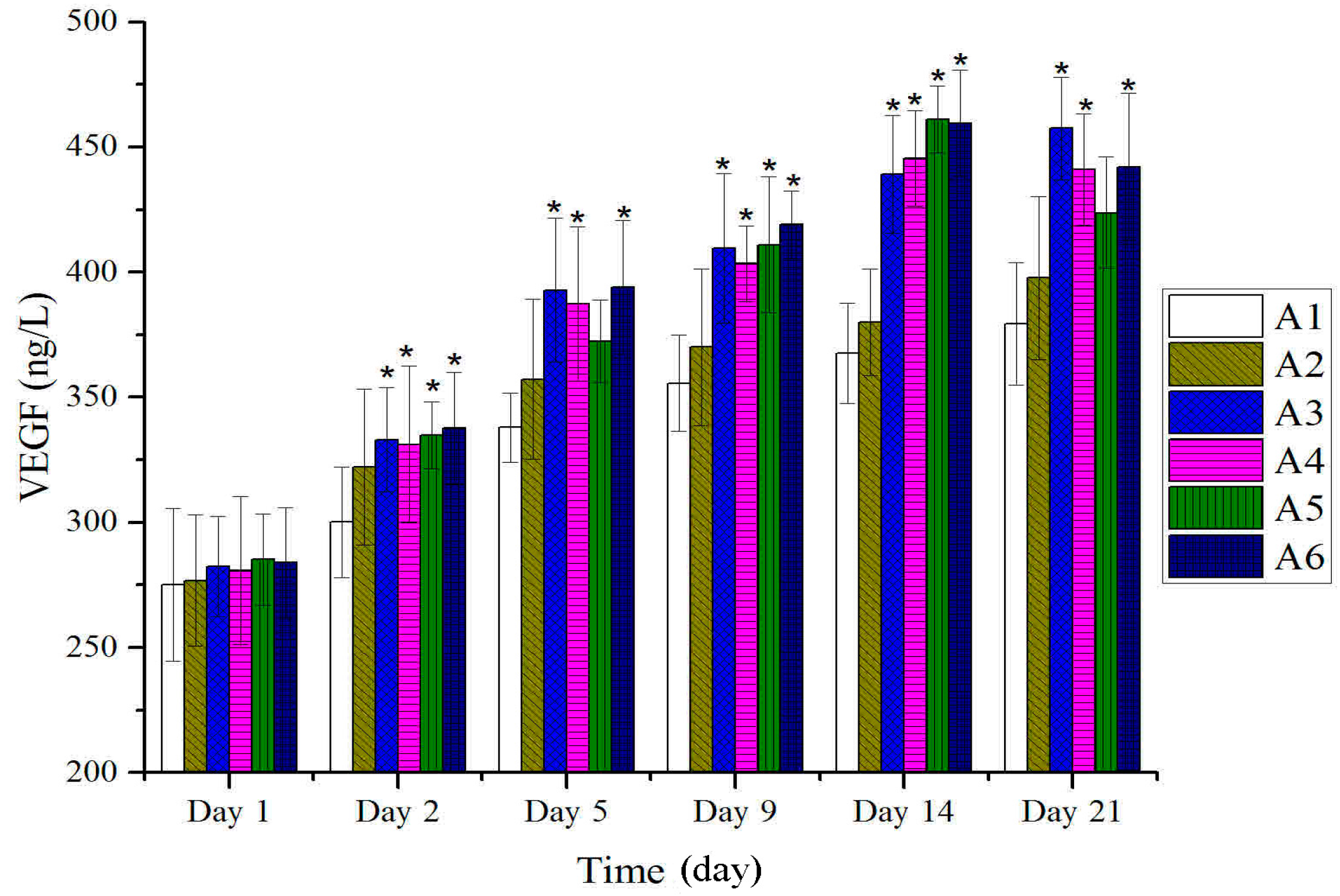
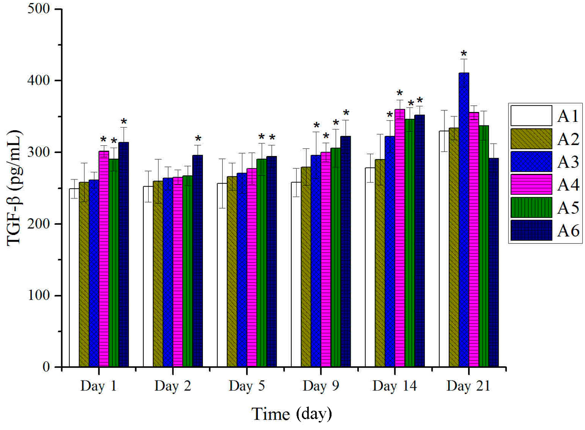
2.4. Expressions of TGF-β and VEGF in Tissues after Treatment with BBO in Rats

As shown in Figure 7, at the first period of burn (first day), the expressions of TGF-β of treated groups with BBO were significantly different compared to those of the control and vehicle groups. However, the levels of TGF-β reduced before the burn-skin dropped. With the beginning of re-epithelialization, the expressions of TGF-β were significantly higher than those of control and vehicle groups, and reached the peak at 14th day.
Figure 7. Expressions of TGF-β in tissues after treatment with BBO in rats. Values are expressed as mean ± S.D. (n = 4 animals). *: p < 0.05 vs. control.
Molecules 20 17166 g007
As shown in Figure 8, expressions of VEGF of all the treated groups with BBO showed no significantly difference compared to those of the control group and vehicle group at the early stage after burn injury. A significant increase in expressions of VEGF was observed at the start of re-epithelialization, and until the wound healing (p < 0.05). Thus, the expression of VEGF increased significantly to enhance the burn-wound healing.
Figure 8. Expressions of VEGF in tissues after treatment with BBO in rats. Values are expressed as mean ± S.D. (n = 4 animals). *: p < 0.05 vs. control.
Molecules 20 17166 g008

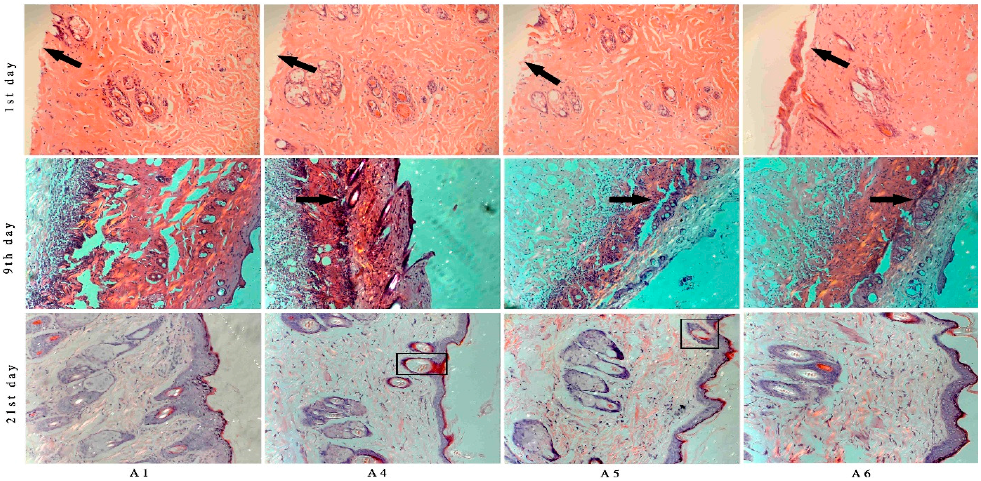
2.5. Histological Analysis

The burned tissues were measured by histological analysis to confirm the burn degree. The pathological sections showed that there were neither dermis nor epidermis; deep second-degree burn was obtained [32,33].
The histological sections from BBO treated rats showed more collagen fibers and fibroblasts cells on ninth day after burn injury compared with the control group (Figure 9). On the 21st day after burn injury, the wounded tissue treated with different concentration of BBO was almost (>90%) covered with healthy epithelial tissue and the new epidermis. The rats under different concentrations of BBO treatments showed complete epithelialization of the burn injury area, and the sebaceous gland in the section was observed (Figure 9).

2.6. Discussion

Burn is a common injury in daily clinical practice. Despite the advanced clinical care and reduced mortality of scalded patients [30,31], some problems still existed such as burn wound infection, delayed wound healing and hypertrophic scarring [34]. Patients were also faced with high economic costs as well. Alternative or complementary medicines, such as traditional Chinese medicine or ethnic drugs, have been used to improve this situation [35].
In this study on rats, different concentrations of BBO have been used and showed that these could significantly accelerate the burn-wound healing. The anti-inflammatory was a main response in the body after burn injuries. The tissue water content was a key index in anti-inflammatory, and its decrease displayed inflammatory was weakened. The percentage of tissue water content was detected and significantly decreased compared to the control group on the second day after burn, especially the 50% BBO group on the fifth day after burn (p < 0.01). The levels of inflammatory factors such as IL-1 and TNF-α on plasma were analyzed at the early stage of burn (Figure 4 and Figure 5). Previous studies suggested that IL-1 and TNF-α accelerate the anti-inflammatory at low concentration at the early stage of burn, and high concentration could cause systemic inflammatory response syndrome [36,37,38,39]. These results confirmed significant decrease in the levels of IL-1 and TNF-α of rats after treatment with BBO on plasma (p < 0.05, respectively), and especially the 50% BBO was observed at fifth day after burn (p < 0.01). In the present study, the treated groups with BBO were found to promote anti-inflammatory activity by reducing the levels of inflammatory factors.
Figure 9. Histopathology of burned tissues samples after treatment with different concentration of BBO at Days 1, 9 and 21 (stained with Hematoxylin-Eosin). On the first day after burn, the pathological sections showed that there were neither dermis nor epidermis (arrows at first day). At the ninth day, the scabs were dropped, and some new epithelial tissues were observed (arrows at ninth day). At the 21st day, the wound was covered by healthy epithelial tissue and the new epidermis, and generated the sebaceous and sweat gland (blocks at 21st day).
Molecules 20 17166 g009
Many types of cytokines and growth factors are responsible for inflammation, re-epithelialization, wound contraction, the formation of granulation tissue and neovascularization during the burn-wound healing process [40]. TGF-β plays a key role through regulating different cellular functions, and enhancing granulation tissue formation and collagen formation [41]. In addition, TGF-β has also been reported to be able to promote wound contraction through its direct induction of alpha smooth muscle act in expression in fibroblasts [42,43]. At the same time, VEGF as an important growth factor in pathological situations, such as tissue repair, involves neovascularization and increased vascular permeability [44,45]. In addition, VEGF also mediates vascular hyper permeability and promotes the secretion of both active growth factors and cytokines necessary for wound repair [46,47,48,49]. These results suggested that the expressions of TGF-β and VEGF followed the beginning of re-epithelialization and significantly higher in the BBO treated groups, and reached the peak on the 14th day (Figure 7 and Figure 8).
A previous study by Werner S showed that bFGF was confirmed as a trigger in the wound repair to enhance the expression of TGF-β and VEGF [44]. Further, it was also an important growth factor in the early phase of burn injury, to improve the quality of skin grafting in burn patients. In this study, the rats treated with BBO significantly enhanced the expression of bFGF at the early stage of burn injury, stimulating the formation of collagenous fiber and the expressions of TGF-β and VEGF (Figure 6). It is important to mention that BBO could accelerate the burn-wound healing by influencing the expression of growth factors and cytokines.
This investigation suggested that three different concentrations of BBO were able to promote burn-wound healing, and the rate of healing in the treated group with 10% BBO was faster than in 50% BBO group (Figure 3). It may be influenced by ethanol (80%), which was a good germicide and vehicle. However, the results of vehicle group showed that ethanol (80%) could not significantly accelerate the wound healing. It revealed that germicide as an auxiliary with could produce better therapeutic activity only with the help of BBO. Compared with the quality of skin that recovered from burn injury, the skin treated by 100% and 50% BBO groups was superior to that of other treated groups. The new skin has been covered with healthy epithelial tissue and the new epidermis, and generated the sebaceous and sweat gland with little visible scars (Figure 9). The skin thickness of new epidermis with BBO groups was significantly thinner compared to the control group (Figure 9). It may be related to the secretion of cytokines and growth factors and this needs further investigation [50,51,52].
Though some papers published by other authors reported that the BBO excited different components caused by location and extraction procedure, the main composition was uniform, for example l-borneol, d-camphor and caryophyllene, which makes sure that BBO is uniformly efficacious as an effective burn-healing agent [26,27,28,53]. Previous investigation showed that l-borneol and d-camphor were the main constituents in BBO [28]. l-borneol has been reported to be able to promote permeability and is anti-inflammatory, inhibiting the expressions of TNF-α, IL-1 and IL-6 [54,55,56,57,58]. However, the contact between l-borneol and growth factors is not reported. It may also be the main effect ingredient that could accelerate anti-inflammatory effects at the early stage of burn injury healing. Possibly, the healing efficacy of BBO may probably be due to the synergistic action of its multiple phytoconstituents, and further phytochemical studies are required to isolate the active compound(s) responsible for adding these pharmacological activities.

3. Experimental Section

3.1. Materials

B. balsamifera leaves was collected in Luodian (Guizhou, China) and BBO was provided by Guizhou AiYuan Ecological Medicine Development Ltd. (Guizhou, China). The oil yield was about 0.01% of fresh B. balsamifera leaves and extracted by steam distillation. The samples were stored at 4 °C until use and the composition were analyzed in previous investigation [28].

3.2. Experimental Animals

Healthy male and female Sprague-Dawley rats were obtained from Changsha Tianqin Biotech Ltd. (Changsha, China). The rats, weighing between 220 and 250 g, were housed in polypropylene cage and maintained in standard laboratory conditions of temperature (24 ± 2 °C), light-dark cycle 12 h:12 h and humidity (60% ± 5%). They were fed on standard pellet diet and water ad libitum. Before administering any treatment, rats were kept off feed for 7 days. After treatment, food was made available ad libitum. Then, the animals were sacrificed under anesthesis at the end of the experiment.

3.3. Animal Model

Rats were anesthetized with 10% chloral hydrate with a dose of 350 mg kg−1 of body, administered by an intra-peritoneal injection. The dorsal fur was removed by 10% sodium sulfide. Briefly, a 2.5 cm in diameter steel rod (25 cm in length) was heated to 100 °C in boiling water for 15 min, then it was applied for 7 s on the removed dorsal skin of the rat and created two full thickness of burn injury. Lactated Ringer’s solution (5 mL of body) was immediately injected into peritoneal cavity to prevent shock [59]. All animals were randomly assigned to 6 groups of 24 each. After 1 h, all the animals were administered treatment (Table 1) and applied once daily for 21 days.
Table 1. Animal groups (n = 24/group), treatments, doses and route of administration of different concentration or fractions of BBO TDD.
Group No.TreatmentsDoses (mg/kg)
A1Control0
A280% ethanol (vehicle)1500
A3Moist exposed burn ointment (standard)1500
A4BBO (100%)1500
A5BBO (50%)1500
A6BBO (10%)1500

3.4. General Observation

The rate of healing, scabs dropped time, granulation and re-epithelialization time were observed every three days for 21 days after burn. The full-thickness of burn area (about 100 mg) was removed from different treated rats by sacrificing the animals on the 1st, 2nd, 5th, 9th, 14th, and 21st day post-burn creation, and weight was measured after bake for 24 h at 80 °C to obtain the tissue water content. The rate of healing was assessed by digitized planimetry.

3.5. Sample Collection

Blood samples (about 3 mL) were collected from rats by vein puncture in the mandibular region under anesthesia on the 1st, 2nd, 5th, 9th, 14th, 21st day post-burn creation, and centrifuged at 4000 rpm for 10 min at 15 °C for plasma. The blood samples were stored at −80 °C in aliquots until analysis.
The full thickness of burn area was removed from different treated rats by sacrificing the animals on the 1st, 2nd, 5th, 9th, 14th, and 21st day post-burn creation, one aliquot (about 100 mg) was processed by standard histological techniques, and others samples were stored at −80 °C in aliquots until analysis.

3.6. Clinical Chemistry

Plasma levels of TNF-α, IL-1 were analyzed with the TNF-α adduct ELISA Kit (Nanjing Jiancheng Bioengineering Institute, Nanjing, China) and the IL-1 Assay kit (Nanjing Jiancheng Bioengineering Institute, Nanjing, China). The frozen full-thickness samples were processed by standard ELISA techniques. The samples were subsequently centrifuged and the supernatant isolated for analysis. The enzyme-linked immunoassay (Nanjing Jiancheng Bioengineering Institute, Nanjing, China) was performed according to the manufacturer’s instructions. The tissue supernatant was analyzed with antibody magnetic beads in duplicate. Three different cytokines and growth factors were assessed, namely, TGF-β, VEGF, and bFGF.

3.7. Histopathologic Evaluation

The full-thickness tissue was processed routinely and embedded in paraffin blocks. Tissue sections were prepared (6 μm) and stained with hematoxylin and eosin (H & E). The slides were assessed using a light microscope (200×).

3.8. Statistical Analysis

The results were expressed as mean ± standard deviation (SD). Data were analyzed by one-way ANOVA followed by Duncan’s multiple range test using Statistical Package for Social Sciences 18.0 (SPSS, Chicago, IL, USA). Values were considered statistically significant when p-value was less than 0.05.

4. Conclusions

The study showed that the BBO facilitated wound healing in experimental animal model by influencing the anti-inflammatory response, and the expressions of cytokines and growth factors in burn injury repair process. BBO could upgrade the quality of skin that recovered from burn injuries. The biological data obtained in the present study should be useful for the further development of new burn-treating drugs. However, there is a need for further studies in order to analyze the active ingredients in the BBO and to completely elucidate the mechanisms of actions of these active ingredients.

Acknowledgments

This research was funded by the National Natural Science Funds of China (No. 81374065), the Fundamental Scientific Research Funds for Chinese Academy of Tropical Agricultural Sciences—Crops Genetic Resources Institute (no. 1630032014016) and the Non-Profit Funds for CATAS-TCGRI (no. 201421). The authors thank Zou Chunli, for his company supplying Blumea balsamifera oil for this study. The authors thank Guan Qinghua for the technical guidance. The authors would like to thank Jiang Dongquan and his company staff for animal supply services.

Author Contributions

Zuo-Wang Fan: Experiments, interpretation of data and wrote the manuscript. Kai Wang: interpretation of data and revising the article. Fu-Lai Yu: References management and revising the article. Quan Yang: Obtained fundings. Qing-Song Ma: Experiments. Xiao-Ting Li: Experiments. Jin Zou: Experiments. Wen-Qing Zhang: Experiments. Li-Fen Wu: Experiments. Yu-Xin Pang and Dan Wang: Contributed to conception and design of the article, obtained funding, and overall responsibility.

Conflicts of Interest

The authors declare no conflict of interest.

References

  1. Wasserman, D. Criteria for burn severity: Epidemiology, prevention, organization of management. Pathol. Biol. 2002, 50, 65–73. [Google Scholar]
  2. Ma, X.L. The experience of nursing the severe burn patients with sepsis. Chin. J. Med. Guide 2012, 3, 481. (In Chinese) [Google Scholar] [CrossRef]
  3. Li, A. The Treatment of Burns, 2nd ed.; People’s Medical Publishing House: Beijing, China, 1995. [Google Scholar]
  4. Tyack, Z.F.; Ziviani, J. What influences the functional outcome of children at 6 months post-burn? Burns 2003, 29, 433–444. [Google Scholar] [CrossRef]
  5. Griffiths, H.R.; Thornton, K.L.; Clements, C.M.; Burge, T.S.; Kay, A.R.; Young, A.E. The cost of a hot drink scald. Burns 2006, 32, 372–374. [Google Scholar] [CrossRef] [PubMed]
  6. Atiyeh, B.S.; Costagliola, M.; Hayek, S.N. Effect of silver on burn wound infection control and healing: Review of the literature. Burns 2007, 33, 139–148. [Google Scholar] [CrossRef] [PubMed]
  7. Aziz, Z.; Abu, S.F.; Chong, N.J. A systematic review of silver-containing dressings and topical silver agents (used with dressings) for burn wonds. Burns 2012, 38, 307–318. [Google Scholar] [CrossRef] [PubMed]
  8. Fraser, J.F.; Cuttle, L.; Kempf, M. Cytotoxicity of topical anti-microbial agents used in burn wounds in Australasia. ANZ J. Surg. 2004, 74, 139–142. [Google Scholar] [CrossRef] [PubMed]
  9. Kearns, R.D.; Cairns, C.B.; Holmes, J.H.; Rich, P.B.; Cairns, B.A. Chemical burn care: A review of best practices. EMS World 2014, 43, 40–45. [Google Scholar] [PubMed]
  10. Rowan, M.P.; Cancio, L.C.; Burmeister, D.M.; Rose, L.F.; Natesan, S.; Chan, R.K.; Christy, R.J.; Chung, K.K. Burn wound healing and treatment: Review and advancements. Crit. Care 2015, 19, 243. [Google Scholar] [CrossRef] [PubMed]
  11. Gupta, A.; Upadhyay, N.K.; Sawhney, R.C.; Kumar, R. A poly herbal formulation accelerates normal and impaired diabetic wound healing. Wound Repair Regen. 2008, 16, 784–790. [Google Scholar] [CrossRef] [PubMed]
  12. Shetty, S.; Udupa, S.; Udupa, L. Evaluation of antioxidant and wound healing effects of alcoholic and aqueous extract of Ocimum sanctum Linn in rats. Evid. Based Complement. Altern. Med. 2008, 5, 95–101. [Google Scholar] [CrossRef] [PubMed]
  13. Nayak, B.S.; Sandiford, S.; Maxwell, A. Evaluation of the wound- healing activity of ethanolic extract of Morinda citrifolia L. leaf. Evid. Based Complement. Altern. Med. 2009, 6, 351–356. [Google Scholar] [CrossRef] [PubMed]
  14. Phan, T.T.; Hughes, M.A.; Cherry, G.W. Enhanced proliferation of fibroblasts and endothelial cells treated with an extract of the leaves of Chromolaena odorata (Eupolin), an herbal remedy for treating wounds. Plast. Reconstr. Surg. 1998, 101, 756–765. [Google Scholar] [CrossRef] [PubMed]
  15. Pang, Y.X.; Wang, W.Q.; Mo, T.H.; Yuan, Y. Clonal diversity and structure in natrual populations of Blumea balsamifera. Guihaia 2010, 2, 13–15. (In Chinese) [Google Scholar]
  16. Yuan, Y.; Pang, Y.X.; Wang, W.Q.; Yu, J.B. Investigation on the plants resources of Blumea balsamifera (L.) DC. in China. J. Trop. Org. 2011, 2, 78–82. (In Chinese) [Google Scholar]
  17. Guan, L.L.; Pang, Y.X.; Wang, D.; Wu, K.Y. Research progress on Chinese minority medicine of Blumea balsamifera L. DC. J. Plant Genet. Res. 2012, 4, 695–698. [Google Scholar]
  18. Jiangsu New Medical College. Dictionary of Chinese Materia Medica; Shanghai Science and Technology Press: Shanghai, China, 1977. [Google Scholar]
  19. Guizhou Institute of Traditional Chinese Medicine. Guizhou Chinese Medicine Resources; Chinese Medical Science Press: Beijing, China, 1992; p. 262. [Google Scholar]
  20. Li, J.; Zhao, G.Z.; Chen, H.H.; Wang, H.B.; Qin, S.; Zhu, W.Y.; Xu, L.H.; Jiang, C.L.; Li, W.J. Antitumour and antimicrobial activities of endophytic streptomycetes from pharmaceutical plants in rainforest. Lett. Appl. Microbiol. 2008, 47, 574–580. [Google Scholar] [CrossRef] [PubMed]
  21. Osaki, N.; Koyano, T.; Kowithayakorn, T.; Hayashi, M.; Komiyama, K.; Ishibashi, M. Sesquiterpenoids and plasmin-inhibitory flavonoids from Blumea balsamifera. J. Nat. Prod. 2005, 68, 447–449. [Google Scholar] [CrossRef] [PubMed]
  22. Fazilatun, N.; Nornisah, M.; Zhari, I. Superoxide radical scavenging properties of extracts and flavonoids isolated from the leaves of Blumea balsamifera. Pharm. Biol. 2005, 43, 15–20. [Google Scholar] [CrossRef]
  23. Chen, M.; Qin, J.J.; Fu, J.J.; Hu, X.J.; Liu, X.H.; Zhang, W.D.; Jin, H.Z. Blumeaenes A-J, sesquiterpenoid esters from Blumea balsamifera with NO inhibitory activity. Planta Med. 2010, 76, 897–902. [Google Scholar] [CrossRef] [PubMed]
  24. Hasegawa, H.; Yamada, Y.; Komiyama, K.; Hayashi, M.; Ishibashi, M.; Yoshida, T.; Koyano, T.; Kam, T.S.; Murata, K.; Sugahara, K.; et al. Dihydroflavonol BB-1, an extract of natural plant Blumea balsamifera, abrogates TRAIL resistance in leukemia cells. Blood 2006, 107, 679–688. [Google Scholar] [CrossRef] [PubMed]
  25. Norikura, T.; Kojima-Yuasa, A.; Shimizu, M.; Huang, X.D.; Xu, S.H.; Kametani, S.; Rho, S.N.; Kennedy, D.O.; Matsui-Yuasa, I. Anticancer activities and mechanisms of Blumea balsamifera extract in hepatocellular carcinoma Cells. Am. J. Chin. Med. 2008, 36, 411–424. [Google Scholar] [CrossRef] [PubMed]
  26. Zhou, X.; Yang, X.S.; Zhao, C. Chemical components of volatile oil from foliumet caumen Blumea balsamifera originated from Guizhou. J. Instrum. Anal. 2001, 20, 76–78. (In Chinese) [Google Scholar]
  27. Md Nazrul, I.B.; Jasim, U.C.; Jaripa, B. Chemical components in volatile oil from Blumea balsamifera (L.) DC. Bangladesh J. Bot. 2009, 38, 107–109. [Google Scholar]
  28. Pang, Y.X.; Fan, Z.W.; Wang, D.; Yang, Q.; Wang, K.; Chen, X.L.; Hu, X.; Yu, F.L.; Chen, Z.X. External application of the volatile oil from Blumea balsamifera may be safe for liver—A study on its chemical composition and hepatotoxicity. Molecules 2014, 19, 18479–18492. [Google Scholar] [CrossRef] [PubMed]
  29. Chinese Pharmacopeia Commission. Pharmacopoeia of the People’s Republic of China; China Medical Science Press: Beijing, China, 2010; p. 82. [Google Scholar]
  30. Pang, Y.X.; Wang, D.; Fan, Z.W.; Chen, X.L.; Yu, F.L.; Hu, X.; Wang, K.; Yuan, L. Blumea balsamifera—A phytochemical and pharmacological review. Molecules 2014, 19, 9453–9477. [Google Scholar] [CrossRef] [PubMed]
  31. Pang, Y.X.; Wang, D.; Hu, X.; Wang, H.; Fu, W.; Fan, Z.W.; Chen, X.L.; Yu, F.L. Effect of volatile oil from Blumea balsamifera (L.) DC. leaves on wound healing in mice. J. Tradit. Chin. Med. 2014, 34, 716–724. [Google Scholar] [CrossRef]
  32. Dorsett-Martin, W.A. Rat models of skin wound healing: A review. Wound Repair Regen. 2004, 12, 591–599. [Google Scholar] [CrossRef] [PubMed]
  33. Jandera, V.; Hudson, D.A.; de Wet, P.M.; Innes, P.M.; Rode, H. Cooling the burn wound: Evaluation of different modalities. Bums 2000, 26, 265–270. [Google Scholar] [CrossRef]
  34. Thomas, G.W.; Rael, L.T.; Bar-Or, R.; Shimonkevitz, R.; Mains, C.W.; Slone, D.S.; Craun, M.L.; Bar-Or, D. Mechanisms of delayed wound healing by commonly used antiseptics. J. Trauma 2009, 66, 82–90. [Google Scholar] [CrossRef] [PubMed]
  35. Cho Lee, A.R.; Leem, H.; Lee, J.; Park, K.C. Reversal of silver sulfadiazine-impaired wound healing by epidermal growth factor. Biomaterials 2005, 26, 4670–4676. [Google Scholar] [CrossRef] [PubMed]
  36. Arturson, G. Pathophysiology of the burn wound and pharmacological treatment. The Rudi Hermans lecture, 1995. Burns 1996, 22, 255–274. [Google Scholar] [CrossRef]
  37. Lootens, L.; Brusselaers, N.; Beele, H.; Monstrey, S. Keratinocytes in the treatment of severe burn injury: An update. Int. Wound J. 2012, 10, 6–12. [Google Scholar] [CrossRef] [PubMed]
  38. Franchi, L.; Eigenbrod, T.; Nunez, G. Cutting edge: TNF-alpha mediates sensitization to ATP and silica via the NLRP3 inflammasome in the absence of microbial stimulation. J. Immuol. 2009, 183, 792–796. [Google Scholar] [CrossRef] [PubMed]
  39. Leung, L.; Cahill, C.M. TNF-alpha and neuropathic pain—A review. J. Neuroinflamm. 2010, 27, 2–11. [Google Scholar]
  40. Ashcroft, G.S.; Jeong, M.J.; Ashworth, J.J.; Hardman, M.; Jin, W.; Moutsopoulos, N.; Wild, T.; McCartney-Francis, N.; Sim, D.; McGrady, G.; et al. Tumor necrosis factor-alpha (TNF-α) is a therapeutic target for impaired cutaneous wound healing. Wound Repair Regen. 2012, 20, 38–49. [Google Scholar] [CrossRef] [PubMed]
  41. Lim, J.S.; Yoo, G. Efects of adipose-derived stromal cells and of their extract on wound healing in a mouse model. J. Korean Med. Sci. 2010, 25, 746–751. [Google Scholar] [CrossRef] [PubMed]
  42. Akita, S.; Akino, K.; Imaizumi, T.; Hirana, A. A basic fibroblast growth factor improved the quality of skin grafting in burn patient. Burns 2005, 31, 855–858. [Google Scholar] [CrossRef] [PubMed]
  43. Werner, S.; Krieg, T.; Smola, H. Keratinocyte-fibroblast interactions in wound healing. J. Investig. Dermatol. 2007, 127, 998–1008. [Google Scholar] [CrossRef] [PubMed]
  44. Desmouliere, A.; Geinoz, A.; Gabbiani, F.; Gabbiani, G. Transforming growth factor-beta 1 induces alpha-smooth muscle act in expression in granulation tissue myofibroblasts and in quiescent and growing cultured fibroblasts. J. Cell Biol. 1993, 122, 103–111. [Google Scholar] [CrossRef] [PubMed]
  45. Kulkarni, A.B.; Huh, C.G.; Becker, D.; Geiser, A.; Lyght, M.; Flanders, K.C.; Roberts, A.B.; Sporn, M.B.; Ward, J.M.; Karlsson, S. Transforming growth factor beta 1 null mutation in mice causes excessive inflammatory response and early death. Proc. Natl. Acad. Sci. USA 1993, 90, 770–774. [Google Scholar] [CrossRef] [PubMed]
  46. Leung, D.W.; Cachianes, G.; Kuang, W.J.; Goeddel, D.V.; Ferrara, N. Vascular endothelial growth factor is a secreted angiogenic mitogen. Science 1989, 246, 1306–1309. [Google Scholar] [CrossRef] [PubMed]
  47. Nissen, N.N.; Polverini, P.J.; Koch, A.E.; Volin, M.V.; Gamelli, R.L.; DiPietro, L.A. Vascular endothelial growth factor mediates angiogenic activity during the proliferative phase of wound healing. Am. J. Pathol. 1998, 152, 1445–1452. [Google Scholar] [PubMed]
  48. Infanger, M.; Schmidt, O.; Kossmehl, P.; Grad, S.; Ertel, W.; Grimm, D. Vascular endothelial growth factor serum level is strongly enhanced after burn injury and correlated with local and general tissue edema. Burns 2004, 30, 305–311. [Google Scholar] [CrossRef] [PubMed]
  49. Corral, C.J.; Siddiqui, A.; Wu, L.; Farrell, C.L.; Lyons, D.; Mustoe, T.A. Vascular endothelial growth factor is more important than basic fibroblastic growth factor during is chemic wound healing. Arch. Surg. 1999, 134, 200–205. [Google Scholar] [CrossRef] [PubMed]
  50. Santos, S.C.; Miguel, C.; Domingues, I.; Calado, A.; Zhu, Z.; Wu, Y.; Dias, S. VEGF and VEGFR-2 (KDR) internalization is required for endothelial recovery during wound healing. Exp. Cell Res. 2007, 313, 1561–1574. [Google Scholar] [CrossRef] [PubMed]
  51. Liu, H.H.; Lin, S.; Xiao, D.; Zheng, X.; Gu, Y.; Guo, S. Evaluation of the wound healing potential of resina Draconis (Dracaena cochinchinensis) in animal models. Evid. Based Complement. Altern. Med. 2013. [Google Scholar] [CrossRef] [PubMed]
  52. Wilgus, T.A. Immune cells in the healing skin wound: Influential players at each stage of repair. Pharmacol. Res. 2008, 58, 112–116. [Google Scholar] [CrossRef] [PubMed]
  53. Jiang, Z.L.; Zhou, Y.; Ge, W.C.; Yuan, K. Phytochemical compositions of volatile oil form Blumea balsamifera and their biological activities. Pharmacogn. Mag. 2014, 10, 346–352. [Google Scholar] [PubMed]
  54. Fu, W.J.; Wang, D.; Pang, Y.X.; Wang, D.; Huang, Z.; Nie, H.; Yu, F.L.; Zhang, Y.B. Effect of Blumea balsamifera oil on percutaneous absorption of salbutamol sulfate. Chin. J. Exp. Tradit. Med. Formula 2013, 19, 174–177. (In Chinese) [Google Scholar]
  55. Almeida, J.R.; Souza, G.R.; Silva, J.C.; Saraiva, S.R.; Junior, R.G.; Quintans, J.S.; Barrrto, R.S.; Bonjardim, L.R.; Cavalcanti, S.C.; Quintans, L.J., Jr. Borneol, a bicyclic monoterpene alcohol, reduces nociceptive behavior and inflammatory response in mice. Sci. World J. 2013. [Google Scholar] [CrossRef] [PubMed]
  56. Chien, T.C.; Lo, S.F.; Ho, C.L. Chemical composition and anti-inflammatory activity of Chamaecyparis obtuse f.formasana wood essential oil from Taiwan. Nat. Prod. Commun. 2014, 9, 723–729. [Google Scholar] [PubMed]
  57. Wu, H.Y.; Tang, Y.; Gao, L.Y.; Sun, W.X.; Yang, S.B.; Zhang, Z.P.; Liao, Q.G.; Luo, C.X.; Zhu, D.Y. The synergetic effect of edaravone and borneol in the rat model of ischemic stroke. Eur. J. Pharmacol. 2014, 740, 522–531. [Google Scholar] [CrossRef] [PubMed]
  58. Xia, Y.; Zuo, J.; Li, X. Chen, J.W. Anti-arthritic and anti-oxidative effect of ethyl acetate fraction of Blumea balsamlfera residues in rat adjuvant-induced arthritis. Zhongguo Zhong Yao Za Zhi 2014, 39, 3819–3823. [Google Scholar] [PubMed]
  59. Su, Y.; Zhu, W.J.; Fang, M.; Zhou, L.L. Study on protective effect and mechanism of Fufang Tangshang Gao on deep grade II skin burn in rats. Zhongguo Zhong Yao Za Zhi 2012, 37, 2143–2146. (In Chinese) [Google Scholar] [PubMed]
  • Sample Availability: Samples of the compounds are not available from the authors.

