Article

Steroidal Saponins from the Roots and Rhizomes of Tupistra chinensis

1
Shaanxi Collaborative Innovation Center of Chinese Medicinal Resource Industrialization, Shaanxi Province Key Laboratory of New Drugs and Chinese Medicine Foundation Research, Shaanxi Rheumatism and Tumor Center of TCM Engineering Technology Research, School of Pharmacy, Shaanxi University of Chinese Medicine, Xianyang 712046, China
2
The First Hospital of Xi'an, Xi'an 710002, China
3
School of Pharmaceutical Sciences, Xi'an Medical University, Xi'an 710021, China
*
Authors to whom correspondence should be addressed.
These authors contributed equally to this work.
Molecules 2015, 20(8), 13659-13669; https://doi.org/10.3390/molecules200813659
Submission received: 25 June 2015 / Revised: 22 July 2015 / Accepted: 23 July 2015 / Published: 28 July 2015
(This article belongs to the Section Natural Products Chemistry)

Abstract

:
Two new furostanol saponins 12 and a new spirostanol saponin 3 were isolated together with two known furostanol saponins 45 from the roots and rhizomes of Tupistra chinensis. Their structures were characterized as 1β,2β,3β,4β,5β,26-hexahydroxyfurost-20(22),25(27)-dien-5,26-O-β-d-glucopyranoside (1), 1β,2β,3β,4β,5β,6β,7α,23ξ,26-nona-hydroxyfurost-20(22),25(27)-dien-26-O-β-d-glucopyranoside (2), (20S,22R)-spirost-25 (27)-en-1β,3β,5β-trihydroxy-1-O-β-d-xyloside (3), tupisteroide B (4) and 5β-furost-Δ25(27)-en-1β,2β,3β,4β,5β,7α,22ξ,26-octahydroxy-6-one-26-O-β-d-glucopyranoside (5), respectively, by extensive use of spectroscopic techniques and chemical evidence. Additionally, the in vitro cytotoxic activity of 14 was evaluated on human A549 and H1299 tumor cell lines, and compound 3 exhibited cytotoxicity against A549 cells (IC50 86.63 ± 2.33 μmol·L−1) and H1299 cells (IC50 88.21 ± 1.34 μmol·L−1).

1. Introduction

Tupistra chinensis Baker., a species in the Tupistra genus of the Liliaceae family, is used as an endemic herbal medicine, known as “Kai-Kou-Jian”, in the Qinba Mountains of Shaanxi Province in China [1]. The roots and rhizomes of T. chinensis are commonly used as folk medicine to treat throat irritation, rheumatic diseases and snake-bites [2,3]. Modern pharmacological experiments have showed that the extracts of this species possess significant antitumor activities [4,5], moreover, two main kinds of components—cardenolides and saponins—were isolated from T. chinensis [3,6,7]. As part of our research project to find more diverse bioactive leading compounds from the medicinal herbs of the Qinba Mountains [8,9,10,11], the chemical constituents and pharmacological studies of T. chinensis were investigated, and two new furostanol saponins, 1β,2β,3β,4β,5β,26-hexahydroxyfurost-20(22),25(27)-dien-5,26-O-β-d-glucopyranoside (1), 1β,2β,3β,4β,5β,6β,7α,23ξ,26-nonahydroxyfurost-20(22),25(27)-dien-26-O-β-d-glucopyranoside (2), and a new spirostanol saponin (20S,22R)-spirost-25(27)-en-1β,3β,5β-trihydroxy-1-O-β-d-xyloside (3) were obtained from the roots and rhizomes of T. chinensis together with the two known compounds tupisteroide B (4) and 5β-furost-Δ25(27)-en-1β,2β,3β,4β,5β,7α,22ξ,26-octahydroxy-6-one-26-O-β-d-glucopyranoside (5) (Figure 1). The cytotoxic activity of 14 was evaluated on human A549 and H1299 tumor cells.
Figure 1. Structures of compounds 15.
Molecules 20 13659 g001

2. Results and Discussion

Compound 1 was obtained as a white amorphous powder, which showed positive reactions in the Liebermann-Burchard, Ehrlich and Molisch reactions, suggesting that 1 was a furostanol glycoside. Its molecular formula was determined as C39H62O17 from the HR-ESI-MS peak at m/z 801.3855 [M − H]. The 1H-NMR spectrum showed three methyl protons at δH 0.67 (3H, s), 1.70 (3H, s) and 1.58 (3H, s), two exo-methylene protons (δH 5.35 (1H, brs) and 5.04 (1H, brs)), as well as signals for two anomeric protons at (δH 5.28 (d, J = 7.8 Hz) and 4.89 (1H, d, J = 7.7 Hz)). The 13C-NMR spectrum displayed 39 carbon signals, 27 of which belonged to the aglycone carbons, while the remaining signals were assignable to two glucosyl moieties (δC 103.8, 75.8, 78.5, 71.7, 78.6 and 62.6, and δC 97.4, 76.2, 78.6, 71.9, 78.8 and 62.8). Among carbon signals of the aglycone, δC 146.2 and 111.6 were due to an olefinic bond group, δC 14.3, 13.7 and 11.7 were due to three methyl groups, and δC 77.8, 68.1, 75.2, 67.6, 87.4, 84.4, 64.5 and 71.7 were due to eight oxygenated carbon groups, which indicated that 1 was a furostanol saponin with multiple hydroxyl groups. The structure of 1 was finally determined by analysis of its 2D NMR data (see Figure 2). The HMQC experiment allowed for the assignments of the proton and protonated carbon resonances in the NMR spectra of 1. HMQC correlations of (δH 5.35 (H-27a) and 5.04 (H-27b)) to δC 111.6, showed the appearance of a terminal olefinic bond at C-27. Then, HMBC correlations of H-27/C-24, C-25 and C-26, H-24/C-22, C-23, C-25 and C-26, H-26/C-24, C-25 and C-27, indicated that the appearance of an isopentene group, linked at C-22 of the tetrahydrofuran ring of the furostanol saponin. Moreover, HMBC correlations of H-19/C-1, C-5, C-9 and C-10, H-3/C-1, C-2 and C-5, and H-6/C-4 and C-5, indicated that all hydroxyl groups were linked at C-1–C-5 of the A ring of the furostanol saponin (see Figure 2). Furthermore, the remaining HMBC correlations of H-18/C-12, C-13, C-14 and C-17, H-16/C-13, C-17, C-20 and C-22, H-21/C-17, C-20 and C-22, were assigned (see Figure 2). Therefore, the aglycone of 1 was identified as 1, 2, 3, 4, 5, 26-hexanol-furost-20 (22),25(27)-dien. In addition, the HMBC correlation signals of H-Glc-1′/C-5 and H-Glc-1′′/C-26, indicated that glucosyl groups were connected as (Glc-1′′-O-C-26) and (Glc-1′-O-C-5) (see Figure 2). The two glucosyl moieties were identified as d-glucose by acid hydrolysis of 1, followed by TLC comparison with a reference compound and optical rotation determination [12], and judged to be in a β-configuration [13] from the coupling constants of the anomeric protons (7.8 Hz and 7.7 Hz, respectively). In the NOESY spectrum of 1, the NOE correlations of Me-19/H-8, H-9/H-4, H-4/H-3 and H-2, and H-2/H-1 were observed (see Figure 2), indicated α-axial configurations of H-1, H-2, H-3 and H-4, and β-orientation of Me-19, 1-OH, 2-OH, 3-OH, 4-OH and 5-OH, which supported the A/B cis ring junction pattern; the NOE correlations of Me-19/H-8, H-8/Me-18, and H-14/H-9, H-16 and H-17, supported the B/C and C/D trans ring junction pattern; and the NOE correlations of Me-18/H-15b, H-15a/H-16 and H-17, and H-17/Me-21, suggested an α-orientation of Me-21 (see Figure 2). Therefore, compound 1 was identified as 1β,2β,3β,4β,5β,26-hexahydroxyfurost-20(22),25(27)-dien-5,26-O-β-d-glucopyranoside.
Figure 2. Key HMBC and NOESY correlations of the compound 1.
Molecules 20 13659 g002
Compound 2 was obtained as a white amorphous powder, which showed positive reactions in the Liebermann-Burchard, Ehrlich, and Molisch tests, suggesting that 2 was a furostanol glycoside. The molecular formula C33H52O15 was deduced from the HR-ESI-MS peak at m/z 711.3198 [M + Na]+. Comparison of the HR-ESI-MS and NMR data of 2 and 1, indicated almost similar NMR spectroscopic features, except for the number of oxygenated methine groups. In the 13C-NMR spectrum of 2, only one glucosyl moiety (δC 104.2, 75.6, 80.0, 72.1, 79.0, 63.2) was recognized, however, nine oxygenated carbon groups of the aglycone at δC 79.1, 67.7, 76.1, 70.2, 78.6, 74.0, 72.5, 64.8 and 72.7 were identified. Meanwhile, the spectroscopic features of 2 were similar to those of tupisteroide B (4), indicating that seven hydroxyl groups were linked at C-1–C-7 of the furostanol saponin, which was confirmed by the 1H-1H COSY correlation of H-1/H-2/H-3/H-4 and H-6/H-7 and the HMBC correlation of H-19/C-1, C-5, C-9 and C-10, and H-6/C-4 and C-5 (see Figure 3). The 26-OH was connected with the glucosyl moiety from the correlation signals of H-Glc-1′/C-26 in the HMBC spectra (see Figure 3). The remaining hydroxyl group was deduced to be linked at C-23, from one oxygen-bearing methine signal occurring at δC 64.8 in 2, instead of a methylene carbon (C-23) at δC 34.3 in 4, which was correlated with a proton signal at δH 5.13 (dd, J = 6.0, 8.0 Hz, H-23) in the HMQC spectrum, and the correlation signals of H-23/H-24 in the 1H-1H COSY spectrum, the correlation signals of H-23/C-20, C-22, C-24 and C-25, H-24/C-22, C-23, C-25, C-26 and C-27, and H-27/C-24, C-25 and C-26 in the HMBC spectrum (see Figure 3). In addition, the glucosyl moiety was identified as β-d-glucose by the acid hydrolysis procedure and the coupling constant analysis of the anomeric proton (J = 7.8 Hz), according to the same protocol as that described for 1. Thus, the planar structure of 2 was deduced as 1,2,3,4,5,6,7,23,26-nonanolfurost-20(22),25(27)-dien-26-O-β-d-glucose. In the NOESY spectrum of 2, the NOE correlations of Me-19/H-8, H-4/H-2, H-3 and H-9, and H-2/H-1 were observed, indicating α-axial configurations of H-1, H-2, H-3, and H-4, and β-orientation of Me-19, 1-OH, 2-OH, 3-OH, 4-OH and 5-OH, which supported the A/B cis ring junction pattern (see Figure 3). Besides, NOE correlation of H-7/H-8 was observed and no correlation signals was occurred between Me-19/H-6, which indicated α-axial configuration of 7-OH and β-orientation of 6-OH (see Figure 3). Finally, the NOE correlations of H-8/Me-19 and Me-18, and H-14/H-16 and H-17, supported the B/C and C/D trans ring junction pattern; and the NOE correlations of Me-18/H-15b, H-15a/H-16 and H-17, and H-17/Me-21, suggested the α-orientation of Me-21 (see Figure 3). Therefore, compound 2 was identified as 1β,2β,3β,4β,5β,6β,7α,23ξ,26-nonahydroxyfurost-20(22),25(27)-dien-26-O-β-d-glucopyranoside.
Figure 3. Key HMBC, 1H-1H COSY and NOESY correlations of the compound 2.
Molecules 20 13659 g003
Compound 3 was obtained as a white amorphous powder, and the molecular formula of C32H50O9 was established by the HR-ESI-MS signal at m/z 579.3590 [M + H]+. The 13C-NMR spectrum exhibited 32 carbon signals, 27 of which were attributed to the aglycone carbons, while the remaining signals were assignable to a characteristic of a xylosyl moiety (δC 104.1, 75.8, 78.9, 71.5 and 68.1), which was identified as β-d-xylose by the coupling constant analysis of the anomeric proton (J = 7.2 Hz), the acid hydrolysis procedure, TLC comparison, and the optical rotation determination. Among the aglycone carbon signals, the quaternary carbon signal at δC 109.9 (see, Table 1), was identified as an acetal carbon (C-22), a characteristic signal of spirostanol or norspirostanol saponin [14]. In HMBC spectrum, the anomeric proton [4.81 (1H, d, J = 7.2 Hz)] of the xylose was correlated with δC 82.5, which was confirmed as C-1 for the HMQC correlation of δH 4.26 (H-1)/δC 82.5 (C-1), 1H-1H COSY correlations of H-1/H-2/H-3/H-4, and HMBC correlations of H-19/C-1, C-5, C-9 and C-10 (see, Figure 4). Moreover, HMBC correlations of H-18/C-12, C-13, C-14 and C-17, H-21/C-17, C-20 and C-22, H-23/C-22, and H-27/C-24, C-25 and C-26, were observed (see, Figure 4).The above data indicated the planar structure of 3 as spirost-25(27)-en-1,3,5-trihydroxy-1-O-β-d-xyloside. In the NOESY spectrum of 3 (see, Figure 4), the NOE correlations of Me-19/H-8, H-3/H-2a and H-4, H-2a/H-1, and H-4a/H-7a and H-9, indicated α-axial configurations of H-1 and H-3, and β-orientation of Me-19, 1-OH, 3-OH and 5-OH, which supported the A/B cis ring junction pattern; the NOE correlations of H-8/Me-19 and Me-18, and H-14/H-9 and H-7a, supported the B/C and C/D trans ring junction pattern; the NOE correlations of Me-18/H-15b and H-20, H-15a/H-16 and H-17, and H-17/Me-21, suggested α-orientation of Me-21. These spectra data was almost similar to those of (20S,22R)-1β,3β,5β-trihydroxyspirost-25(27)-en-5-O-β-d-glucopyranoside [8], expect for the site of glycosylation. Therefore, compound 3 was elucidated as (20S, 22R)-spirost-25(27)-en-1β,3β,5β-trihydroxy-1-O-β-d-xyloside.
Table 1. 1H-NMR and 13C-NMR spectral data of compounds 13.
Position123
δc aδH a (J in Hz)δc bδH b (J in Hz)δc cδH c (J in Hz)
177.84.25 (brs)79.14.29 (brs)82.54.26 (brs)
268.14.38 (brs)67.74.33 (brs)30.42.53 (H-2a, ca.) 1.85 (H-2b, ca.)
375.24.70 (brs)76.14.77 (brs)67.84.59 (brs)
467.64.08 (brs)70.25.33 (brs)40.02.40 (H-4a, ca.) 2.04 (H-4b, ca.)
587.4-78.6-74.7-
624.91.93 (ca.), 2.80 (ca.)74.05.03 (brs)36.31.54 (ca.), 1.90 (ca.)
728.51.1 (ca.), 1.51 (ca.)72.54.49 (brs)29.20.98 (H-7a, ca.) 1.51 (H-7b, ca.)
834.41.59 (ca.)34.82.62 (ca.)35.41.67 (ca.)
946.61.19 (ca.)37.82.05 (ca.)46.31.15 (ca.)
1046.2-46.3-44.9-
1121.91.41 (ca.), 1.44 (ca.)21.91.61 (ca.), 1.67 (ca.)22.31.14(ca.),1.38 (ca.)
1239.71.62 (d, 12.0), 1.15 (ca.)40.01.70 (d, 12.0), 1.24 (ca.)40.51.73 (d, 12.5), 1.13 (ca.)
1343.3-43.8-41.2-
1454.30.76 (ca.)48.91.96 (ca.)56.71.12 (ca.)
1531.02.48 (H-15a, ca.) 2.38 (H-15b, ca.)34.72.58 (H-15a, ca.) 1.65 (H-15b, ca.)32.72.07 (H-15a, ca.) 1.48 (H-15b, ca.)
1684.44.77 (q, 7.5)85.14.87 (ca.)81.94.62 (q, 7.2)
1764.52.42 (ca.)65.32.57 (ca.)63.51.88 (ca.)
1814.30.67 (s)14.60.81 (s)17.00.87 (s)
1913.71.70 (s)16.01.99 (s)14.41.59 (s)
20103.9-105.9-42.42.00(ca.)
2111.71.58 (s)12.11.74 (s)15.51.10 (d, 8.0)
22151.8-154.2-109.9-
2334.31.45 (ca.), 2.04 (ca.)64.85.13 (dd, 6.0, 8.0)33.71.81 (ca.)
2424.62.37 (ca.), 2.47 (ca.)40.32.88 (H-24a, dd, 6.0, 14.3), 3.10 (H-24b, dd, 8.0, 14.3)29.42.26 (ca.) 2.74 (ca.)
25146.2-144.4-144.9-
2671.74.58 (d, 13.0) 4.34 (d, 13.0)72.74.75 (d, 13.0) 4.61 (d, 13.0)65.54.50 (d, 12.1) 4.07 (d, 12.1)
27111.65.35 (H-27a, s) 5.04 (H-27b, s)114.65.47 (H-27a, s) 5.28 (H-27b, s)109.24.81(H-27a, s) 4.84 (H-27b, s)
1'97.45.28 (d, 7.8)104.25.0 (d, 7.8)104.14.81 (d, 7.2)
2'76.23.95 (ca.)75.64.12 (ca.)75.83.99 (ca.)
3'78.64.01 (ca.)80.04.36 (ca.)78.94.21 (ca.)
4'71.94.02 (ca.)72.14.27 (ca.)71.54.23 (ca.)
5'78.84.22 (ca.)79.03.96 (ca.)68.13.78 (t, 10.5), 4.42 (dd, 4.5, 11.5)
6'62.84.52 (ca.), 4.21 (ca.)63.24.58 (dd, 2.0, 11.8), 4.41 (dd, 5.5, 11.8)--
1''103.84.89 (d, 7.7)----
2''75.84.03 (ca.)----
3''78.54.22 (ca.)----
4''71.74.19 (ca.)----
5''78.63.92 (ca.)----
6''62.64.52 (ca.), 4.35 (ca.)----
a δ in pyridine-d5, in ppm from TMS; coupling constants (J) in Hz; 1H-NMR at 500 MHz and 13C-NMR at 125 MHz; b δ in pyridine-d5, 1H-NMR at 600 MHz and 13C-NMR at 150 MHz; c δ in pyridine-d5, 1H-NMR at 400 MHz and 13C-NMR at 100 MHz.
Figure 4. Key HMBC, 1H-1H COSY and NOESY correlations of the compound 3.
Molecules 20 13659 g004
Additionally, the known furostanol saponins were identified by comparison of their spectroscopic data with those reported in the literature as tupisteroide B (4) [15] and 5β-furost-Δ25(27)-en-1β,2β,3β,4β,5β,7α,22ξ,26-octaol-6-one-26-O-β-d-glucopyranoside (5) [16].
The cytotoxic activity of 14 towards the A549 and H1299 tumor cell lines was measured by the MTT method. Compound 3 exhibited cytotoxicity against A549 cells (IC50 86.63 ± 2.33 μmol·L−1) and H1299 cells (IC50 88.21 ± 1.34 μmol·L−1, see Table 2 and Table 3). Considering 3 is a spirostanol saponin, our results showed the cytotoxic activity of this type of steroidal saponin as mentioned in the literature [8,17,18,19].
Table 2. Activities of compounds 14 on proliferation of the H1299 cells.
Comp.1 μM3 μM10 μM30 μM100 μMIC50 μM
11.93 ± 0.95 **13.50 ± 1.81 **14.69 ± 1.41 **16.53 ± 1.26 **16.90 ± 0.69 **>100
23.95 ± 2.09 **5.75 ± 1.48 **11.50 ± 3.22 **16.17 ± 1.50 **20.04 ± 1.36 **>100
34.55 ± 1.10 **8.04 ± 1.94 **13.47 ± 0.61 **17.39 ± 0.73 **55.74 ± 0.87 **88.21 ± 1.34
44.01 ± 0.86 **9.26 ± 0.44 **11.46 ± 2.91 **13.47 ± 1.49 **26.07 ± 0.99 **>100
5-FU3.07 ± 0.525.21 ± 0.2817.39 ± 1.1147.88 ± 1.3871.96 ± 2.4938.65 ± 1.59
The data are expressed as mean ± SD of three independent experiments (** p < 0.01 vs. control).
Table 3. Activities of compounds 14 on proliferation of the A549 cells.
Comp.1 μM3 μM10 μM30 μM100 μMIC50 μM
13.75 ± 1.24 **11.62 ± 1.88 **12.83 ± 2.02 **14.35 ± 0.77 **20.19 ± 3.63 **>100
24.17 ± 1.30 **7.68 ± 1.27 **11.07 ± 1.57 **13.80 ± 2.05 **23.11 ± 0.74 **>100
33.95 ± 0.95 **7.90 ± 1.67 **13.05 ± 1.75 **20.60 ± 2.40 **56.17 ± 1.98 **86.63 ± 2.33
42.93 ± 1.18 **6.65 ± 0.94 **7.01 ± 2.47 **13.21 ± 1.40 **24.75 ± 1.62 **>100
5-FU6.97 ± 0.829.03 ± 1.2123.76 ± 1.2242.18 ± 1.2269.24 ± 2.0542.78 ± 1.63
The data are expressed as mean ± SD of three independent experiments (** p < 0.01 vs. control).

3. Experimental Section

3.1. General Information

The IR spectra were recorded on a TENSOR-27 instrument (Bruker, Rheinstetten, Germany). ESI-MS was performed on a Quattro Premier instrument (Waters, Milford, MA, USA). The HR-ESI-MS spectra were recorded on an Agilent Technologies 6550 Q-TOF (Santa Clara, CA, USA). 1D and 2D NMR spectra were recorded on Bruker-AVANCE 400, Bruker-AVANCE 500 and Bruker-AVANCE 600 instrument (Bruker, Rheinstetten, Germany) with TMS as an internal standard. The analytical HPLC was performed on a Waters 2695 Separations Module coupled with a 2996 Photodiode Array Detector and a Accurasil C18 column (4.6 mm × 250 mm, 5 mm particles, Ameritech, Chicago, IL, USA). Semipreparative HPLC was performed on a system comprising an LC-6AD pump (Shimadzu, Kyoto, Japan) equipped with a SPD-20A UV detector and a Ultimate XB-C18 (10 mm × 250 mm, 5 mm particles) or YMC-Pack-ODS-A (10 mm × 250 mm, 5 mm particles). D101 was from Sunresin New Materials Co. Ltd. (Xi’an, China). Silica gel was purchased from Qingdao Haiyang Chemical Group Corporation (Qingdao, China).

3.2. Plant Material

The roots and rhizomes of T. chinensis Baker were collected from the Taibai region of Qinba Mountains in Shaanxi Province, China, in August 2010, and identified by senior experimentalist Jitao Wang. A voucher specimen (herbarium No. 20100816) has been deposited in the Medicinal Plants Herbarium (MPH), Shaanxi University of Chinese Medicine, Xianyang, China.

3.3. Extraction and Isolation

The air-dried and powdered underground parts of T. chinensis (1.5 kg) were extracted with 65% EtOH (15 L) three times at 80 °C. The combined EtOH extracts were evaporated to 6 L, and applied to a resin D101 column, eluting with H2O, 20% EtOH, 60% EtOH, and 95% EtOH to give four fractions (Fr.1–Fr.4). Fr.3 (75 g) was subjected to column chromatography (CC) on silica gel, eluting with gradient solvent system (CHCl3–MeOH–H2O, 100:0:0–0:50:50) to yield nine fractions (Fr.3-1–Fr.3-9). Fr.3-6 (5 g) was separated over silica gel using CHCl3–MeOH (100:1–50:50) as eluent to obtain eight fractions (Fr.3-6-1–Fr.3-6-8). Fr.3-6-5 (150 mg) and Fr.3-6-7 (370 mg) were purified by HPLC (YMC-Pack-ODS-A, 10 mm × 250 mm, 5 μm particles, flow rate: 1.0 mL∙min−1) with CH3OH–H2O (45:55) as mobile phase to afford 1 (23 mg; tR = 35 min), 2 (15 mg; tR = 27 min), 3 (20 mg; tR = 43 min), 4 (27 mg; tR = 47 min) and 5 (1.8 mg; tR = 65 min).

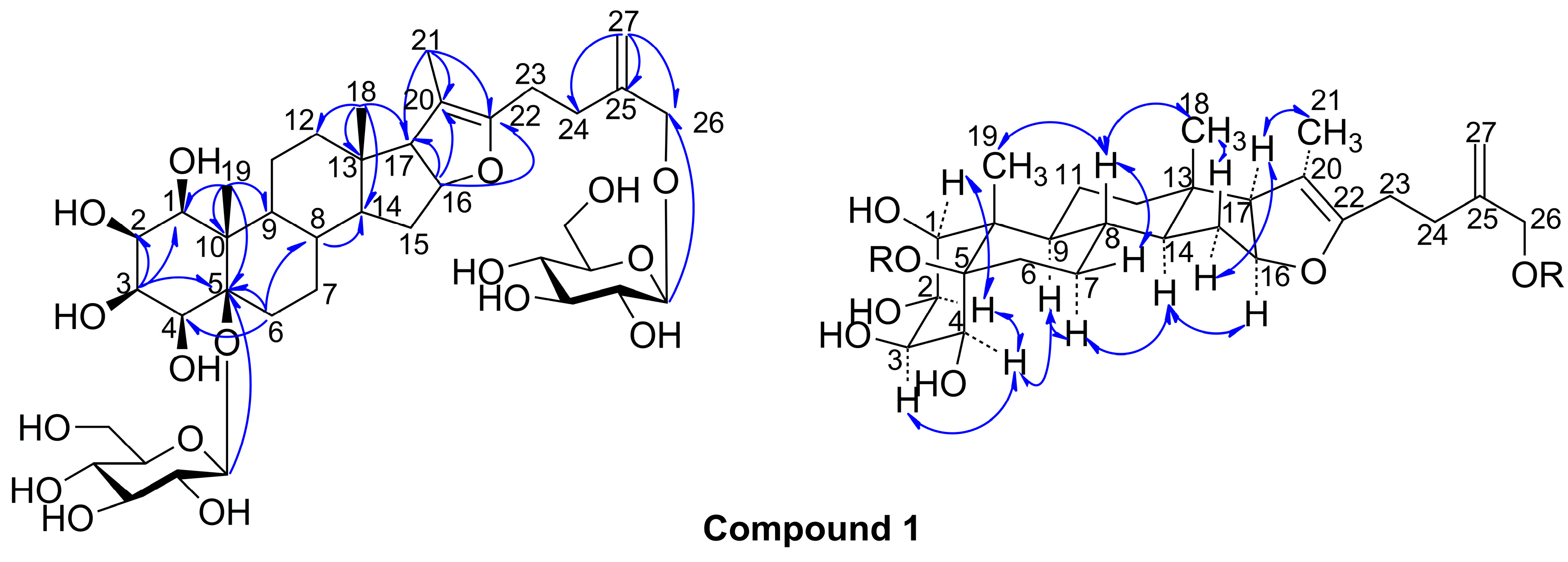
3.4. 1β,2β,3β,4β,5β,26-Hexahydroxyfurost-20(22),25(27)-dien-5,26-O-β-d-glucopyranoside (1)

A white amorphous powder, IR (KBr) νmax: 3450, 2980, 1694, 1025, 907, 804, 772 cm−1. 1H-NMR (500 MHz, pyridine-d5) and 13C-NMR (125 MHz, pyridine-d5) spectral data, see Table 1; m/z 801.3855 [M − H] (calcd. for C39H61O17, 801.3909).

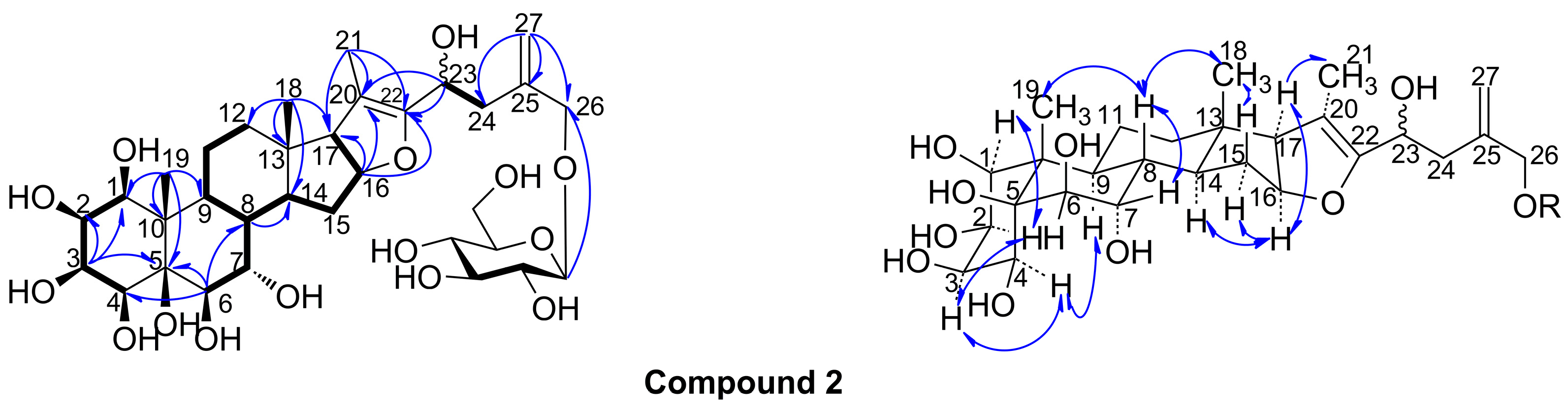
3.5. 1β,2β,3β,4β,5β,6β,7α,23ξ,26-Nonahydroxyfurost-20(22),25(27)-dien-26-O-β-d-glucopyranoside (2)

A white amorphous powder, IR (KBr) νmax: 3475, 2980, 1742, 1062, 904, 804 cm−1. 1H-NMR (600 MHz, pyridine-d5) and 13C-NMR (150 MHz, pyridine-d5) spectral data, see Table 1; m/z 711.3198 [M + Na]+ (calcd. for C33H52O15Na, 711.3204).

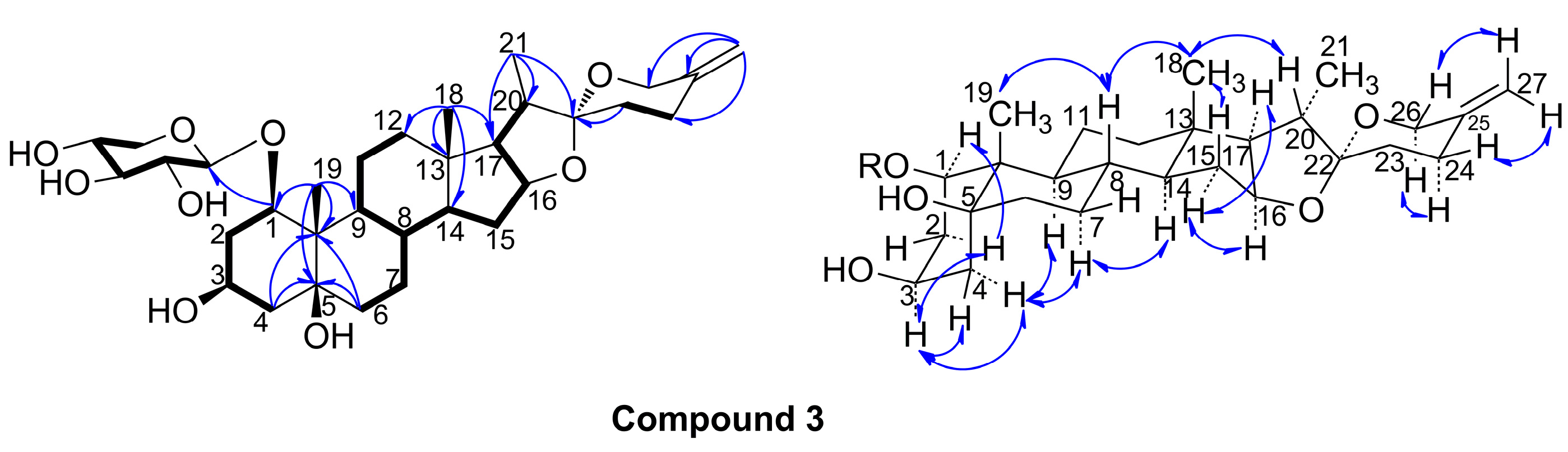
3.6. (20S,22R)-Spirost-25(27)-en-1β,3β,5β-trihydroxy-1-O-β-d-xyloside (3)

A white amorphous powder; IR (KBr) νmax: 3306, 2922, 1650, 1042, 989, 917, 892, 876 cm−1; 1H-NMR (400 MHz, pyridine-d5) and 13C-NMR (100 MHz, pyridine-d5) spectral data, see Table 1; m/z 579.3590 [M + H]+ (calcd. for C32H51O9, 579.3633).

3.7. Acid Hydrolysis of Compounds 1, 2, 3 and Absolute Sugar Configuration Determination

The solutions of compounds 1 (3 mg), 2 (3 mg) and 3 (5 mg) were hydrolyzed with 2N HCl (5 mL) for 5 h at 80 °C, respectively. The reaction mixtures were concentrated and dried by N2, and then water (5 mL) was added and the mixtures were extracted with EtOAc (3 × 5 mL). The aqueous layers of 1 and 2 were subjected to CC over silica gel eluted with MeCN–H2O (8:1) to yield d-glucose, which was determined by TLC comparison (MeCN–H2O, 6:1) with the authentic sugar and the optical rotation determination [ α ] D 20 +49.2 (c 0.16, H2O). The aqueous layer of 3 was subjected to CC over silica gel eluted with MeCN–H2O (8:1–15:1) to yield d-xylose, which was identified by TLC comparison with the authentic sugar and the optical rotation determination [ α ] D 20 +17.9 (c 0.14, H2O).

3.8. Cytotoxicity Assay

The cytotoxic activity assays towards the A549 and H1299 tumor cell lines were measured by the MTT method in vitro, using 5-fluorouracil as positive control. Briefly, 1 × 10⁴ mL−1 cells were seeded into 96-well plates and allowed to adhere for 24 h. Compounds 14 were dissolved in DMSO and diluted with complete medium to five concentration levels (from 0.001 mmol·L−1 to 0.1 mmol·L−1) for inhibition rate determination. After incubation at 37 °C for 4 h, the supernatant was removed before adding DMSO (100 μL) to each well. 5-Fluorouracil (5-Fu) was used as positive control. The inhibition rate (IR) and IC50 were calculated. Values are mean ± SD, n = 3, ** p < 0.01 vs. DMEM control. Compound 3 exhibited cytotoxicity against A549 cells (IC50 86.63 ± 2.33 μmol·L−1) and H1299 cells (IC50 88.21 ± 1.34 μmol·L−1), while the positive control of 5-Fu exhibited cytotoxicity against A549 cells (IC50 42.78 ± 1.63 μmol·L−1) and H1299 cells (IC50 38.65 ± 1.59 μmol·L−1), respectively, (see Table 2 and Table 3).

Supplementary Materials

IR, HR-ESI-MS, 1H-NMR 13C-NMR and 2D NMR spectra for compounds 13 can be accessed at: https://www.mdpi.com/1420-3049/20/08/13659/s1.

Acknowledgments

This project was financially supported by the National Natural Science Foundations of China (grant No. 81102805, 81373978), the Open Projects Program of the Key Laboratory of Tibetan Medicine Research, Chinese Academy of Sciences (grant No. 2014-TMR-01), the Innovative Research Team in TCM Material Foundation and Key Preparation Technology (grant No. 2012KCT-20), the Innovation Projects of Science and Technology in Shaanxi Province (grant No. 2011KTCQ03-02, 2013KTCQ03-14), and the Key Program of Shaanxi University of Chinese Medicine (grant No. 2015PY09).

Author Contributions

Every author has participated in the research and did his or her individual contribution to the article: Y.L. and X.W. conducted the experiments and collected the data; D.Z. and Y.J. planned and coordinated the experiments; H.H. and F.W. carried out the cytotoxicity biology experiments; X.Y. and Z.T. analysed the data; X.S. designed the study, and Z.Y. planned and oversaw the research project and drafted the paper. Finally, All authors read and approved the final manuscript.

Conflicts of Interest

The authors declare no conflict of interest.

References

  1. Song, X.; Liu, H. Research and Application of “Qi-Medicines” in Taibai Mountains; People’s Medical Publishing House: Beijing, China, 2011. [Google Scholar]
  2. Wu, X.; Fan, J.; Ouyang, Z.; Ning, R.; Guo, W.; Shen, Y.; Sun, Y.; Xu, Q. Tupistra chinensis extract attenuates murine fulminant hepatitis with multiple targets against activated T lymphocytes. J. Pharm. Pharmacol. 2014, 66, 453–465. [Google Scholar]
  3. Pan, Z.; Li, Y.; Liu, J.; Ning, D.; Li, D.; Wu, X.; Wen, Y. A cytotoxic cardenolide and a saponin from the rhizomes of Tupistra chinensis. Fitoterapia 2012, 83, 1489–1493. [Google Scholar]
  4. Cai, J.; Zhu, Z.; Yu, C.; Lei, L.; Wu, S. Saponin from Tupistra chinensis Baker inhibits mouse sarcoma S-180 cell proliferation in vitro and implanted solid tumor growth in mice. J. South. Med. Univ. 2007, 27, 188–190. [Google Scholar]
  5. Li, Q.; Zou, K.; Wang, Y. Experimental study on vitro anti-tumor action of “Kai-Kou-Jian” extract. Chin. J. Ethnomed. Ethnopharm. 2007, 86, 164–167. [Google Scholar]
  6. Liu, C.; Guo, Z.; Xue, Y.; Cheng, J.; Huang, N.; Zhou, Y.; Cheng, F.; Zou, K. Five new furostanol saponins from the rhizomes of Tupistra chinensis. Fitoterapia 2012, 83, 323–328. [Google Scholar]
  7. Huang, W.; Zhang, H.; Zou, K.; Chen, J.; Li, X.; Liu, C.; Huang, N. Total saponins of Tupistra chinensis induces apoptosis in A549 cells. Neoplasma 2012, 59, 613–621. [Google Scholar]
  8. Song, X.; Li, Y.; Zhang, D.; Jiang, Y.; Wang, W.; Song, B.; Tang, Z.; Cui, J.; Yue, Z. Two new spirostanol saponins from the the roots and rhizomes of Tupistra chinensis. Phytochem. Lett. 2015, 13, 6–10. [Google Scholar]
  9. Chai, J.; Song, X.; Wang, X.; Mei, Q.; Li, Z.; Cui, J.; Tang, Z.; Yue, Z. Two new compounds from the roots and rhizomes of Trillium tschonoskii. Phytochem. Lett. 2014, 10, 113–117. [Google Scholar]
  10. Li, H.; Pan, X.; Mei, Q.; Song, X.; Pei, Y.; Yue, Z. Isolation and identification of chemical constituents from root and rhizoma of Trillium tschonoskii Maxim. J. Shenyang Pharm. Univ. 2013, 30, 509–516. [Google Scholar]
  11. Yue, Z.; Qin, H.; Li, Y.; Sun, Y.; Wang, Z.; Yang, T.; Liu, L.; Wang, M.; Feng, F.; Mei, Q. Chemical constituents of the root of Jasminum giraldii. Molecules 2013, 18, 4766–4775. [Google Scholar]
  12. Hudson, C.; Dale, J. Studies on the forms of D-glucose and their mutarotation. J. Am. Chem. Soc. 1917, 39, 320–328. [Google Scholar]
  13. Zhang, X.; Chen, C.; Yang, J.; Ni, W.; Liu, H. New minor spirostane glycosides from Ypsilandra thibetica. Helv. Chim. Acta 2012, 95, 1087–1093. [Google Scholar]
  14. Yan, W.; Ohtani, K.; Kasai, R.; Yamasaki, K. Steroidal saponins from fruits of Tribulus terrestris. Phytochemistry 1996, 42, 1417–1422. [Google Scholar]
  15. Liu, C.; Guo, Z.; Xue, Y.; Zhang, H.; Zhang, H.; Zou, K.; Huang, N. Tupisteroide A–C, three new polyhydroxylated steroidal constituents from the roots of Tupistra chinensis. Magn. Reson. Chem. 2012, 50, 320–324. [Google Scholar]
  16. Xu, L.; Zou, K.; Wang, J.; Wu, J.; Zhou, Y.; Dan, F.; Yang, J. New polyhydroxylated furostanol saponins with inhibitory action against NO production from Tupistra chinensis Rhizomes. Molecules 2007, 12, 2029–2037. [Google Scholar]
  17. Li, Y.; Fan, L.; Sun, Y.; Miao, X.; Zhang, F.; Meng, J.; Han, J.; Zhang, D.; Zhang, R.; Yue, Z.; et al. Paris saponin VII from trillium tschonoskii reverses multidrug resistance of adriamycin-resistant MCF-7/ADR cells via P-glycoprotein inhibition and apoptosis augmentation. J. Ethnopharmcol. 2014, 154, 728–734. [Google Scholar]
  18. Li, Y.; Sun, Y.; Fan, L.; Zhang, F.; Meng, J.; Han, J.; Guo, X.; Zhang, D.; Zhang, R.; Yue, Z.; et al. Paris saponin VII inhibits growth of colorectal cancer cells through Ras signaling pathway. Biochem. Pharmacol. 2014, 88, 150–157. [Google Scholar]
  19. Pan, W.; Chang, F.; Wei, L.; Wu, Y. New flavans, spirostanol sapogenins, and a pregnane genin from Tupistra chinensis and their cytotoxicity. J. Nat. Prod. 2003, 66, 161–168. [Google Scholar] [CrossRef] [PubMed]
  • Sample Availability: Samples of the compounds 35 are available from the authors.

