Article

Bioactive Cembranoids from the South China Sea Soft Coral Sarcophyton elegans

1
School of Pharmaceutical Sciences, Sun Yat-sen University, Guangzhou 510006, China
2
East China Sea Fisheries Research Institute, Chinese Academy of Fishery Sciences, Shanghai 200090, China
*
Author to whom correspondence should be addressed.
Molecules 2015, 20(7), 13324-13335; https://doi.org/10.3390/molecules200713324
Submission received: 13 June 2015 / Accepted: 13 July 2015 / Published: 22 July 2015
(This article belongs to the Section Natural Products Chemistry)

Abstract

:
Four new cembranoids, sarcophelegans A–D (14) and six known analogues (510) were isolated from the South China Sea soft coral Sarcophyton elegans. Their structures were elucidated through detailed spectroscopic analysis, and the absolute configuration of 1 was confirmed by single-crystal X-ray diffraction. The antimigratory potential of compounds 110 were evaluated and compounds 2 and 6 were found to inhibit human breast tumor MDA-MB-231 cell migration at 10 μM.

1. Introduction

Cembranoids are a group of highly functionalized dierpenoids with a 14-membered carbon ring, an isopropyl residue, and three methyls [1]. Since the first representative, cembrene, was isolated from the pine tree Pinus albicaulis Engelm in 1962, hundreds of cembranoids have been reported from plants, insects, alligators, and especially from marine organisms [1,2]. Although cembranoids are indisputably not uniquely marine, their striking presence in soft corals, especially in the genus Sarcophyton (Alcyoniidae) [3,4], outstrips their occasional occurrence in other taxa. In recent years, the significant biological activity of cembranoids in terms of antimicrobial, anti-cancer and anti-inflammation effects, together with their fascinating architectures, have attracted great interest from natural product [5,6] and pharmaceutical chemists [7,8].
The soft coral species of the genus Sarcophyton are widely distributed along tropic and subtropic oceans. So far, around 30 species of this genus from different locations have been chemically examined [9]. Previous chemical investigations of S. elegans have led to the isolation of several cembranoids, tetracyclic diterpenoids, steroids, and carotenoids [9,10,11,12,13,14,15,16]. As part of our continuing efforts to discover structurally intriguing and bioactivity-significant metabolites from South China Sea marine invertebrates [17,18,19], we undertook a detailed chemical analysis of S. elegans, collected in the Xisha Islands, South China Sea, which led to the isolation of four new cembranoids (14) and six known compounds (510) (Figure 1). The antimigratory potential of compounds 110 was evaluated and compounds 2 and 6 were found to inhibit human breast tumor MDA-MB-231 cell migration at 10 μM. Herein, details of the isolation, structure elucidation, and antimigratory activity of these compounds are described.
Figure 1. Compounds 110 isolated from Sarcophyton elegans.
Molecules 20 13324 g001

2. Results and Discussion

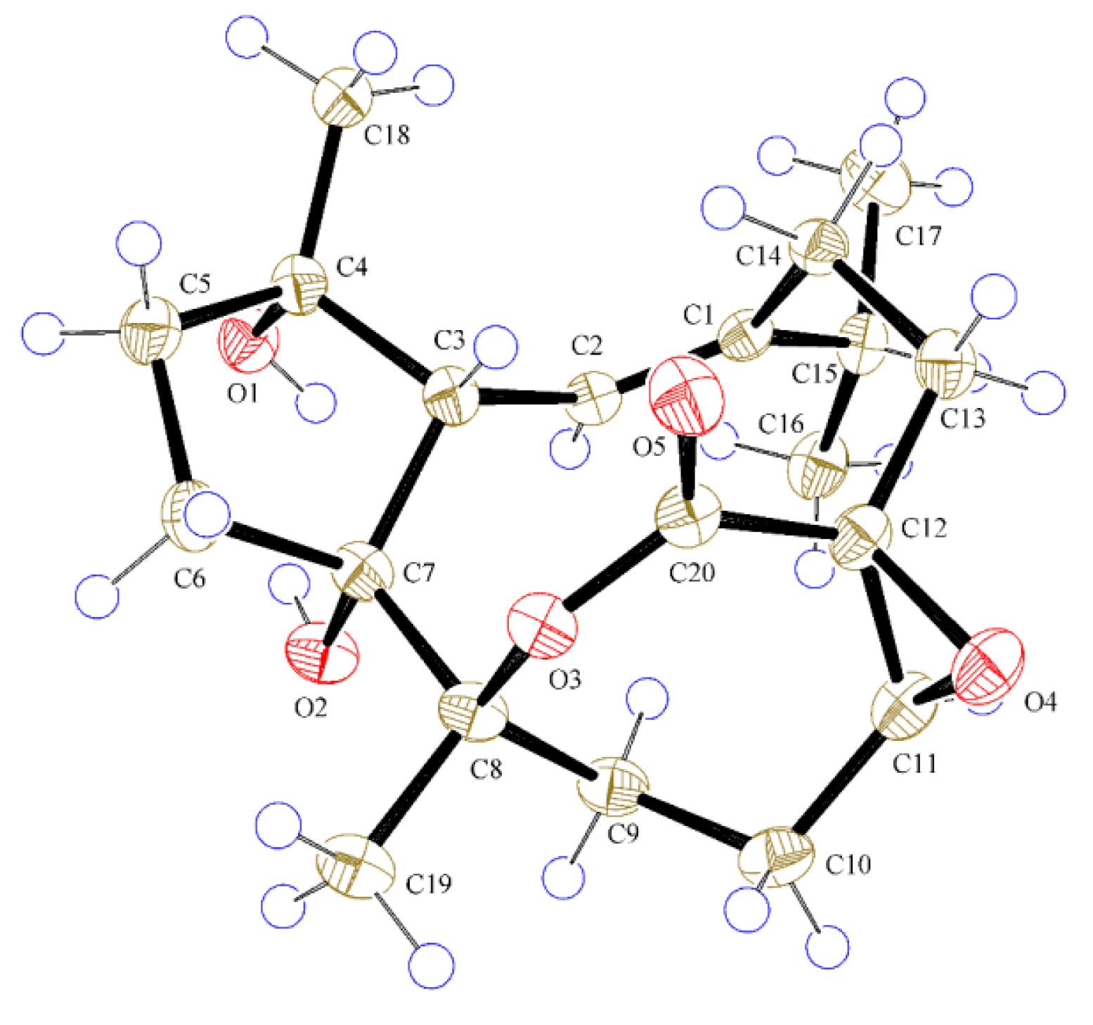
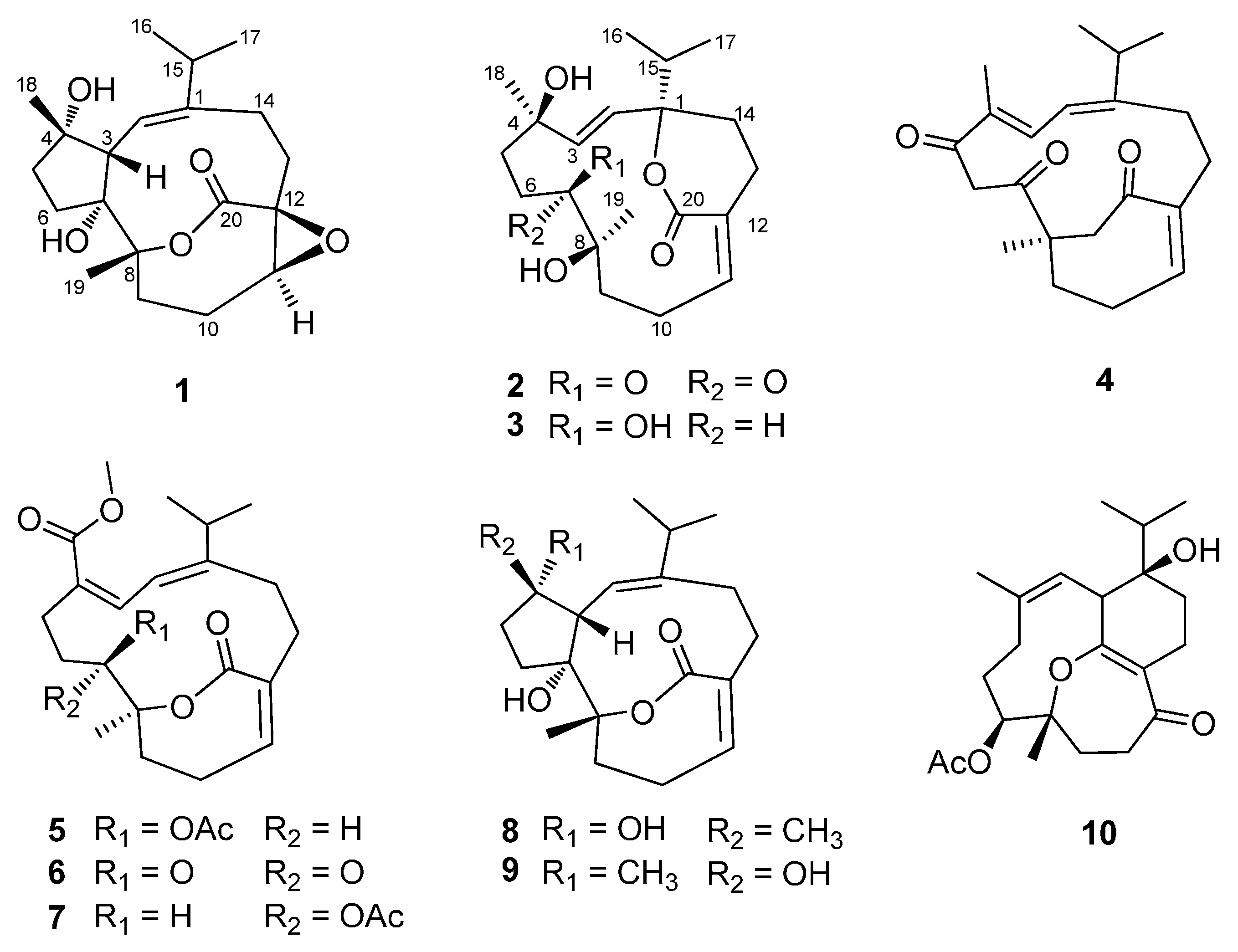
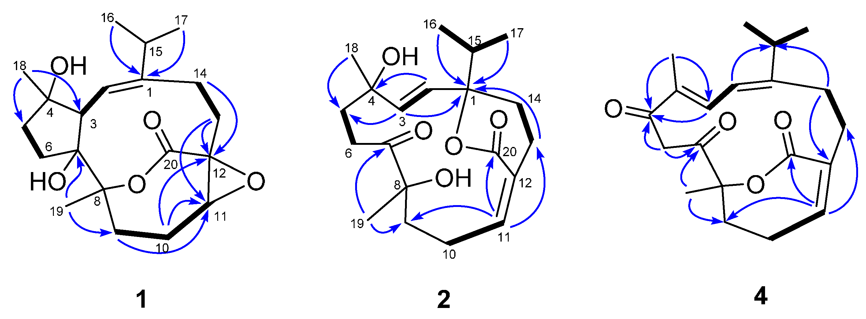
2.1. Structural Elucidation of New Compounds

The soft coral of S. elegans (1 kg, wet weight) was freeze-dried, ground, and extracted with a mixture of CH2Cl2/MeOH (v/v, 1:1) at room temperature. After removal of solvent in vacuo, the residue was suspended in H2O and then partitioned sequentially with petroleum ether (PE) and EtOAc. Various column chromatographic separations of the EtOAc extract afforded compounds 110.
Compound 1, a colorless crystal, had the molecular formula C20H30O5, as determined by HRESIMS at m/z 333.2059 [M − H2O + H]+ (calcd 333.2066), corresponding to six degrees of unsaturation. The IR spectrum exhibited absorption bands for hydroxyl (3451 cm−1) and carbonyl (1716 cm−1) functionalities. The 1H-NMR data (Table 1) of 1 showed two methyl singlets [δH 1.10 (3H, s, CH3-18) and 1.34 (3H, s, CH3-19)], an isopropyl group [δH 1.09 (3H, d, J = 6.9 Hz, CH3-17), 1.19 (3H, d, J = 6.9 Hz, CH3-16), and 2.34 (1H, m, H-15)], an oxygenated methine [δH 3.13 (1H, dd, J = 7.0, 7.0 Hz, H-11)], an olefinic proton [δH 5.69 (1H, d, J = 11.3 Hz, H-2)], and a series of aliphatic methylene multiplets. The 13C-NMR data (Table 2), in combination with DEPT experiments, resolved 20 carbon resonances attributable to an ester carbonyl group (δC 173.9), a trisubstituted double bond (δC 120.1, 147.1), four sp3 oxygenated quaternary carbons, three sp3 methines (one oxygenated), six sp3 methylenes, and four methyls. The above-mentioned data implied that 1 possessed most of the structural features of cembranoid diterpenes, which showed high similarity to those of co-isolated sarsolilide B (8) [20]. In comparison with 8, the signals for Δ11 in 8 were replaced by the signals for an epoxy in 1H 3.13 (1H, dd J = 7.0, 7.0 Hz); δC 65.6 (CH) and 60.4 (C)], indicating that 1 was an 11,12-epoxy derivative of 8. This was confirmed by HMBC correlations from both H-10 and H-13 to C-11 and C-12, as well as the downfield-shifted carbonyl at C-20 (δC 173.9 in 1; δC 170.3 in 8) (Figure 2). The relative configuration of 1 was assigned to be the same as that of 8 by comparing their 1D NMR and NOESY data. In particular, the NOESY correlation between H-11 and H-13a indicated the epoxy ring was cis-oriented (Figure S6). Finally, the successful performance of the X-ray crystallographic analysis using anomalous scattering of Cu Kα radiation verified the proposed structure and also allowed unambiguous assignment of the absolute configuration of 1 as drawn in Figure 3. Thus, compound 1 was determined as depicted and given the trivial name sarcophelegan A.
Figure 2. Key 1H-1H COSY (▬) and HMBC ( ) correlations for 1, 2, and 4.
Molecules 20 13324 g002
Figure 3. ORTEP depiction for X-ray crystal structures of 1.
Molecules 20 13324 g003
Table 1. 1H-NMR spectroscopic data of 14 (400 MHz, J in Hz, δ in ppm).
Position1 a2 a3 a4 b
25.69, d (11.3)5.55, d (16.7)5.54, d (16.3)6.23 d (11.4)
32.35, m (overlapped)5.69, d (16.7)5.74, d (16.3)6.72, brd (11.4)
5a1.98, m1.77, m (overlapped)1.83, m (overlapped)
5b1.87, m1.55, ddd (14.2, 10.5, 7.4)1.46, m
6a2.21, m3.04, ddd (20.9, 10.5, 7.4)1.70, m4.90, d (16.8)
6b1.90, m2.44, m (overlapped)1.38, m3.29, d (16.8)
7 3.25, brd (11.0, 1.4)
9a2.82, dd (14.4, 14.4)1.96, m1.88, m2.40, m (overlapped)
9b1.73, dd (14.4, 7.6)1.77, m (overlapped)1.79, m2.01, m
10a2.31, m3.40, m3.44, m2.73, m
10b1.38, m2.17, m2.05, m2.31, m
113.13, dd (7.0, 7.0)5.67, m 6.19, dd (10.1, 5.1)6.10, dd (4.1, 4.1)
13a2.62, m2.45, m (overlapped)2.60, m3.19, m
13b1.30, m 2.55, m1.92, m
14a2.51, m2.12, m2.16, m2.56, dd (13.8, 13.8)
14b2.12, m1.76 m1.84, m (overlapped)2.23, dd (13.8, 7.8)
152.34, m (overlapped)1.86, m1.87, m2.41, m (overlapped)
161.19, d (6.9)0.98, d (6.8)0.98, d (7.1)1.07, d (6.8)
171.09, d (6.9)0.95, d (6.8)0.96, d (7.1)1.09, d (6.8)
181.10, s1.39, s1.33, s1.83, s
191.34, s1.21, s1.15, s1.54, s
a Measured in CD3OD; b Measured in CDCl3.
Table 2. 13C-NMR spectroscopic data of 14 (100 MHz, δ in ppm).
Position1 a2 a3 a4 b
1147.1, C87.8, C88.0, C158.8, C
2120.1, CH128.2, CH128.3, CH119.4, CH
352.0, CH140.4, CH140.8, CH137.8, CH
483.4, C72.8, C73.4, C133.3, C
538.5, CH236.5, CH241.0, CH2195.1, C
634.7, CH235.2, CH225.4, CH245.7, CH2
788.9, C219.3, C76.0, CH204.3, C
891.9, C79.7, C75.8, C86.1, C
929.9, CH242.0, CH239.0, CH233.3, CH2
1024,1, CH225.9, CH225.5, CH227.2, CH2
1165.6, CH150.3, CH149.5, CH143.6, CH
1260.4, C125.1, C125.5, C130.9, C
1333.0, CH225.0, CH225.8, CH237.1, CH2
1425.8, CH227.5, CH227.5, CH227.6, CH2
1534.0, CH38.4, CH38.9, CH36.3, CH
1621.3, CH317.4, CH317.4, CH322.4, CH3
1723.9, CH317.1, CH317.1, CH321.7, CH3
1826.2, CH329.3, CH330.2, CH310.9, CH3
1925.1, CH328.5, CH324.4, CH328.9, CH3
20173.9, C168.8, C169.5, C165.8, C
a Measured in CD3OD; b Measured in CDCl3.
Compound 2 possessed a molecular formula of C20H30O5 as determined by HRESIMS at m/z 373.1986 [M + Na]+, which was compatible with its 1D NMR data. The 1H-NMR data of 2 (Table 1) showed signals for two methyl singlets [δH 1.21, (3H, s, CH3-19) and 1.39 (3H, s, CH3-18)], an isopropyl group [δH 0.95 (3H, d, J = 6.8 Hz, CH3-17), 0.98 (3H, d, J = 6.8 Hz, CH3-16), and 1.86 (1H, m, H-15)], two trans-olefinic protons [δH 5.55 (1H, d, J = 16.7 Hz, H-2) and 5.69 (1H, d, J = 16.7 Hz, H-3)], an olefinic proton [δH 5.67 (1H, m, H-11)], and a series of aliphatic methylene multiplets. The 20 carbon resonances were classified by DEPT experiments as a ketone carbonyl group (δC 219.3), an ester carbonyl group (δC 168.8), two double bonds (δC 125.1, 128.2, 140.4, and 150.3), three sp3 oxygenated quaternary carbons, a sp3 methine, six sp3 methylenes, and four methyls (Figure S8). The above-mentioned information was similar to that of sartrolide E [21]. However, analysis of HSQC and HMBC data (Figures S10 and S11) revealed that the chemical shift of C-1 (δC 76.7) in sartrolide E was downfield-shifted to δC 87.8 in 2, while C-8 was upfield-shifted from δC 87.0 to δC 79.7 (Figure 2), indicating that the linkage of the lactone ring from C-12 to C-8 in sartrolide E was migrated to C-1 in 2. This was further supported by comparison of the C-1 and C-8 chemical shifts of 2 with those of a known analogue, laevigatlactone E [22], sharing the similar lactone linkage as that in 2C 87.9, C-1 and δC 73.2, C-8, in laevigatlactone E).
The relative configuration of 2 was determined on the basis of the NOESY experiment (Figure S12). The NOESY correlation observed between H-11 and H-13 suggested the E geometry for the Δ11. The crucial NOE correlations between H-2/CH3-16 and H-2/CH3-18 revealed that the isopropyl group and CH3-18 were co-facial and were arbitrarily designated as α-oriented, while the interactions of H-5a with CH3-18 and CH3-19 indicated that the CH3-18 and CH3-19 were both α-oriented (Figure 4). Thus, compound 2 was deduced as shown and named sarcophelegan B.
Figure 4. Selected NOESY correlations of 2 (↔).
Molecules 20 13324 g004
Compound 3 displayed the HRESIMS ion at m/z 375.2147 [M + Na]+, consistent with a molecular formula of C20H32O5, two mass units more than that of 2. The 1H and 13C NMR data of 3 (Table 1 and Table 2) were very similar to those of 2 except for the presence of an additional oxygenated methine (δH 3.25; δC 76.0) in 3 instead of the ketone group (δC 219.3, C-7) in 2, indicating that 3 was a 7-hydrogenated derivative of 2. HMBC correlation from CH3-19 and H2-5 to the oxygenated methine (δC 76.0) confirmed the location of the hydroxyl group at C-7. This was also supported by the upfield-shifted signals of C-6 and C-8 in 3 with respect to those in 2C 25.4, C-6; 75.8, C-8 in 3; δC 35.2, C-6; 79.7, C-8 in 2). The relative configurations at C-1, C-4, and C-8 of 3 were assigned to be the same as those of 2 by comparing their 1D NMR and NOESY data. The 7-OH was designated as β by the NOE correlation between H-7 and CH3-19, as in the Chem3D molecular modeling study, the 7β-OH isomer of 3 display a distance of 2.573 Å between H-7 and CH3-19, while the 7α-OH isomer showed a large distance of 3.738 Å (Figure S25). Thus, compound 3 was deduced as shown and named sarcophelegan C.
Compound 4, a colorless oil, exhibited a molecular formula of C20H26O4 as determined by HRESIMS and the data of 13C-NMR. The 1H- and 13C-NMR spectra of 4 (Figures S19 and S20) showed signals for two ketone signals (δC 195.1 and 204.3), an α,β-unsaturated-ε-lactone (δC 86.1, 130.9, 143.6, and 165.8), an isopropyl group [δC 21.7, 22.4, and 36.3; δH 1.07 (3H, d, J = 6.8 Hz, 1.09 (3H, d, J = 6.8 Hz)), and 2.41 (1H, m)], two double bonds [δC 119.4, 133.3, 137.8, and 158.8; δH 6.23 (1H, d, J = 11.4 Hz) and 6.72 (1H, brd, J = 11.4 Hz)], and two methyl singlets [δC 10.9 and 28.9; δH 1.54 (3H, s) and1.83 (3H, s)]. These data showed high similarity to those of (1Z,5S,9E,11E)-5,9-dimethyl-12-isopropyl-6-oxocyclotetradeca-1,9,11-triene-1,5-carbolactone [23], a cembranoid diterpene previously reported from the same genus, except for the presence of an additional carbonyl group (δC 195.1), which indicated that 4 was a carbonylated derivative of (1Z,5S,9E,11E)-5,9-dimethyl-12-isopropyl-6-oxocyclotetradeca-1,9,11-triene-1,5-carbolactone. HMBC correlations from CH3-18 and H-3 to the carbonyl carbon revealed that the carbonyl group was located at C-5. This was further supported by the downfield-shifted H-3 signal in 4 with respect to that in (1Z,5S,9E,11E)-5,9-dimethyl-12-isopropyl-6-oxocyclotetradeca-1,9,11-triene-1,5-carbolactone (δH 6.72 in 2; δH 6.07 in (1Z,5S,9E,11E)-5,9-dimethyl-12-isopropyl-6-oxocyclotetradeca-1,9,11-triene-1,5-carbolactone). Detailed 2D NMR analyses [1H-1H COSY, HSQC, and HMBC (Figures S21-23)] permitted the establishment of the gross structure of 4 as depicted in Figure 2. The absolute configuration of the only chiral center C-8 in 4 was proposed as S based on comparison of its specific rotation ( [ α ] D 20 + 190.8) with (1Z,5S,9E,11E)-5,9-dimethyl-12-isopropyl-6-oxocyclotetradeca-1,9,11-triene-1,5-carbolactone ( [ α ] D 20 + 177), which was also supported by the biogenetic origin of this skeleton. Interestingly, cembranoids with C-3 and C-7 cyclization exclusively give R configuration at C-8 [20]. Compound 4 was given the trivial name sarcophelegan D.
The known compounds emblide (5) [4], ketoemblide (6) [24], sarcrassin D (7) [4], sarsolilides B (8) [20], sarsolilide C (9) [20], and dihydrosarsolenone (10) [20] were identified by comparison of their NMR and MS data with those in the literature.

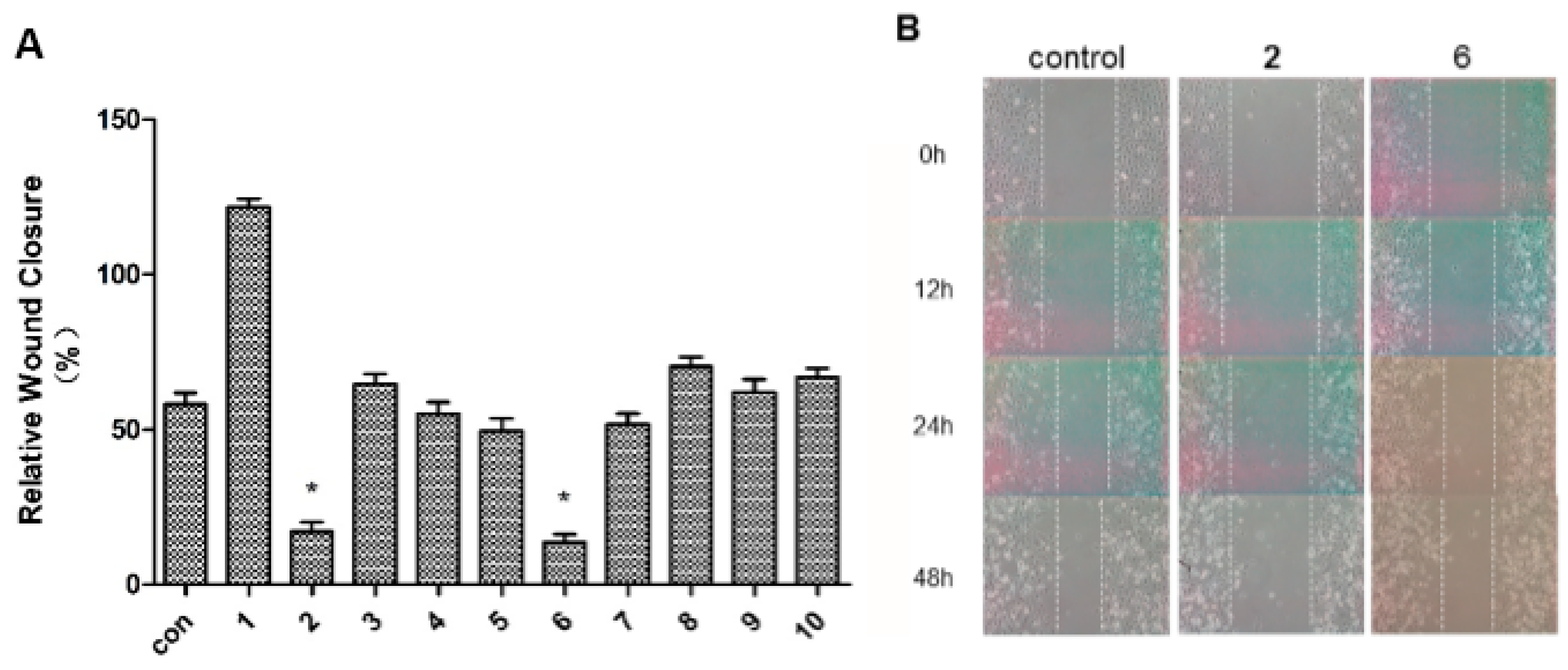
2.2. Antimigratory Activity

Metastasis is one of the major biological characteristics of cancer cells. The wound-healing assay is a simple and widely used tool to investigate in vitro directional cell migration [5,25,26]. The effects of compounds 110 on the migration of human breast cancer MDA-MB-231 cells were evaluated using wound-healing assays. The ability of the compounds to inhibit the migration of the cancer cells into the wound is measured by comparing the original wound width before assay with the wound width after 48 h incubation [relative wound closure = (W0 − W48)/W0]. The higher antimigratory activity of the compound is, the smaller the wound-relative closure value it generates.
Among 110, compounds 2 and 6 had the greatest capability to inhibit the migration of MDA-MB-231 cells while others did not show evident activity in comparison with control. Furthermore, compounds 2 and 6 inhibited the cell migration in a time dependent manner (Figure 5B).
Figure 5. Wound-healing assays on compounds 110 with human breast tumor cell MDA-MB-231. (A) The antimigration effects of 110 at 10 μM on the tumor cells (* p < 0.05); (B) The incubation of 2 and 6 with tumor cells at 0, 12, 24, and 48 h (left: control; middle: 2 at 10 μM; right: 6 at 10 μM).
Molecules 20 13324 g005

3. Experimental Section

3.1. General Experimental Procedures

X-ray data were collected using an Agilent Xcalibur Nova X-ray diffractometer (Agilent, Santa Clara, CA, USA). Melting points were measured on an X-4 melting instrument and are uncorrected. Optical rotations were measured on a Perkin-Elmer 341 polarimeter (Perkin-Elmer, Waltham, MA, USA). UV spectra were recorded on a Shimadzu UV-2450 spectrophotometer (Shimadzu, Kyoto, Japan). IR spectra were determined on a Bruker Tensor 37 infrared spectrophotometer (Bruker, Karlsruhe, Germany). NMR spectra were measured on a Bruker AM-400 spectrometer (Bruker, Karlsruhe, Germany) at 25 °C. ESIMS was measured on a Finnigan LCQ Deca instrument (Thermo Finnigan, San Jose, CA, USA), and HRESIMS was performed on a Waters Micromass Q-TOF (Waters, Milford, MA, USA). A Shimadzu LC-20 AT equipped with a SPD-M20A PDA detector (Shimadzu, Kyoto, Japan) was used for HPLC. A YMC-pack ODS-A column (250 ´ 10 mm, S-5 μm, 12 nm) (YMC, Tokyo, Japan) was used for semipreparative HPLC separation. Wound closure was monitored and photographed with a Nikon Eclipse inverted microscope. Silica gel (300−400 mesh, Qingdao Marien Chemical Co., Ltd., Qingdao, Shandong, China), reversed-phase C18 (Rp-C18) silica gel (12 nm, S-50 μm, YMC Co., Ltd., Kyoto, Japan), Sephadex LH-20 gel (Amersham Biosciences, Piscataway, NJ, USA), and MCI gel (CHP20P, 75−150 μm, Mitsubishi Chemical Industries Ltd. Tokyo, Japan) were used for column chromatography (CC). All solvents used were of analytical grade (Guangzhou Chemical Reagents Co., Ltd., Guangzhou, China).

3.2. Animal Material

The soft coral S. elegans were collected from the Xisha Islands in the South China Sea, in October 2014, at a depth of 8–10 m of water. The biological material was frozen immediately until used and was identified by Cheng-Qi Fan from East China Sea Fisheries Research Institute. A voucher specimen (accession number: HLRZ201410) has been deposited at the School of Pharmaceutical Sciences, Sun Yat-sen University, Guangzhou, China.

3.3. Extraction and Isolation

The frozen samples (1 kg, wet weight) were extracted with CH2Cl2/MeOH (1:1, 3 × 1 L) at room temperature. After removal of solvent in vacuo, the residue (16 g) was suspended in H2O (200 mL) and partitioned sequentially to give dried petroleum ether (2 g) and EtOAc (4 g) extracts. The EtOAc extract was subjected to silica gel column chromatography eluted with a CH2Cl2/MeOH gradient (100:1→10:1) to afford five fractions (I–V). Fr. II (460 mg) was subjected to Rp-C18 silica gel CC eluted with MeOHH2O (6:4 to 10:0), followed by a Sephadex LH-20 and eluted with EtOH to afford 2 (11 mg), 5 (72 mg), 6 (45 mg), and 7 (3.7 mg). Fr. III (1.4 g) was chromatographed over Sephadex LH-20 (CH2Cl2/MeOH, v/v, 1:1), followed by Rp-C18 silica gel eluted with a CH3CN/H2O gradient (5:5→10:0) to obtain four sub-fractions (Fr. IIIa–IIId). Fr. IIIb was further separated by HPLC equipped with an ODS-18 column using CH3CN/H2O (65:35, v/v; 3 mL·min−1) to afford 1 (8.1 mg, tR 8.5 min), 8 (6 mg, tR 11 min), and 9 (18 mg, tR 13.5 min). Fr. IIId was purified by repeating the HPLC conditions described above to yield 3 (41 mg, tR 13 min) and 4 (3.7 mg, tR 17 min). Fr. IIIc was chromatographed with silica gel CC (CH2Cl2/MeOH, 40:1) to give 10 (25.2 mg).

3.4. Cell Culture

Human breast tumor cells (MDA-MB-231) were obtained from the Institute of Chinese Academy of Medical Sciences, Beijing, China. MDA-MB-231 cells were cultured in RPMI-1640 containing 10% FBS in cell culture flasks under a humidified 5% CO2 and 95% air atmosphere at 37 °C.

3.5. Wound-Healing Assays

The method used to detect migration by wound-healing assay was previously described [5,25,26]. Briefly, the cells were allowed to grow to 90% confluence in 6-well plates. Once the monolayer was developed, a wound was made by scrapping with a 100 μL pipet tip to create a denuded zone (gap) of constant width. Subsequently, cellular debris was washed with 2‰ FBS, and the MDA-MB-231 cells were exposed to 10 μM of compounds 110. Wound closure was monitored and photographed at 0, 12 h, 24 h, and 48 h with a Nikon Eclipse inverted microscope. Wound width was measured immediately before (W0) and after the 48 h (W48) incubation. To quantify the migrated cells, pictures of the initial wounded monolayers were compared with the corresponding pictures of cells at the end of the incubation. Artificial lines fitting the cutting edges were drawn on pictures of the original wounds and overlaid on the pictures of cultures after incubation. Figure 5A represents wound closure values for different compounds (110), relative to the control (time 0).

3.6. Statistical Analysis

Data were expressed as the mean ± SD of at least three independent experiments. To compare three or more groups, one-way analysis of variance (ANOVA) was used followed by Newman-Keuls post hoc test. Statistical analysis was performed using GraphPad Prism software (5.01, GraphPad Software Inc., San Diego, CA, USA).
Sarcopelegan A (1): Colorless crystals; mp 187–189 °C; [ α ] D 20 +16.7 (c 0.23, MeOH); UV (MeOH) λmax (log ε) 208.4 (6.82) nm; IR (KBr) νmax 3451, 2958, 2925, 1716, 1237 cm−1; 1H- and 13C-NMR data see Table 1 and Table 2; HRESIMS m/z 333.2059 (calcd for C20H29O4 [M − H2O + H]+, 333.2066).
Sarcopelegan B (2): Colorless oil; [ α ] D 20 −3.7 (c 0.46, MeOH); UV (MeOH) λmax (log ε) 230.4 (6.82) nm; IR (KBr) νmax 3396, 2933, 1699, 1381, 983 cm−1; 1H- and 13C-NMR data see Table 1 and Table 2; HRESIMS m/z 373.1986 (calcd for C20H30O5Na [M + Na]+, 373.1991).
Sarcopelegan C (3): Colorless oil; [ α ] D 20 −10.0 (c 0.14, MeOH); UV (MeOH) λmax (log ε) 232.2 (7.17), 210.0 (7.05) nm; IR (KBr) νmax 3357, 2966, 2929, 1694, 1071 cm−1; 1H- and 13C-NMR data see Table 1 and Table 2; HRESIMS m/z 375.2147 (calcd for C20H32O5Na [M + Na]+, 375.2143).
Sarcopelegan D (4): Colorless oil; [ α ] D 20 +190.8 (c 0.20, MeOH); UV (MeOH) λmax (log ε) 298.2 (7.10), 219.8 (6.98) nm; IR (KBr) νmax 3454, 2961, 2067, 1621, 1270 cm−1; 1H- and 13C-NMR data see Table 1 and Table 2; HRESIMS m/z 331.1901 (calcd for C20H27O4 [M + H]+, 331.1909).
Crystal data for compound (1): C20H30O5, M = 350.44, 0.5 × 0.1 × 0.2 mm3, space group P65 (No. 170), V = 2769.03(4) Å3, Z = 6, Dc = 1.261 g·cm−3, F000 = 1140, Xcalibur, Onyx, Nova, Cu Kα radiation, λ = 1.54184 Å, T = 293(2) K, 2θmax = 143.5°, 35758 reflections collected, 3601 unique (Rint = 0.0507). Final GooF = 1.043, R1 = 0.0285, wR2 = 0.0757, R indices based on 3523 reflections with I > 2 sigma (I) (refinement on F2), 232 parameters, 1 restraint. Lp and absorption corrections applied, μ = 0.723 mm−1. Flack parameter = −0.02 (11). Crystallographic data for the structure of 1 have been deposited in the Cambridge Crystallographic Data Centre (deposition number: CCDC 1401385). The data can be obtained free of charge via http://www.ccdc.cam.ac.uk/conts/retrieving.html (or from the CCDC, 12 Union Road, Cambridge CB2 1EZ, UK; Fax: +44 1223 336033; E-mail: [email protected]).

4. Conclusions

Four new cembranoids and six known analogues were isolated from the South China Sea soft coral S. elegans, collected from the Xisha Islands. Their structures were elucidated through detailed spectroscopic analysis, and the absolute configuration of 1 was confirmed by single-crystal X-ray diffraction. The antimigratory potential of compounds 110 were evaluated, two of which were found to inhibit human breast tumor MDA-MB-231 cell migration at 10 μM. The current research not only expanded the members of the cembranoid family, but may also provide some diterpene prototypes for further development of anti-cancer leads with antimigratory properties.

Supplementary Materials

1D and 2D NMR spectra of 14 were provided. These materials can be accessed at: https://www.mdpi.com/1420-3049/20/07/13324/s1.

Acknowledgments

The authors thank the Science and Technology Planning Project of Guangdong Province (No. 2013B021100009), the Guangdong Natural Science Funds for Distinguished Young Scholars (No. 2014A030306047), and the National High Technology Research and Development Program of China (863 Projects, No. 2015AA020928) for providing financial support to this work.

Author Contributions

S.Y. designed the research; X.L., J.Z. and Q.L. performed the experimental work; X.L., G.T., H.W., and C.F. wrote the manuscript. All authors discussed, edited and approved the final version.

Conflicts of Interest

The authors declare no conflict of interest.

References

  1. Li, Y.; Peng, L.; Zhang, T. Progress of studies on the natural cembranoids from the soft coral species of Sarcophyton genus. Med. Chem. Bioact. Nat. Prod. 2006, 19, 257–300. [Google Scholar]
  2. Gross, H.; Konig, G.M. Terpenoids from marine organisms: Unique structures and their pharmacological potential. Phytochem. Rev. 2006, 5, 115–141. [Google Scholar] [CrossRef]
  3. Jia, R.; Guo, Y.W.; Chen, P.; Yang, Y.M.; Mollo, E.; Gavagnin, M.; Cimino, G. Biscembranoids and their probable biogenetic precursor from the Hainan soft coral Sarcophyton tortuosum. J. Nat. Prod. 2007, 70, 1158–1166. [Google Scholar] [CrossRef] [PubMed]
  4. Zhang, C.X.; Li, J.; Su, J.Y.; Liang, Y.J.; Yang, X.P.; Zheng, K.C.; Zeng, L.M. Cytotoxic diterpenoids from the soft coral Sarcophyton crassocaule. J. Nat. Prod. 2006, 69, 1476–1480. [Google Scholar] [CrossRef] [PubMed]
  5. Sawant, S.S.; Youssef, D.T.A.; Reiland, J.; Ferniz, M.; Marchetti, D.; El Sayed, K.A. Biocatalytic and antimetastatic studies of the marine cembranoids sarcophine and 2-epi-16-deoxysarcophine. J. Nat. Prod. 2006, 69, 1010–1013. [Google Scholar] [CrossRef] [PubMed]
  6. Gross, H.; Wright, A.D.; Beil, W.; Konig, G.M. Two new bicyclic cembranolides from a new Sarcophyton species and determination of the absolute configuration of sarcoglaucol-16-one. Org. Biomol. Chem. 2004, 2, 1133–1138. [Google Scholar] [CrossRef] [PubMed]
  7. Kim, H.; Lee, H.; Kim, J.; Kim, S.; Kim, D. A general strategy for synthesis of both (6Z)- and (6E)-cladiellin diterpenes:  Total syntheses of (−)-cladiella-6,11-dien-3-ol, (+)-polyanthellin A, (−)-cladiell-11-ene-3,6,7-triol, and (−)-deacetoxyalcyonin acetate. J. Am. Chem. Soc. 2006, 128, 15851–15855. [Google Scholar] [CrossRef] [PubMed]
  8. Crimmins, M.T.; Stauffer, C.S.; Mans, M.C. Total syntheses of (+)-vigulariol and (−)-sclerophytin A. Org. Lett. 2011, 13, 4890–4893. [Google Scholar] [CrossRef] [PubMed]
  9. Bie, W.; Deng, Z.W.; Xu, M.J.; Lin, W.H. Structural elucidation of a new cembranoid diterpene from the Chinese soft coral Sarcophyton sp. J. Chin. Pharm. Sci. 2008, 17, 221–224. [Google Scholar]
  10. Xi, Z.; Bie, W.; Chen, W.; Liu, D.; Leen, V.O.; Proksch, P.; Lin, W. Sarcophytolides G–L, new biscembranoids from the soft coral Sarcophyton elegans. Helv. Chim. Acta 2013, 96, 2218–2227. [Google Scholar] [CrossRef]
  11. Xi, Z.; Bie, W.; Chen, W.; Liu, D.; Leen, V.O.; Proksch, P.; Lin, W. Sarcophyolides B–E, new cembranoids from the soft coral Sarcophyton elegans. Mar. Drugs 2013, 11, 3186–3196. [Google Scholar] [CrossRef] [PubMed]
  12. Minh, C.V.; Kiem, P.V.; Nguyen, H.N.; Nguyen, X.C.; Nguyen, P.T.; Nguyen, H.D.; Quang, T.H.; Nguyen, T.D.; Thung, D.C.; Thuy, D.T.T. Carotenoids from the soft coral Sarcophyton elegans. Tap Chi Hoa Hoc. 2010, 48, 627–631. [Google Scholar]
  13. Bishara, A.; Rudi, A.; Benayahu, Y.; Kashman, Y. Three biscembranoids and their monomeric counterpart cembranoid, a biogenetic Diels-Alder precursor, from the soft coral Sarcophyton elegans. J. Nat. Prod. 2007, 70, 1951–1954. [Google Scholar] [CrossRef] [PubMed]
  14. Anjaneyulu, A.S.R.; Gowri, P.M.; Venugopal, M.J.R.V.; Sarada, P.; Murthy, M.V.R.K.; Rao, G.V.; Murthy, P.S.N.; Rao, C.V.; Kumar, G. Novel diterpenoids from the Indian Ocean soft coral Sarcophyton elegans. J. Ind. Chem. Soc. 1999, 76, 656–659. [Google Scholar]
  15. Moldowan, J.M.; Tursch, B.M.; Djerassi, C. 24ξ-Methylcholestane-3β,5α,6β,25-tetrol 25-monoacetate, a novel polyhydroxylated steroid from an alcyonarian. Steroids 1974, 24, 387–398. [Google Scholar] [CrossRef]
  16. Moldowan, J.M.; Tan, W.L.; Djerassi, C. 24ξ-Methylcholestane-3β,5α,6β,12β,25-pentol 25-monoacetate, a novel polyoxygenated marine sterol. Steroids 1975, 26, 107–128. [Google Scholar] [CrossRef]
  17. Cheng, Z.B.; Deng, Y.L.; Fan, C.Q.; Han, Q.H.; Lin, S.L.; Tang, G.H.; Luo, H.B.; Yin, S. Prostaglandin derivatives: nonaromatic phosphodiesterase-4 inhibitors from the soft coral Sarcophyton ehrenbergi. J. Nat. Prod. 2014, 77, 1928–1936. [Google Scholar] [CrossRef] [PubMed]
  18. Sun, Z.H.; Cai, Y.H.; Fan, C.Q.; Tang, G.H.; Luo, H.B.; Yin, S. Six new tetraprenylated alkaloids from the South China Sea Gorgonian Echinogorgia pseudossapo. Mar. Drugs 2014, 12, 672–681. [Google Scholar] [CrossRef] [PubMed]
  19. Cheng, Z.B.; Xiao, H.; Fan, C.Q.; Lu, Y.N.; Zhang, G.; Yin, S. Bioactive polyhydroxylated sterols from the marine sponge Haliclona crassiloba. Steroids 2013, 78, 1353–1358. [Google Scholar] [CrossRef] [PubMed]
  20. Liang, L.F.; Kurtan, T.; Mandi, A.; Gao, L.X.; Li, J.; Zhang, W.; Guo, Y.W. Sarsolenane and capnosane diterpenes from the Hainan soft coral Sarcophyton trocheliophorum Marenzeller as PTP1B Iinhibitors. Eur. J. Org. Chem. 2014, 2014, 1841–1847. [Google Scholar] [CrossRef]
  21. Liang, L.F.; Lan, L.F.; Orazio, T.S.; Guo, Y.W. Sartrolides A–G and bissartrolide, new cembranolides from the South China Sea soft coral Sarcophyton trocheliophorum Marenzeller. Tetrahedron 2013, 69, 7381–7386. [Google Scholar] [CrossRef]
  22. Zou, G.A.; Ding, G.; Su, Z.H.; Yang, J.S.; Zhang, H.W.; Peng, C.Z.; Aisa, H.A.; Zou, Z.M. Lactonecembranoids from Croton laevigatus. J. Nat. Prod. 2010, 73, 792–795. [Google Scholar] [CrossRef] [PubMed]
  23. Bowden, B.F.; Coll, J.C.; Willis, R.H. Studies of Australian soft corals. XXVII. Two novel diterpenes from Sarcophyton glaucum. Aust. J. Chem. 1982, 35, 621–627. [Google Scholar] [CrossRef]
  24. Uchio, Y.; Nitta, M.; Nakayama, M.; Iwagawa, T.; Hase, T. Ketoemblide and sarcophytolide, two new cembranolides with ε-lactone function from the soft coral Sarcophyta elagans. Chem. Lett. 1983, 4, 613–616. [Google Scholar] [CrossRef]
  25. Huang, G.J.; Yang, C.M.; Chang, Y.S.; Amagaya, S.; Wang, H.C.; Hou, W.C.; Huang, S.S.; Hu, M.L. Hispolon suppresses SK-Hep1 human hepatoma cell metastasis by inhibiting matrix metalloproteinase-2/9 and urokinase-plasminogen activator through the PI3K/Akt and ERK signaling pathways. J. Agric. Food Chem. 2010, 58, 9468–9475. [Google Scholar] [CrossRef] [PubMed]
  26. Lu, W.Q.; Liu, X.F.; Cao, X.W.; Xue, M.Z.; Liu, K.D.; Zhao, Z.J.; Shen, X.; Jiang, H.L.; Xu, Y.F.; Huang, J.; et al. Shafts: A hybrid approach for 3D molecular similarity calculation. 2. Prospective case study in the discovery of diverse p90 ribosomal S6 protein kinase 2 inhibitors to suppress cell migration. J. Med. Chem. 2011, 54, 3564–3574. [Google Scholar] [CrossRef] [PubMed]
  • Sample Availability: Samples of the compounds 110 are available from the authors.

