Article

Effect of Culture Conditions on Metabolite Production of Xylaria sp.

1
Hubei Key Laboratory of Natural Products Research and Development, College of Biological and Pharmaceutical Sciences, China Three Gorges University, Yichang 443002, China
2
The First College of Clinical Medical Science, China Three Gorges University, Yichang 443002, China
*
Authors to whom correspondence should be addressed.
Molecules 2015, 20(5), 7940-7950; https://doi.org/10.3390/molecules20057940
Submission received: 10 March 2015 / Revised: 26 April 2015 / Accepted: 28 April 2015 / Published: 30 April 2015
(This article belongs to the Section Natural Products Chemistry)

Abstract

:
Seeking a strategy for triggering the cryptic natural product biosynthesis to yield novel compounds in the plant-associated fungus Xylaria sp., the effect of culture conditions on metabolite production was investigated. A shift in the production of five known cytochalasin-type analogues 15 to six new α-pyrone derivatives, xylapyrones A–F (compounds 611), from a solid to a liquid medium was observed. These compounds were identified by analysis of 1D and 2D NMR and HRMS data. Compounds 13 showed moderate cytotoxicity against HepG2 and Caski cancer cell lines with IC50 values ranging from 25 to 63 μM and compounds 411 were found to be inactive, with IC50 values >100 μM.

1. Introduction

Natural products from microorganisms are a vital source for innovative therapeutic agents and drug leads [1]. Unfortunately, the high rediscovery rate of known compounds in traditional screening methods has completely frustrated researchers [2]. Secondary metabolism of microbes is regulated by large amounts of genes encoding biosynthetic enzymes and therefore a variety of secondary metabolites should be produced [3,4]. In fact, only a minority of pathway genes is expressed under standardized laboratory conditions and many valuable compounds are overlooked. In order to exploit the full metabolic potential of microorganisms, many regulatory strategies to activate cryptic pathways to facilitate the discovery of new natural products through modification of culture conditions [5], external cues [6], stress [7], co-cultures [8] and genomic approaches [9] were described in the literature. In our approach, by altering easily accessible culture conditions from a solid to a liquid medium, a shift in the production of reported cytochalasin-type analogues 15 to hitherto unknown α-pyrone derivatives 611 was observed in the plant-associated fungus Xylaria sp. This paper describes the isolation, structure elucidation, and cytotoxic activities of the isolated compounds 111 (Figure 1).
Figure 1. Structures of compounds 111.
Molecules 20 07940 g001

2. Results and Discussion

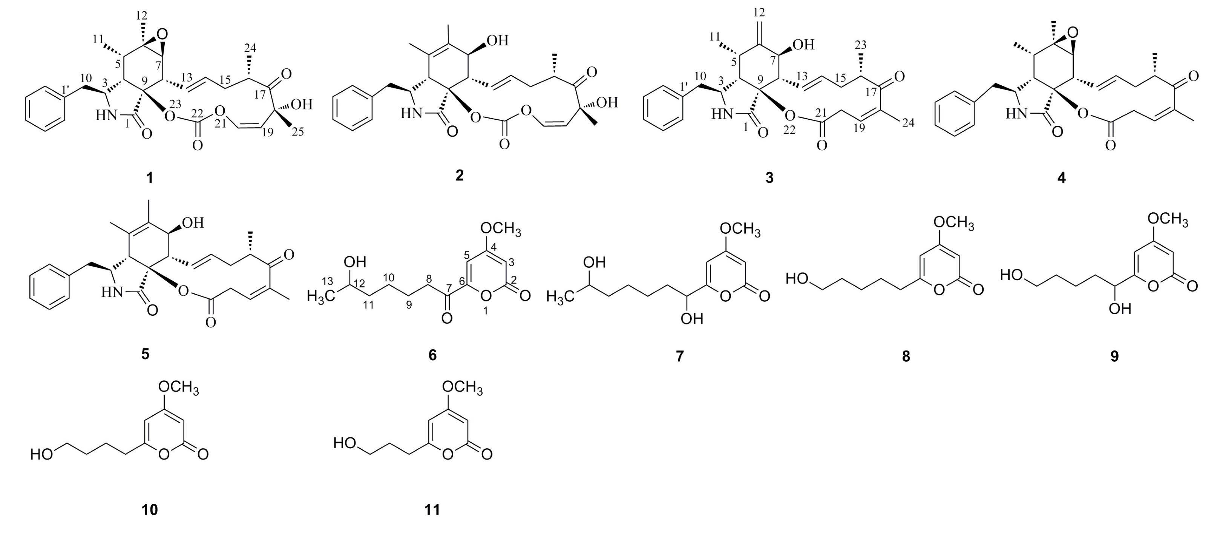
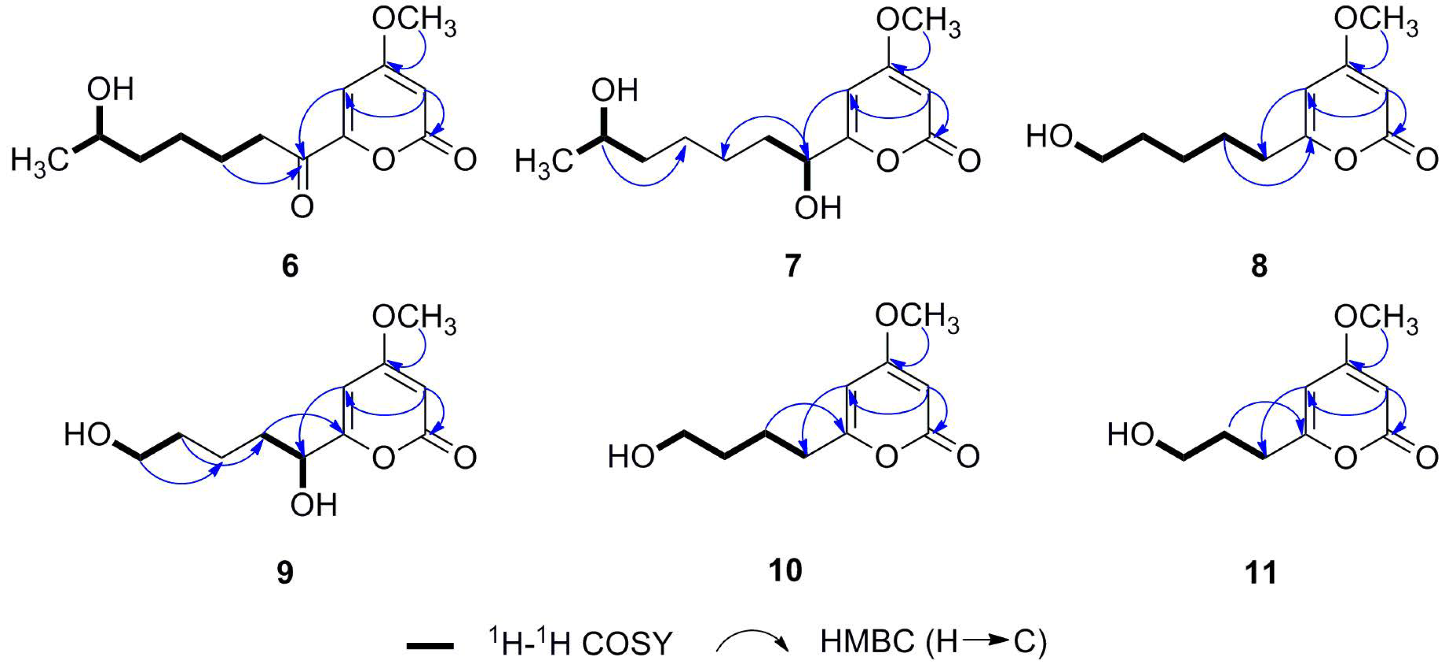
2.1. Structure Determination

The known compounds cytochalasin E (1), cytochalasin K (2), cytochalasin Z16 (3), rosellichalasin (4) and cytochalasin Z17 (5) were identified by comparison of the corresponding 1H- and 13C-NMR data with published results [10,11,12].
Compound 6 was obtained as a white amorphous powder. The molecular formula was determined to be C13H18O5 by HREIMS, implying five degrees of unsaturation. Inspection of the 1H-NMR spectrum (Table 1 and Supporting Information) reveals signals for two meta-coupled aromatic protons at δH 6.91 and 5.96, three protons at δH 3.88 (3H, s), one oxygenated methine proton at δH 3.56, one methylene proton at δH 2.85, three methylene proton in the higher field at δH 1.54 (m, 2H), 1.35 (m, 2H) and 1.28 (m, 2H), and one exchangeable proton at δH 4.32. The 13C-NMR and DEPT spectra (Table 1 and Supporting Information) displayed two carbonyl carbons at δC 193.0 and 161.6, two olefinic methine carbons at δC 104.6 and 93.5, two sp2 quaternary carbon at δC 169.5 and 153.9, a methoxy carbon at δC 56.9, an oxygenated methine carbon at δC 65.6, four methylene carbons at δC 38.7, 37.2, 24.7 and 23.0, and a methyl carbon at δC 23.6. The α-pyrone ring was elucidated as 4-methoxy-2-one with a substitutent at C-6 by analysis of HMBC correlations between H1-3 (δH 5.96)/C-2 (δC 161.6), C-4 (δC 169.5), C-5 (δC 104.6), H1-5 (δH 6.91)/C-3 (δC 93.5), C-4 (δC 169.5), C-6 (δC 153.9), and H3-OCH3H 3.88)/C-4 (δC 169.5) (Figure 2). COSY correlations (Figure 2) from H3-13 (δH 1.03), OH-12 (δH 4.32) via H1-12 (δH 3.56), H2-11 (δH 1.35), H2-10 (δH 1.28) and H2-9 (δH 1.54) to H2-8 (δH 2.85) show the presence of a 2-hexanol group , which is attached to a carbonyl group as indicated by HMBC correlations from H2-9 (δH 1.54) and H2-8 (δH 2.85) to C-7 (δC 193.0), while H1-5 (δH 6.91) give HMBC correlations (Figure 2) to C-7 (δC 193.0) implying that C-7 (δC 193.0) is attached to C-6 (δC 153.9). The structure of compound 6 was therefore determined to correspond to the previously unreported compound 4-methoxy-6-(6-hydroxy-1-oxoheptyl)-2H-pyran-2-one, named xylapyrone A.
Table 1. 1H- and 13C-NMR spectral data (400 MHz, DMSO) of compounds 68.
Positionp678
δH (mult., J in Hz)δC (mult.)δH (mult., J in Hz)δC (mult.)δH (mult., J in Hz)δC (mult.)
2 161.6, qC 163.3, qC 164.1, qC
35.96, d(2.0)93.5, CH5.54, d (2.2)87.5, CH5.52, d (2.2)87.7, CH
4 169.5, qC 171.0, qC 171.5, qC
56.91, d(2.0)104.6, CH6.10, d (2.2)97.7, CH6.05, d (2.2)99.9, CH
6 153.9, qC 167.8, qC 166.0, qC
7 193.0, qC4.22, dt (5.0, 7.6)68.9, CH2.44, t (7.5)33.1, CH2
82.85, t (7.2)37.2, CH21.64, m/1.53, m34.7, CH21.55, dt (7.5)26.6, CH2
91.54, m23.0, CH21.30, m25.1, CH21.30, m25.3, CH2
101.28, m24.7, CH21.30, m24.7, CH21.42, m32.6, CH2
111.35, m38.7, CH21.30, m38.9, CH23.38, dd (6.4, 11.5)61.0, CH2
123.56, m65.6, CH3.54, m65.6, CH
131.03, d (6.1)23.6, CH31.01, d (6.2)23.6, CH3
4-OCH33.88, s56.9, CH33.81, s56.3, CH33.79, s56.7, CH3
12-OH4.32, d (4.9) 4.29, d (5.0)
7-OH 5.58, d (5.4)
11-OH 4.36, t (5.1)
Compound 7 was also obtained as a white amorphous powder and the molecular formula was deduced to be C13H20O5 from the molecular ion peak at m/z 256.1308 [M]+ (calcd for 256.1311) in the HREIMS, having two more hydrogen atoms than 6. Detailed NMR spectra (Table 1 and Supporting Information) revealed the existence of the same 4-methoxy-α-pyrone framework as in 6 and that a carbonyl group in 6 has been reduced to a hydroxyl group in 7. The carbonyl group resonating at δC 193.0 (C-7) in the NMR spectra of 6 was absent in those of 7, and instead an oxygenated methine signal at δH 4.22 (H1-7) and δC 68.9 (C-7) were observed in 7. These observations suggested the hydroxylation at C-7 and this was verified by the HMBC correlations from H1-7 (δH 4.22) to C-5 (δC 97.7), C-6 (δC 167.8), C-8 (δC 34.7) and C-9 (δC 25.1) (Figure 2). Thus, the structure of 7 was determined to be as shown in Figure 1, and the compound was named xylapyrone B.
Figure 2. Key HMBC and COSY correlations of compounds 611 in DMSO.
Molecules 20 07940 g002
Compound 8 has the molecular formula C11H16O4 suggested by HREIMS data. 1H- and 13C-NMR data (Table 1 and Supporting Information) showed that 8 had the same basic 4-methoxy-α-pyrone structure with a substituent at C-6, as 6 and 7. Five methylenes at δH 3.38, δH 2.44, δH 1.55, δH 1.42 (2H, m) and δH 1.30 (2H, m) were observed in the 1H-NMR spectra. The corresponding carbon signals at δC 61.0, 33.1, 26.6, 32.6 and 25.3 were present in the 13C-NMR spectra. The COSY correlations (Figure 2) of five methylenes suggested the side chain was pentanol. Therefore, the structure of xylapyrone C was unambiguously elucidated as 8.
Compound 9 was obtained as a white amorphous solid. Its HREIMS exhibits a peak at m/z 228.0991 [M]+ (calcd for 228.0998), indicating a molecular formula C11H16O5 with one more oxygen atom than 8 [C11H16O4]. The NMR spectroscopic data (Table 2 and Supporting Information) indicated that they were structurally related, and that they differed in their oxidation status. Detailed analysis of the 1H- and 13C-NMR spectra revealed that the methylene unit at δH 2.44 and δC 33.1(CH2) of 8 was replaced by an oxygenated methine unit at δH 4.23 (1H, brs) and δC 69.5(CH) in 9. Unexpectedly, no HMBC correlation from H1-7 (δH 4.23) to any other carbon signal was observed, so the location of the hydroxyl group was deduced to be C-7 according to the COSY correlations from OH-7 (δH 5.60) via H1-7 (δH 4.23) to H2-8 (δH 1.65 and 1.54) and a strong HMBC correlation from H2-8 (δH 1.54) to C-6 (δC 168.3) (Figure 2). Consequently, the structure of 9 was identified as shown in Figure 1 and named xylapyrone D.
The structures of xylapyrone E (10) and xylapyrone F (11) were determined straightforwardly as they were very similar to those of 8. Their NMR data (Table 2 and Supporting Information) and HREIMS suggested the difference of their structures is that side-chains attached to C-6 in 8, 10 and 11 are pentanol, butanol and propanol, respectively.
Table 2. 1H- and 13C-NMR spectral data (400 MHz, DMSO-d6) of compounds 911.
Position91011
δH (mult., J in Hz)δC (mult.)δH (mult., J in Hz)δC (mult.)δH (mult., J in Hz)δC (mult.)
2 163.8, qC 164.2, qC 164.1, qC
35.55, d (2.0)88.1, CH5.53, d (2.2)87.7, CH5.52, d (2.2)87.7, CH
4 171.5, qC 171.5, qC 171.5, qC
56.11, d (2.0)98.3, CH6.06, d (2.2)99.9, CH6.04, d (2.2)99.8, CH
6 168.3, qC 166.0, qC 166.0, qC
74.23, brs69.5, CH2.46, t (7.4)32.9, CH22.48, t (7.8)30.0, CH2
81.65, m/1.54, m35.0, CH21.60, m23.3, CH21.70, m30.0, CH2
91.34, m21.7, CH21.45, m32.1, CH23.42, dd (6.0, 11.0)60.1, CH2
101.40, m32.8, CH23.42, dd (6.0, 11.0)60.7, CH2
113.37, dd (6.0, 11.1)61.1, CH2
12
13
4-OCH33.81, s56.8, CH33.81, s56.7, CH33.79, s56.7, CH3
7-OH5.60, brs
11-OH4.35, t (5.0)
10-OH 4.43, t (5.0)
9-OH 4.55, t (5.0)

2.2. Cytotoxic Properties

Isolated compounds 111 were evaluated for in vitro cytotoxicity against two cancer cell lines HepG2 and Caski. Mitomycin was employed as positive control. Of the tested compounds, compounds 13 showed moderate cytotoxicity, with IC50 values ranging from 25 to 63 μM, and compounds 411 were found to be inactive, with IC50 values >100 μM (Table 3). The most cytotoxic compound is cytochalasin E (1). Cytochalasin-type compounds have been reported as promising lead compounds for anticancer drug discovery that reduce the proliferation of different cancer cell lines such as P388, A549 and KB cells [10,11]. α-Pyrone natural products are widespread in Nature and have a broad spectrum of biological activities [13]. The literature [14,15,16] has suggested that structural variations of the side chains attached to α-pyrones ring may make significant differences to the cytotoxicity, which is worth further investigation.
Table 3. Cytotoxicities of compounds 111 against two cancer cell lines a.
1234567891011
HepG2255945>100>100>100>100>100>100>100>100
Caski296353>100>100>100>100>100>100>100>100
a Results are expressed as IC50 values in μM. An average value of three independent experiments is reported. Hep-G2 and Caski are human liver cancer cell line and human cervical cancer cell line, respectively.

3. Experimental Section

3.1. General Experimental Procedures

NMR spectra (1H at 400 MHz, 13C at 100 MHz) were measured on a Bruker Ultrashield TM Plus 400 MHz spectrometer (Faellanden, Switzerland) with tetramethylsilane as an internal standard and chemical shifts were recorded as δ values. EI and HREIMS spectra were recorded on DSQ II (Thermo Fisher Scientific Inc., Waltham, MA, USA) and MAT95XP (Thermo Electron Corp., Madison, WI, USA) mass spectrometers, respectively. Semipreparative HPLC was performed on a Dionex Ultimate 3000 system (Thermo Fisher Scientific Inc.) using a semipreparative C18 (COSMOSIL 5 μm, 10 mm × 250 mm) column coupled with an diode-array detector. TLC was performed on precoated silica gel GF254 (Merck, Darmstadt, Germany) plates (PTLC, Merck). Silica gel (200–300 mesh) for column chromatography was produced by Qingdao Marine Chemical Factory (Qingdao, China). Sephadex LH-20 was produced by Pharmacia Biotech (Uppsala, Sweden). UV spectra were taken on a UV-3100PC spectrometer (Hitachi, Ltd., Tokyo, Japan).

3.2. Isolation and Identification of the Strain

The endophytic fungus Xylaria sp. BM9 was isolated from a piece of fresh tissue from the inner part of a medicinal plant leaf of Saccharum arundinaceum Retz., collected from the Yichang (Hubei Province, China) in April 2011. The fungus was deposited as Xylaria sp. BM9 (GenBank accession numbers KC184129) at the Hubei Key Laboratory of Natural Products Research and Development, College of Chemistry and Life Sciences, China Three Gorges University, Yichang, China.

3.3. Seed and Mass Cultures of the Strain

The fungus Xylaria sp. BM9 was maintained on potato dextrose agar. Agar plugs containing the fungal strain were inoculated in 500 mL Erlenmeyer flasks, each containing 200 mL of potato dextrose broth. Flask cultures were incubated at 28 °C on a rotary shaker at 130 rpm for 3 days as seed culture.
Liquid fermentation was performed by the following procedure: each of the seed cultures (200 mL) was transferred into 500 mL Erlenmeyer flasks containing 200 mL of potato dextrose broth. These flasks were incubated at 28 °C on a rotary shaker at 130 rpm for 14 days. After fermentation, the culture (25 L) was centrifuged to yield the supernatant and a mycelial cake. The supernatant was extracted three times with an equal volume of EtOAc, the extracts were combined and solvent was removed under reduced pressure. The mycelial cake was immersed in 2 L of acetone and the organic layers were collected and removed under reduced pressure. Two residues were combined for purification.
Solid fermentation was carried out in 500 mL Erlenmeyer flasks containing 40 g rice, each flask was inoculated with the seed cultures (5.0 mL) and incubated at 25 °C for 30 days. The fermented material was soaked in EtOAc, and the organic solvent was evaporated to dryness under vacuum to afford the crude extract.

3.4. Extraction and Isolation of Compounds

The liquid fermentation residue (4.0 g) was chromatographed on a silica gel (200–300 mesh) column and eluted with petroleum ether–acetone (1:0, 9:1, 4:1, 2:1, 1:1, and 1:2, v/v) to yield six fractions (Fr. 1–Fr. 6). Fr. 3 (1.2 g) was further separated by semi-preparative reversed-phase HPLC on an ODS semi-preparative C18 column (COSMOSIL 5 μm, 10 mm × 250 mm) eluted with 30% MeCN/H2O to afford 6 (2.8 mg), 7 (5.4 mg), 8 (3.2 mg), 9 (1.5 mg), 10 (4.9 mg) and 11 (1.9 mg).
The solid fermentation residue (50 g) was chromatographed on a silica gel (200–300 mesh) column and eluted with petroleum ether–acetone (1:0, 9:1, 4:1, 2:1, 1:1, and 1:2, v/v) to yield nine fractions (Fr.1-Fr.9). Fr.2 (2.5 g) was further separated by chromatography on a Sephadex LH-20 column eluted with CHCl3–MeOH (v/v = 1/1) to yield seven fractions (Fr. 2.1–Fr. 2.7). Fr. 2.3 (200 mg) was further separated by semi-preparative reversed-phase HPLC on an ODS semi-preparative C18 column (COSMOSIL 5 μm, 10 mm × 250 mm) eluted with 75% MeCN/H2O to afford 3 (11.3 mg), 4 (12.9 mg) and 5 (14.6 mg). Fr. 2.5 (200 mg) was successively subjected to semi-preparative reversed-phase HPLC eluted with 85% MeCN/H2O to afford 2 (1.9 mg). The Fr. 4 (1.8 g) was purified by column chromatography over Sephadex LH-20 using an equal ratio mixture of methanol and chloroform as eluent to give 1 (40 mg).
Cytochalasin E (1): colorless needles; C28H33NO7; UV (MeOH) λmax (logε) 224 (2.78), 258 (2.46) nm; 13C-NMR (acetone-d6): δ 212.9 (C-17), 170.3 (C-1), 150.1 (C-22), 142.4 (C-20), 137.4 (C-1'), 132.1 (C-14), 131.1 (C-2' & 6'), 129.5 (C-13), 129.1 (C-3' & 5'), 127.4 (C-4'), 121.7 (C-19), 87.8 (C-9), 77.6 (C-18), 61.2 (C-7), 57.7 (C-6), 53.6 (C-3), 47.5 (C-4), 47.0 (C-8), 44.5 (C-10), 41.1 (C-16), 40.0 (C-15), 36.9 (C-5), 25.0 (C-25), 20.4 (C-24), 19.6 (C-12), 13.1 (C-11); ESIMS m/z 518 [M+Na]+, 1013 [2M+Na]+.
Cytochalasin K (2): White amorphous solid; C28H33NO7; UV (MeOH) λmax (logε) 224 (3.38), 258 (2.42) nm; 13C-NMR (CDCl3): δ 211.5 (C-17), 170.0 (C-1), 149.0 (C-22), 142.5 (C-20), 136.7 (C-1'), 133.6 (C-14), 131.8 (C-6), 129.4 (C-13), 129.3 (C-2' & 6'), 129.0 (C-3' & 5'), 127.2 (C-4'), 125.3 (C-5), 120.5 (C-19), 86.2 (C-9), 77.2 (C-18), 70.1 (C-7), 59.0 (C-3), 50.0 (C-8), 48.4 (C-4), 44.2 (C-10), 41.0 (C-16), 39.0 (C-15), 24.6 (C-25), 20.2 (C-24), 17.7 (C-11), 14.0 (C-12); ESIMS m/z 518 [M+Na]+, 1013 [2M+Na]+.
Cytochalasin Z16 (3): White amorphous solid; C28H33NO5; UV (MeOH) λmax (logε) 244 (3.73) nm; 13C-NMR (CDCl3): δ 206.1 (C-17), 171.3 (C-1), 170.0 (C-21), 149.1 (C-6), 143.7 (C-18), 138.1 (C-1'), 137.9 (C-14), 132.9 (C-19), 130.2 (C-2' & 6'), 129.8 (C-3' & 5'), 128.0 (C-4'), 126.6 (C-13), 115.8 (C-12), 84.1 (C-9), 69.9 (C-7), 54.4 (C-3), 50.8 (C-8), 49.9 (C-4), 44.6 (C-10), 40.6 (C-15), 40.3 (C-16), 37.2 (C-20), 32.7 (C-5), 18.5 (C-23), 15.0 (C-11), 13.5 (C-24); ESIMS m/z 486 [M+Na]+, 949 [2M+Na]+.
Rosellichalasin (4): White amorphous solid; C28H33NO5; UV (MeOH) λmax (logε) 244 (3.75) nm; 13C-NMR (CDCl3): δ 205.4 (C-17), 171.6 (C-1), 169.0 (C-21), 143.0 (C-18), 136.5 (C-1'), 135.2 (C-14), 131.7 (C-19), 129.5 (C-2' & 6'), 128.8 (C-3' & 5'), 127.1 (C-4'), 125.6 (C-13), 84.5 (C-9), 60.1 (C-3), 57.1 (C-6), 53.8 (C-7), 48.9 (C-4), 46.9 (C-8), 44.1 (C-10), 39.7 (C-5), 39.6 (C-15), 36.4 (C-20), 35.9 (C-16), 19.5 (C-12), 17.3 (C-11), 12.8 (C-23), 12.7 (C-24); ESI-MS m/z 486 [M+Na]+, 949 [2M+Na]+.
Cytochalasin Z17 (5): White amorphous solid; C28H33NO5; UV (MeOH) λmax (logε) 242 (3.71) nm; 13C-NMR (CDCl3): δ 205.5 (C-17), 171.6 (C-1), 168.9 (C-21), 143.2 (C-18), 137.1 (C-14), 137.0 (C-1'), 132.9 (C-6), 131.5 (C-19), 129.2 (C-2' & 6'), 128.9 (C-3' & 5'), 127.1 (C-4'), 126.4 (C-13), 125.1 (C-5), 83.7 (C-9), 69.6 (C-7), 59.2 (C-3), 50.0 (C-8), 49.6 (C-4), 43.7 (C-10), 40.1 (C-16), 39.7 (C-15), 37.4 (C-20), 17.5 (C-11), 17.2 (C-23), 14.1 (C-12), 13.0 (C-24); ESI-MS m/z 486 [M+Na]+, 949 [2M+Na]+.
Xylapyrone A (6): white amorphous powder; UV (MeOH) λmax (logε) 201 (0.16), 280 (2.94) nm; 1H-NMR (DMSO-d6) and 13C-NMR (DMSO-d6) spectral data see Table 1. HREIMS m/z 254.1147 [M]+ (calcd for C13H18O5, 254.1154).
Xylapyrone B (7): white amorphous powder; UV (MeOH) λmax (logε) 201 (0.14), 280 (2.93) nm; 1H-NMR (DMSO-d6) and 13C-NMR (DMSO-d6) spectral data see Table 1. HREIMS m/z 256.1308 [M]+ (calcd for C13H20O5, 256.1311).
Xylapyrone C (8): white amorphous powder; UV (MeOH) λmax (logε) 201 (0.15), 280 (2.93) nm; 1H-NMR (DMSO-d6) and 13C-NMR (DMSO-d6) spectral data see Table 1. HREIMS m/z 212.1045 [M]+ (calcd forC11H16O4, 212.1049).
Xylapyrone D (9): white amorphous solid; UV (MeOH) λmax (logε) 201 (0.14), 280 (2.98) nm; 1H-NMR (DMSO-d6) and 13C-NMR (DMSO-d6) spectral data see Table 2. HREIMS m/z 228.0991 [M]+ (calcd for C11H16O5, 228.0998).
Xylapyrone E (10): pale yellow oil; UV (MeOH) λmax (logε) 201 (0.15), 280 (2.99) nm; 1H-NMR (DMSO-d6) and 13C-NMR (DMSO-d6) spectral data see Table 2. HREIMS m/z 198.0886 [M]+ (calcd for C10H14O4, 198.0892).
Xylapyrone F (11): white amorphous powder; UV (MeOH) λmax (logε) 201 (0.17), 280 (2.99) nm; 1H-NMR (DMSO-d6) and 13C-NMR (DMSO-d6) spectral data see Table 2. HREIMS m/z 184.0729 [M]+ (calcd for C9H12O4, 184.0736).

3.5. Cytotoxicity Test

The cancer cell lines CaSki and HepG2 were obtained from the Shanghai Institute of Cell Biology, Chinese Academy of Science (Shanghai, China). All cells were maintained in RPMI-1640 medium supplemented with 10% fetal calf serum, 25 mM HEPES buffer, 2 mmol/L l-glutamine, 100 µg/mL streptomycin, and 100 U/mL penicillin. Cultures were incubated in a humidified atmosphere of 5% CO2 at 37 °C. Cells (1 × 10⁴/well) were seeded in supplemented culture medium (100 μL/well) in a 96-well plate and incubated for 24 h. The medium was then replaced with a test compound-containing medium, and the cells were further incubated for 48 h. All experiments were run in parallel with controls (0.1% DMSO without test compounds) and the cell viabilities were evaluated by MTT assays. The absorbance of formazan formed was measured at 570 nm by a microplate reader. The concentrations resulting in 50% inhibition of cell proliferation/survival (IC50) as measured by this assay are given in Table 3. Each experiment was repeated three times.

4. Conclusions

The Xylaria genus is an ubiquitous filamentous fungus, often isolated from marine environments and terrestrial sources, that can produce various types of secondary metabolites, including typical cytochalasins [17], terpenoids [18], benzofurans [19], xanthones [20], and cyclopeptides [21]. Long-chain α-pyrones are widespread in Nature and most of the PKS genes have been involved in the biosynthesis of their derivatives from acetate units [22,23]. A few papers have reported α-pyrone metabolites from Xylaria sp. Pukachaisirikul et al. described the isolation of one known α-pyrone derivative [24]. In this paper, five known cytochalasin-type analogues 15 were isolated as the major metabolites from a solid rice medium culture of Xylaria sp. BM9. However, applying liquid culture conditions to Xylaria sp. BM9 resulted in the identification of six new α-pyrone derivatives, xylapyrones A–F (compounds 611), with different biogenetic origin, in which its PKS genes for the biosynthesis of complex liquids were actived. Xylapyrones A–F were evaluated for in vitro cytotoxicity against two cancer cell lines (Hep-G2 and Caski) and found to be inactive. Further bioassay evaluation of the compounds against fungi and other targets are ongoing, so it is an issue that we don’t have sufficient amounts of these compounds to determine the absolute stereochemistry of compounds 6, 7 and 9.

Supplementary Materials

Supplementary materials can be accessed at: https://www.mdpi.com/1420-3049/20/05/7940/s1.

Acknowledgments

This work was supported by grants from the National Natural Science Foundation of China (31100080 and 21272136), Science Foundation of China Three Gorges Universtiy (KJ2011B003 and KJ2014B021), Yichang Science Research and Development Project (A13301-56), and The Education Department of Hubei Province Key Project (D20141201).

Author Contributions

All authors performed the biological and chemical experiments. H. Zhang especially contributed to the isolation of the strain, fermentation, and purification of compounds 111. X. Tu finished the identification of fungi Xylaria sp. BM9. Z. Deng and K. Zou managed and supervised the project, elucidated the structure of compounds 111 and prepared and corrected the manuscript. Z. Guo and N. Huang recorded and analyzed the NMR data to determine their chemical structures. H. He and Y. Peng evaluated the cytotoxicity of compounds 111. Z. Deng wrote the paper. All authors read and approved the final manuscript.

Conflicts of Interest

The authors declare no conflict of interest.

References

  1. Cragg, G.M.; Grothaus, P.G.; Newman, D.J. Impact of Natural Products on Developing New Anti-Cancer Agents. Chem. Rev. 2009, 109, 3012–3043. [Google Scholar] [CrossRef] [PubMed]
  2. Hertweck, C.; Scherlach, K. Triggering Cryptic Natural Product Biosynthesis in Microorganisms. Org. Biomol. Chem. 2009, 7, 1753–1760. [Google Scholar] [CrossRef] [PubMed]
  3. Singh, B.K.; Macdonald, C.A. Drug Discovery from Uncultivable Microorganisms. Drug Discov. Today 2010, 15, 792–799. [Google Scholar] [CrossRef] [PubMed]
  4. Yang, X.L.; Zhang, J.Z.; Luo, D.Q. The Taxonomy, Biology and Chemistry of the Fungal Pestalotiopsis genus. Nat. Prod. Rep. 2012, 29, 622–641. [Google Scholar] [CrossRef] [PubMed]
  5. Paranagama, P.A.; Wijeratne, E.M.K.; Gunatilake, A.A.L. Uncovering Biosynthetic Potential of Plant-Associated Fungi: Effect of Culture Conditions on Metabolite Production by Paraphaeosphaeria quadriseptata and Chaetomium chiversii. J. Nat. Prod. 2007, 70, 1939–1945. [Google Scholar] [CrossRef] [PubMed]
  6. Zhang, H.Q.; Deng, Z.S.; Guo, Z.Y.; Tu, X.; Wang, J.Z.; Zou, K. Pestalafuranones F–J, Five New Furanone Analogues from the Endophytic Fungus Nigrospora sp. BM-2. Molecules 2014, 19, 819–825. [Google Scholar] [CrossRef] [PubMed]
  7. Bode, H.B.B.; Walker, M.; Zeeck, A. Structure and Biosynthesis of Mutolide, a Novel Macrolide from a UV Mutant of the Fungus F-24'707. Eur. J. Org. Chem. 2000, 8, 1451–1456. [Google Scholar] [CrossRef]
  8. Ola, A.R.B.; Thomy, D.; Lai, D.W.; Brötz-Oesterhelt, H.; Proksch, P. Inducing Secondary Metabolite Production by the Endophytic Fungus Fusarium tricinctum through Coculture with Bacillus subtilis. J. Nat. Prod. 2013, 76, 2094–2099. [Google Scholar] [PubMed]
  9. Udwary, D.W.; Zeiqler, L.; Asolkar, R.N.; Singan, V.; Lapidus, A.; Fenical, W.; Jensen, P.R.; Moore, B.S. Genome Sequencing Reveals Complex Secondary Metabolome in the Marine Actinomycete Salinispora tropica. Proc. Natl. Acad. Sci. USA 2007, 104, 10376–10381. [Google Scholar] [CrossRef] [PubMed]
  10. Liu, H.; Gu, Q.Q.; Zhu, W.M.; Cui, C.B.; Fan, G.T.; Fang, Y.C.; Zhu, J.T.; Liu, H.B. 10-Phenyl-[12]-cytochalasins Z7, Z8 and Z9 from the Marine-Derived Fungus Spicariaelegans. J. Nat. Prod. 2006, 69, 871–875. [Google Scholar] [CrossRef] [PubMed]
  11. Zhang, H.W.; Zhang, J.; Hu, S.; Zhang, Z.J.; Zhu, C.J.; Ng, S.W.; Tan, R.X. Ardeemins and Cytochalasins from Aspergillu sterreus Resding in Artemisia annua. Planta Med. 2010, 76, 1616–1621. [Google Scholar] [CrossRef] [PubMed]
  12. Li, H.; Xiao, J.; Gao, Y.Q.; Tang, J.J.; Zhang, A.L.; Gao, J.M. Chaetoglobosins from Chaetomium globosum, an endophytic fungus in Ginkgo biloba, and their phytotoxic and cytotoxic activities. J. Agric. Food Chem. 2014, 62, 3734–3741. [Google Scholar] [CrossRef] [PubMed]
  13. McGlacken, G.P.; Fairlamb, I.J.S. 2-Pyrone natural products and mimetics: Isolation, characterization and biological acitivity. Nat. Prod. Rep. 2005, 22, 369–385. [Google Scholar] [CrossRef] [PubMed]
  14. Shin, H.J.; Lee, H.-S.; Lee, J.S.; Shin, J.; Lee, M.A.; Lee, H.-S.; Lee, Y.-J.; Yun, J.; Kang, J.S. Violapyrones H and I, New Cytotoxic Compounds Isolated from Streptomyces sp. Associated with the Marine Starfish Acanthaster planci. Mar. Drugs 2014, 12, 3283–3291. [Google Scholar] [CrossRef] [PubMed]
  15. Liu, D.; Li, X.M.; Meng, L.; Li, C.S.; Gao, S.S.; Shang, Z.; Proksch, P.; Huang, C.G.; Wang, B.G. Nigerapyrones A-H, α-Pyrone Derivatives from the Marine Mangrove-Derived Endophytic Fungus Aspergillus niger MA-132. J. Nat. Prod. 2011, 74, 1787–1791. [Google Scholar] [CrossRef] [PubMed]
  16. Zhang, J.Y.; Jiang, Y.; Cao, Y.; Liu, J.; Zhang, D.; Chen, X.; Han, L.; Jiang, C.L.; Huang, X.S. Violapyrones A-G, α-Pyrone Derivatives from Streptomyces violascens Isolated from Hylobates hoolock Feces. J. Nat. Prod. 2013, 76, 2126–2130. [Google Scholar] [CrossRef]
  17. Zhang, Q.; Xiao, J.; Sun, Q.Q.; Qin, J.C.; Pescitelli, G.; Gao, J.M. Characterization of cytochalasins from the endophytic Xylaria sp. and their biological functions. J. Agric. Food Chem. 2014, 62, 10962–10969. [Google Scholar] [CrossRef]
  18. Isaka, M.; Srisanoh, U.; Sappan, M.; Kongthong, S.; Srikitikulchai, P. Eremophilane and eudesmane sesquiterpenoids and a pimarane diterpenoid from the wood-decay fungus Xylaria sp. BCC 5484. Phytochem. Lett. 2012, 5, 78–82. [Google Scholar] [CrossRef]
  19. Xu, F.; Zhang, Y.; Wang, J.J.; Pang, J.Y.; Huang, C.H.; Wu, X.Y.; She, Z.G.; Vrijmoed, L.L.P.; Jones, E.B.G.; Lin, Y.C. Benzofuran Derivatives from the Mangrove Endophytic Fungus Xylaria sp. (#2508). J. Nat. Prod. 2008, 71, 1251–1253. [Google Scholar] [CrossRef] [PubMed]
  20. Healy, P.C.; Hocking, A.; Tran-Dinh, N.; Pitt, J.I.; Shivas, R.G.; Mitchell, J.K.; Kotiw, M.; Davis, R.A. Xanthones from a microfungus of the genus Xylaria. Phytochemistry 2004, 65, 2373–2378. [Google Scholar] [CrossRef] [PubMed]
  21. Wu, W.; Dai, H.Q.; Bao, L.; Ren, B.; Lu, J.C.; Luo, Y.M.; Guo, L.D.; Zhang, L.X.; Liu, H.W. Isolation and Structural Elucidation of Proline-Containing Cyclopentapeptides from an Endolichenic Xylaria sp. J. Nat. Prod. 2011, 74, 1303–1308. [Google Scholar] [CrossRef] [PubMed]
  22. Thomas, J.S. The 13C-NMR spectrum of a pyrone metabolite of aspergillus melleus. Biosynthetic incorporation of singly and doubly labelled [13C]-acetate. Tetrahedron Lett. 1975, 16, 175–178. [Google Scholar] [CrossRef]
  23. Terry, D.C.; Gerald, A.P. Pyrones. VIII. Biosynthetic investigations of the fungal metabolite phacidin. Can. J. Chem. 1982, 60, 133–137. [Google Scholar] [CrossRef]
  24. Rukachaisirikul, V.; Khamthong, N.; Sukpondma, Y.; Pakawatchai, C.; Phongpaichit, S.; Sakayaroj, J.; Kirtikara, K. An [11]Cytochalasin Derivative from the Marine-Derived Fungus Xylaria sp. PSU-F100. Chem. Pharm. Bull. 2009, 57, 1409–1411. [Google Scholar] [CrossRef] [PubMed]
  • Sample Availability: Samples of the compounds are not available from the authors because bioactivity tests of those compounds are going on.

