Article

Anti-Ageing Effects of Sonchus oleraceus L. (pūhā) Leaf Extracts on H2O2-Induced Cell Senescence

1
School of Pharmacy, University of Otago, Dunedin 9056, New Zealand
2
Department of Pharmacy, University of Copenhagen, Copenhagen 2100, Denmark
*
Author to whom correspondence should be addressed.
Molecules 2015, 20(3), 4548-4564; https://doi.org/10.3390/molecules20034548
Submission received: 21 January 2015 / Revised: 3 March 2015 / Accepted: 3 March 2015 / Published: 12 March 2015

Abstract

:
Antioxidants protect against damage from free radicals and are believed to slow the ageing process. Previously, we have reported the high antioxidant activity of 70% methanolic Sonchus oleraceus L. (Asteraceae) leaf extracts. We hypothesize that S. oleraceus extracts protect cells against H2O2-induced senescence by mediating oxidative stress. Premature senescence of young WI-38 cells was induced by application of H2O2. Cells were treated with S. oleraceus extracts before or after H2O2 stress. The senescence- associated β-galactosidase (SA-β-gal) activity was used to indicate cell senescence. S. oleraceus extracts showed higher cellular antioxidant activity than chlorogenic acid in WI-38 cells. S. oleraceus extracts suppressed H2O2 stress-induced premature senescence in a concentration-dependent manner. At 5 and 20 mg/mL, S. oleraceus extracts showed better or equivalent effects of reducing stress-induced premature senescence than the corresponding ascorbic acid treatments. These findings indicate the potential of S. oleraceus extracts to be formulated as an anti-ageing agent.

Graphical Abstract

1. Introduction

Ageing or senescence is a complex and inevitable biological process, which is not only attributed to individual genetic variation but also to external factors such as environmental conditions, nutrition, alcohol and diseases [1,2,3,4]. There are three theories that have been proposed to explain ageing: genetic, neuroendocrine and damage-accumulation theories, which are all important and interrelated [5]. Of these theories, the most widely accepted is the free radical theory that ageing and its related diseases result from accumulated oxidative damage to cell constituents and tissues caused by excessive exposure to free radicals [6]. Reactive species are generated during normal cell metabolism [7] or to protect humans from immediate death from infections [8]. These reactive species can cause lipid peroxidation, denature proteins and fragment DNA due to oxidative stress, and further increase the mutation rate, especially when the antioxidant defenses cannot remove the excess free radicals [5,7,8]. Increasing oxidative stress is commonly associated with ageing and age-related diseases [9].
Antioxidants, which mediate the imbalance between intracellular antioxidant defenses and oxidative damage by reducing the reactive oxygen species (ROS) levels, are believed to be able to reduce stress-induced premature senescence or slow down replicative senescence. Investigations with ascorbic acid and vitamin E [10,11,12,13], or plant-derived antioxidants [14,15,16,17] confirm this claim. Due to the reported carcinogenesis and hepatoxicity of synthetic antioxidants [18], antioxidants from natural origins are preferable in applications for medications and food additives. Both normal human fibroblasts and H2O2-stressed fibroblasts experienced an extended lifespan in the presence of cyanidin, the most prevalent anthocyanin in plants [19]. Likewise in in vivo studies, protocatechuic acid from Alpinia oxyphylla fruits successfully enhanced endogenous antioxidant levels and therefore their activity, and down-regulated malondialdehyde (a biomarker of ageing) in aged rats [14].
Cellular senescence can occur due to failure in cell proliferation, or exposure to a variety of cellular stresses, such as oxidizing or DNA damaging agents, or expression of activated oncogenes [3,20]. A variety of cellular senescence features have been identified, amongst which are cellular morphology [2,21], telomere length [2,22], gene expression [23] and senescence-associated β-galactosidase (SA-β-gal) activity [24,25]. During ageing, senescence-related genes can be over-expressed to induce senescent morphogenesis [23]. Many senescence-associated, over-expressed proteins can be utilized as biomarkers, such as transforming growth factor-β1, ICFBP-3 mRNA, p16 tumor suppressor protein and β-galactosidase [23,24,25]. β-Galactosidase is a collective name for enzymes that cleave non-reducing β-d-galactose residues from glycoproteins, sphingolipids and keratin sulphate in β-d-galactosides [20]. In senescent cells, the over-expression of β-galactosidase enables the detection of β-galactosidase activity at sub-optimal pH [3,24]. This enzyme activity is not detectable in actively proliferating cells. There are two main methods to measure the SA-β-gal activity [3]. It can be cytochemically or histochemically detected using the chromogenic substrate 5-bromo-4-chloro-3-indoyl β-d-galactopyranoside. SA-β-gal positive (blue-stained) cells are manually counted and expressed as the percentage of total cell population [24]. The cytochemical assay is simple and allows detection in tissue samples. On the other hand, it is subjective and the procedure is time-consuming. The second method is fluorescence-based using the fluorogenic substrates 5-dodecanoylaminofluorescein-di-β-d-galactopyranoside (C12FDG) or fluorescein-di-β-d-galacto-pyranoside (FDG) lysosome alkalinizaiton. SA-β-gal positive activity can be detected and quantified using a flow cytometer, microfluidics analyzer or fluorescence microscope [3]. The fluorescence-based methods permit quantitative measurement of single cells in the population. Compared with cytochemical assays, fluorescent assays are more sensitive and accurate with higher throughput.
Sonchus oleraceus L. (family Asteraceae) is well-known for its high content of antioxidants and antioxidant activity [26,27,28,29,30]. In New Zealand, it is commonly known as pūhā, where it is a dietary and traditional medicinal plant in Māori culture. We have previously identified three major antioxidants in methanolic leaf extracts of S. oleraceus: caftaric acid, chlorogenic acid and chicoric acid, with chicoric acid having the highest concentration [31]. To the authors’ knowledge, there is no report of the potential anti-ageing effects of S. oleraceus leaf extracts, neither in vitro nor in vivo. We have previously shown that S. oleraceus leaf extracts are absorbed into cells in vitro and can exert an antioxidant effect, but it remains unclear if the antioxidants in S. oleraceus leaves are beneficial for cells in combating oxidative senescence. Demonstration of antioxidant activity in healthy cells is a useful screening method for the bioactivity of novel compounds, however to be useful as therapeutic agents, these compounds must show effects in a disease state. Herein, we investigated the effects of S. oleraceus leaf extracts on H2O2-stressed human lung fibroblasts. SA-β-gal activity was used as the biomarker indicating premature senescence of the cells.

2. Results and Discussion
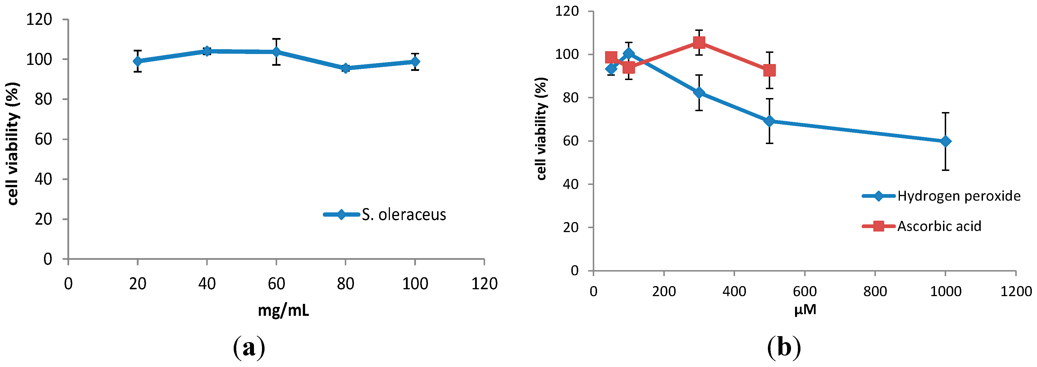
2.1. Cell Viability

WI-38 cells retained greater than 90% viability after 3 h incubation with leaf extracts from S. oleraceus (Figure 1a). It is assumed that concentrations of the leaf extract lower than 20 mg/mL would also be non-toxic. With addition of H2O2, cell viability decreased with increasing concentration (Figure 1b). Therefore, a concentration of 100 µM or lower H2O2 was chosen for further experiments.
Figure 1. Viability of WI-38 cells after treatment with (a) various concentrations of S. oleraceus leaf extracts from 20 to 100 mg/mL and (b) 50–1000 μM H2O2 or 50–500 μM ascorbic acid for 3 h. Data represent triplicate treatments and are expressed as mean ± SD (n = 3).
Molecules 20 04548 g001

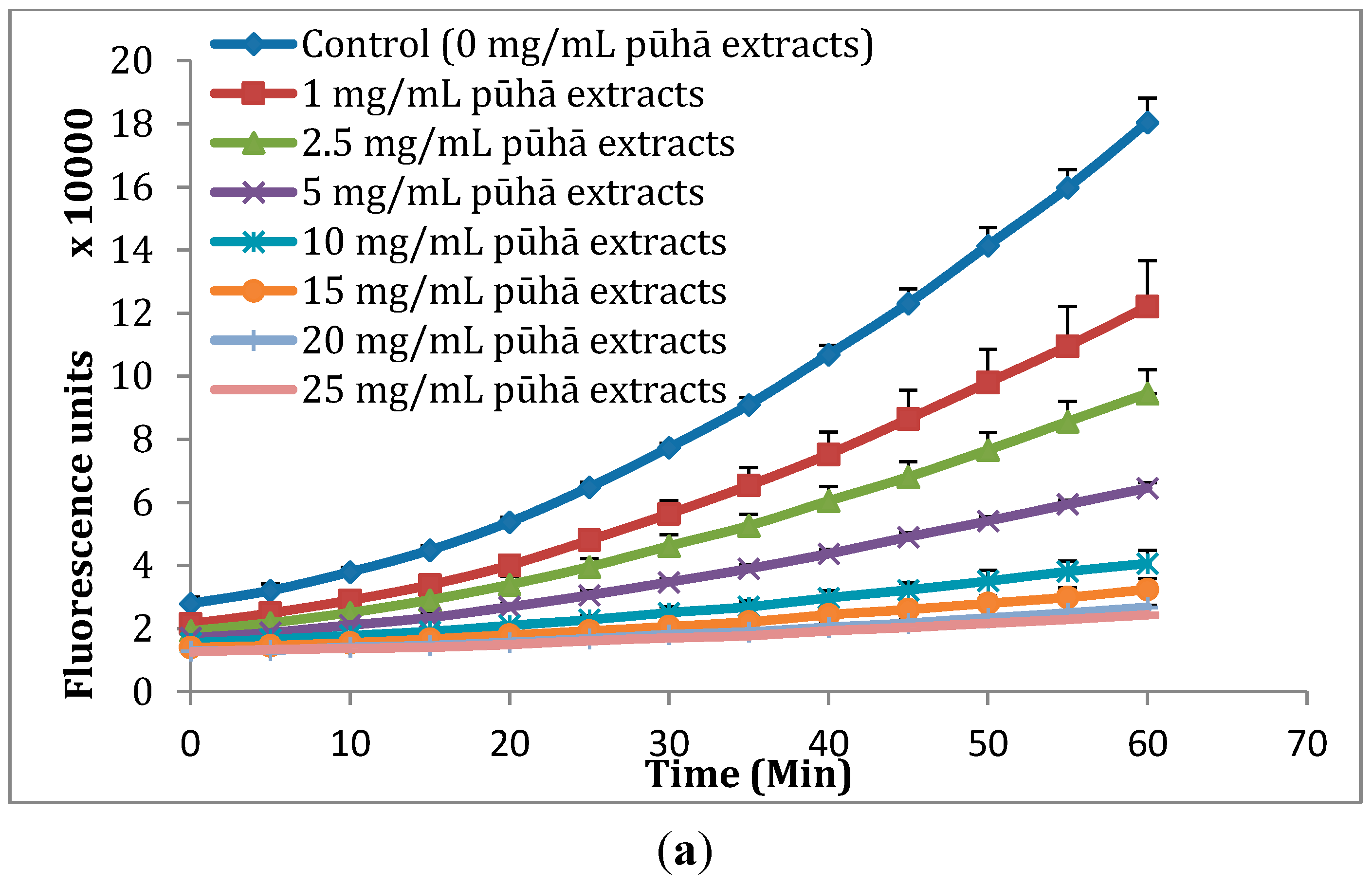
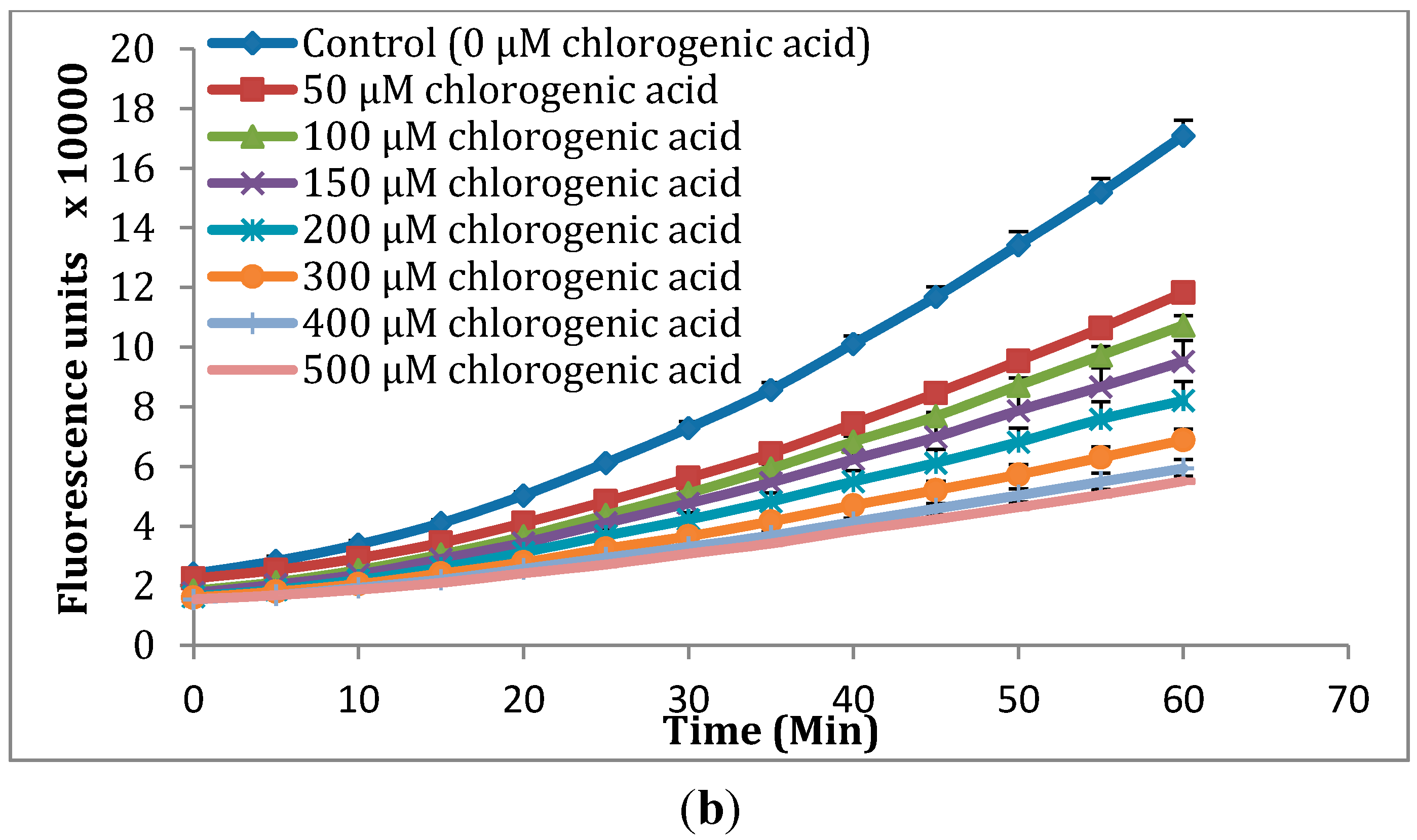
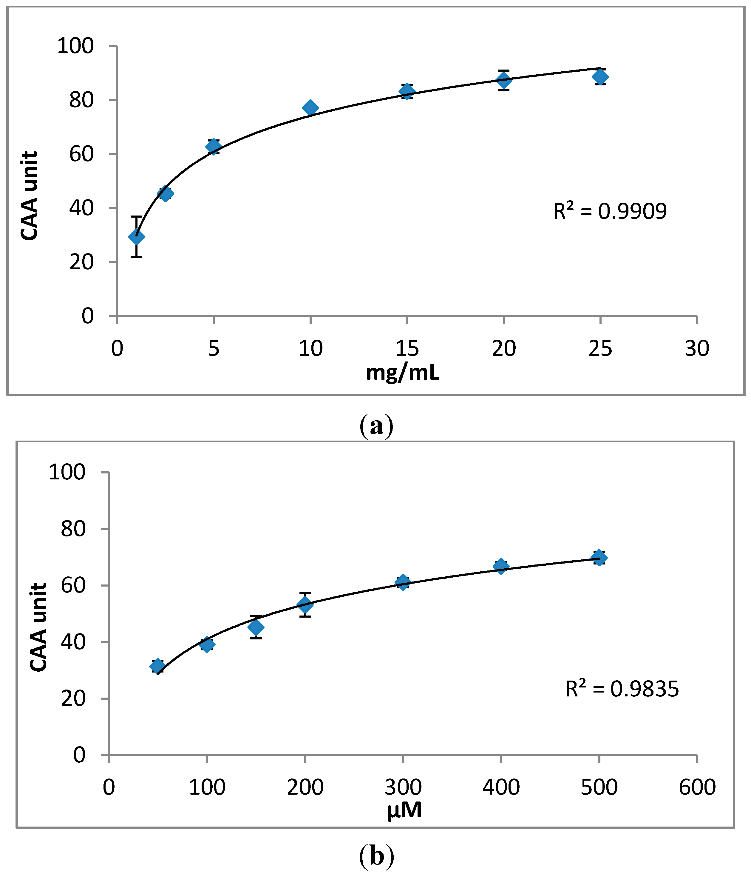
2.2. Cellular Antioxidant Activity (CAA) Assay

The CAA assay quantifies the antioxidant activity by measuring the ability of applied compounds to prevent oxidation in cells. In the CAA assay, DCFH-DA is converted to fluorescent DCF by peroxyl radicals [32]. The fluorescence intensity is proportional to the ROS concentration in the cells. The curves in the Figure 2 indicate the formation of the fluorescent DCF over 60 min. The flatter the curve, the less DCFH is oxidised. Using the CAA assay we found that the oxidation of DCFH to DCF was reduced by S. oleraceus leaf extracts in a dose-dependent manner within the concentration range of 1–25 mg/mL (Figure 2a). Intracellular ROS due to normal cell metabolism also would cause the oxidation of DCFH. However, the present results are processed data after subtracting a blank (oxidation of DCFH by normal cell metabolism) from raw data. Chlorogenic acid also decreased the formation of fluorescence with increasing concentration from 50 to 500 μM (Figure 2b). For the two other major compounds identified in S. oleraceus leaf extracts, caftaric acid and chicoric acid, the fluorescence was completely quenched (data not shown). Comparing S. oleraceus extracts with chlorogenic acid, S. oleraceus leaf extracts showed much higher activity with an EC50 value of 2.85 mg/mL (50.16 μM equivalent of total concentration of the three key antioxidants) than that of chlorogenic acid with an EC50 value of 166.34 μM. 1 mg/mL S. oleraceus leaf extracts and 50 μM chlorogenic acid showed similar inhibition of the fluorescence (Figure 2). This also can be confirmed by the CAA units obtained from the CAA assay (Figure 3). The CAA unit from the CAA assay indicates the intra-cellular activity of the samples. 1 mg/mL of S. oleraceus leaf extracts (containing 17.6 μM of the three key active compounds in total) exhibited a trend of higher activity, with 29.44 ± 7.44 CAA units, than that of 50 μM chlorogenic acid (31.34 ± 1.79 CAA units) (Figure 3). Within the concentration ranges studied, S. oleraceus leaf extracts showed higher potency than chlorogenic acid alone.
Figure 2. Peroxyl radical-induced oxidation of DCFH to DCF in WI-38 cells and inhibition of oxidation by (a) S. oleraceus leaf extracts and (b) chlorogenic acid with time. The fluorescence intensity indicates the oxidation of DCFH. The curves shown in each graph are from a single experiment (mean + SD, n = 3).
Molecules 20 04548 g002aMolecules 20 04548 g002b
Figure 3. Dose–response curves for inhibition of peroxyl radical-induced DCFH oxidation by (a) S. oleraceus leaf extracts and (b) chlorogenic acid. The data were expressed as CAA unit indicating the intra-cellular antioxidant activity of samples. The curves shown are each from a single experiment (mean ± SD, n = 3).
Molecules 20 04548 g003

2.3. Effects of S. oleraceus Leaf Extracts on Stress-Induced Premature Senescence in WI-38 Cells

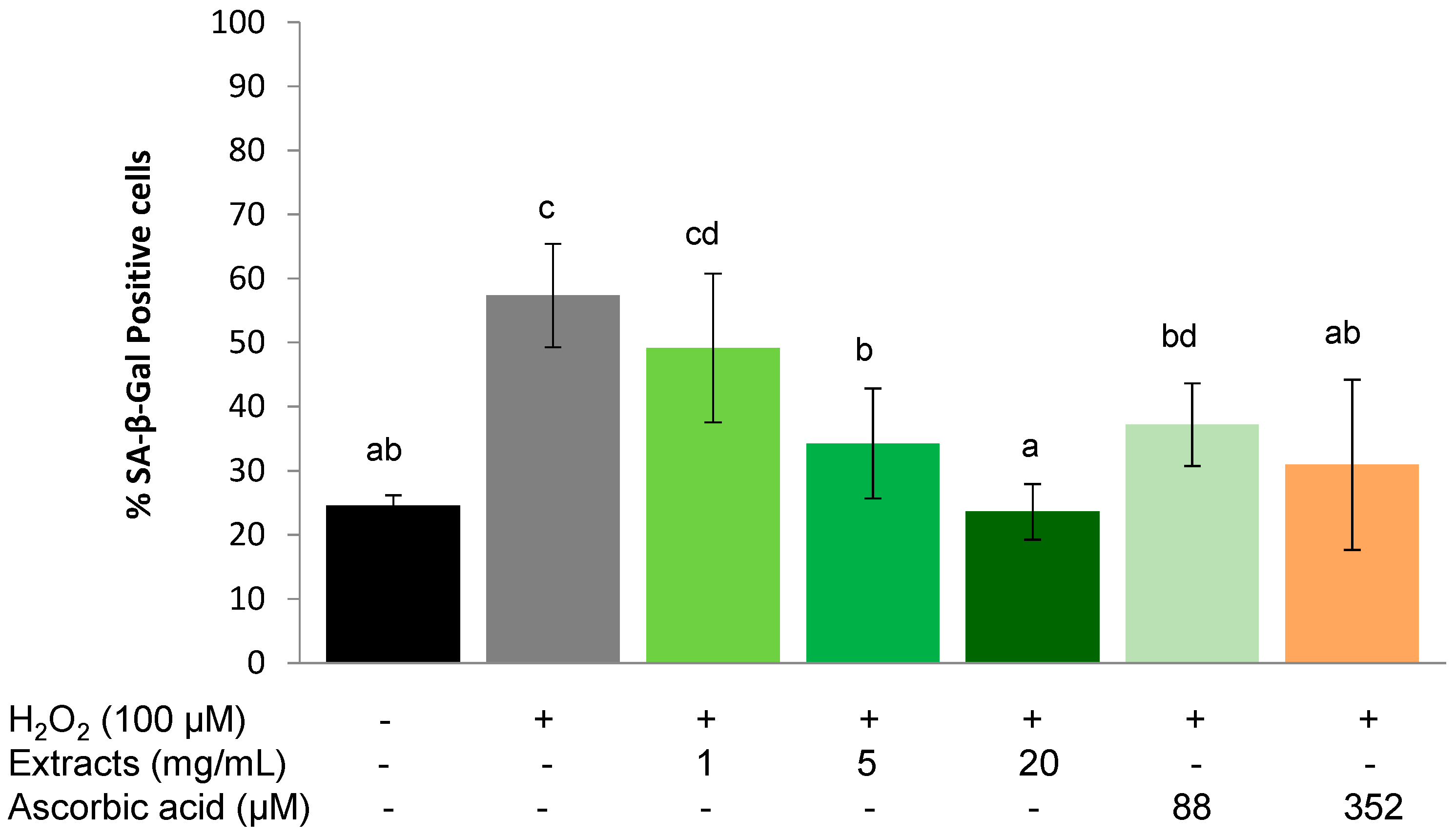
2.3.1. Protective Effect

The SA-β-Gal positive cell population increased significantly (p < 0.01) from 24.6 ± 1.6 to 57.4% ± 8.1% after being stressed with 100 µM H2O2 (Figure 4). There was a concentration-dependent effect of the S. oleraceus leaf extracts on the proportion of SA-β-Gal positive cells (Figure 4), and a trend for pre-treatment with 1 mg/mL S. oleraceus leaf extracts to reduce the percentage of SA-β-Gal positive cells. At 5 mg/mL, S. oleraceus leaf extracts reduced two thirds of H2O2 stress-induced SA-β-Gal activity. Stress-induced premature senescence was completely suppressed by 20 mg/mL S. oleraceus leaf extracts and was equivalent to the control group that did not receive the H2O2 stress (Figure 4). Compared to cells with corresponding ascorbic acid treatments (88 and 352 µM), 5 and 20 mg/mL S. oleraceus leaf extracts had a tendency to show better protective effects, but these data were not statistically significant.
Figure 4. Protective effect of S. oleraceus leaf extracts on H2O2 treated WI-38 cells. Cells were treated with 1, 5 or 20 mg/mL S. oleraceus leaf extracts or 88 or 352 µM ascorbic acid for 1 h before being stressed with 100 µM H2O2. Data are shown as percentage of SA-β-Gal positive cells (mean ± SD, n = 6). Bars with no letters in common are significantly different (p < 0.05).
Molecules 20 04548 g004

2.3.2. Recovery Effect

A similar trend as for the protective effect was observed for the ability of S. oleraceus extracts to assist in the recovery of WI-38 cells from the application of exogenous stress. 1 h treatment with 100 µM H2O2 significantly (p < 0.01) increased SA-β-Gal positive cells from 21.55% ± 4.12% to 33.40% ± 4.25%. However, this increase was lower than that of H2O2 treatment in the protective experiment (Figure 4). S. oleraceus extracts (except 1 mg/mL) and ascorbic acid reduced the SA-β-Gal activity caused by H2O2, and showed a concentration-dependent effect. When the concentration of S. oleraceus extracts was increased to 5 or 20 mg/mL, SA-β-Gal activity induced by H2O2 was significantly (p < 0.05) reduced.
Furthermore, 5 and 20 mg/mL S. oleraceus extracts showed significantly (p < 0.05) better recovery ability than the corresponding ascorbic acid treatments, although 352 µM ascorbic acid also significantly reduced the SA-β-Gal activity (p < 0.01) compared to H2O2 stress (Figure 5).
Figure 5. Recovery effect of S. oleraceus leaf extracts on H2O2 treated WI-38 cells. Cells were treated with 1, 5 or 20 mg/mL S. oleraceus leaf extracts or 88 or 352 µM ascorbic acid for 1 h following a 1 h incubation with 100 µM H2O2 as exogenous stress. Data are shown as percentage of SA-β-Gal positive cells (mean ± SD, n = 6). Bars with no letters in common are significantly different (p < 0.05).
Molecules 20 04548 g005
Figure 6. Protective effect of S. oleraceus leaf extracts on WI-38 cells chronically treated with H2O2. Cells were treated with 1, 5 or 20 mg/mL S. oleraceus leaf extracts or 88 or 352 µM ascorbic acid for 1 h at day 1 before being stressed with 30 µM H2O2 1 h per day for five consecutive days. Data are shown as percentage of SA-β-Gal positive cells (mean ± SD, n = 6). Bars with no letters in common are significantly different (p < 0.05).
Molecules 20 04548 g006

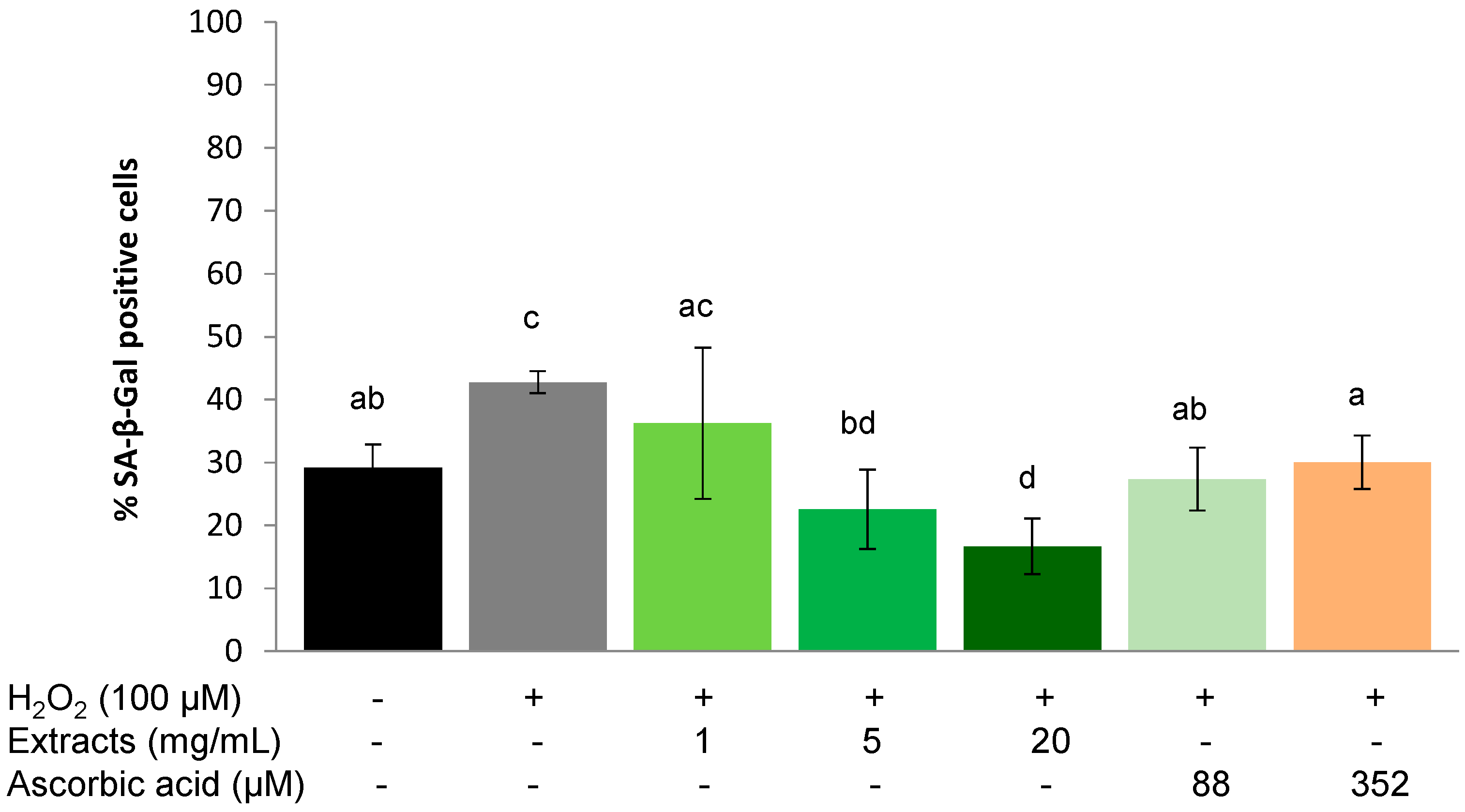
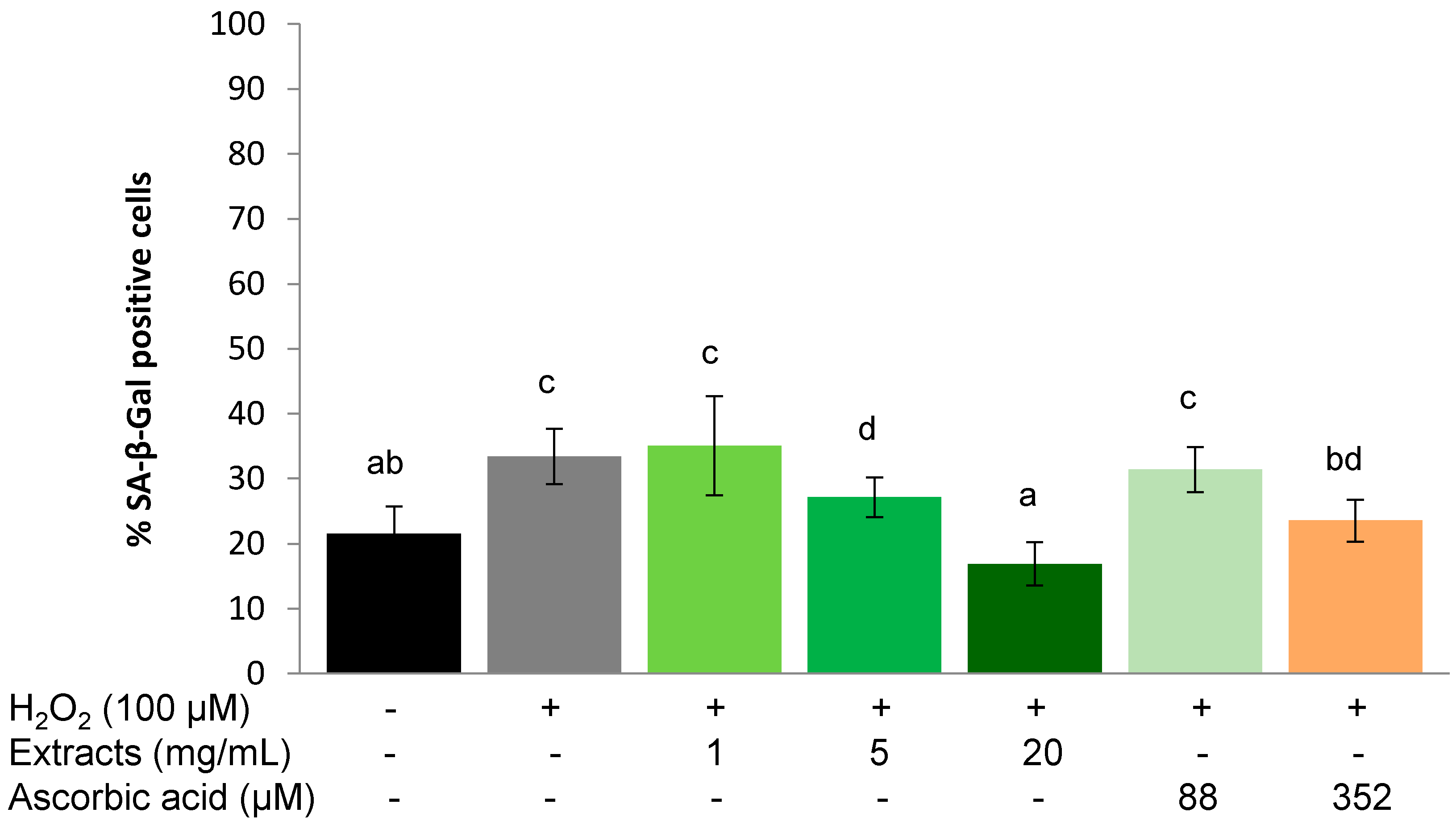
2.3.3. Protective Effect on Chronic Stress

SA-β-Gal positive cells increased significantly (p < 0.01) after consecutive stress of 30 µM of H2O2 each day for 5 days (Figure 6). 1 h treatment with S. oleraceus leaf extracts (except 1 mg/mL) or ascorbic acid significantly reduced the SA-β-Gal positive cell population, indicating a sustained protective effect of one acute dose of these antioxidants from repeated exposure to low H2O2 levels (Figure 6). In line with the acute protective and recovery effects, a concentration-dependent effect of S. oleraceus leaf extracts was also observed (Figure 6). The highest concentration of S. oleraceus leaf extracts tested was also more effective at reducing SA-β-Gal activity than the equivalent ascorbic acid treatment.

2.4. Discussion

There is growing interest in the use of natural antioxidants for human health benefits or even as food preservatives due to the safety issues raised by the use of synthetic antioxidants [33]. Many natural products have been reported to have anti-ageing activity, including herbal extracts such as Rhodiola rosea (Crassulaceae), Rosa damascene (Rosaceae), Damnacanthus officinarum (Rubiaceae), spinach leaf and green tea extracts [34]. S. oleraceus, a traditional medicinal plant worldwide, is well-known for its high antioxidant content. It is reported that S. oleraceus leaf extracts are non-cytotoxic on HepG2 cells below a concentration of 100 mg/mL [30]. To our knowledge, there is no report on the application of S. oleraceus leaf extracts on normal human diploid cells. In the present study, no cytotoxicity of S. oleraceus leaf extracts up to 100 mg/mL was observed in WI-38 cells (Figure 1).
The measurement of antioxidant activity is an important part of screening plant extracts that have the potential to be developed into supplements. Previously, we reported high antioxidant activity of S. oleraceus leaf extracts determined by the DPPH free radical assay [30,35]. However, a high antioxidant activity as measured by in vitro chemical assays does not necessarily correlate to relevant in vivo activity because of the limited predictability in complex biological systems. For instance, extracts from plum fruit had the same chemical antioxidant activity using the ORAC assay as blackberry extracts, but significantly lower cellular activity than blackberry extracts [36]. Nevertheless, some studies do show some correlations between chemical and cellular measurements of antioxidant activity of plant samples [37,38]. Cellular antioxidant activity is an important and useful parameter that indicates antioxidant activity of samples, and is more biologically representative compared to test tube chemical antioxidant activity measurements. The CAA assay developed by Wolfe and Liu [32] utilizes peroxyl radicals to assess antioxidant activity and takes into account cellular uptake, distribution and metabolism by incubating cells with antioxidants prior to exogenous application of oxidative stress. Using this assay in the present study, it is observed that S. oleraceus leaf extracts were absorbed by and exhibited antioxidant activity in WI-38 cells with an EC50 value of 2.85 mg/mL (Figure 2). This activity is lower than the reported value for blueberry (10.81 mg/mL) [32]. This indicates the high potency of S. oleraceus extracts, with values also higher than a range of vegetables [39], including broccoli, lettuce and tomatoe; and a range of other fruits [36], including apple and kiwifruit. 1 mg/mL S. oleraceus leaf extracts with 17.6 µM of the total key antioxidants had a comparably high activity to that of 50 µM chlorogenic acid, suggesting that the antioxidant extract matrix is more effective than chlorogenic acid alone. It is also possible that chlorogenic acid is not the major contributor to the cellular activity of S. oleraceus leaf extract.
Ageing is inevitable, but processes that are induced by external factors can be slowed by down-regulating oxidative stress in the body, for instance, by supplementing with antioxidants. Hwang et al. [13] demonstrated that ascorbic acid extends the replicative life span of human embryonic fibroblasts by decreasing cellular ROS to reduce mitochondrial and DNA damage. In the present study, ascorbic acid and S. oleraceus extracts containing an antioxidant mixture also exhibited an anti-ageing effect by reducing stress-induced premature senescence (Figure 4, Figure 5, and Figure 6). However, long-term ascorbic acid supplementation has shown conflicting effects in animal studies. A diet of an antioxidant combination showed no increase in the life span of rats [40]. The authors proposed that compensatory reductions in endogenous protection mechanisms might be involved when high dietary doses of vitamin C were administered. For this reason, the concentration of ascorbic acid used for the present study has been carefully chosen. Cells with stress-induced, premature senescence share similar characteristics to those with replicative senescence, such as morphology, cell cycle regulation, increased ROS generation and SA-β-gal activity [2]. Stress-induced premature senescence can be induced by various sub-lethal stresses including H2O2, hyperoxia, UV radication or tert-butylhydroperoxide [2,9,41]. Human fibroblasts undergo finite proliferation before irreversible growth arrest in vitro [42]. Human diploid fibroblasts have become a classical model for studying cellular ageing and identifying ageing-related changes in human cells. Based on these features, H2O2 stressed human fibroblast cells are considered a suitable model to assess effects of anti-ageing agents [9,19,43]. SA-β-gal activity is one of the most popular biomarkers for cell senescence [44,45]. The SA-β-gal activity in the present study increased significantly in the presence of H2O2, suggesting that cells experienced senescence. Both S. oleraceus extracts and ascorbic acid suppressed SA-β-gal activity caused by H2O2, indicating the anti-senescence properties of S. oleraceus extracts and ascorbic acid. 88 and 352 µM ascorbic acid (equivalent to 5 and 20 mg/mL S. oleraceus extracts, respectively) did not show better ability than the corresponding S. oleraceus extracts. This implied that S. oleraceus extracts at a concentration of 20 mg/mL and above might have better ability of protecting or recovering stress-induced premature senescence than ascorbic acid.
From the above, it is clear that S. oleraceus leaf extracts can protect human cells from stress-induced senescence. However, if this effect is long lasting or if frequent treatments are required to maintain this effect remain unknown. In view of this, a chronic stress experiment was carried out to further explore the anti-ageing effect of S. oleraceus leaf extracts. The results demonstrated a similar effect pattern as that of acute stresses (Figure 6). This suggested that S. oleraceus extracts can not only assist cells to recover from acute H2O2 stress, but can also protect cells from chronic stress applied for five consecutive days as tested in the present study. Similar to the present results, a mixture of cocoa and green tea extracts, rich in polyphenols, and vitamin E significantly reduced UV stress-induced premature senescence from 51.67% to 38.81% in human dermal fibroblasts [46]. Oxidative DNA damage has been demonstrated to be proportional to the age of WI-38 human fibroblast cells [9] and was proposed to be another biomarker for cellular senescence. H2O2-induced DNA damage [43] thus showed senescent-like growth arrest [47] in human fibroblasts. In accordance with previous studies, despite the use of a different biomarker in the present study, H2O2 significantly increased stress-induced premature senescence (Figure 4, Figure 5, and Figure 6). It is reported that antioxidant treatments counteracted the effect of stressors, including H2O2, and restored DNA synthesis [48]; and therefore can protect the cells from premature ageing. Further studies are needed to understand how S. oleraceus leaf extracts mediate the effects of the external stress factors on cells. For example, utilizing the comet assay [49] to characterize the extent of DNA damage. An understanding of the cellular components that are damaged by application of stress would help to define which of the cellular processes are involved during recovery from stress.
The protective and recovery ability of S. oleraceus extracts against H2O2 stress-induced premature senescence in the present study was encouraging for the use of S. oleraceus leaves as a potent anti-ageing, nutritional supplement. In the development of oral supplements, it is imperative to characterize the in vivo stability of bioactive components in the gastrointestinal tract environment. Further, to ensure the therapeutic effect of the S. oleraceus leaf extracts following oral administration, formulation strategies that increase the in vivo stability, absorption and bioavailability should be investigated.

3. Experimental Section

3.1. Plant Material

S. oleraceus seeds were collected from plants growing wild in Oamaru, New Zealand (45°05.346' S, 170°58.861' E) and identified by Mr John Steel, a botanist in the Department of Botany at the University of Otago, New Zealand. Voucher specimens (OTA 061166 and OTA 061167) are lodged at the Otago Regional Herbarium in the Department of Botany at the University of Otago, New Zealand. Seeds were germinated in a 50:50 potting compost:sand mix and grown for three weeks in a greenhouse, and the seedlings transplanted into 1 L pots in the same soil mix as above. Tap water was supplied every two days. Artificial light was provided 24 h a day in addition to normal natural daylight. The temperature inside the greenhouse was maintained at 25 °C. Fully expanded mature leaves from the middle of eight-week-old S. oleraceus plants were harvested and freeze-dried before being blended into a powder.

3.2. Preparation of Leaf Extracts

Freeze-dried S. oleraceus leaf powder was extracted with 70% methanol (v/v) to obtain a 10% (fresh w/v) mixture as described by Ou et al. [31]. After stirring for approximately 0.5 h at room temperature, the extraction mixture was centrifuged at 9700 g for 10 min. Solvent was removed by centrifugal evaporation (SVC-200H SpeedVac® Concentrator, Savant, Farmingdale, NY, USA). The dried extracts were stored at −20 °C before further analysis.

3.3. Preparation of Extract Samples and Chemical Solutions

S. oleraceus extracts were prepared from freeze-dried leaf powder as described above. Chlorogenic acid (99% GR) (Acros Organics, Geel, Belgium) and ascorbic acid (Sigma®, St. Louis, MO, USA) and extract solutions were prepared immediately prior to use. For cytotxicity measurements, methylene blue stain solution consisted of 98% HBSS (v/v), 0.5% glutaraldehyde and 0.6% methylene blue (w/v) (Koch-Light laboratories Ltd., UK). Elution solution was made of 49% phosphate buffered saline solution (PBS) (Gibco®, Grand Island, NY, USA), 50% ethanol and 1% acetic acid (v/v) (BDH Aristar®, Edmonton, AB, Canada).
For the Cellular Antioxidant Activity (CAA) assay, a 20 mM stock solution of 2',7'-dichloro-fluorescin diacetate (DCFH-DA) (Sigma®, Rehovot, Israel) in methanol was prepared and stored at −20 °C. A 200 mM 2,2'-azobis(2-methylpropionamidine) dihydrochloride (ABAP) (Sigma®) stock solution in Hank’s balanced salt solution (HBSS) (Sigma®) was prepared, and aliquots were stored at −20 °C. Chlorogenic acid was dissolved in purified water before further dilution in treatment medium (Basal Medium Eagle (BME) (Gibco®) with 2 mM GlutaMAX (Gibco®) and 25 mM 2-[4-(2-hydroxyethyl)piperazin-1-yl]ethanesulfonic acid (HEPES) (Sigma®). Dried S. oleraceus extracts were re-dissolved and further diluted to required working concentrations in treatment medium.
For the ageing study, dried leaf extracts were re-dissolved in treatment media resulting in a concentration of 100 mg/mL (fresh weight). 1 mM stock solution of ascorbic acid was prepared in purified ionized water immediately prior to experiments. The filtered S. oleraceus extracts and ascorbic acid stock solution were further diluted in treatment media to working concentrations. 30% H2O2 was diluted to required working concentrations using treatment media. Bafilomycin A1 (>95%) (Merck Sydney, Australia) was dissolved in dimethyl sulfoxide (DMSO) (Sigma®, Lyon, France) to a final concentration of 0.1 mM. For experimental application, the bafilomycin A1 stock was diluted to 100 nM in cBME (BME supplemented with 10% Fetal bovine serum (FBS) (Gibco®, Auckland, New Zealand), 2 mM GlutaMAX plus 25 mM HEPES, pH 7.2). 5-Dodecanoylaminofluorescein Di-β-d-galacto-pyranoside (C12FDG) (Setareh Biotech, Eugene, OR, USA) stock solution was also prepared in DMSO to a final concentration of 20 mM. The 20 mM stock solution of C12FDG was diluted 1:10 with fresh cBME to make a 2 mM working solution immediately before its addition into the cells. The final concentration of C12FDG obtained in cells was 33 μM. Fluorescence-activated cell sorting (FACS) buffer was prepared by dissolved 1% (w/v) bovine serum albumin (Gibco®, New Zealand) and 0.01% (w/v) sodium azide (Sigma®) in PBS.

3.4. Cell Culture

WI-38 human lung diploid fibroblast cells (Coriell Cell Repositories, Camden, NJ, USA) were cultured in cBME at 37 °C with 5% CO2. The medium was changed every 3–4 days. Cells used for this study were at population doubling level (PDL) 19–27. The PDL of the cell culture was determined as follows: current PDL = last PDL + 3.32 × Log (harvested cell population/seeded cell population).

3.5. Cell Viability

Cell viability was measured using the method of Wolfe et al. [32] with modifications. WI-38 cells were seeded in a 96-well plate at a density of 5 × 10⁴ cells per cm2 and cultured in cBME. After 24 h incubation, cells were treated with various concentrations of S. oleraceus extracts or ascorbic acid in treatment media for 3 h or 24 h, or treated with H2O2 only for 2 h. Cells were then washed with 100 µL HBSS and 50 µL methylene blue staining solution was added into each well. After another hour of incubation at 37 °C, the dye was washed off using fresh deionized water and the plate was air-dried following elution.

3.6. Cellular Antioxidant Activity (CAA) Assay

The cellular antioxidant activity was measured using a modified method from Wolf et al. [32]. WI-38 cells were seeded at 5 × 10⁴ cells per cm2 on a 96-well plate in 100 µL of cBME, excluding the outside wells of the plate. After incubation for 24 h, the cBME was removed and the wells were washed with PBS. Triplicate wells were treated with 100 µL of S. oleraceus extracts and 25 µM DCFH-DA dissolved in treatment media and incubated for 1 h. Cells were then washed with 100 µL PBS, before 600 µM ABAP was applied to the cells in 100 µL of HBSS. Fluorescence was read with emission at 538 nm and excitation at 485 nm every 5 min for 1 h at 37 °C (POLARstar Omega microplate reader, BMG Labtech, Cary, NC, USA).

3.7. Quantification of CAA

After blank subtraction from the fluorescence readings, the area under the curve of fluorescence versus time was integrated to calculate the CAA unit as:
CAA unit = 100 − (∫SA/∫CA) × 100
where ∫SA and ∫CA are the integrated areas under the curve of sample and control, respectively [32]. The median effective concentration (EC50) was determined for the S. oleraceus extracts and chlorogenic acid from the dose-response curve where the CAA unit is equal to 50.

3.8. Stress-Induced Premature Senescence in WI-38 Cells

3.8.1. Protective Effect of S. oleraceus Leaf Extracts

To investigate if S. oleraceus leaf extracts can protect human cells from exposure to oxidative stress, WI-38 cells were seeded at a density of 5 × 10⁴ cells per cm2 in a 24-well plate. After 24 h incubation, cells were treated with 1, 5 or 20 mg/mL (fresh weight) S. oleraceus leaf extracts or 88 or 352 µM ascorbic acid in treatment media for 1 h. 88 or 352 µM ascorbic acid were chosen as the equivalent concentrations to the total concentrations of the three key antioxidants in 5 and 20 mg/mL extracts, respectively. Then cells were washed with HBSS before being stressed with 100 µM H2O2 in treatment media for another 1 h. Fresh cBME was added after the cells were washed with HBSS. Cells without any treatment were used as unstained controls. Cells with H2O2 treatment only were used as positive controls. After 48 h rest from stress, cells were treated for FACS measurement as detailed below.

3.8.2. Recovery Ability of S. oleraceus Leaf Extracts

The recovery ability of S. oleraceus leaf extracts was conducted in the same way as the protective effect study described above with one modification: WI-38 cells were stressed with 100 µM H2O2 for 1 h before being treated with 1, 5 or 20 mg/mL (fresh weight) S. oleraceus leaf extracts or 88 or 352 µM ascorbic acid.

3.8.3. Protective Effect from Chronic Stress

WI-38 cells were treated for 1 h with 1, 5 or 20 mg/mL (fresh weight) S. oleraceus leaf extracts or 80 or 352 µM ascorbic acid before chronic stress. Chronic stress was applied by adding 30 µM of H2O2 for 1 h per day 5 days after a single treatment of S. oleraceus leaf extracts or ascorbic acid. General procedures were otherwise the same as described above in the protective effect experiment.

3.9. Analysis of Senescence-Associated β-Galactosidase (SA-β-gal) Activity in WI-38 Cells

The SA-β-gal activity was determined as described by Debacq-Chainiaux et al. [3] with modifications. After 48 h rest from stress, cells in 24-well plates were treated with 100 nM bafilomycin A1 for 1 h in fresh cBME (0.5 mL) at 37 °C to induce lysosomal alkalinisation to facilitate measurement of the SA-β-gal activity at pH 6. 8.25 µL of 2 mM C12FDG working solution (C12FDG is a fluorogenic membrane-permeable substrate of β-galactosidase) was added to the media to give a final concentration of approximately 33 µM and the incubation was continued for another 1 h. The solution was removed and the cell monolayers were washed twice for approximately 30 s with 0.5 mL of PBS at room temperature. Cells were then trypsinized (TrypLE™) (Gibco®) and harvested followed by centrifugation at 100 g for 10 min at 4 °C. Cells were re-suspended in ice-cold FACS buffer and analysed immediately in a FACScalibur flow cytometer (FACSCanto II, Becton Dickinson, Franklin Lakes, NJ, USA). FlowJo software (Version 9.5.2, TreeStar, Inc., Ashland, OR, USA) was used for data analysis. Propidium iodide (BD Biosciences, San Jose, CA, USA) was used as dead/living cell indicator for all the cell samples.

3.10. Statistics

Either a Student’s t-test using Microsoft Excel (Version 2007) or analysis of variance with post-hoc Tukey’s multiple comparisons using SPSS Statistics from IBM (International Business Machines Corp., Armonk, NY, USA, Version 20) were used to establish significant differences between the groups. p-values < 0.05 were considered to be significant.

4. Conclusions

In summary, our results demonstrate that S. oleraceus extracts possessed high cellular antioxidant activity, compared to chlorogenic acid or other plant samples reported in literature. H2O2 stress-induced premature senescence can be significantly suppressed by S. oleraceus extracts at 5 mg/mL or above, and this anti-ageing effect is concentration-dependent. When compared to the corresponding ascorbic acid treatments, 5 and 20 mg/mL S. oleraceus extracts showed better or equivalent effects. These data suggest the therapeutic potential of S. oleraceus extracts as an anti-ageing agent.

Acknowledgments

The authors thank New Zealand Pharmacy Education Research Fund for the financial support. The authors wish to express their sincere appreciation to Kevin Gould for supply of the seeds and Susan McKenzie, Department of Botany, University of Otago, for helping with growing the plants.

Author Contributions

A.McD. and T.R. conceived and designed the experiments; performed the experiments; Z.Q.O. and A.McD. analyzed the data; Z.Q.O., A.McD. and T.R. wrote the paper. All authors have read and approved this manuscript.

Conflicts of Interest

The authors declare no conflict of interest.

References

  1. Getoff, N. Anti-aging and aging factors in life. The role of free radicals. Radiat. Phys. Chem. 2007, 76, 1577–1586. [Google Scholar] [CrossRef]
  2. Toussaint, O.; Medrano, E.E.; von Zglinicki, T. Cellular and molecular mechanisms of stress-induced premature senescence (sips) of human diploid fibroblasts and melanocytes. Exp. Gerontol. 2000, 35, 927–945. [Google Scholar] [CrossRef] [PubMed]
  3. Debacq-Chainiaux, F.; Erusalimsky, J.D.; Campisi, J.; Toussaint, O. Protocols to detect senescence-associated beta-galactosidase (SA-β-gal) activity, a biomarker of senescent cells in culture and in vivo. Nat. Protoc. 2009, 4, 1798–1806. [Google Scholar] [CrossRef] [PubMed]
  4. Von Zglinicki, T. Role of oxidative stress in telomere length regulation and replicative senescence. Annu. N. Y. Acad. Sci. 2000, 908, 99–110. [Google Scholar]
  5. Halliwell, B.; Gutteridge, J.M.C. Free Radicals in Biology and Medicine, 4th ed.; Oxford University Press: New York, NY, USA, 2007. [Google Scholar]
  6. Harman, D. Aging: A theory based on free radical and radiation chemistry. J. Gerontol. 1956, 11, 298–300. [Google Scholar] [CrossRef] [PubMed]
  7. Gutteridge, J.M.C.; Halliwell, B. Metals and oxygen: Respiration, oxidation and oxygen toxicity. In Antioxidants in Nutrition, Health, and Disease; Oxford University Press: Oxford, UK; New York, NY, USA, 1994; pp. 24–39. [Google Scholar]
  8. Ames, B.N.; Shigenaga, M.K.; Hagen, T.M. Oxidants, antioxidants, and the degenerative diseases of aging. Proc. Natl. Acad. Sci. USA 1993, 90, 7915–7922. [Google Scholar] [CrossRef] [PubMed]
  9. Wolf, F.I.; Torsello, A.; Covacci, V.; Fasanella, S.; Montanari, M.; Boninsegna, A.; Cittadini, A. Oxidative DNA damage as a marker of aging in WI-38 human fibroblasts. Exp. Gerontol. 2002, 37, 647–656. [Google Scholar] [CrossRef] [PubMed]
  10. Packer, L.; Smith, J.R. Extension of the lifespan of cultured normal human diploid cells by vitamin E. Proc. Natl. Acad. Sci. USA 1974, 71, 4763–4767. [Google Scholar] [CrossRef] [PubMed]
  11. Fusco, D.; Colloca, G.; Lo Monaco, M.R.; Cesari, M. Effects of antioxidant supplementation on the aging process. Clin. Interv. Aging 2007, 2, 377–387. [Google Scholar] [PubMed]
  12. Masaki, H. Role of antioxidants in the skin: Anti-aging effects. J. Dermatol. Sci. 2010, 58, 85–90. [Google Scholar] [CrossRef] [PubMed]
  13. Hwang, W.-S.; Park, S.-H.; Kim, H.-S.; Kang, H.-J.; Kim, M.-J.; Oh, S.-J.; Park, J.-B.; Kim, J.; Kim, S.C.; Lee, J.-Y. Ascorbic acid extends replicative life span of human embryonic fibroblast by reducing DNA and mitochondrial damages. Nutr. Res. Pract. 2007, 1, 105–112. [Google Scholar] [CrossRef] [PubMed]
  14. Zhang, X.; Shi, G.F.; Liu, X.Z.; An, L.J.; Guan, S. Anti-ageing effects of protocatechuic acid from Alpinia on spleen and liver antioxidative system of senescent mice. Cell Biochem. Funct. 2011, 29, 342–347. [Google Scholar] [CrossRef] [PubMed]
  15. Wiegant, F.A.C.; Surinova, S.; Ytsma, E.; Langelaar-Makkinje, M.; Wikman, G.; Post, J.A. Plant adaptogens increase lifespan and stress resistance in C. elegans. Biogerontology 2009, 10, 27–42. [Google Scholar] [CrossRef]
  16. Yang, X.; Zhang, P.; Wu, J.; Xiong, S.; Jin, N.; Huang, Z. The neuroprotective and lifespan-extension activities of Damnacanthus officinarum extracts in Caenorhabditis elegans. J. Ethnopharmacol. 2012, 141, 41–47. [Google Scholar] [CrossRef] [PubMed]
  17. Fan, D.; Hodges, D.M.; Zhang, J.; Kirby, C.W.; Ji, X.; Locke, S.J.; Critchley, A.T.; Prithiviraj, B. Commercial extract of the brown seaweed Ascophyllum nodosum enhances phenolic antioxidant content of spinach (Spinacia oleracea L.) which protects Caenorhabditis elegans against oxidative and thermal stress. Food Chem. 2011, 124, 195–202. [Google Scholar]
  18. Safer, A.M.; Al-Nughamish, A.J. Hepatotoxicity induced by the anti-oxidant food additive, butylated hydroxytoluene (BHT), in rats: An electron microscopical study. Histol. Histopathol. 1999, 14, 391–406. [Google Scholar] [PubMed]
  19. Choi, M.J.; Kim, B.K.; Park, K.Y.; Yokozawa, T.; Song, Y.O.; Cho, E.J. Anti-aging effects of cyanidin under a stress-induced premature senescence cellular system. Biol. Pharm. Bull. 2010, 33, 421–426. [Google Scholar] [CrossRef] [PubMed]
  20. Noppe, G.; Dekker, P.; de Koning-Treurniet, C.; Blom, J.; van Heemst, D.; Dirks, R.W.; Tanke, H.J.; Westendorp, R.G.; Maier, A.B. Rapid flow cytometric method for measuring senescence associated β-galactosidase activity in human fibroblasts. Cytom. A 2009, 75, 910–916. [Google Scholar] [CrossRef]
  21. Hwang, E.; Yoon, G.; Kang, H. A comparative analysis of the cell biology of senescence and aging. Cell. Mol. Life Sci. 2009, 66, 2503–2524. [Google Scholar] [CrossRef] [PubMed]
  22. Von Zglinicki, T.; Saretzki, G.; Ladhoff, J.; D’Adda di Fagagna, F.; Jackson, S.P. Human cell senescence as a DNA damage response. Mech. Ageing Dev. 2005, 126, 111–117. [Google Scholar] [CrossRef] [PubMed]
  23. Debacq-Chainiaux, F.; Pascal, T.; Boilan, E.; Bastin, C.; Bauwens, E.; Toussaint, O. Screening of senescence-associated genes with specific DNA array reveals the role of IGFBP-3 in premature senescence of human diploid fibroblasts. Free Radic. Bio. Med. 2008, 44, 1817–1832. [Google Scholar] [CrossRef]
  24. Dimri, G.P.; Lee, X.; Basile, G.; Acosta, M.; Scott, G.; Roskelley, C.; Medrano, E.E.; Linskens, M.; Rubelj, I.; Pereira-Smith, O. A biomarker that identifies senescent human cells in culture and in aging skin in vivo. Proc. Natl. Acad. Sci. USA 1995, 92, 9363–9367. [Google Scholar] [CrossRef] [PubMed]
  25. Itahana, K.; Campisi, J.; Dimri, G. Methods to detect biomarkers of cellular senescence. In Biological Aging; Tollefsbol, T., Ed.; Humana Press: Totowa, NJ, USA, 2007; Volume 371, pp. 21–31. [Google Scholar]
  26. Guil-Guerrero, J.L.; Gimenez-Gimenez, A.; Rodriguez-Garcia, I.; Torija-Isasa, M.E. Nutritional composition of Sonchus species (S. asper L., S. oleraceus L. and S. tenerrimus L.). J. Sci. Food Agric. 1998, 76, 628–632. [Google Scholar]
  27. Yin, J.; Si, C.-L.; Wang, M.-H. Antioxidant activity of flavonoids and their glucosides from Sonchus oleraceus L. J. Appl. Biol. Chem. 2008, 51, 57–60. [Google Scholar] [CrossRef]
  28. Gould, K.S.; Thodey, K.; Philpott, M.; Ferguson, L.R. Antioxidant activities of extracts from traditional māori food plants. N. Z. J. Bot. 2006, 44, 1–4. [Google Scholar] [CrossRef]
  29. Xia, D.-Z.; Yu, X.-F.; Zhu, Z.-Y.; Zou, Z.-D. Antioxidant and antibacterial activity of six edible wild plants (Sonchus spp.) in china. Nat. Prod. Res. 2011, 25, 1893–1901. [Google Scholar] [CrossRef]
  30. McDowell, A.; Thompson, S.; Stark, M.; Ou, Z.-Q.; Gould, K.S. Antioxidant activity of puha (Sonchus oleraceus L.) as assessed by the cellular antioxidant activity (CAA) assay. Phytother. Res. 2011, 25, 1876–1882. [Google Scholar]
  31. Ou, Z.-Q.; Schmierer, D.M.; Rades, T.; Larsen, L.; McDowell, A. Application of an online post-column derivatization HPLC-DPPH assay to detect compounds responsible for antioxidant activity in Sonchus oleraceus L. leaf extracts. J. Pharm. Pharmacol. 2013, 65, 271–279. [Google Scholar] [CrossRef]
  32. Wolfe, K.L.; Liu, R.H. Cellular antioxidant activity (CAA) assay for assessing antioxidants, foods, and dietary supplements. J. Agric. Food Chem. 2007, 55, 8896–8907. [Google Scholar] [CrossRef] [PubMed]
  33. Yin, J.; Kwon, G.-J.; Wang, M.-H. The antioxidant and cytotoxic activities of Sonchus oleraceus L. extracts. Nutr. Res. Pract. 2007, 1, 189–194. [Google Scholar] [CrossRef]
  34. Argyropoulou, A.; Aligiannis, N.; Trougakos, I.P.; Skaltsounis, A.-L. Natural compounds with anti-ageing activity. Nat. Prod. Rep. 2013, 30, 1412–1437. [Google Scholar] [CrossRef] [PubMed]
  35. Ou, Z.-Q.; Schmierer, D.M.; Strachan, C.J.; Rades, T.; McDowell, A. Influence of postharvest processing and storage conditions on key antioxidants in pūhā (Sonchus oleraceus L.). J. Pharm. Pharmacol. 2014, 66, 998–1008. [Google Scholar] [CrossRef]
  36. Wolfe, K.L.; Kang, X.M.; He, X.J.; Dong, M.; Zhang, Q.Y.; Liu, R.H. Cellular antioxidant activity of common fruits. J. Agric. Food. Chem. 2008, 56, 8418–8426. [Google Scholar] [CrossRef] [PubMed]
  37. Eberhardt, M.V.; Kobira, K.; Keck, A.-S.; Juvik, J.A.; Jeffery, E.H. Correlation analyses of phytochemical composition, chemical, and cellular measures of antioxidant activity of broccoli (Brassica oleracea Var.Italica). J. Agric. Food Chem. 2005, 53, 7421–7431. [Google Scholar] [CrossRef]
  38. Roy, M.K.; Juneja, L.R.; Isobe, S.; Tsushida, T. Steam processed broccoli (Brassica oleracea) has higher antioxidant activity in chemical and cellular assay systems. Food Chem. 2009, 114, 263–269. [Google Scholar] [CrossRef]
  39. Song, W.; Derito, C.M.; Liu, M.K.; He, X.; Dong, M.; Liu, R.H. Cellular antioxidant activity of common vegetables. J. Agric. Food Chem. 2010, 58, 6621–6629. [Google Scholar] [CrossRef] [PubMed]
  40. Selman, C.; McLaren, J.S.; Meyer, C.; Duncan, J.S.; Redman, P.; Collins, A.R.; Duthie, G.G.; Speakman, J.R. Life-long vitamin C supplementation in combination with cold exposure does not affect oxidative damage or lifespan in mice, but decreases expression of antioxidant protection genes. Mech. Ageing Dev. 2006, 127, 897–904. [Google Scholar] [CrossRef] [PubMed]
  41. Debacq-Chainiaux, F.; Borlon, C.; Pascal, T.; Royer, V.; Eliaers, F.; Ninane, N.; Carrard, G.; Friguet, B.; de Longueville, F.; Boffe, S.; et al. Repeated exposure of human skin fibroblasts to UVB at subcytotoxic level triggers premature senescence through the TGF-beta1 signaling pathway. J. Cell Sci. 2005, 118, 743–758. [Google Scholar]
  42. Hayflick, L.; Moorhead, P.S. The serial cultivation of human diploid cell strains. Exp. Cell Res. 1961, 25, 585–621. [Google Scholar] [CrossRef] [PubMed]
  43. Kang, K.A.; Lee, K.H.; Zhang, R.; Piao, M.J.; Chae, S.; Kim, K.N.; Jeon, Y.J.; Park, D.B.; You, H.J.; Kim, J.S.; et al. Caffeic acid protects hydrogen peroxide induced cell damage in WI-38 human lung fibroblast cells. Biol. Pharm. Bull. 2006, 29, 1820–1824. [Google Scholar]
  44. Kurz, D.J.; Decary, S.; Hong, Y.; Erusalimsky, J.D. Senescence-associated (beta)-galactosidase reflects an increase in lysosomal mass during replicative ageing of human endothelial cells. J. Cell Sci. 2000, 113, 3613–3622. [Google Scholar] [PubMed]
  45. Zhou, Y.; Dong, Y.; Xu, Q.G.; Zhu, S.Y.; Tian, S.L.; Huo, J.J.; Hao, T.T.; Zhu, B.W. Mussel oligopeptides protect human fibroblasts from hydrogen peroxide (H2O2)-induced premature senescence. Arch. Gerontol. Geriatr. 2014, 58, 293–299. [Google Scholar] [CrossRef] [PubMed]
  46. Jorge, A.T.; Arroteia, K.F.; Lago, J.C.; de Sa-Rocha, V.M.; Gesztesi, J.; Moreira, P.L. A new potent natural antioxidant mixture provides global protection against oxidative skin cell damage. Int. J. Cosmet. Sci. 2011, 33, 113–119. [Google Scholar] [CrossRef] [PubMed]
  47. Chen, Q.M.; Bartholomew, J.C.; Campisi, J.; Acosta, M.; Reagan, J.D.; Ames, B.N. Molecular analysis of H2O2-induced senescent-like growth arrest in normal human fibroblasts: P53 and Rb control G1 arrest but not cell replication. Biochem. J. 1998, 332, 43–50. [Google Scholar] [PubMed]
  48. San Miguel, S.M.; Opperman, L.A.; Allen, E.P.; Zielinski, J.; Svoboda, K.K.H. Bioactive polyphenol antioxidants protect oral fibroblasts from ROS-inducing agents. Arch. Oral. Biol. 2012, 57, 1657–1667. [Google Scholar] [CrossRef] [PubMed]
  49. Collins, A.R.; Oscoz, A.A.; Brunborg, G.; Gaivao, I.; Giovannelli, L.; Kruszewski, M.; Smith, C.C.; Stetina, R. The comet assay: Topical issues. Mutagenesis 2008, 23, 143–151. [Google Scholar] [CrossRef] [PubMed]
  • Sample Availability: Not available.

