Article

Inhibitory Effect of Triterpenoids from Dillenia serrata (Dilleniaceae) on Prostaglandin E2 Production and Quantitative HPLC Analysis of Its Koetjapic Acid and Betulinic Acid Contents

1
Drug Herbal Research Centre, Faculty of Pharmacy, Universiti Kebangsaan Malaysia, Jalan Raja Muda Abdul Aziz, Kuala Lumpur 50300, Malaysia
2
Faculty of Applied Sciences, Universiti Teknologi Mara, Shah Alam 40450, Malaysia
3
Laboratory of Natural Products Chemistry, Faculty of Pharmacy, Halu Oleo University, Kendari 93232, Indonesia
*
Author to whom correspondence should be addressed.
Molecules 2015, 20(2), 3206-3220; https://doi.org/10.3390/molecules20023206
Submission received: 13 November 2014 / Accepted: 30 January 2015 / Published: 16 February 2015
(This article belongs to the Section Natural Products Chemistry)

Abstract

:
The crude methanol extracts and fractions of the root and stem barks of Dillenia serrata Thunb. showed 64% to 73% inhibition on the production of prostaglandin E2 (PGE2) in lipopolysaccharide-induced human whole blood using a radioimmunoassay technique. Three triterpenoids isolated from the root bark of the plant, koetjapic (1), 3-oxoolean-12-en-30-oic (2), and betulinic (3) acids, exhibited significant concentration-dependent inhibitory effects on PGE2 production with IC50 values of 1.05, 1.54, and 2.59 μM, respectively, as compared with the positive control, indomethacin (IC50 = 0.45 μM). Quantification of compounds 1 and 3 in the methanol extracts and fractions were carried out by using a validated reversed-phase high performance liquid chromatography (RP-HPLC) method. The ethyl acetate fraction of the stem bark showed the highest content of both compound 1 (15.1%) and compound 3 (52.8%). The strong inhibition of the extracts and fractions on cyclooxygenase-2 (COX-2) enzymatic activity was due to the presence of their major constituents, especially koetjapic and betulinic acids.

1. Introduction

Prostaglandin E2 (PGE2), a lipid mediator of prostanoids, is derived through the oxidative metabolism of arachidonic acid (AA) via the cyclooxygenase (COX) pathway [1]. PGE2 is abundantly produced in the human body and involved in controlling a variety of fundamental biological functions, including reproductive, neuronal, metabolic, and immune functions [1,2,3]. Despite of its constitutive functions, stimulation of PGE2 via the cyclooxygenase-2 (COX-2) pathway is recognized to be a pro-inflammatory mediator associated with inflammatory symptoms (i.e., redness, swelling, and pain) [1,4]. These two opposing functions of PGE2 are mediated by the four E-prostanoid (EP) receptors, classified into the EP1 to EP4 subtypes [5,6]. Inhibition of PGE2 biosynthesis would therefore be expected to result in analgesic, anti-pyretic, and anti-inflammatory effects [7].
The genus Dillenia is comprised of about 100 species of shrubs and woody trees found in the seasonal tropics and subtropics of Asia, Australia, and Oceania [8]. Species of this family are used in traditional medicines [9], especially for gastrointestinal disorders [10]. The astringent preparation of the plants is used in the treatment of diarrhea [11]. Several species of Dillenia also produce edible fruits [8,12]. Dillenia serrata Thunb. is a small tree endemic to Indonesia (Sulawesi) [8,13] that produces a sweetish-acid edible fruit [8]. The vernacular names of D. serrata in Indonesia are Dongi (Manado), Dengilo (Manado), Dengen (Sulawesi), Songi (Sulawesi), and Menampa (Tembuku). D. serrata grows in the lowland primary forest in alluvial, sandy to clayey locations at 200 m above sea level [8,13]. Traditionally, the native make use of the fruit and stem bark of this plant, as well as the wood. The yellow fruit is used to acidify foods and can be consumed directly. Meanwhile, the decoction of stem bark is used orally to treat blood vomiting [14]. To the best of our knowledge, the phytochemistry and pharmacology of D. serrata have yet to be established and remain to be explored. Meanwhile, other Dillenia species such as D. indica, D. papuana, D. pentagyna, D. philippinensis, D. parviflora, and D. retusa have already been reported to contain several types of triterpenoids [15,16,17,18,19], flavonoids, and flavonoid glycosides [20,21,22], as well as phenolic compounds [21,23]. Bioactivities such as antimicrobial [18,24,25,26], anti-inflammatory [27], antinociceptive and antioxidant [26,28], antidiabetic and hypolipidemic [29], anti-leukemic [30], anti-tumor [23,31], anti-hypertension [32], and anti-protozoal [33] have been attributed to these species.
The present study is an attempt to isolate and elucidate the structure of the chemical constituents of D. serrata, as well as to examine their inhibitory activity on PGE2 production in human whole blood. In addition, we also quantified the detectable koetjapic acid (1) and betulinic acid (3) in the plant extracts, both of which have been reported to possess significant biological activities, including antibacterial [34,35], anticancer [30,36,37,38,39], anti-inflammatory [40,41] and anti-HIV properties [42,43].

2. Results and Discussion

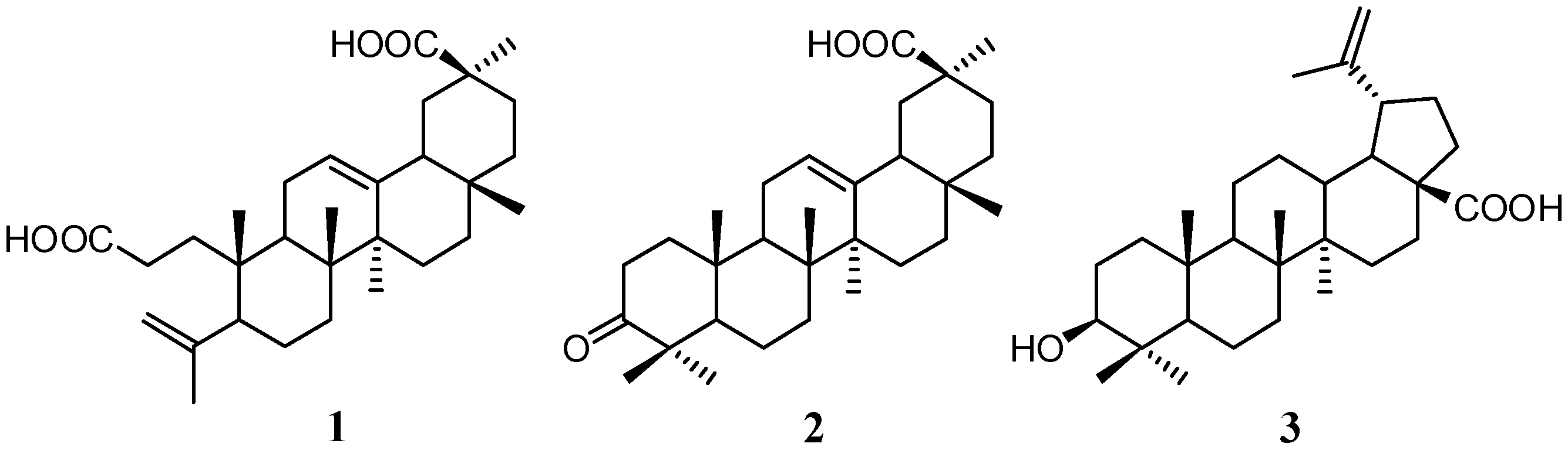
2.1. Isolation and Characterization of Compounds

After successive partition of the crude methanol extract of the root bark with petroleum ether and ethyl acetate, serial chromatography yielded two known oleanene-type triterpenoids, koetjapic acid (1) and 3-oxoolean-12-en-30-oic acid (2) and a lupene derivative, betulinic acid (3). Compound 3 was identified as a major compound of this plant. Koetjapic acid (1, also known as a seco-triterpenoid) was unexpectedly identified in the extracts of D. serrata. This compound was first reported from Sandoricum koetjape (Meliaceae) by Kaneda et al. [44] and, to the best of our knowledge, the occurrence of koetjapic acid (1) in Dillenia species has not been previously reported. The presence of seco-triterpenoids seems to be a common feature of Dillenia species, as reported by previous works [18,19]. Figure 1 displays the structures of these isolated triterpenoids. Identification of all triterpenoids was accomplished using a combination of physicochemical and spectroscopic experiments, viz. IR, 1D NMR as well as 2D NMR and HRESI-MS. All obtained values were similar to those reported in the literature [18,44,45].
Figure 1. Chemical structure of the isolated triterpenoids from D. serrata.
Molecules 20 03206 g001

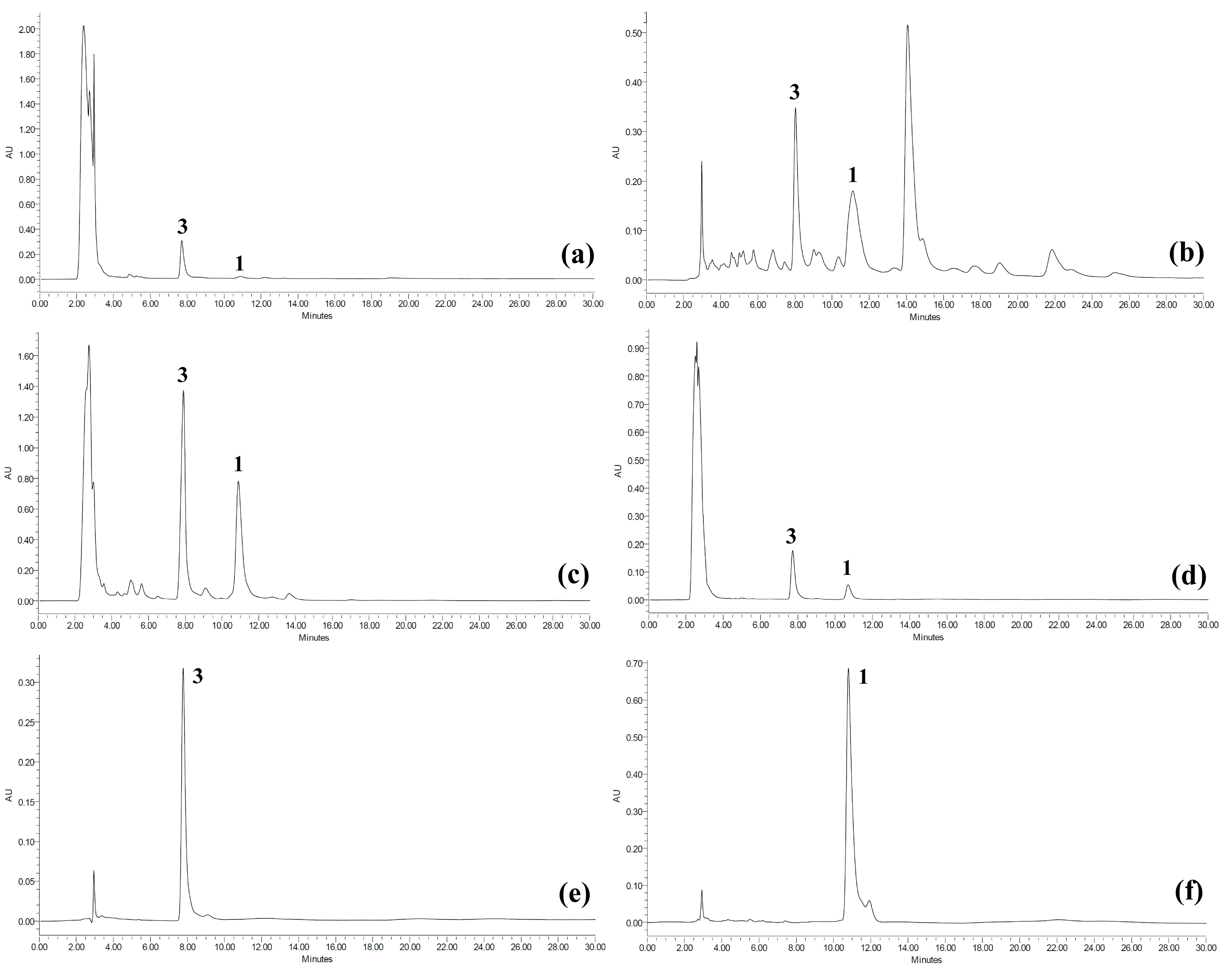
2.2. Quantitative Analysis of Koetjapic Acid (1) and Betulinic Acid (3)

Previous HPLC studies on quantification of betulinic acid (3) in plants have been reported in some papers [46,47,48]. Bae et al. [46] quantified betulinic acid (3) in Ziziphus fructus using a Nova-Pak C18 column, eluted with phosphate buffer (Na2HPO4 0.05 M, pH 2.5)-methanol at a ratio of 19:81 and showed retention time at 28 min. Considering that the background noise resulted from methanol, Oliveira et al. [47] improved the quantification of this compound in Doliocarpus schottianus by using acetonitrile instead of methanol. The condition was isocratic with acetonitrile-water pH 3.0 (9:1) and the retention time was at 11.5 min. Kumar et al. [30] applied this method for quantification of betulinic acid (3) from D. indica in a study on anticancer activity of this plant. A modification of the method was also performed using a Diamonsil C18 column, eluted with acetonitrile-water (86:14) for quantification of betulinic acid (3) in Betula platyphylla and showed that the retention time was at 16.5 min [48]. To the best of our knowledge, quantitative analysis of koetjapic acid (1) has not previously been reported.
In our study, a pH modification of a method [47] was conducted in order to shorten the retention time of betulinic acid (3), resulting in a rapid analysis. The method was also able to give a good separation for koetjapic acid (1) at a retention time of 10.801 min (see Figure 2). Validation of the reversed phase HPLC method for quantification of these compounds was determined by regression equation, coefficient correlation (r2), limit of detection (LOD) and limit of quantification (LOQ) (see Experimental Section). The calibration curves plotted were linear over the concentration range of 62.5 to 1000 μg/mL. LOD and LOQ values were found to be reliable for the method according to Bretnall et al. [49]. In addition, the precision of HPLC method regarding reproducibility and repeatability was satistacfory as indicated by the relative standard deviation (RSD) not greater than 5.0% [50] of peak area and retention time by intraday and interday analyses. Thus, the linearity, precision and accuracy of this modification were acceptable for quantitative analysis of koetjapic acid (1) and betulinic acid (3) in D. serrata.
Figure 2. HPLC chromatograms for (a) crude MeOH extract, (b) petroleum ether fraction, (c) ethyl acetate fraction, (d) MeOH fraction, (e) isolated betulinic acid (3) and (f) isolated koetjapic acid (1).
Molecules 20 03206 g002
Distribution of detectable amounts of koetjapic acid (1) and betulinic acid (3) in the crude methanol extracts and fractions of D. serrata was determined by using the validated HPLC method. As shown in Table 1, koetjapic acid (1) and betulinic acid (3) were mainly concentrated in the ethyl acetate fraction while they were found in least amounts in the methanol fractions. From the quantitative analysis, we also found that betulinic acid (3) preferentially accumulated in the stem bark rather than the root bark. Koetjapic acid (1) together with betulinic acid (3) are suggested as the chemical markers of D. serrata. Betulinic acid has been reported from several genera of the Dilleniaceae family, including Dillenia, Wormia, and Acrotrema [16]. The presence of a considerable amount of betulinic acid in these genera suggests that this compound may be a chemotaxonomic marker of the family. Koetjapic acid (1) can also be used as an additional marker since its presence in this plant seems unique among the Dillenia species.
Table 1. Concentrations of koetjapic acid (1) and betulinic acid (3) in the crude methanol extracts and fractions of Dillenia serrata.
SampleCodeConcentration of 1 (mg/g) *Concentration of 3 (mg/g) *
Root bark
Crude MeOHCRB21.31 ± 1.9981.76 ± 1.40
Petroleum ether fractionRB-PEF46.32 ± 0.61128.20 ± 3.91
Ethyl acetate fractionRB-EAF117.62 ± 1.54424.26 ± 2.97
MeOH fractionRB-MF19.78 ± 1.4190.93 ± 2.53
Stem bark
Crude MeOHCSB4.27 ± 1.09102.59 ± 2.99
Petroleum ether fractionSB-PEF45.37 ± 1.68137.58 ± 3.09
Ethyl acetate fractionSB-EAF151.29 ± 2.40528.08 ± 1.11
MeOH fractionSB-MF8.49 ± 0.5058.81 ± 1.21
* Value given in mean ± SD (n = 3).

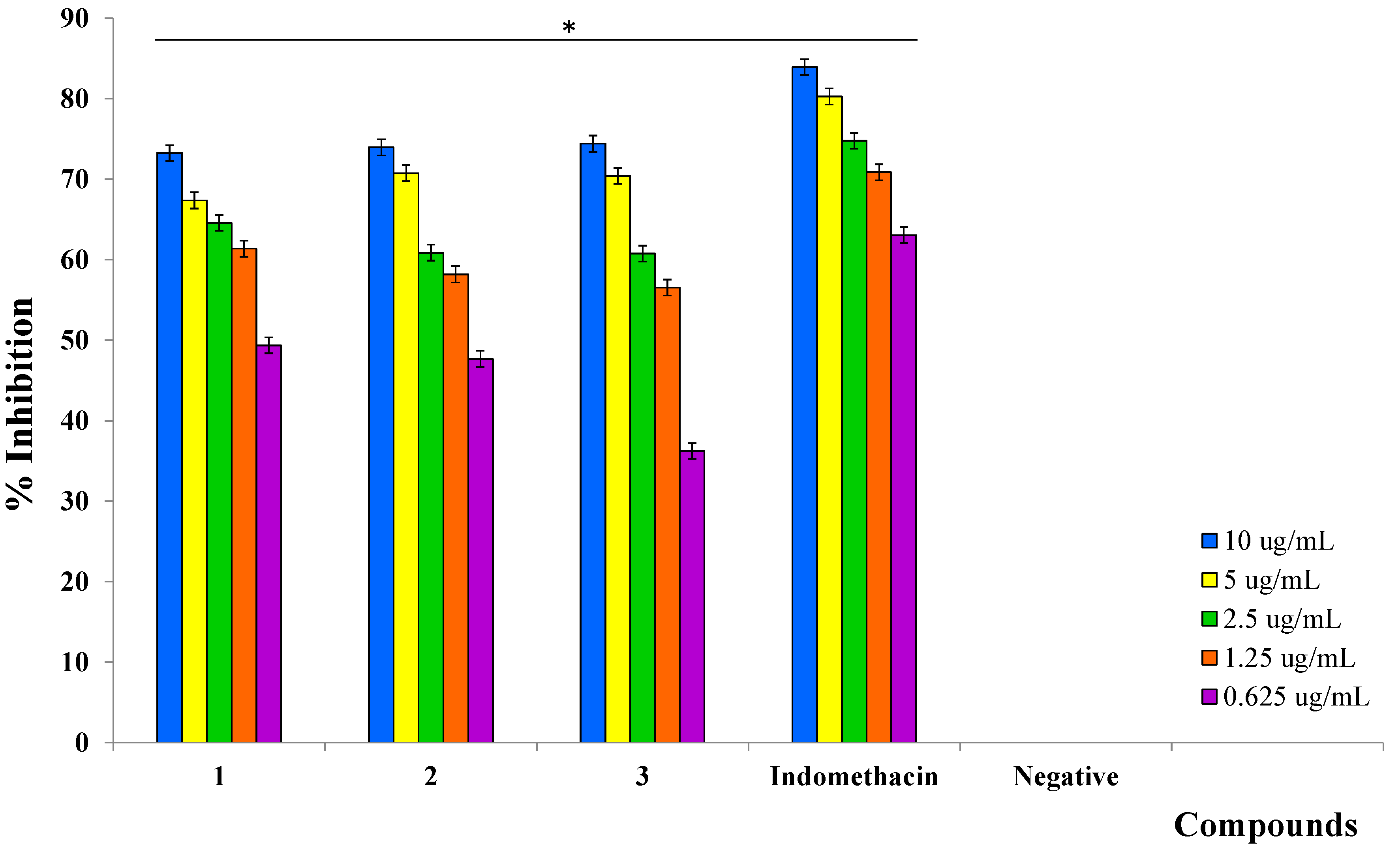
2.3. Inhibition of Prostaglandin E2 (PGE2)

Production of PGE2 induced by lipopolysaccharide (LPS) in human whole blood has been measured as a reflection of cyclooxygenase-2 (COX-2) activity of blood cells such as monocytes [51]. The inhibition of PGE2 production in human whole blood can be expressed as an inhibition of the enzymatic activity of COX-2 and/or inhibition of the expression of COX-2 protein. Crude methanol extracts and fractions of D. serrata root and stem bark were investigated for their ability to inhibit PGE2 in LPS-induced human whole blood and showed various percentage inhibitions at a concentration of 10 μg/mL (Table 2). This inhibitory activity indicated the presence of bioactive compounds in the crude methanol extracts and fractions of D. serrata.
The three triterpenoids 13 isolated from the root bark of D. serrata were also able to inhibit PGE2 production induced by LPS. This concentration-dependent inhibition was observed at five serial concentrations ranging from 10 to 0.625 μg/mL (Figure 3). All compounds showed significant inhibitory activity on PGE2 production as indicated by their IC50 values (Table 3). Of the three compounds, koetjapic acid (1) was identified as a promising inhibitor, with an IC50 value of 1.05 μM, comparable to that of a potent cyclooxygenase inhibitor, indomethacin (0.45 μM). In contrast to our study, koetjapic acid (1) was considered inactive in mouse ear inflammation model induced by tetradecanoylphorbol acetate [52]. Betulinic acid (3) had a higher IC50 value (2.59 μM) among the three compounds. The inhibition of LPS-induced PGE2 production by betulinic acid has been reported in human peripheral blood mononuclear cells (hPBMCs), and this inhibition was due to the suppression of COX-2 protein expression induced by LPS [53]. On the other hand, Wenzig et al. [54] investigated inhibition of COX-2 enzyme activity by betulinic acid and the results showed that the inhibition against this enzyme was not significant (IC50 ˃ 125 μM). Based on both studies, we can suggest that betulinic acid (3) plays role as an inhibitor of LPS induced expression of COX-2 protein, hence it can inhibits the production of PGE2 induced by LPS. Koetjapic acid (1) and 3-oxoolean-12-en-30-oic acid (2) may also probably inhibiting the COX-2 enzyme by similar mechanisms. However, phospholipase A2 (PLA2) may also be a potential target for these three compounds.
Table 2. Percentage inhibition (%) and IC50 values of the crude MeOH extracts and fractions of D. serrata on production of PGE2 in LPS-induced human whole blood.
SampleCode% Inhibition (10 µg/mL) *,†IC50 (µg/mL) *
Root bark
Crude MeOHCRB73.03 ± 0.771.80 ± 0.09
Petroleum ether fractionRB-PEF72.36 ± 1.180.23 ± 0.15
Ethyl acetate fractionRB-EAF73.86 ± 2.902.00 ± 0.13
MeOH fractionRB-MF65.01 ± 0.213.33 ± 0.53
Stem bark
Crude MeOHCSB71.88 ± 1.800.96 ± 0.32
Petroleum ether fractionSB-PEF64.26 ± 0.984.61 ± 0.09
Ethyl acetate fractionSB-EAF64.06 ± 1.621.31 ± 0.33
MeOH fractionSB-MF69.02 ± 0.901.24 ± 0.25
Indomethacin (positive control) 83.90 ± 0.270.16 ± 0.02
* Value given in mean ± SD (n = 3); p < 0.05.
Figure 3. Inhibitory effects of compounds 1, 2, and 3 on PGE2 production by LPS-stimulated human whole blood at various concentrations (value given in mean ± SD; n = 3; * p < 0.05).
Molecules 20 03206 g003
Since betulinic acid (3) was found as a major compound in all the crude extracts and fractions, the LPS-induced PGE2 production inhibitory activity might be contributed mainly by this compound. However, as shown in Table 2, various IC50 values of the crude methanol extracts and fractions of D. serrata were found not to correlate with either the amounts of koetjapic acid (1) or betulinic acid (3). For example, both RB-PEF and SB-PEF fractions contained almost the same concentrations of compound 1 and 3, but gave very different IC50 values (0.23 and 4.61 μg/mL, respectively) on inhibition of LPS-induced PGE2 production. This result indicated that there may be other potent inhibitors present in fraction RB-PEF. Similarly, fractions SB-EAF and SB-MF possessed close IC50 values (1.31 and 1.24 μg/mL, respectively), but they contained very different amounts of compounds 1 and 3. Interestingly, fraction RB-MF with IC50 of 3.33 μg/mL contains about 0.06 μg (1.98%) of compound 1 and 0.30 μg (9.09%) of compound 3, and these amount were considerably lower than the IC50 values of pure compound 1 and 3. Based on these results, we suggest that substantial amounts of koetjapic (1) and betulinic (3) acids in the crude methanol extracts and fractions of D. serrata may affect LPS-induced PGE2 production. However, the presence of other compounds in the extracts and fractions may also contribute to the activity, either in inhibiting or assisting LPS-induced PGE2 production.
Table 3. IC50 values of compounds 1, 2, and 3 on PGE2 production by LPS-stimulated human whole blood.
CompoundsIC50 (µM) *
11.05 ± 0.02
21.54 ± 0.05
32.59 ± 0.07
Indomethacin (positive control)0.45 ± 0.02
* Value given in mean ± SD (n = 3).
Koetjapic acid (1) has various pharmacological activities, including growth inhibition of multi-drug resistant bacteria Staphylococcus aureus and Pseudomonas aeruginosa (minimum inhibitory concentration, MIC of 12.5 and 6.25 μg/mL, respectively) [34], inhibition on Epstein-Barr virus antigen activation [36] and inhibition of DNA polymerase beta (IC50 20 μM) [37]. This compound also preserved high viability of the Raji cells [36]. Meanwhile, Nick et al. [18] investigated the antibacterial activity of 3-oxoolean-12-en-30-oic acid (2) from D. papuana that inhibited the growth of Bacillus subtilis, Escherichia coli, and Micrococcus luteus. Some papers have reported the anti-inflammatory, antileukemic and anti-HIV activities of betulinic acid (3) [38,39,40,41,42,43]. Treatment of hPBMCs with this compound alone did not elicit any PGE2 production and was non-cytotoxic, as indicated by trypan blue assay [53].

3. Experimental Section

3.1. General Information

Standard PGE2, Anti-PGE2 sera (developed in rabbit), lipopolysaccharide (LPS) from E. coli 0111:B4 and dextran from Leuconostoc mesentroides were purchased from Sigma (St. Louis, MO, USA). Radiolabelled PGE2 ([3H]-PGE2; 50 μCi/mmol) and scintillation cocktail (Ultima Gold MV) were purchased from PerkinElmer (Boston, MA, USA). Bovine serum albumin (BSA), ethylenediamine tetraacetic acid (EDTA), activated charcoal and analytical and HPLC grades of solvents (petroleum ether, ethyl acetate, methanol, and acetonitrile) were purchased from Merck (Darmstadt, Germany). Chromatography techniques (Thin Layer Chromatography; TLC, Vacuum Liquid Chromatography; VLC and Column Chromatography; CC) were performed using Merck Si-gel. Melting point was observed using a Stuart Melting Point SMP10. UV-Vis and IR spectra were recorded on a Shimadzu UV1800 and a PerkinElmer GX IR (ATR), respectively. High-resolution electrospray ionization mass spectrometry (HRESI-MS) was performed on a Bruker MicroTOF-Q mass spectrometer. Spectra of 1H-NMR (600 MHz) and 13C-NMR (150 MHz) were recorded on a Bruker Advance NMR. Radioactivity was measured using a Tri-Carb 3110 TR PerkinElmer Liquid Scintillation Analyzer. HPLC technique was performed on a Waters 2535 Quaternary Gradient Module HPLC using an XBridgeTM RP C-18 column (4.6 × 250 mm, 5 μm).

3.2. Plant Material

The barks of root and stem of D. serrata were collected from secondary forest in Onewila village, a region of Southeast Sulawesi, Indonesia and authenticated by Herbarium Bogoriense, Bogor, Indonesia (voucher number: BO-1902181).

3.3. Extraction and Isolation of Compounds

The dried root barks (1.15 kg) and stem barks (2.5 kg) were macerated with MeOH (4 and 8 L, respectively) for 24 h. The extracts were filtered and the solvent was evaporated under vacuum. The steps were executed three times to yield 500 g crude (43.5%) extract of root barks (CRB) and 600 g crude (24%) extract of stem barks (CSB). 250 g of CRB was dissolved in methanol (2.5 L) and left to stand overnight to re-crystallize compound 3. Compound 3 (993.4 mg) was collected using a vacuum filter and the residue was evaporated. The remaining CRB was dissolved in a small volume of MeOH (~100 mL) and partitioned three times with petroleum ether (PE) and ethyl acetate (EtOAc) (~500 mL), to give PE (1.5 g) and EtOAc (29.7 g) soluble fractions, respectively. The PE fraction was subjected onto VLC with hexane-EtOAc to give 28 sub-fractions. Sub-fractions were combined into 7 fractions (F1–F7) based on TLC analysis. Compound 2 (9.0 mg) was precipitated from F3. The EtOAc fraction was then subjected onto VLC with hexane-EtOAc to yield 32 sub-fractions. Compound 1 (102.2 mg) was precipitated from sub-F29 and sub-F31. For HPLC and bioactivity samples, 10 g of each crude extract was suspended in methanol and partitioned successively with petroleum ether, ethyl acetate and methanol. Six fractions were then evaporated under vacuum to yield residues of petroleum ether (RB-PEF) (0.6 g, 6%), ethyl acetate (RB-EAF) 1.9 g, 19%), and methanol (RB-MF) (7.2 g, 72%) fractions of root barks followed by petroleum ether (SB-PEF) (0.3 g, 3%), ethyl acetate (SB-EAF) (1.7 g, 17%), and methanol (SB-MF) (7.9 g, 79%) fractions of stem barks. These residues were stored in a refrigerator at 4 °C until analyses.
Koetjapic acid (3,4-seco-olean-4(23), 12-diene-3,30-dioic acid) (1); 102.2 mg; white prisms (MeOH); mp 296–298°; UV (EtOH) λmax nm (log ε): 203 (3.88); IR (ATR) ʋmax cm−1: 3440, 2978, 2860, 1706, 1702, 1698, 1694, 1454, 1387, 1281, 1230, 1192, 906; HRESI-MS m/z: [M+Na]+ 493.3279 (calc. for C30H46O4, 470.3396). 1H-NMR (600 MHz; DMSO-d6) δH (ppm): 0.74 (s, H3-28), 0.84 (Hα-16), 0.88 (s, H3-25), 0.96 (s, H3-26), 0.99 (bs, Hα-15), 1.07 (s, H3-29), 1.15 (s, H3-27), 1.25 (Hα-7), 1.27 (H2-22), 1.31 (H2-6), 1.39 (Hα-2), 1.45 (Hβ-2), 1.52 (Hβ-7), 1.61 (Hα-19), 1.70 (Hβ-15), 1.72 (s, H3-24), 1.73 (Hβ-19), 1.76 (Hα-9), 1.78 (Hα-11, H2-21), 1.89 (Hβ-11, Hβ-18), 1.99 (Hα-5, Hβ-16), 2.06 (m, Hα-1), 2.26 (m, Hβ-1), 4.66 (s, Ha-23), 4.85 (s, Hb-23), 5.18 (t, H-12) and 12.04 (2OH-3,30); 13C-NMR (150 MHz; DMSO-d6) δC (ppm): 17.0 (C-26), 19.6 (C-25), 23.6 (C-11), 23.9 (C-24), 24.5 (C-6), 26.0 (C-27), 26.2 (C-15), 26.8 (C-16), 28.5 (C-1), 28.6 (C-28), 28.7 (C-29), 31.1 (C-21), 31.3 (C-7), 32.1 (C-17), 34.3 (C-2), 37.7 (C-9), 38.5 (C-22), 39.1 (C-10), 39.5 (C-8), 42.1 (C-14), 42.8 (C-19), 43.6 (C-20), 48.3 (C-18), 49.8 (C-5), 113.9 (C-23), 122.2 (C-12), 144.8 (C-13), 147.6 (C-4), 175.3 (C-3) and 178.5 (C-30). NMR spectral data were identical to those given in [44].
3-Oxoolean-12-en-30-oic acid (2); 9 mg; white crystalline solid (MeOH); mp 270–272°; UV (EtOH) λmax nm (log ε): 205 (4.00); IR (ATR) ʋmax, cm−1: 3498; 2968–2861; 1705; 1698; 1695; HRESI-MS m/z: [M+Na+16]+ 493.3300 (calc. for C30H46O3, 454.34470); 1H-NMR (600 MHz; DMSO-d6) δH (ppm): 0.74 (s, H3-28), 0.86 (Hα-16), 0.96 (s, H3-24, H3-26), 0.99 (s, H3-25), 0.99 (Hα-15), 1.00 (s, H3-23), 1.06 (s, H3-29), 1.13 (s, H3-27), 1.29 (Hα-21, H2-22), 1.33 (Hα-5), 1.35 (Hα-7), 1.41 (Hα-1), 1.46 (H2-6), 1.53 (Hβ-7), 1.60 (Hα-19), 1.64 (Hα-9), 1.70 (Hβ-19), 1.73 (Hβ-15), 1.79 (Hβ-21), 1.80 (Hβ-1), 1.88 (H2-23), 1.91 (Hβ-18), 1.98 (Hβ-16), 2.29 (Hα-2), 2.53 (Hβ-2) and 5.19 (t, H-12) and 12.10 (bs, OH-30); 13C-NMR (150 MHz; DMSO-d6) δC (ppm): 15.3 (C-25), 16.9 (C-26), 19.6 (C-6), 21.6 (C-24), 23.6 (C-11), 26.0 (C-27), 26.2 (C-15), 26.7 (C-23), 26.8 (C-16), 28.6 (C-28), 28.7 (C-29), 31.1 (C-21), 32.0 (C-17), 32.1 (C-7), 34.2 (C-2), 36.6 (C-10), 38.5 (C-22), 39.0 (C-1), 39.7 (C-8), 41.7 (C-14), 42.8 (C-19), 43.6 (C-20), 46.6 (C-9), 47.2 (C-4), 48.3 (C-18), 54.7 (C-5), 122.2 (C-12), 144.9 (C-13), 178.5 (C-30) and 216.7 (C-3). NMR spectral data were identical to those in [18].
Betulinic acid (3β-hydroxy-20(29)-lupen-28-oic acid) (3); 993.4 mg; white crystalline needles (MeOH); mp 296–301°; UV (EtOH) λmax nm (log ε): 206 (3.96); IR (ATR) ʋmax, cm−1: 3446, 2940; 2870, 1684, 1681, 1456, 1360, 1236, 1043, 886; HRESI-MS m/z: [M−H] 455.35252 (calc. for C30H48O3, 456.36035.); 1H-NMR (600 MHz; DMSO-d6) δH (ppm): 0.64 (s, H3-24), 0.75 (s, H3-26), 0.84 (Hα-1), 0.85 (s, H3-25), 0.86 (s, H3-23), 0.92 (s, H3-27), 0.97 (Hα-12), 1.09 (Hα-15), 1.15 (Hβ-11), 1.24 (Hα-9), 1.30 (Hβ-6, Hβ-7, Hα-21), 1.37 (Hα-11, Hα-16), 1.44 (H2-2, Hα-6, Hβ-15, Hα-22), 1.50 (t, Hα-18), 1.55 (Hβ-1), 1.60 (Hβ-12), 1.64 (s, H3-30), 1.79 (Hβ-21, Hβ-22), 2.11 (Hβ-16), 2.21 (td, J1 = J3 = 3.6, J2 = 2.4, Hβ-13), 2.95 (Hα-3, Hβ-19), 4.30 (bs, OH-3), 4.55 (d, J = 0.6, Ha-29), 4.68 (d, J = 1.8, Hb-29) and 12.10 (bs, OH-28); 13C-NMR (150 MHz; DMSO-d6) δC (ppm): 14.8 (C-27), 16.2 (C-25), 16.3 (C-24), 16.4 (C-26), 18.4 (C-6), 19.4 (C-30), 20.9 (C-11), 25.5 (C-12), 27.6 (C-2), 28.6 (C-23), 29.7 (C-15), 30.5 (C-21), 32.2 (C-16), 34.4 (C-7), 36.8 (C-22), 37.2 (C-10), 38.0 (C-13), 38.7 (C-1), 38.9 (C-4), 40.7 (C-8), 42.5 (C14), 47.1 (C-19), 48.9 (C-18), 50.4 (C-9), 55.3 (C-5), 55.9 (C-17), 77.2 (C-3), 110.1 (C-29), 150.8 (C-20) and 177.7 (C-28). NMR spectral data were identical with reference [45].

3.4. Quantification of Koetjapic Acid (1) and Betulinic Acid (3) Using HPLC

HPLC analysis was performed based on the method described by Oliveira et al. [47] with slight modification. HPLC (Waters 2535) equipped with a reversed-phased column C-18 (4.6 × 250 mm, 5 μm; XBridge, Waters, Dublin, Ireland) and photodiode array detector (Waters 2998) were used. Koetjapic acid (1) and betulinic acid (3) (1 mg/mL each) in methanol were injected (20 μL) three times and separated isocratically with acetonitrile-water (9:1) pH 2.5 (with trifluoroacetic acid) at a flow rate of 1 mL/min and 3000–3500 psi pressure. The compounds were detected at 210 nm [30,49].
The HPLC method for koetjapic acid (1) and betulinic acid (3) was validated by determination of linearity, precision and accuracy in accordance with ICH guidelines [55]. Linearity was evaluated from the linear regression equation and correlation coefficient (r2) of calibration curves constructed for both compounds within the concentration range of 62.5 to 1000 μg/mL (Table 4).
Table 4. Validation parameters of HPLC method for koetjapic acid (1) and betulinic acid (3).
CompoundConc. 1Intra-day precision 2Inter-day precision 4Equation (r2)LOD 5LOQ 5
Rt 3Area
Day 1Day 2Day 3Day 1Day 2Day 3Rt 3Area
Koetjapic acid (1)62.51.512.160.064.513.623.681.201.48y = 13523x − 44326 (0.9994)1.895.75
1252.582.521.483.091.152.083.571.89
2502.500.561.571.330.954.042.263.41
5000.681.980.561.582.042.193.822.11
10000.940.831.741.621.271.592.492.66
Betulinic acid (1)62.50.480.441.314.923.823.452.292.46y = 4851x − 42307 (0.9999)9.2327.97
1251.071.501.114.533.982.282.183.53
2500.500.880.661.032.832.822.403.32
5001.691.071.571.152.084.962.552.58
10000.830.890.832.862.572.861.554.03
1 μg/mL; 2 n = 3; 3 Retention time; 4 n = 9; 5 ng/mL.
Precision was determined by the LOD and LOQ by injecting a series of known concentrations of the compounds. The values of LOD and LOQ were calculated from the relative standard deviation (RSD) and slope (S) of the calibration curves. The accuracy of the method regarding reproducibility and repeatability was evaluated by intra- and inter-day variation on three consecutive days with three repetitions each. The reproducibility and repeatability were demonstrated by the RSD of peak area and retention time.
The content of koetjapic acid (1) and betulinic acid (3) in the D. serrata extracts and fractions was quantified using the validated HPLC method. Precise amount of samples (10 mg for each of CRB, RB-PEF, RB-EAF, RB-MF, CSB, SB-PEF, SB-EAF, and SB-MF) were sonicated in methanol (1 mL) and filtered through a 0.45 μm filter. An aliquot of 20 μL of each sample was injected onto the HPLC.

3.5. Radioimmunoassay for Prostaglandin E2 (PGE2)

The inhibition of PGE2 production indicated by the concentration of PGE2 in human whole blood was measured according to the validated radioimmunoassay (RIA) method [51]. The application of human blood was permitted by the Ethics Committee of Universiti Kebangsaan Malaysia (UKM) with approval number NF-016-2013.
Human whole blood was drawn using aseptic vein puncture from the same donors of healthy volunteers when they had not taken any medicine or supplements during the last two weeks and fasted for 8 h prior to blood being withdrawn. The blood sample was prevented from coagulation by adding 10% (v/v) of 2% EDTA in a polypropylene tube. Duplicate 1 mL aliquots of EDTA-whole blood samples were transferred into test tubes and incubated with 10 μL of sample or indomethacin (1 mg/mL in 1:1 of DMSO-ethanol) for 15 min (37 °C) before LPS addition. The effects of samples or indomethacin on PGE2 production were studied by incubating each sample with whole blood-EDTA in the presence of LPS (10 μg/mL in 0.9% normal saline) for 24 h. For IC50, the concentration of samples were adjusted in five serial dilutions over a concentration range of 0.625 to 10 μg/mL. After incubation at 37 °C for 24 h, the plasma was separated by centrifugation at 2600 × g for 15 min at 4 °C. RIA buffer (phosphate buffered saline [0.01 M, pH 7.4] containing 0.1% BSA and 0.1% sodium azide) was used as the standard diluent of the assay. The plasma (100 μL) was added to anti-PGE2 (100 μL; diluted with ratio of 1:50,000) and [3H]-PGE2 (100 μL; 5000 cpm) and incubated for 18–24 h at 4 °C. After incubation, dextran-charcoal (200 μL) was added to the mixture and incubated for 10 min at 0 °C. The supernatant was then separated by centrifugation at 3000× g for 15 min at 4 °C and pipetted (300 μL) into liquid scintillation cocktail (3 mL). The radioactivity was measured using a liquid scintillation analyzer.
Concentration of PGE2 (pg/0.1 mL) in the blood was calculated using a semi-logarithmic graph of standard PGE2. Previously, standard PGE2 (1 mg/mL) had been serially diluted to concentrations ranging from 2.45 to 400 pg/0.1 mL. The interference of compounds in the crude methanol extracts and fractions towards RIA method was checked by adding crude extracts and fractions to the standards and found not to interfere with the measurements. The average count per min (cpm) values of standards and samples (B) resulting from antibody-antigen (labeled PGE2) binding in the plasma were subtracted from the non-specific binding (Nc) together with the total binding between antibody and antigen (Bo). The normalized percent bound (% B/Bo) was then determined using Equation (1):
% B B o = ( B N c B O N c ) 100
% I = ( 1 [ Concentration of PGE 2  in samples or standard ] [ Concentration of PGE 2  in control ] ) 100 %
The calculated % B/Bo values were plotted against their respective concentrations of standard PGE2 in picograms (pg) semi-logarithmically. Thus, the interpolation of % B/Bo values for samples and indomethacin using PGE2 standard curve resulted in the determination of PGE2 concentration in the blood. The percentage inhibition (% I) was then calculated using Equation (2).

3.6. Statistical Analysis

The HPLC analysis and bioassay were performed in triplicate and the data were expressed as means ± SD. Empower software (Waters) was used to construct a calibration curve of koetjapic and betulinic acids as well as their quantification. The bioactivity data were analyzed using Statistical Package for Social Sciences (SPSS) software Version 17. Data were analyzed using one way ANOVA analysis with a probability p < 0.05 representing a significant difference as compared to control. GraphPad Prism 5 was used to determine the IC50 value of active extracts and compounds.

4. Conclusions

Our study revealed that D. serrata possesses a promising inhibitory effect on LPS-induced PGE2 production. Three triterpenoids from this plant, koetjapic acid (1), 3-oxoolean-12-en-30-oic acid (2), and betulinic acid (3), were found to inhibit LPS-induced PGE2 production concentration-dependently. Although substantial amounts of koetjapic (1) and betulinic (3) acids in the crude methanol extracts and fractions of D. serrata may affect LPS-induced PGE2 production, the presence of other compounds in the extracts and fractions may also contribute to the observed activity. Further studies need to be carried out to investigate the effect of these compounds on COX-2 especially in vitro prostaglandin biosynthesis catalysed by COX-2 in cell free assays and expression of COX-2 protein.

Acknowledgments

The authors are very grateful for the financial support from Universiti Kebangsaan Malaysia (UKM) grant (GUP-2012-001).

Author Contributions

J.J and C.W.S designed and performed research, analyzed the data as well as wrote and edited the manuscript; N.A designed research; J.A.J and I.J edited and revised the manuscript; N.-A.A performed research and analyzed the data; K.M, F.B and H.F.M assisted the experimental works; I.S supplied plant sample. All authors read and approved the final manuscript.

Conflicts of Interest

The authors declare no conflict of interest.

References

  1. Milatovic, D.; Montine, T.J.; Aschner, M. Prostanoid signaling: dual role for prostaglandin E2 in neurotoxicity. NeuroToxicology 2011, 32, 312–319. [Google Scholar] [CrossRef] [PubMed]
  2. Nakagawa, T. Roles of prostaglandin E2 in the cochlea. Hear. Res. 2011, 276, 27–33. [Google Scholar] [CrossRef] [PubMed]
  3. Legler, D.F.; Bruckner, M.; Uetz-von Allmen, E.; Krause, P. Prostaglandin E2 at new glance: Novel insights in functional diversity offer therapeutic chances. Int. J. Biochem. Cell Biol. 2010, 42, 198–201. [Google Scholar] [CrossRef] [PubMed]
  4. Harris, S.G.; Padilla, J.; Koumas, L.; Ray, D.; Phipps, R.P. Prostaglandins as modulators of immunity. TRENDS Immunol. 2002, 23, 144–150. [Google Scholar] [CrossRef] [PubMed]
  5. Narumiya, S.; Sugimoto, Y.; Ushikubi, F. Prostanoid receptors: Structures, properties, and functions. Physiol. Rev. 1999, 79, 1193–1226. [Google Scholar] [PubMed]
  6. Jabbour, H.N.; Sales, K.J. Prostaglandin receptor signaling and function in human endometrial pathology. TRENDS Endocrin. Met. 2004, 15, 398–404. [Google Scholar] [CrossRef]
  7. Offermanns, S.; Rosenthal, W. Encyclopedia of Molecular Pharmacology, 2nd ed.; Springer: New York, NY, USA, 2008. [Google Scholar]
  8. Lim, T.K. Dillenia Serrata. Edible Medicinal and Non-Medicinal Plants; Springer: Dordrecht, The Netherlands, 2012; Volume 2. [Google Scholar]
  9. Hedberg, I.; Hedberg, O.; Madati, P.J.; Mshigeni, K.E.; Mshiu, E.N.; Samuelsson, G. Inventory of plants used in traditional medicine in Tanzania. II. Plants of the families Dilleniaceae-Opiliaceae. J. Ethnopharmacol. 1983, 9, 105–128. [Google Scholar]
  10. Grosvenor, P.W.; Gothard, P.K.; McWilliam, N.C.; Supriono, A.; Gray, D.O. Medicinal plants from Riau Province, Sumatra, Indonesia. Part 1: Uses. J. Ethnopharmacol. 1995, 45, 75–95. [Google Scholar] [CrossRef]
  11. Burkill, I.H. A Dictionary of the Economic Products of the Malay Peninsula; Ministry of Agriculture and Cooperatives: Kuala Lumpur, Malaysia, 1966. [Google Scholar]
  12. Kerrigan, R.A.; Craven, L.A.; Dunlop, C.R. Dilleniaeae. In Flora of the Darwin Region; Short, P.S., Cowie, I.D., Eds.; Northern Territory Herbarium, Department of Natural Resources, Environment, the Arts and Sport: Palmerston, Australia, 2011. [Google Scholar]
  13. Jansen, P.C.M.; Jukema, J.; Oyen, L.P.A.; van Lingen, T.G. Dillenia serrata Thunb. In Plant Resources of South-East Asia No. 2: Edible Fruit and Nuts; Verheij, E.W.M., Coronol, R.E., Eds.; Prosea Foundation: Bogor, Indonesia, 1992. [Google Scholar]
  14. Windadri, F.I.; Rahayu, M.; Uji, T.; Rustiami, H. Pemanfaatan tumbuhan sebagai bahan obat oleh masyarakat lokal suku Muna di kecamatan Wakarumba, kabupaten Muna, Sulawesi Utara. Biodiversitas 2006, 7, 333–339. [Google Scholar] [CrossRef]
  15. Bhattacharjee, S.R.; Chatterjee, A. Betulinic acid and betulin, the triterpenoid constituents of Dillenia indica. J. Indian Chem. Soc. 1962, 39, 276–284. [Google Scholar]
  16. Pavanasasivam, G.; Sultanbawa, M.U.S. Chemical investigation of Ceylonese plants. IX. Betulinic acid in the Dilleniaceae and a review of its natural distribution. Phytochemistry 1974, 13, 2002–2006. [Google Scholar]
  17. Dan, S.; Dan, S.S. Triterpenoids of Indian Dilleniaceae. J. Indian Chem. Soc. 1980, 57, 760. [Google Scholar]
  18. Nick, A.; Wright, A.D.; Rali, T.; Sticher, O. Antibacterial triterpenoids from Dillenia papuana and their structure-activity relationships. Phytochemistry 1995, 40, 1691–1695. [Google Scholar] [CrossRef] [PubMed]
  19. Macahig, R.A.S.; Matsunami, K.; Otsuka, H. Chemical studies on an endemic Philippine plant: Sulfated glucoside and seco-A-ring triterpenoids from Dillenia philippinensis. Chem. Pharm. Bull. 2011, 59, 397–401. [Google Scholar] [CrossRef] [PubMed]
  20. Bate-Smith, E.C.; Harborne, J.B. Differences in flavonoid content between fresh and herbarium leaf tissue in Dillenia. Phytochemistry 1981, 10, 1055–1058. [Google Scholar] [CrossRef]
  21. Pavanasasivam, G.; Sultanbawa, M.U.S. Flavonoids of some Dilleniaceae species. Phytochemistry 1975, 14, 1127–1128. [Google Scholar] [CrossRef]
  22. Gurni, A.A.; Kubitzki, K. Flavonoid chemistry and systematics of the Dilleniaceae. Biochem. Syst. Ecol. 1981, 9, 109–114. [Google Scholar] [CrossRef]
  23. Li, X.; Li, C.; Cui, C.; Li, M.; Fan, M. Chemical constituents of Dillenii kerrii and their activities on antitumor and anti-hypoxia in vitro. Chin. J. Med. Chem. 2009, 19, 130–134. [Google Scholar] [CrossRef]
  24. Nick, A.; Wright, A.D.; Sticher, O.; Rali, T. Antibacterial triterpenoid acids from Dillenia papuana. J. Nat. Prod. 1994, 57, 1245–1250. [Google Scholar] [CrossRef] [PubMed]
  25. Muliawan, S.Y. Effect of Dillenia suffruticosa extract on dengue virus type 2 replication. Univ. Med. 2008, 27, 1–5. [Google Scholar]
  26. Parvin, M.N.; Rahman, M.S.; Islam, M.S.; Rashid, M.A. Chemical and biological investigations of Dillenia indica Linn. Bangladesh J. Pharmacol. 2009, 4, 122–125. [Google Scholar]
  27. Yeshwante, S.B.; Juvekar, A.R.; Nagmoti, D.M.; Wankhede, S.S.; Shah, A.S.; Pimprikar, R.B.; Saindane, D.S. Anti-inflammatory activity of methanolic extracts of Dillenia indica L. leaves. Pharmacology 2009, 1, 63–66. [Google Scholar]
  28. Abdille, M.H.; Sigh, R.P.; Jayaprakasha, G.K.; Jena, B.S. Antioxidant activity of the extracts from Dillenia indica fruits. Food Chem. 2005, 90, 891–896. [Google Scholar] [CrossRef]
  29. Kumar, S.; Kumar, V.; Prakash, O. Antidiabetic, hypolipidemic and histopathological analysis of Dillenia indica (L.) leaves extract on alloxan induced diabetic rats. Asian Pac. J. Trop. Med. 2011, 4, 347–352. [Google Scholar] [CrossRef]
  30. Kumar, D.; Mallick, S.; Vedasiromoni, J.R.; Pal, B.C. Anti-leukemic activity of Dillenia indica L. Fruit and quantification of betulinic acid by HPLC. Phytomedicine 2010, 17, 431–435. [Google Scholar]
  31. Rosangkima, G.; Rongpi, T.; Prasad, S.B. Role of glutathione and glutathione-related enzymes in the antitumor activity of Dillenia pentagyna in Dalton’s lymphoma-bearing mice. Int. J. Cancer Res. 2008, 4, 92–102. [Google Scholar] [CrossRef]
  32. Nyman, U.; Joshi, P.; Madsen, L.B.; Pedersen, T.B.; Pinstrup, M.; Rajasekharan, S.; George, V.; Pushpangadan, P. Ethnomedical information and in vitro screening for angiotensin-converting enzyme inhibition of plants utilized as traditional medicines in Gujarat, Rajasthan and Kerala (India). J. Ethnophamacol. 1998, 60, 247–263. [Google Scholar] [CrossRef]
  33. Nguyen-Pouplin, J.; Tran, H.; Tran, H.; Phan, T.A.; Dolecek, C.; Farrar, J.; Tran, T.H.; Caron, P.; Bodo, B.; Grellier, P. Antimalarial and cytotoxic activities of ethnopharmacologically selected medicinal plants from South Vietnam. J. Ethnopharmacol. 2007, 109, 417–427. [Google Scholar] [CrossRef] [PubMed]
  34. Muhammad, I.; El Sayed, K.; Mossa, J.S.; Al-Said, M.S.; El-Feraly, F.S.; Clark, A.M.; Hufford, C.D.; Oh, S.; Mayer, A.M.S. Bioactive 12-oleanene triterpene and secotriterpene acids from Maytenus undata. J. Nat. Prod. 2000, 63, 605–610. [Google Scholar] [CrossRef] [PubMed]
  35. Wachter, G.A.; Valcic, S.; Flagg, M.L.; Franzblau, S.G.; Montenegro, G.; Suarez, E.; Timmermann, B.N. Antitubercular activity of pentacyclic triterpenoids from plants of Argentina and Chile. Phytomedicine 1999, 6, 341–345. [Google Scholar] [CrossRef] [PubMed]
  36. Ismail, I.S.; Ito, H.; Mukainaka, T.; Higashihara, H.; Enjo, F.; Tokuda, H.; Nishino, H.; Yoshida, T. Ichthyotoxic and anticarcinogenic effects of triterpenoids from Sandoricum koetjape bark. Biol. Pharm. Bull. 2003, 26, 1351–1353. [Google Scholar] [CrossRef] [PubMed]
  37. Sun, D.-H.; Starck, S.R.; Locke, E.P.; Hecht, S.M. DNA polymerase beta inhibitors from Sandoricum koetjape. J. Nat. Prod. 1999, 62, 1110–1113. [Google Scholar] [CrossRef] [PubMed]
  38. Pisha, E.; Chai, H.; Lee, I.S.; Chagwedera, T.E.; Farnsworth, N.R.; Cordell, A.C.; Beecher, C.W.W.; Fong, H.H.S.; Kinghorn, A.D.; Brown, D.M.; et al. Discovery of betulinic acid as a selective inhibitor of human melanoma that functions by induction of apoptosis. Nat. Med. 1995, 1, 1046–1051. [Google Scholar]
  39. Schmidt, M.L.; Kuzmanoff, K.L.; Ling-Indeck, L.; Pezzuto, J.M. Betulinic acid induces apoptosis in human neuroblastoma cell lines. Eur. J. Cancer 1997, 33, 2007–2010. [Google Scholar] [CrossRef] [PubMed]
  40. Safayhi, H.; Sailer, E.-R. Anti-inflammatory actions of pentacyclic triterpenes. Planta Med. 1997, 63, 487–493. [Google Scholar] [CrossRef] [PubMed]
  41. Huguet, A.-I.; Recio, M.C.; Manez, S.; Giner, R.M.; Rios, J.-L. Effect of triterpenoids on the inflammation induced by protein kinase C activators, neuronally acting irritants and other agents. Eur. J. Pharmacol. 2000, 410, 69–81. [Google Scholar] [CrossRef] [PubMed]
  42. Fujioka, R.E.; Kashiwada, Y.; Kilkuskie, R.E.; Cosentino, M.; Ballas, L.M.; Jiang, J.B.; Janzen, W.P.; Chen, I.-S. Anti-HIV principles from Syzygium claviflorum and the anti-HIV activity of structurally related triterpenoids. J. Nat. Prod. 1994, 57, 243–247. [Google Scholar] [CrossRef] [PubMed]
  43. Hashimoto, F.; Kashiwada, Y.; Cosentino, L.M.; Chen, C.-H.; Garret, P.E.; Lee, K.-H. Anti-AIDS agents-XXVII. Synthesis and anti-HIV activity of betulinic acid and dihydrobetulinic acid derivatives. Bioorganic Med. Chem. 1997, 5, 2133–2143. [Google Scholar]
  44. Kaneda, N.; Pezzuto, J.M.; Kinghorn, A.D.; Farnsworth, N.R.; Santisuk, T.; Tuchinda, P.; Udchachon, J.; Reutrakul, V. Plant anticancer agents, L.1 Cytotoxic triterpenes from Sandoricum koetjape stems. J. Nat. Prod. 1992, 55, 654–659. [Google Scholar]
  45. Peng, C.; Bodenhausen, G.; Qiu, S.; Fong, H.H.S.; Farnsworth, N.R.; Yuanm, S.; Zheng, C. Computer-assisted structure elucidation: Application of CISOC-SES to the resonance assignment and structure generation of betulinic acid. Magn. Reson. Chem. 1998, 36, 267–278. [Google Scholar] [CrossRef]
  46. Bae, K.H.; Lee, S.M.; Lee, E.S.; Lee, J.S.; Kang, J.S. Isolation and quantitative analysis of betulinic acid and alphitolic acid from Zyziphi fructus. Yakhak Hoeji 1996, 40, 558–562. [Google Scholar]
  47. Oliveira, B.H.; Santos, C.A.M.; Espíndola, A.P.D.M. Determination of the triterpenoid, betulinic acid, in Doliocarpus schottianus by HPLC. Phytochem. Anal. 2002, 13, 95–98. [Google Scholar] [CrossRef] [PubMed]
  48. Zhao, G.; Yan, W.; Cao, D. Simultaneous determination of betulin and betulinic acid in white birch bark using RP-HPLC. J. Pharm. Biomed. Anal. 2007, 43, 959–962. [Google Scholar] [CrossRef] [PubMed]
  49. Bretnall, A.E.; Clarke, G.S. Validation of analytical test methods. In Separation Scince and Technology; Ahuja, S., Ed.; Academic Press: Amsterdam, The Netherlands, 2011; Volume 10. [Google Scholar]
  50. Crowther, J.B. Validation of pharmaceutical methods. In Handbook of Modern Pharmaceutical Analysis; Ahuja, S., Scypinski, S., Eds.; Academic Press: San Diego, CA, USA, 2001; Volume 3. [Google Scholar]
  51. Patrignani, P.; Panara, M.R.; Greco, A.; Fusco, O.; Natoli, C.; Iacobelli, S.; Cipollone, F.; Ganci, A.; Créminon, C.; Maclouf, J.; et al. Biochemical and pharmacological characterization of the cyclooxygenase activity of human blood prostaglandin endoperoxide synthases. J. Pharmacol. Exp. Ther. 1994, 271, 1705–1712. [Google Scholar]
  52. Rasadah, M.A.; Khozirah, S.; Aznie, A.A.; Nik, M.M. Anti-inflammatory agents from Sandoricum koetjape Merr. Phytomedicine 2004, 11, 261–263. [Google Scholar] [CrossRef] [PubMed]
  53. Viji, V.; Helen, A.; Luxmi, V.R. Betulinic acid inhibits endotoxin-stimulated phosphorylation cascade and pro-inflammatory prostaglandin E2 production in human peripheral blood mononuclear cells. Br. J. Pharmacol. 2011, 162, 1291–1303. [Google Scholar] [CrossRef] [PubMed]
  54. Wenzig, E.M.; Widowitz, U.; Kunert, O.; Chrubasik, S.; Bucar, F.; Knauder, E.; Bauer, R. Phytochemical composition and in vitro pharmacological activity of two rose hip (Rosa canina L.) preparations. Phytomedicine 2008, 15, 826–835. [Google Scholar] [CrossRef]
  55. ICH. Validation of Analytical Procedures: Text and Methodology; ICH Harmonized Tripartite Guidelines, 2005. Available online: http://www.ich.org (accessed on 1 December 2013).
  • Sample Availability: Samples of the compounds 13 are available from the authors.

