Article

Potassium Hexacyanoferrate (III)-Catalyzed Dimerization of Hydroxystilbene: Biomimetic Synthesis of Indane Stilbene Dimers

1
State Key Laboratory of Bioactive Substance and Function of Natural Medicines, Institute of Materia Medica, Chinese Academy of Medical Sciences & Peking Union Medical College, Beijing 100050, China
2
School of Chemical Engineering, Northwest University, Xi’an 710069, China
3
Chinese Pharmaceutical Association, Beijing 100022, China
*
Authors to whom correspondence should be addressed.
Molecules 2015, 20(12), 22662-22673; https://doi.org/10.3390/molecules201219872
Submission received: 16 November 2015 / Revised: 7 December 2015 / Accepted: 9 December 2015 / Published: 18 December 2015
(This article belongs to the Section Bioorganic Chemistry)

Abstract

:
Using potassium hexacyanoferrate (III)–sodium acetate as oxidant, the oxidative coupling reaction of isorhapontigenin and resveratrol in aqueous acetone resulted in the isolation of three new indane dimers 4, 6, and 7, together with six known stilbene dimers. Indane dimer 5 was obtained for the first time by direct transformation from isorhapontigenin. The structures and relative configurations of the dimers were elucidated using spectral analysis, and their possible formation mechanisms were discussed. The results indicate that this reaction could be used as a convenient method for the semi-synthesis of indane dimers because of the mild conditions and simple reaction products.

Graphical Abstract

1. Introduction

Stilbene dimers with indane skeletons possess a wide range of biological activity [1,2] and novel structures which are difficult to achieve by common organic reactions on account of their intricate architectures and chiral centers. As many of these compounds are exclusively obtained by extraction from natural sources, the studies of biological properties are limited by their extreme scarcity. This has made the synthesis of stilbene dimers, especially indane dimers a popular research topic [3]. Indane stilbene dimers such as quadragularin A, pallidol, ampelopsin F, paucifloral F, ampelopsin D, and caraphenol C have been successfully synthesized [4,5,6,7,8,9,10]. However, the complexity of the synthetic routes has hindered further studies on these indane derivatives, and the search for simple and convenient synthetic routes to obtain abundant samples is of significant interest. As a conventional inorganic one-electron oxidant, potassium hexacyanoferrate (III) (K3Fe(CN)6) has been reported to generate resveratrol trans-dehydrodimer, ε-viniferin and indane dimers in the oxidative coupling reaction of resveratrol (1) [11,12]. However, a detailed study of this reaction has yet to be reported. In our previous paper [13], we reported that the transformation of isorhapontigenin (2) with K3Fe(CN)6/sodium acetate (NaOAc) as oxidant yielded shegansu B (3) as the major product peak (65.2%) and another peak (about 10%) according to high-performance liquid chromatography (HPLC). Further investigation showed that the latter peak comprised two indane dimers, 4 and 5, which indicates that the reaction is amenable for the formation of indane dimers. To substantiate this hypothesis, studies on the oxidative coupling reaction of resveratrol employing the same oxidants were conducted, resulting in the isolation of five resveratrol indane dimers 610, and the benzofuran derivative resveratrol trans-dehydrodimer 11 (Figure 1). Among the dimers, 4, 6 and 7 are new indane dimers. This paper reports on the oxidative coupling of 1 and 2 in aqueous acetone with K3Fe(CN)6/NaOAc as oxidant, the isolation and structural identification of the products, and the discussion of the mechanisms of formation of all the products.
Figure 1. Structures of compounds 311.
Molecules 20 19872 g001

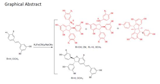
2. Results and Discussion

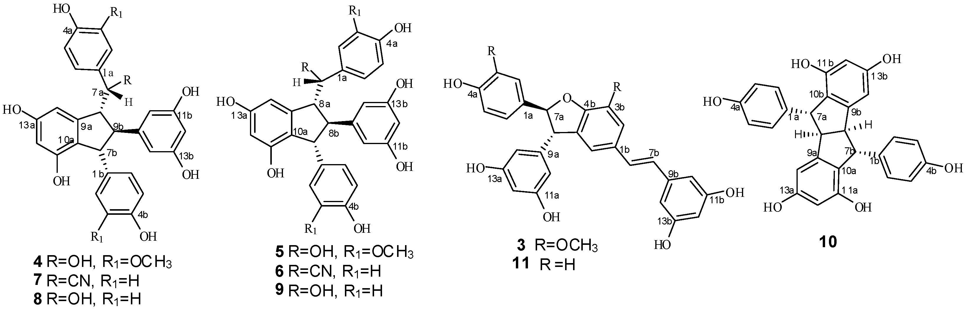
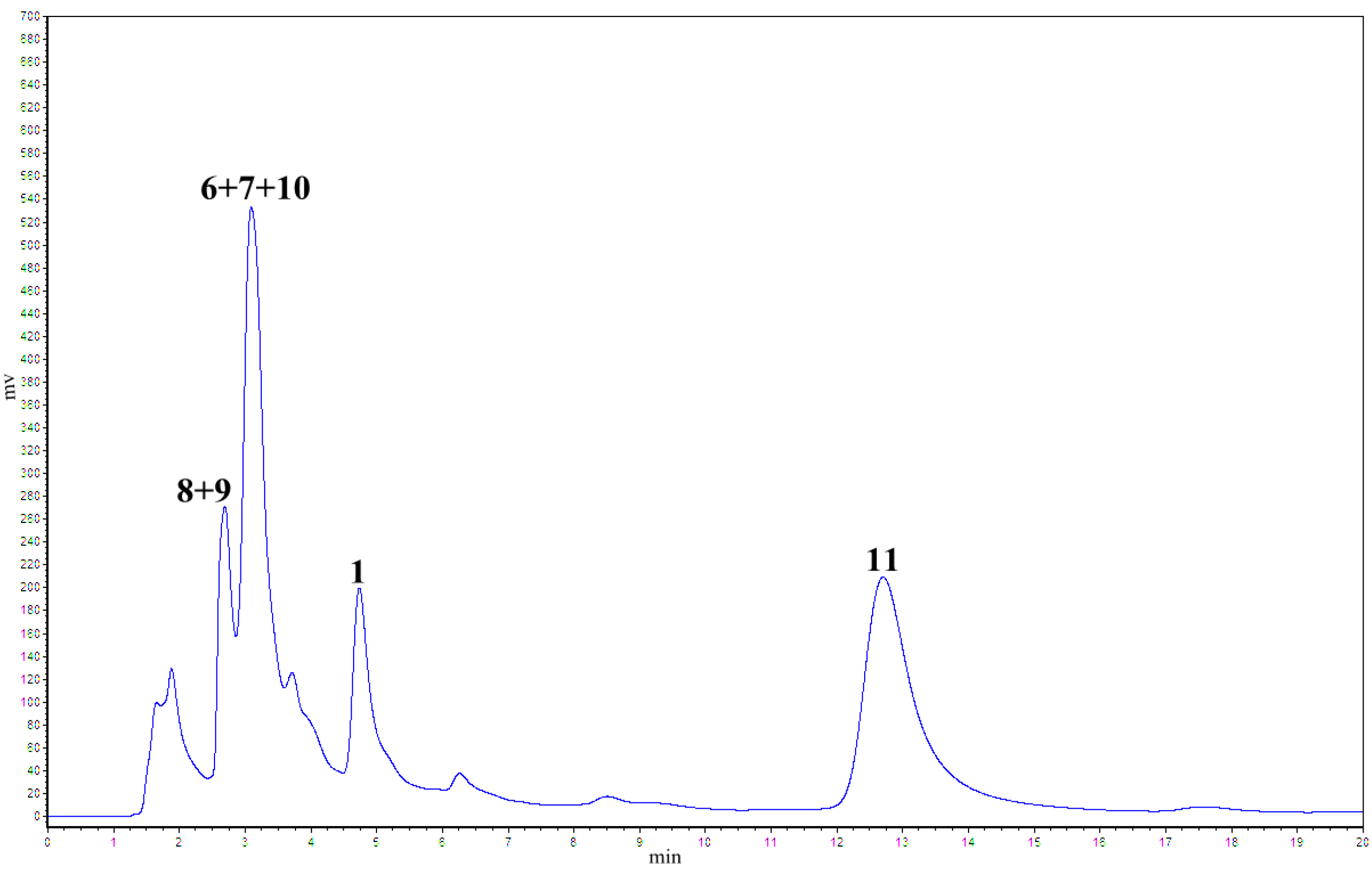
2.1. Treatment of 2 with Potassium Hexacyanoferrate (III)/Sodium Acetate

As reported in a previous paper [13], the oxidative coupling reaction of 2 in aqueous acetone using K3Fe(CN)6/NaOAc as oxidant at room temperature generated a major product peak 3 in 65.2% yield and a peak of another product obtained in 10% yield with a retention time of 4.2 min in the HPLC chromatogram (Figure 2).
Figure 2. Analysis of isorhapontigenin oxidation products by K3Fe(CN)6/NaOAc (32% CH3CN/H2O, λ = 230 nm, 1 mL/min).
Molecules 20 19872 g002
This finding indicates that the reaction mainly produced two types of products as compared with the complex products seen in the common oxidative coupling reactions of hydroxylstilbene [13,14,15,16]. Furthermore, several reports of K3Fe(CN)6 molecules catalyzing the oxidative coupling reaction of hydroxystilbene can be found in the literature [11,12]. However, to the best of our knowledge, a detailed investigation on this reaction, especially for the K3Fe(CN)6/NaOAc oxidant system, has yet to be reported. In this study, isorhapontigenin was treated with K3Fe(CN)6/NaOAc in aqueous acetone at room temperature, followed by silica gel column chromatography, preparative HPLC, and semi-preparative HPLC to obtain a major product 3 in 52.2% yield, as well as two indane dimers 4 and 5 (Figure 1) in 6.0% and 3.3% yield, respectively. Among these dimers, 5 possesses the same structure as the natural product gnetuhainin I [17], and compound 4, an isomer of 5, is a new isorhapontigenin dimer. These indane dimers were obtained for the first time by direct transformation from isorhapontigenin. The results imply that these reaction conditions which led to a total yield of about 10% for the indane dimers should be beneficial to the formation of carbon-carbon bonds.

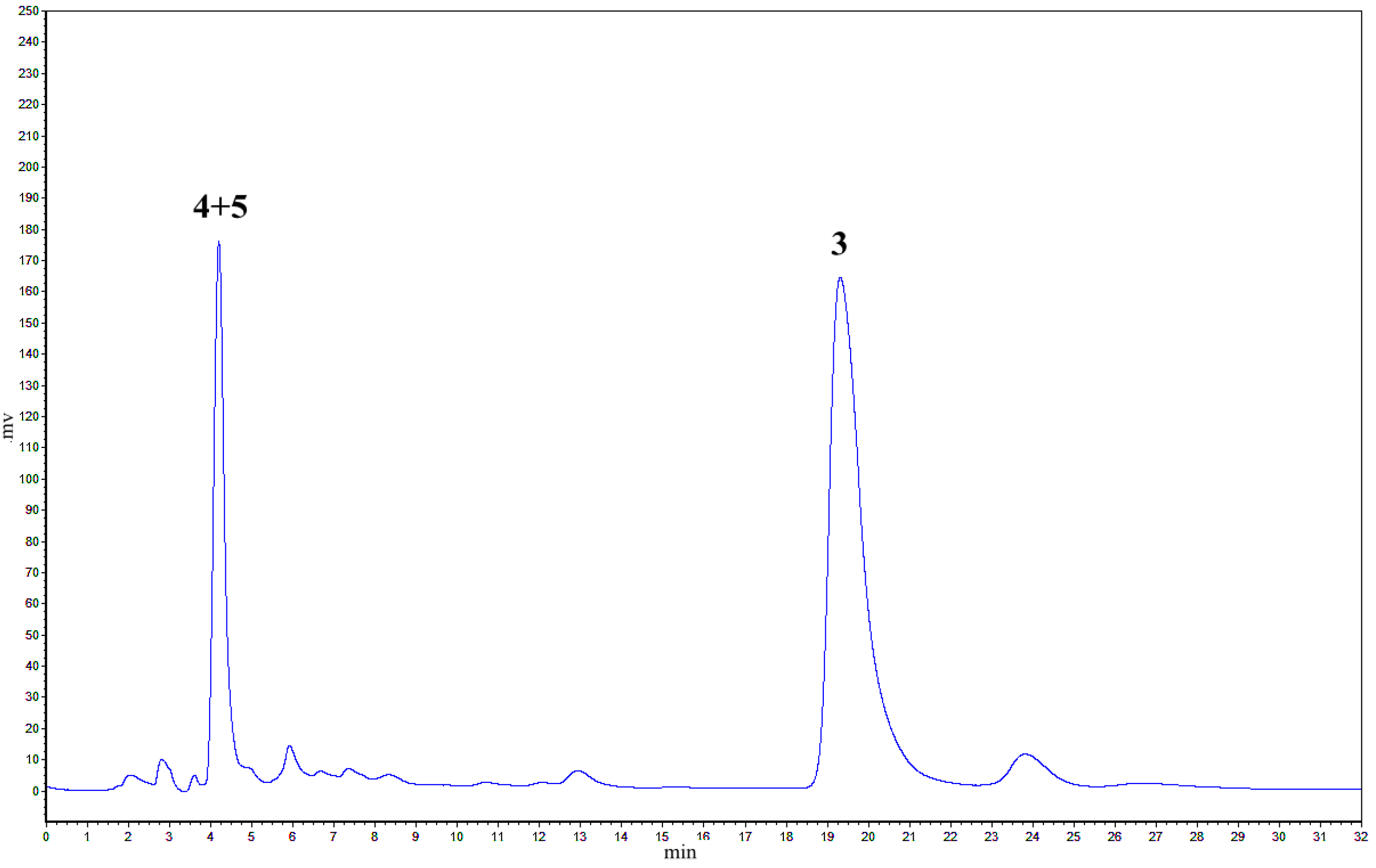
2.2. Treatment of 1 with Potassium Hexacyanoferrate (III)/Sodium Acetate

To substantiate the above hypothesis, we conducted a further study on the oxidative coupling reaction of resveratrol catalyzed by K3Fe(CN)6/NaOAc in aqueous acetone under reflux for 60 h. This approach was used because when resveratrol was treated at room temperature for one week, only small amounts of products were observed, and large amounts of unchanged resveratrol were recovered. Under the optimized conditions, two major products were observed on TLC plates, whereas other than the major products, several peaks ascribed to indane isomers of resveratrol dimer on the basis of previous results, appeared in the HPLC chromatogram (Figure 3). Therefore, by application of the above conditions, the oxidative coupling reaction of 1000 mg resveratrol, combined with silica gel column chromatography (CC), preparative HPLC, and semi-preparative HPLC, led to the isolation of six resveratrol dimers: one major benzofuran product 11 (21.1% yield) and five indane dimers 6 (4.9% yield), 7 (5.9%), 8 (1.3%), 9 (2.1%), and 10 (0.8%, Figure 1). Among these dimers, 6 and 7 are new indane stilbene dimers that are substituted by a cyano group. Dimers 8, 9, 10, and 11 possess the same structures as the natural products leachianol G, leachianol F, pallidol, and resveratrol trans-dehydrodimer. The total yield of about 15% for indane products indicates that the indane dimer is one of the major products in this reaction.
Figure 3. Analysis of resveratrol oxidation products by K3Fe(CN)6/NaOAc (52% MeOH/H2O, λ = 230 nm, 1 mL/min).
Molecules 20 19872 g003

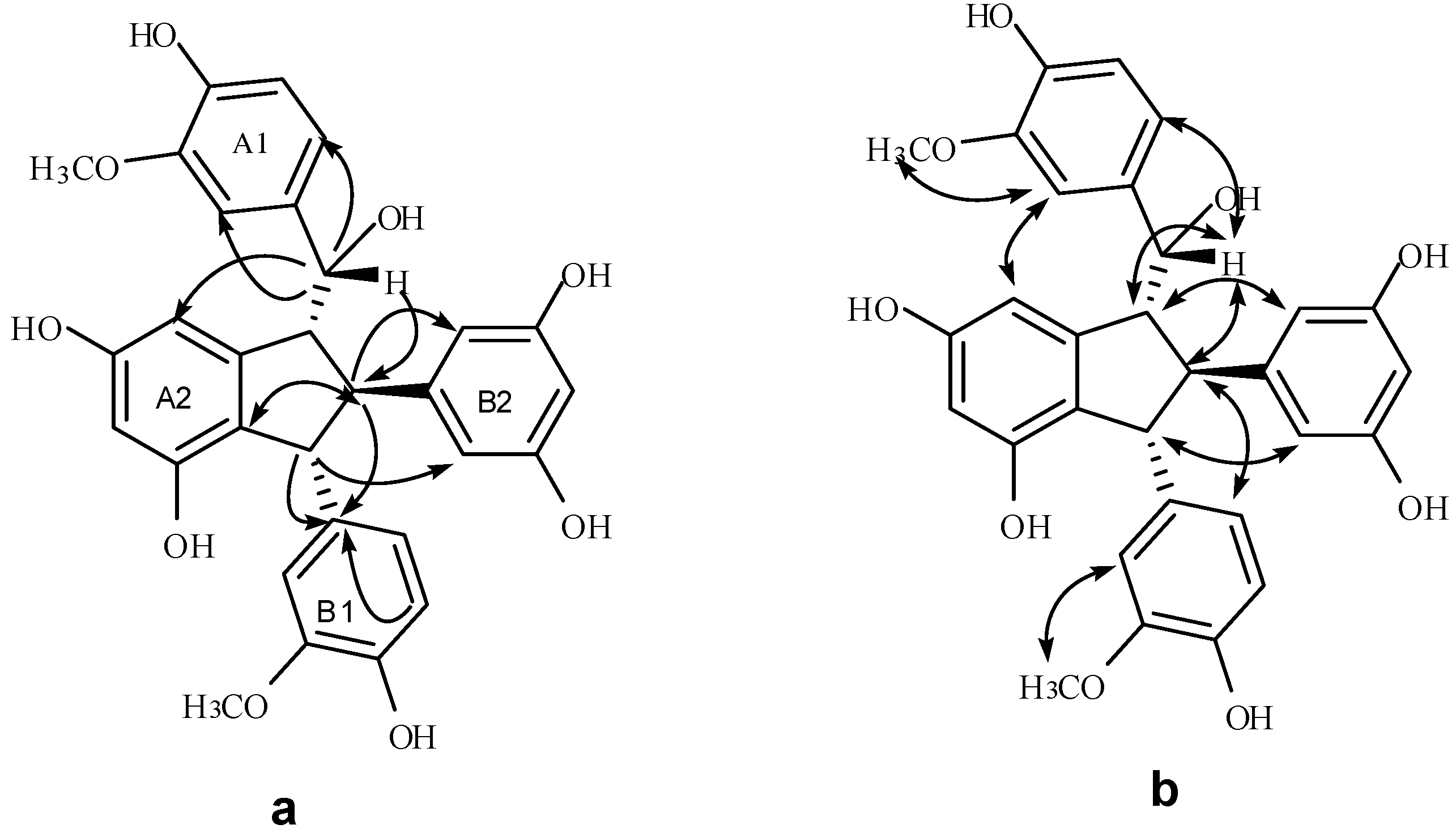
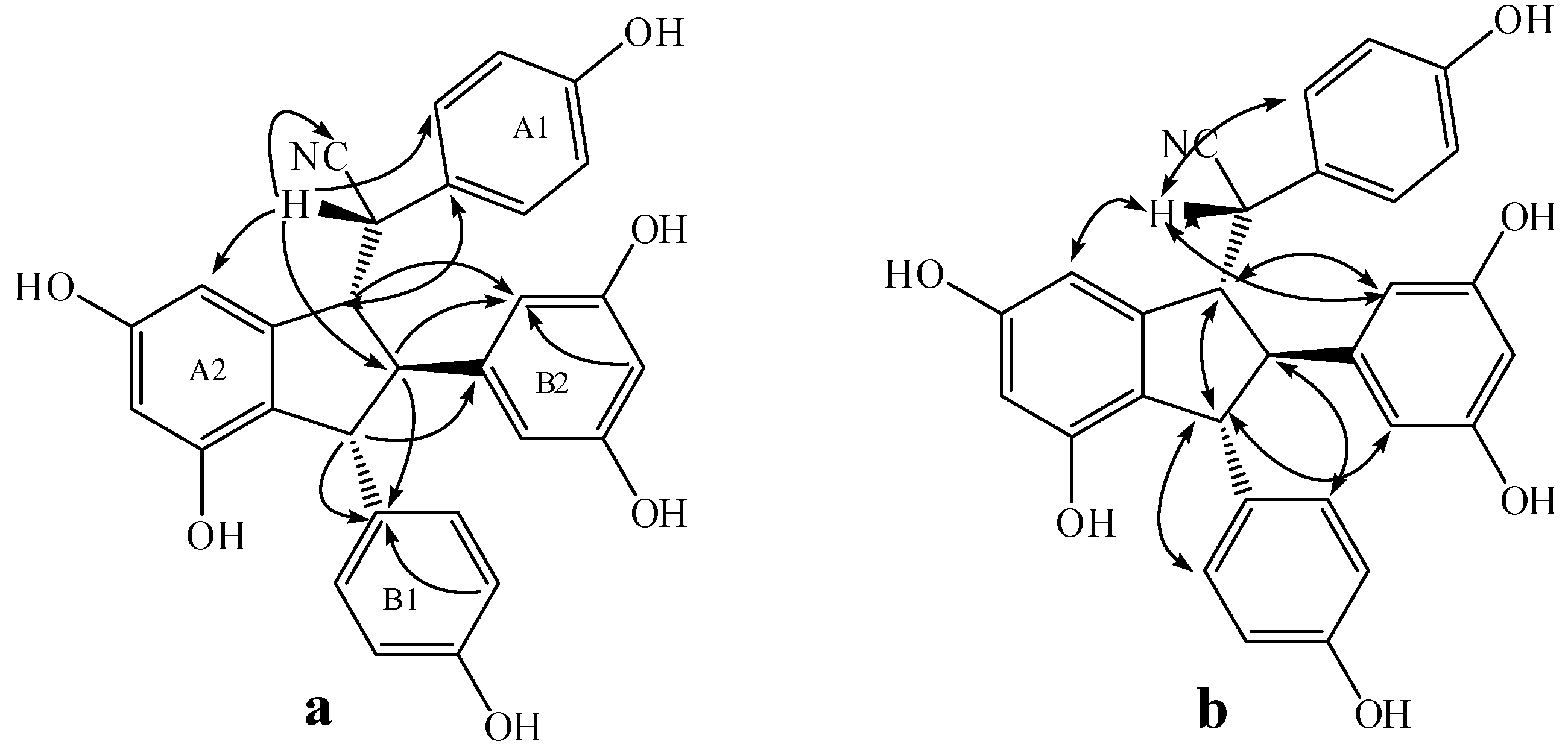
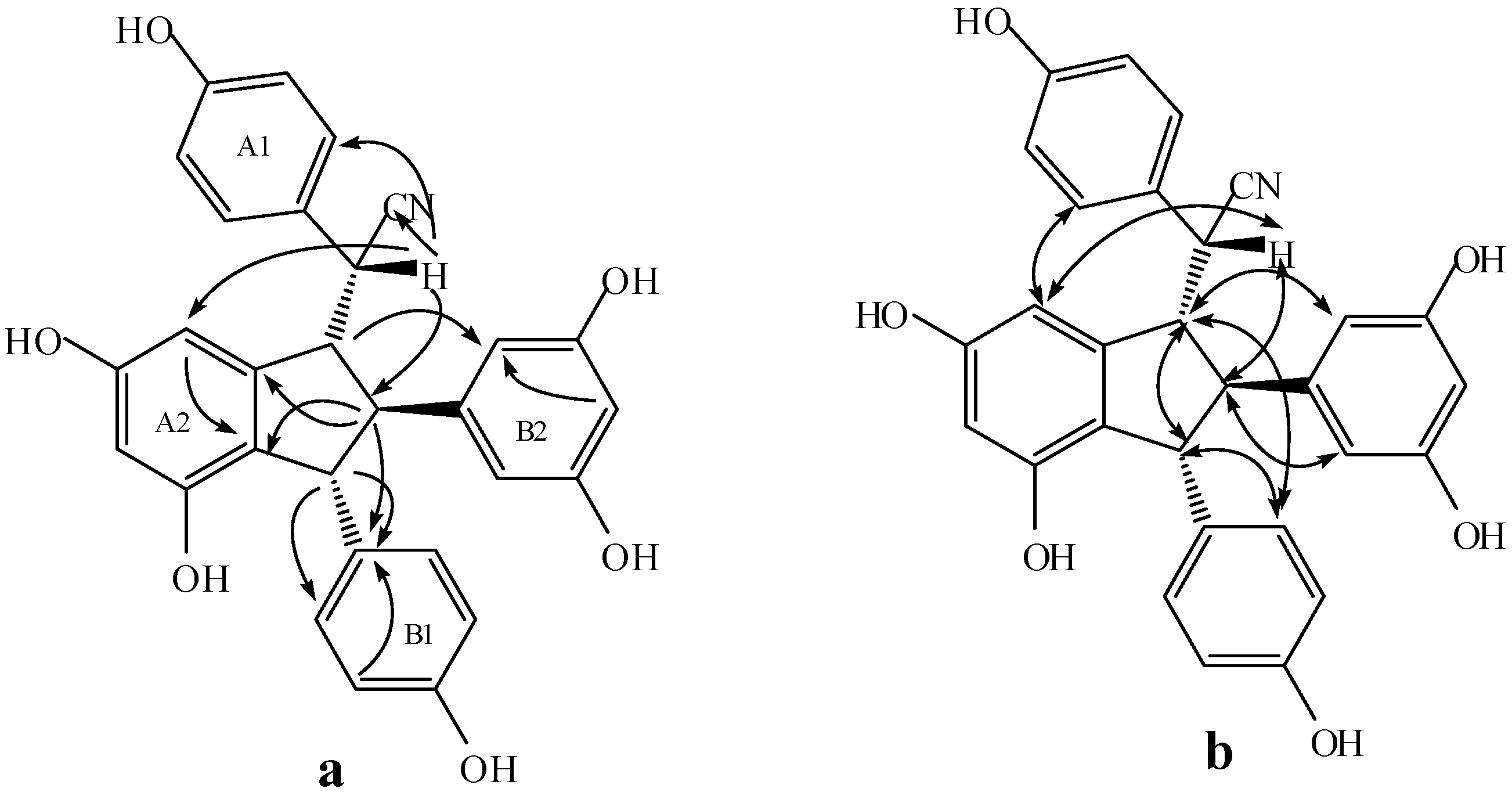
2.3. Structural Identification of New Dimers

Compound 4 was isolated as a brown amorphous powder. The corresponding negative ion HR-ESI-MS (Figure S8) peak at m/z 531.1662 [M − H] (calcd. for C30H27O9, 531.1661) showed the molecular formula of C30H28O9, which together with the 1H- and 13C-NMR spectral data, suggests that 4 should be an isorhapontigenin dimer. The IR spectrum (Figure S10) displays the presence of hydroxyls (3360 cm−1) and aromatic groups (1605 and 1516 cm−1). The 1H-NMR spectrum (Figure S1, Table 1) shows two ABX systems for rings A1 and B1 at δH 6.82 (1H, d, 1.8), 6.61 (1H, d, 7.8), 6.57 (1H, dd, 7.8, 1.8), and 6.37 (1H, d, 1.8), 6.55 (1H, d, 8.4), and 6.30 (1H, dd, 8.4, 1.8), one AB2 system at δH 6.02 (1H, t, 2.4) and 6.03 (2H, d, 2.4) for ring B2, two meta-coupled proton signals at δH 6.08 (1H, d, 1.8) and 6.07 (1H, brs) for ring A2, four multi-coupled aliphatic protons at δH 4.48 (1H, d, 7.2), 4.08 (1H, d, 4.8), 3.49 (1H, t, 6.0), and 3.17 (1H, t, 5.4) ppm, and two methoxyl singlets at δH 3.61 (3H, s), and 3.65 (3H, s). The 13C-NMR spectrum (Figure S2, Table 1) of 4 reveals the presence of four aliphatic carbons at δC 78.0, 61.8, 60.0, and 57.4 ppm, as well as 24 aromatic carbons and two methoxy carbons. The aliphatic carbon at δC 78.0 is due to an alcohol carbon. This group of evidence indicates that compound 4 possesses a similar indane skeleton as 5, as reported in the literature [17]. In addition, downfield shifts of H-2a, H-6a, H-8b, and H-10(14)b and the corresponding upfield shift of H-14a caused by the anisotropic effect of the aromatic ring in comparison to those of 5 proved that 4 should be an 7-epimer of 5 [18]. In the HMBC spectrum of 4 (Figure 4), the correlations among H-2a, H-6a, H-14a and C-7a, which is attached to the hydroxyl group, indicates that C-7a is excluded from the additional ring. The correlations between H-7b, H-8b, H-5b and C-1b verify that the B1 ring should be connected at C-7b. Comparison of the spectral data with those of 5, as well as the analysis of COSY, HMBC and HSQC correlations (Figures S4–S6), determines the planar structure of 4 as shown in Figure 1. The stereochemistry of 4 was determined by analysis of NOESY spectrum (Figure S7 and Figure 4), in which strong NOEs between H-10(14)b with H-7b and H-8a suggests a trans orientation between H-7b and H-8b as well as between H-8b and H-8a. The NOE interactions between H-7a and H-8b revealed a cis relationship of H-7a and H-8b. Accordingly, the structure of 4 was determined as shown in Figure 1.
Table 1. 1H- and 13C-NMR spectroscopic data of 4 and 5 *.
Position45
δCδHδCδH
1a135.5s 136.5s
2a112.4d6.82 (d, 1.8)112.4d6.37 (d, 1.8)
3a148.5s 148.7d
4a146.9s 146.7s
5a115.3d6.61 (d, 7.8)115.74d6.59 (d, 7.8),
6a121.1d6.57 (dd, 7.8, 1.8)120.94d6.39 (dd, 7.8, 1.8)
7a78.0d4.48 (d, 7.2)78.7d4.35 (d, 9.0)
8a61.8d3.49 (t, 6.0)62.0d3.29 (overlap)
9a148.7s 149.5s
10a123.9s 123.0s
11a155.4s 155.1s
12a102.66.08 (d, 1.8)102.6d6.19 (d, 1.8)
13a158.8s 159.2s
14a106.0d6.07 (br s)106.30d6.58 (d, 1.8)
1b139.0s 138.8s
2b112.0d6.37 (d, 1.8)111.9d6.49 (d, 1.8)
3b147.4s 148.5s
4b145.5s 145.6s
5b115.6d6.55 (d, 8.4)115.69d6.62 (d, 7.8)
6b120.0d6.30 (dd, 8.4, 1.8)120.93d6.35 (dd, 7.8, 1.8)
7b57.4d4.08 (d, 4.8)56.4d4.14 (d, 3.0)
8b60.0d3.17 (t, 5.4)60.2d2.74 (t, 3.0)
9b150.7s 151.3s
10(14)b106.9d6.03 (d, 2.4)106.33d5.75 (d, 2.4)
11(13)b159.4s 159.3s
12b101.3d6.02 (t, 2.4)101.2d5.96 (t, 2.4)
3a-OCH356.3q3.61 (s)56.3q3.58 (s)
3b-OCH356.2q3.65 (s)56.1q3.67 (s)
* Data (δ) were measured in CD3OD for 1H at 600 MHz, and for 13C at 150 MHz, The assignments were based on DEPT, 1H-1H COSY, HSQC, HMBC, and NOESY experiments, respectively.
Figure 4. The significant HMBC (a) and NOESY (b) interactions of 4.
Molecules 20 19872 g004
Compound 6 was obtained as a light gray amorphous powder. The corresponding positive ion HR-ESI-MS (Figure S17) peak at m/z 504.1409 [M + Na]+ (calcd. for C29H23NO6Na, 504.1418) yield the molecular formula of C29H23NO6, which indicates that 6 should be a resveratrol dimer derivative. The IR spectrum (Figure S19) displayed the presence of hydroxyls (3393.9 cm−1), cyano group (2248.6 cm−1), and aromatic rings (1610.6, 1513.2, 1468.4 cm−1). The 1H-NMR spectrum (Table 2) shows two A2B2 systems for ring A1 and B1 at δH 6.89 (2H, d, 8.5), 6.79 (2H, d, 8.5), 6.87 (2H, d, 8.5), and 6.76 (2H, d, 8.5), one AB2 system for ring B2 at δH 5.96 (2H, d, 2.0) and 6.17 (1H, t, 2.0), two meta-coupled proton signals for ring A2 at δH 6.38 (1H, d, 2.0) and 6.57 (1H, d, 2.0), and four multi-coupled aliphatic proton signals at δH 4.28 (1H, d, 4.0), 3.76 (1H, d, 8.5), 3.52 (1H, dd, 8.5, 4.0), and 3.01 (1H, t, 4.0). C29H23NO6, the quaternary carbon signal at δC 120.33 in the 13C-NMR data (Figure S12, Table 2) of 6 suggests the presence of a cyano group in the structure of 6. Accordingly, 6 was assumed to contain a leachianol G skeleton with a cyano group replacing a hydroxyl group [19]. In addition to a cyano group, 22 carbon signals in the 13C-NMR representing 28 carbons further support this hypothesis. In the HMBC spectrum (Figure S15, Figure 5), the interactions between H-7a and C-2(6)a, C-14a indicate that ring A1 is located at C-7; the interactions among H-8b, H-12b, and C-10(14)b substantiate that ring B2 is connected at C-8b. Similarly, the correlations of the three proton signals of H-7b, H-8b, and H-3(5)b with C-1b reveal that ring B1 is connected at C-7b. Moreover, in the NOESY spectrum of 6 (Figure S16, Figure 5), the interactions between H-8a and H-7b, H-10(14)b, as well as between H-8b and H-2(6)b suggest a cis-configuration among H-8a, H-7b and ring B2. The interactions between H-7a and H-14a indicate that H-7a should be located near H-14a, and that ring A1 is located near ring B2. Therefore, the structure of 6 is characterized as shown in Figure 1.
Compound 7 was obtained as a light brown amorphous powder. The corresponding positive ion HR-ESI-MS (Figure S26) peak at m/z 504.1431 [M + Na]+ (calcd. for C29H23NO6Na, 504.1418) corresponds to the molecular formula of C29H23NO6. The IR spectrum (Figure S28) revealed the presence of hydroxyls (3337.0 cm−1), a cyano group (2247.8 cm−1), and aromatic rings (1610.9, 1513.5, 1468.2 cm−1). The 1H-NMR spectrum (Figure S20, Table 2) shows two A2B2 systems for rings A1 and B1, one AB2 system for ring B2, two meta-coupled proton signals for ring A2, and four aliphatic proton signals at δH 4.27 (1H, d, 4.5), 3.96 (1H, d, 8.1), 3.58 (1H, dd, 8.1, 4.5), and 3.26 (1H, t, 4.5). Together with the molecular formula C29H23NO6, the quaternary carbon signal at δC 120.33 in the 13C-NMR data (Figure S21, Table 2) indicates the presence of a cyano group in the structure of 7. In combination with the 13C-NMR spectral data and HMBC correlations (Figure S24, Figure 6), these data suggest that 7 should be an 7-epimer of 6.
However, several exceptions in the 1H-NMR spectrum and a slight change in the chemical shift and multiplicity of certain signals were observed (Table 2). Owing to an anisotropic effect of the aromatic ring, the signals at δH 7.06 (2H, d, 8.5) for H-2(6)a shifted slightly downfield whereas the signals at δH 5.95 (1H, brs) for H-14a shifted upfield in comparison with the spectrum of 6. Besides, observation of the downfield shift of H-10(14)b [δH 6.07 (2H, d, 2.0)] and H-8b [δH 3.58 (1H, dd, 8.1, 4.5)] further supports the reversed position of the cyano group and ring A1 at C-7 compared with 6.
The relative stereochemistry of 7 could be determined by analyzing the NOESY spectrum (Figure S25, Figure 6), in which the interactions between H-8a and H-7b, H-10(14)b suggest a cis configuration between H-8a, H-7b and ring B2. Together with the downfield shift of H-10(14)b and upfield shift of H-14a in comparison to those of 6, the interaction between H-14a and H-2(6)a suggests that ring A1 must be located near ring A2, whereas the interaction between H-7a and H-8b indicates that H-7a and H-8b are situated in a cis-orientation. Therefore, the structure of 7 is characterized as shown in Figure 1.
Table 2. 1H- and 13C-NMR spectroscopic data of 6 and 7 *.
Position67
δCδHδCδH
1a125.89s 125.85s
2(6)a129.31d6.89 (2H, d, 8.5)130.72d7.06 (2H, d, 8.5)
3(5)a115.55d6.79 (2H, d, 8.5)116.09d6.74 (2H, d, 8.5)
4a157.12s 158.03s
7a41.16d3.76 (1H, d, 8.5)42.09d3.96 (1H, d, 8.1)
8a59.35d3.01 (1H, t, 4.0)61.31d3.26 (1H, t, 4.5)
9a147.44s 148.66s
10a121.28s 122.54s
11a154.59s 155.23s
12a102.39d6.38 (1H, d, 2.0)103.18d6.30 (1H, d, 1.6)
13a158.49s 159.02s
14a103.90d6.57 (1H, d, 2.0)104.72d5.95 (1H, brs)
1b135.50s 136.82s
2(6)b128.40d6. 87 (2H, d, 8.5)129.21d6.84 (2H, d, 8.5)
3(5)b114.97d6.76 (2H, d, 8.5)115.81d6.71 (2H, d, 8.5)
4b155.72s 156.51s
7b53.84d4.28 (1H, d, 4.0)55.89d4.27 (1H, d, 4.5)
8b56.70d3.52 (1H, dd, 8.5, 4.0)58.07d3.58 (1H, dd, 8.1, 4.5)
9b145.39s 145.53s
10(14)b105.13d5.96 (2H, d, 2.0)106.10d6.07 (2H, d, 2.0)
11(13)b158.49s 159.31s
12b100.75d6.17 (1H, t, 2.0)101.42d6.15 (1H, t, 2.0)
CN120.33s 121.73s
* Data (δ) were measured in CD3COCD3 for 1H at 500 MHz, and for 13C at 125 MHz; The assignments were based on DEPT, HSQC, HMBC, and NOESY experiments, respectively.
Figure 5. The key HMBC (a) and NOESY (b) correlations of 6.
Molecules 20 19872 g005
Figure 6. The key HMBC (a) and NOESY (b) correlations of 7.
Molecules 20 19872 g006
Known compounds 3, 5, 8, 9, 10, and 11 were identified as shegansu B [20], gnetuhainin I [17], leachianol G [21], leachianol F [21], pallidol [22], and resveratrol trans-dehydrodimer [23,24] by comparison of their physical and spectroscopic data with those reported in the literature. Product 5, which possesses an indane skeleton, was obtained for the first time by direct transformation from isorhapontigenin, and all these products would be rather difficult to obtain by common organic reactions. The transformation catalyzed by hexacyanoferrate (III)/sodium acetate was presumed to occur on the basis of a radical reaction. As a result, the obtained dimers should be racemates, which is consistent with the zero values of their optical rotations.

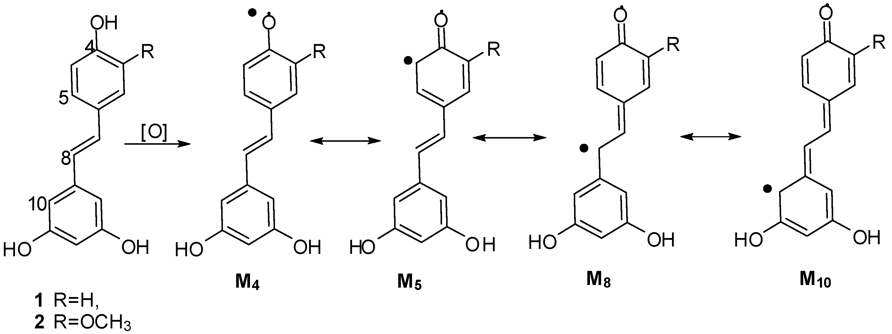
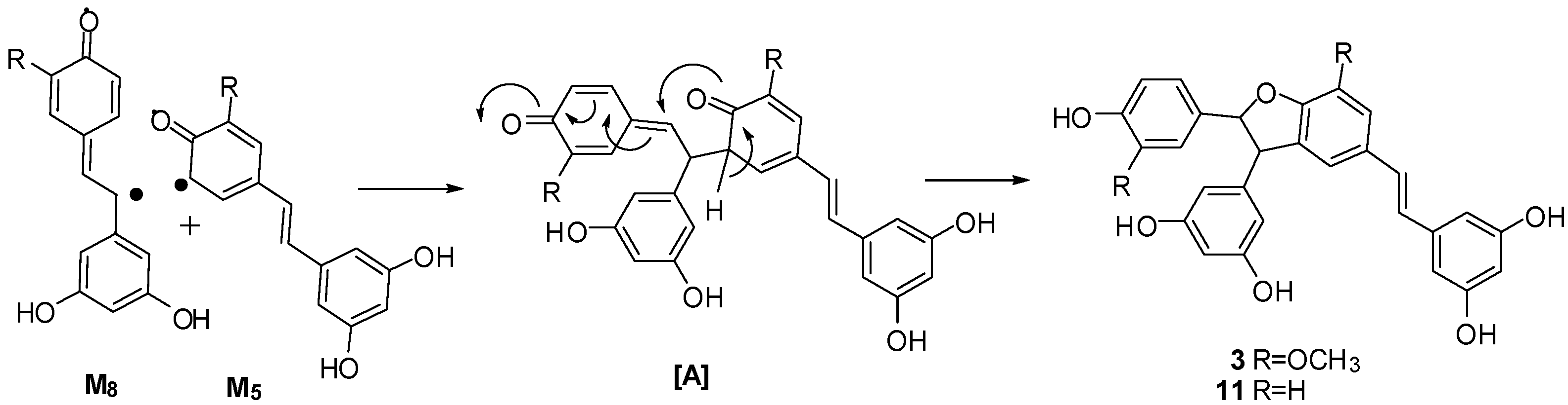
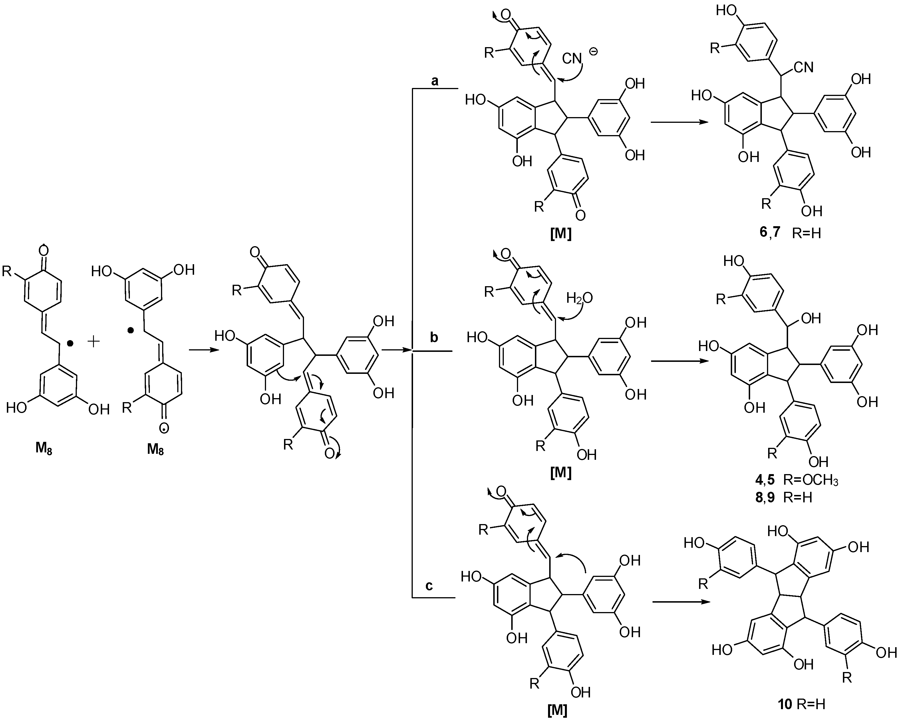
2.4. Discussion of the Probable Coupling Reaction Mechanism

On the basis of the aforementioned structures, the dimerization catalyzed by K3Fe(CN)6/NaOAc was presumed to be based on a radical reaction, induced by K3Fe(CN)6, whereby stilbene monomers 1 and 2 were dehydrogenated and rearranged to yield radicals M4, M5, M8 and M10 (Scheme 1) [11,12,13,14,25,26].
Scheme 1. Plausible radicals of 1 and 2.
Molecules 20 19872 g007
The coupling of radicals M8 and M5 then occurred successively, followed by tautomeric rearrangement and intramolecular nucleophilic attack to the intermediate quinone [A], yielding the dihydrofunan dimers 3 and 11, respectively (Scheme 2). Meanwhile, the coupling of two M8 radicals, followed by intramolecular cyclization, generates intermediate quinone [M]. The difference in the ultimate products is apparently due to the difference in the position and reagent of the nucleophilic attack (Scheme 3). In the case of path a, intermolecular nucleophilic attack to the intermediate quinone [M] of a cyano anion (CN) produced 7 and its isomer 6. In the case of path b, intermolecular nucleophilic attack to the intermediate quinone of a water molecule produced 4, 8 and their isomers 5, 9. In the case of path c, the second intramolecular nucleophilic attack to the intermediate quinone [M] yielded 10. Evidently, all reactions mentioned above should be carried out concertedly. In general, free radical reaction is not stereoselective. In the course of nucleophilic attack reaction, the chances of attack to Re or Si face of intermediates are equal. Therefore, all products are found to be enantiomer pairs.
Scheme 2. Proposed coupling mechanism of compounds 3 and 11.
Molecules 20 19872 g008
Scheme 3. Proposed coupling mechanism of compounds 410.
Molecules 20 19872 g009
Moreover, the comparatively high yield of 6 and 7 results from the high nucleophilicity of the cyano anion, and the low yield of 10 results from the low nucleophilicity of the phenyl group. The dimeric structures indicate that, during the long reaction time, the oxidation reactions of hydroxystilbene through K3Fe(CN)6/NaOAc in aqueous acetone, mainly generate M5 and M8. Product coupling involving M4 and M10 was not detected in the course of the reaction. The sterically less hindered radical M5 would be easily involved in the reaction, possibly accounting for the higher yield of benzofuran products 3 and 11. However, an appropriate account for the distinction of reactivity cannot be proposed based only on this evidence.

3. Experimental Section

3.1. Materials and Instrumentation

Optical rotations were measured on a P2000 polarimeter (JASCO, Tokyo, Japan). UV spectra were obtained on a JASCO P650 spectrometer. IR spectra were recorded on a Nicolet 5700 FT-IR microscope instrument (FT-IR microscope transmission, Thermo Electron Corporation, Madison, WI, USA). 1D and 2D NMR spectra were acquired at 500 or 600 MHz for 1H and 125 or 150 MHz for 13C, respectively, on INOVA 500 MHz (Varian, Inc., Palo Alto, CA, USA), or Bruker AVANCE III HD 600 MHz spectrometers (Bruker Corporation, Karlsruhe, Germany), in acetone-d6 or methanol-d4, with the solvent peaks as references. ESI-MS and HR-ESI-MS data were measured using an AccuToFCS JMST100CS spectrometer (Agilent Technologies, Ltd., Santa Clara, CA, USA). Column chromatography (CC) was performed with silica gel (200–300 mesh, Qingdao Marine Chemical Inc., Qingdao, China). HPLC separation was performed on an instrument consisting of a Waters 515 pump and a Waters 2487 dual λ absorbance detector (Waters Corporation, Milford, MA, USA) with a YMC semi-preparative column (250 mm × 10 mm ID) packed with C18 (5 μM). TLC was carried out with glass precoated silica gel GF254 plates (Qingdao Marine Chemical, Inc.). Spots were visualized under UV light or by spraying with 7% H2SO4 in 95% EtOH followed by heating.

3.2. Treatment of Isorhapontigenin with Potassium Hexacyanoferrate (III)/Sodium Acetate.

To a solution of (E)-isorhapontigenin (2, 100 mg, 0.388 mmol) in acetone cooled to 0 °C in an ice bath, a mixed solution of K3Fe(CN)6 (150 mg, 0.4559 mmol) and NaOAc (140 mg, 1.7073 mmol) in 25 mL of water was added under stirring. The reactant was stirred at 0 °C for 1 h under a N2 atmosphere, and subsequently stirred for another 15 days at room temperature. The reaction mixture was extracted with ethyl acetate and water, the organic layer was washed with brine, water and dried over anhydrous Na2SO4 for 24 h. Then it was concentrated in vacuo to yield a residue that was subjected to silica gel column chromatography eluting with CHCl3–MeOH (10:1, v/v) to give 3 (52.1 mg, 52.2%) and fraction Fr-1 (14.0 mg). Fr-1 was subsequently subjected to semi-preparative Rp-18 HPLC (column, Rp-18, 250 mm × 10 mm I.D., 5 µm, YMC) eluting with acetonitrile in water (25:75, v/v) to yield compounds 4 (6.2 mg, 6.0%) and 5 (3.4 mg, 3.3%), respectively.
Compound 4: brown amorphous powder; [ α ] D 20 0 (c = 0.66, MeOH); UV (MeOH) λmax (log ε): 229 (sh, 4.47), 281 (4.08) nm; IR (film) νmax: 3360, 2973, 1605, 1516, 1464, 1344, 1276, 1151, 1127, 1032, 1004, 841, 822, 699 cm−1; 1H-NMR (600 MHz, CD3OD) and 13C-NMR (150 MHz, CD3OD), see Table 1; ESI-MS m/z: 555.3 [M + Na]+, 571.2 [M + K]+, 531.2 [M − H], 567.2 [M + Cl]; HR-ESI-MS m/z: 531.1662 [M − H] (cacld. for C30H27O9, 531.1661).
Gnetuhainin I (5): brown amorphous powder; [ α ] D 20 0 (c = 0.70, MeOH); 1H-NMR (600 MHz, CD3OD) and 13C-NMR (150 MHz, CD3OD), see Table 1; ESI-MS m/z: 555.2 [M + Na]+, 571.2 [M + K]+, 531.2 [M − H], 567.2 [M + Cl]; HR-ESI-MS m/z: 531.1661 [M − H] (cacld. for C30H27O9, 531.1661).

3.3. Treatment of Resveratrol with Potassium Hexacyanoferrate (III)/Sodium Acetate

To a solution of resveratrol (1, 1000 mg) in acetone (100 ml), a mixed solution of K3Fe(CN)6 (2885.0 mg) and NaOAc (2660.0 mg) in water (125 mL) was added under stirring. The reactant was refluxed for 35 h, after removal of the acetone under reduced pressure, the solution was extracted with ethyl acetate (100 mL × 3), the organic layer was washed with brine, water and dried over anhydrous Na2SO4 for 24 h. The solution was concentrated in vacuo to yield a reaction mixture that was subjected to a silica gel (200–300 mesh, 12.5 g) column and eluted with a gradient of increasing MeOH in CHCl3 (15:1, 10:1, 6:1, 4:1, 2:1, 1:1, v/v) to provide seven fractions, namely, FNR-A~FNR-G. FNR-B (102.1 mg) was unchanged resveratrol, and FNR-C was the main product 11 (187.6 mg, 21.1%). 45 mg FNR-D (132.2 mg) was then separated by semi-preparative Rp-HPLC (column, Rp-18, 250 × 10 mm, 5 µm) eluted using methanol/water (35:65, v/v) to afford 6 (15.8 mg, 4.9%) and 7 (17.7 mg, 5.9%). FNR-F (110.7 mg) was dealt with in the same manner to give 8 (12.1 mg, 1.3%), 9 (19.5 mg, 2.1%) and 10 (7.1 mg, 0.8%), respectively.
Compound 6: a light gray amorphous powder. [ α ] D 20 0 (c = 0.33, MeOH); UV (MeOH) λmax (log ε): 206 (4.48), 228 (4.21, sh), 282 (3.59) nm; IR (film) νmax: 3393.9, 2920.3, 2849.7, 2248.6, 1610.6, 1513.2, 1468.4, 1344.1, 1247.6, 1157.3, 1003.3, 835.7 cm−1; 1H-NMR (500 MHz in acetone-d6) and 13C-NMR (125 MHz in acetone-d6), see Table 2; (+)-ESI-MS m/z: 482 [M + H]+, 504 [M + Na]+, 520 [M + K]+; HR-ESI-MS m/z: 504.1409 [M + Na]+ (cacld. for C29H23NO6Na, 504.1418).
Compound 7: a light brown amorphous powder. [ α ] D 20 0 (c = 0.66, MeOH); UV (MeOH) λmax (log ε): 204 (4.55), 230 (4.22, sh), 283 (3.70), nm; IR (film) νmax: 3337.0, 2916.6, 2850.1, 1610.9, 1513.5, 1468.2, 1342.0, 1247.8, 1155.6, 1006.9, 834.0 cm−1; 1H-NMR (500 MHz in acetone-d6) and 13C-NMR (125 MHz in acetone-d6), see Table 2; (+)-ESI-MS m/z: 482 [M + H]+, 504 [M + Na]+, 520 [M + K]+; HR-ESI-MS m/z: 504.1431 [M + Na]+ (cacld. for C29H23NO6Na, 504.1418).
Leachianol G (8): a brown amorphous powder. [ α ] D 20 0 (c = 1.05, MeOH); 1H-NMR (500 MHz in acetone-d6) data: 7.07 (2H, d, 8.5, H-2(6)a), 6.73 (2H, d, 8.5, H-3(5)a), 4.45 (1H, d, 8.2, H-7a), 3.38 (1H, dd, 8.2, 3.5, H-8a), 6.22 (1H, d, 2.0, H-12a), 5.71 (1H, d, 2.0, H-14a), 6.87 (2H, d, 8.5, H-2(6)b), 6.71 (2H, d, 8.5, H-3(5)b), 4.27 (1H, d, 3.5, H-7b), 3.50 (1H, t, 3.5, H-8b), 6.14 (2H, d,1.5, H-10(14)b), 6.16 (1H, t, 1.5, H-12b). 13C-NMR (125 MHz in acetone-d6): 135.81s (C-1a), 129.48d (C-2(6)a), 115.18d (C-3(5)a), 157.23s (C-4a), 77.25d (C-7a), 62.69d (C-8a), 147.38s (C-9a), 123.04s (C-10a), 154.84s (C-11a), 102.17d (C-12a), 158.49s (C-13a), 105.55d (C-14a), 138.09s (C-1b), 129.31d (C-2(6)b), 115.55d (C-3(5)b), 156.18s (C-4b), 55.93d (C-7b), 59.04d (C-8b), 151.63s (C-9b), 106.07d (C-10(14)b), 159.21s (C-11(13)b), 100.87d (C-12b). (+)-ESI-MS m/z: 473 [M + H]+, 495 [M + Na]+, 511 [M + K]+; HR-ESI-MS m/z: 495.1419 [M + Na]+ (cacld. for C28H24O7Na, 495.1414).
Leachianol F (9): a brown amorphous powder. [ α ] D 20 0 (c = 1.05, MeOH); 1H-NMR (500 MHz in acetone-d6) data: 6. 86 (2H, d, 8.5, H-2(6)a), 6.68 (2H, d, 8.5, H-3(5)a), 4.46 (1H, d, 8.0, H-7a), 3.37 (1H, dd, 8.0, 3.5, H-8a), 6.32 (1H, d, 1.5, H-12a), 6.59 (1H, d , 1.5, H-14a), 6.85 (2H, d, 8.5, H-2(6)b), 6.74 (2H, d, 8.5, H-3(5)b), 4.24 (1H, d, 3.5, H-7b), 2.95 (1H, t, 3.5, H-8b), 5.93 (2H, d, 2.0, H-10(14b), 6.13 (1H, t, 2.0, H-12b), 4.01 (1H, d, 3.5, 7-OH). 13C-NMR (125 MHz in acetone-d6): 136.28s (C-1a), 128.87d (C-2(6)a), 115.34d (C-3(5)a), 156.26s (C-4a), 76.58d (C-7a), 61.91d (C-8a), 148.84s (C-9a), 122.46s (C-10a), 154.96s (C-11a), 102.19d (C-12a), 158.75s (C-13a), 106.12d (C-14a), 137.46s (C-1b), 129.35d (C-2(6)b), 115.51d (C-3(5)b), 157.06s (C-4b), 55.61d (C-7b), 59.47d (C-8b), 150.75s (C-9b), 105.90d (C-10(14)b), 159.14s (C-11(13)b), 100.99d (C-12b). (+)-ESI-MS m/z: 472 [M]+, 495 [M + Na]+, 511 [M + K]+; HR-ESI-MS m/z: 495.1425 [M + Na]+ (cacld. for C28H24O7Na, 495.1414).
Pallidol (10): a brown amorphous powder. [ α ] D 20 0 (c = 1.05, MeOH); 1H-NMR (500 MHz in acetone-d6) δ: 6. 99 (2H, d, 8.5, H-2(6)a), 6.71 (2H, d, 8.5, H-3(5)a), 4.58 (1H, s, H-7a), 3.82 (1H, s, H-8a), 6.20 (1H, d, 1.7, H-10a), 6.63 (1H, d, 1.7, H-10a) , 3.32 (1H, s, OH); 13C-NMR (125 MHz in acetone-d6) δ: 137.74s (C-1a), 129.05d (C-2(6)a), 115.74d (C-3(5)a), 159.27s (C-4a), 60.52d (C-7a), 53.97d (C-8a), 123.19 (C-9a), 102.35s (C-10a), 155.24s (C-11a), 103.28d (C-12a), 156.27s (C-13a), 150.53d (C-14a). (+)-ESI-MS m/z: 455 [M + H]+, 477 [M + Na]+, 493 [M + K]+.
Resveratrol trans-dehydrodimer (11): a brown amorphous powder. 1H-NMR (500 MHz in acetone-d6), δ: 7.24 (2H, d, J = 8.5 Hz, H-2a ,6a), 6.83 (2H, d, J = 8.5 Hz, H-3a, 5a), 5.44 (1H, d, J = 8.0 Hz, H-7a), 4.45 (1H, d, J = 8.0 Hz, H-8a), 6.18 (2H, d, J = 2.0 Hz, H-10a, 14a), 6.27 (1H, t, J = 2.0 Hz, H-12a), 7.25 (1H, brs, H-2b), 6.87 (1H, d, J = 8.5 Hz, H-5b), 7.42 (1H, dd, J = 8.5 Hz, 2.0, H-6b), 7.04 (1H, d, J = 16.5 Hz, H-7b), 6.89 (1H, d, J = 16.5 Hz, H-8b), 6.53 (2H, d, J = 2.0 Hz, H-10b, 14b), 6.27 (1H, t, J = 2.0 Hz, H-12b); 13C-NMR (125 MHz in acetone-d6) δ: 131.71 (C-1a), 128.57 (C-2a, 6a), 116.08 (C-3a, 5a), 158.33 (C-4a), 94.03 (C-7a), 57.80 (C-8a), 145.21 (C-9a), 107.33 (C-10a), 159.66 (C-11a), 102.24 (C-12a), 159.66 (C-13a), 107.33 (C-14a), 132.10 (C-1b), 123.90 (C-2b), 132.52 (C-3b), 160.60 (C-4b), 110.12 (C-5b), 128.61 (C-6b), 129.09 (C-7b), 127.19 (C-8b), 140.76 (C-9b), 105.63 (C-10b,14b), 159.45 (C-11b,13b), 102.57 (C-12b); (+) ESI m/z: 455 [M + H] +, 477 [M + Na]+, 493 [M + K]+; (−) ESI m/z: 453 [M − H], 489 [M + Cl].

4. Conclusions

The oxidative coupling reaction of isorhapontigenin and resveratrol with K3Fe(CN)6/NaOAc in aqueous acetone as oxidant, in combination with silica gel column chromatography and preparative Rp-HPLC resulted in the isolation of nine stilbene dimers. The structures of the nine dimers were determined on the basis of spectral analysis and chemical properties. Products 4, 6, and 7 are new dimers with indane skeletons, product 5 with an indane skeleton was obtained for the first time by direct transformation from isorhapontigenin, and all products would be rather difficult to obtain by common organic reactions.
Results indicated that, under the reaction conditions, the oxidative coupling reaction of two stilbenes yields only two types of stilbene dimers, namely, the benzofuran dimers 3 and 11 with the highest yields, and the indane dimers 4, 5, and 610 in comparatively low yields. Compared with other non-enzymatic oxidants (such as FeCl3, Ag2O, AgOAc, and so on), this reaction seems to generate only two types of radical intermediates, namely, M5 and M8, and only two coupling mechanisms occur to form benzofuran and indane stilbene dimers. Thus, the reaction could be used as a convenient method of synthesizing indane stilbene dimers because of its mild conditions, long reaction time and simple products. To the best of our knowledge, up to now, this is the most detailed report on the potassium hexacyanoferrate (III)-sodium acetate catalyzed biomimetic synthesis of stilbene dimers.

Supplementary Materials

Supplementary materials can be accessed at: https://www.mdpi.com/1420-3049/20/12/19872/s1.

Acknowledgments

This work was financially supported by National Mega-project for Innovative Drugs (grant No. 2012ZX09301002-001-003). We are grateful to the department of Instrumental Analysis, Institute of Materia Medica, Chinese Academy of Medical Sciences and Peking Union Medical College for measuring the IR, UV, NMR, and MS spectra.

Author Contributions

C.-S.Y., W.-S.Z. and J.-S.X. conceived and designed the experiments; J.-S.X., X.-F.W., J.W., J.-Q.Z., J.-F.Z. and Y.-L.K. performed the experiments; J.-S.X., J.-Q.Z. and C.-S.Y. analyzed the data; W.-S.Z., Y.-W.H. and J.W. contributed materials; C.-S.Y. and X.-F.W. wrote the paper.

Conflicts of Interest

The authors declare no conflict of interest.

References

  1. Yao, C.S.; Wang, X.F. Naturally Active Oligostilbenes. J. Asian Nat. Prod. Res. 2016, in press. [Google Scholar]
  2. Lin, M.; Yao, C.S. Natural oligostilbenes. Stud. Nat. Prod. Chem. 2006, 33, 601–644. [Google Scholar]
  3. Li, W.L.; Li, H.F.; Luo, Y.L.; Yang, Y.H.; Wang, N. Biosynthesis of resveratrol dimers by regioselective oxidative coupling reaction. Synlett 2010, 8, 1247–1250. [Google Scholar] [CrossRef]
  4. Snyder, S.A.; Zografos, A.L.; Lin, Y.Q. Total synthesis of resveratrol-based natural products: A chemoselective solution. Angew. Chem. Int. Ed. 2007, 46, 8186–8191. [Google Scholar] [CrossRef] [PubMed]
  5. Snyder, S.A.; Breazzano, S.P.; Ross, A.G.; Lin, Y.Q.; Zografos, A.L. Total synthesis of diverse carbogenic complexity within the resveratrol class from a common building block. J. Am. Chem. Soc. 2009, 131, 1753–1765. [Google Scholar] [CrossRef] [PubMed]
  6. Zhu, J.; Zhong, C.; Lu, H.F.; Li, G.Y.; Sun, X. Toward the synthesis of caraphenol C: Substituted effect on the nazarov cyclyzation of 2-arylchalcones. Synlett 2008, 3, 458–462. [Google Scholar]
  7. Zhong, C.; Zhu, J.; Chang, J.; Sun, X. Concise total synthesis of (±)-isopaucifloral F, (±)-qaudranglarin A and (±)-pallidol. Tetrahedron Lett. 2011, 52, 2815–2817. [Google Scholar] [CrossRef]
  8. Li, W.L.; Li, H.; Li, Y.; Hou, Z.J. Total synthesis of (±)-quadrangularin A. Angew. Chem. Int. Ed. 2006, 45, 7609–7611. [Google Scholar] [CrossRef] [PubMed]
  9. Snyder, S.A.; Gollner, A.; Chiriac, M.I. Regioselective reaction for programmable resveratrol oligomer synthesis. Nature 2011, 474, 461–466. [Google Scholar] [CrossRef] [PubMed]
  10. Velu, S.S.; Buniyamin, I.; Ching, L.K.; Feroz, F.; Noorbatcha, I.; Gee, L.C.; Awang, K.; Wahab, I.A.; Weber, J.F.F. Regio- and stereoselective biomimetic synthesis of oligostilbenoid dimers from resveratrol analogues: Influence of the solvent, oxidant, and substitution. Chem. -Eur. J. 2008, 14, 11376–11384. [Google Scholar] [CrossRef] [PubMed]
  11. Takaya, Y.; Terashima, K.; Ito, J.; He, Y.H.; Tateoka, M.; Yamaguchi, N.; Niwa, M. Biomimic transformation of resveratrol. Tetrahedron 2005, 61, 10285–10290. [Google Scholar] [CrossRef]
  12. Sako, M.; Hosokawa, H.; Ito, T.; Iinuma, M. Regioselective oxidative coupling of 4-hydroxystilbenes: synthesis of resveratrol and ε-viniferin (E)-dehydrodimers. J. Org. Chem. 2004, 69, 2598–2600. [Google Scholar] [CrossRef] [PubMed]
  13. Wang, X.F.; Zhang, Y.; Lin, M.B.; Hou, Q.; Yao, C.S.; Shi, J.G. Biomimetic synthesis of active isorhapontigenin dimers. J. Asian Nat. Prod. Res. 2014, 16, 511–521. [Google Scholar] [CrossRef] [PubMed]
  14. Zhang, Y.; Wang, X.F.; Hou, Q.; Lin, M.B.; Yao, C.S.; Shi, J.G. Preparation of active resveratrol dimeric derivatives by oxidative coupling reaction using AgOAc as oxidant. Chin. J. Org. Chem. 2014, 34, 886–892. [Google Scholar] [CrossRef]
  15. Yao, C.S.; Lin, M.; Yang, Q.Y. Preparation of active gnetol dimers by oxidative coupling reaction and acid-catalyzed dimerization. Chin. J. Org. Chem. 2013, 33, 312–318. [Google Scholar] [CrossRef]
  16. Yao, C.S.; Zhou, L.X.; Lin, M. Preparation on oligostilbenes of isorhapontigenin by oxidative coupling reaction. Chem. Pharm. Bull. 2004, 52, 238–243. [Google Scholar] [CrossRef] [PubMed]
  17. Huang, K.S.; Wang, Y.H.; Li, R.L.; Lin, M. Stilbene dimers from the lianas of Gnetum hainanense. Phytochemistry 2000, 54, 875–881. [Google Scholar] [CrossRef]
  18. Kim, H.J.; Saleem, M.; Seo, S.H.; Jin, C.; Lee, Y.S. Two new antioxidant stilbene dimers, parthenostilbenins A and B from Parthenocissus tricuspidata. Planta Med. 2005, 71, 973–976. [Google Scholar] [CrossRef] [PubMed]
  19. Kawazoe, K.; Shimogai, N.; Takaishi, Y.; Rao, S.; Imakura, Y. Four stilbenes from Salacia lehmbachii. Phytochemistry 1997, 44, 1569–1573. [Google Scholar] [CrossRef]
  20. Zhou, L.X.; Lin, M. A new stilbene dimer-shegansu B from Belamcanda chinensis. J. Asian Nat. Prod. Res. 2000, 2, 169–175. [Google Scholar] [CrossRef] [PubMed]
  21. Ohyama, M.; Tanaka, T.; Iinuma, M. Five resveratrol oligomers from roots of Sophora Leachiana. Phytochemistry 1995, 38, 733–740. [Google Scholar] [CrossRef]
  22. Khan, M.; Nabi, S.G.; Prakashi, S.; Zaman, A. Pallidol, a resveratrol dimer from Cissus pallida. Phytochemistry 1986, 25, 1945–1948. [Google Scholar] [CrossRef]
  23. Roger, P.; Camille, P.; Junlien, B.J.; Raffaele, T.; Kartia, G.; Olivier, V. δ-viniferin, a resveratrol dehydrodimer: One of the major stilbenes synthesized by stressed grapevine leaves. J. Agric. Food Chem. 2003, 51, 5488–5492. [Google Scholar]
  24. Pilar, R.B.; Lorena, M.C.; Jose, M.L.; Francisco, G.C. Kinetic mechanism and product characterization of the enzymatic peroxidation of pterostilbene as model of the detoxification process of stilbene-type phytoalexins. Phytochemistry 2011, 72, 100–108. [Google Scholar]
  25. Ponzoni, C.; Beneventi, E.; Cramarossa, M.R.; Raimondi, S.; Trevisi, G.; Pagnoni, U.M.; Riva, S.; Forti, L. Laccase-catalyzed dimerization of hydroxystilbenes. Adv. Synth. Catal. 2007, 349, 1497–1506. [Google Scholar] [CrossRef]
  26. Wan, X.; Wang, X.B.; Yang, M.H.; Wang, J.S.; Kong, L.Y. Dimerization of piceatannol by momordica charantia peroxidase and α-glucosidase inhibitory activity of the biotransformation products. Bioorg. Med. Chem. 2011, 19, 5085–5092. [Google Scholar] [CrossRef] [PubMed]
  • Sample Availability: Samples of the compounds are not available from the authors.

