Article

Synthesis of Stabilized Myrrh-Capped Hydrocolloidal Magnetite Nanoparticles

by
Ayman M. Atta
1,2,*,
Hamad A. Al-Lohedan
1 and
Sami A. Al-Hussain
1,3
1
Surfactants research chair, Department of Chemistry, College of Science, King Saud University, Riyadh 11541, Kingdom of Saudi Arabia
2
Egyptian Petroleum Research Institute, 1 Ahmad Elzomor St., Nasr city 11727,Cairo, Egypt
3
Faculty of science, Department of Chemistry, Al-Imam Muhammad Bin Saud Islamic University, Riyadh 11632, Kingdom of Saudi Arabia
*
Author to whom correspondence should be addressed.
Molecules 2014, 19(8), 11263-11278; https://doi.org/10.3390/molecules190811263
Submission received: 4 June 2014 / Revised: 16 July 2014 / Accepted: 16 July 2014 / Published: 31 July 2014
(This article belongs to the Section Molecular Diversity)

Abstract

:
Herein we report a new method for synthesizing stabilized magnetic nanoparticle (MNP) colloids. A new class of monodisperse water-soluble magnetite nano-particles was prepared by a simple and inexpensive co-precipitation method. Iron ions and iodine were prepared by the reaction between ferric chloride and potassium iodide. The ferrous and ferric ions were hydrolyzed at low temperature at pH 9 in the presence of iodine to produce iron oxide nanoparticles. The natural product myrrh gum was used as capping agent to produce highly dispersed coated magnetite nanoparticles. The structure and morphology of the magnetic nanogel was characterized by Fourier transform infrared spectroscopy (FTIR) and transmission electron microscopy (TEM), and X-ray diffraction (XRD) was used to examine the crystal structure of the produced magnetite nanoparticles.

1. Introduction

Magnetite (Fe3O4) is a biocompatible mineral with very low toxicity (nanocrystals of magnetite are magnetoreceptors in some animal brains), making it one of the best and most preferred stable materials for future applications in medicine [1]. Unfortunately, significant reduction of magnetization occurs at the surface of Fe3O4 nanoparticles, which affects many of their applications. This obstacle can be overcome by capping the magnetic nanoparticles with various polymers [2,3] and organic acids, which in some cases allows restoration of the surface magnetism [4,5]. The best capping natural materials are obviously organic ones like oleic and aliphatic acids [6], oleic acid with oleyamine [4], stearic acid [7], natural alginic acid biopolymer [8] and gum Arabic (GA), [9,10]. Recently, the use of natural GA has been extended to the field of nanotechnology due to its proven stabilization of nanostructures. GA has been probed for coating iron oxide magnetic nanoparticles [9,10], gold nanoparticles [11], carbon nanotubes [12] and quantum dot nanocolloids [13]. In all these cases, GA has been physically adsorbed via nonspecific interactions on the surface of the different nanostructures, either during their synthesis or adsorbed afterwards on as-produced structures.
The synthesis procedures for metal or metal oxide nanomaterials are relatively complex and expensive in comparison to those used for pure iron oxide nanoparticles [14]. Due to their unique magnetic properties, low cost synthesis [14] and low toxicity [15], Fe3O4 nanoparticles can be used in numerous applications. The literature reports different methods to prepare iron oxide, such as co-precipitation [16], hydrothermal [17] as well as high temperature preparation methods [18]. However, there are many problems facing the applications of iron oxides with magnetic characteristics such as method of preparation used to reduce toxicity [19], dispersability, and low productivity. Recently, electrophysical methods (laser ablation, electric explosion of wires, etc.) have been used to prepare high yield magnetite nanomaterials for use in medicine [20,21,22]. The aim of the present work was to develop a new, simpler, economically feasible method to prepare mono-dispersed and stabilized magnetite nanoparticles at low temperature to facilitate their applications. This method is based on reaction of ferric chloride with potassium iodide in the presence of aqueous ammonium hydroxide solutions. Myrrh is considered as an alternative biopolymer for the coating and stabilization of nanostructures, including iron oxide magnetic nanoparticles (MNPs). In this work, we have used myrrh to cap the surface of iron oxide magnetic nanoparticles (MNPs), either by an adsorption mechanism or formation of covalent bonds between the functional groups of myrrh and the hydroxyl groups of magnetite. It is postulated that the amine or carboxylate groups in myrrh are responsible for its chemical adsorption on the surface of bare magnetite, so in this case the aldehyde, amine and carboxylate functional groups of myrrh, were used to form covalent bonds to the MNPs. The work is completed with a demonstration of the utility of the novel nanoparticles as corrosion inhibitors.

2. Results and Discussion

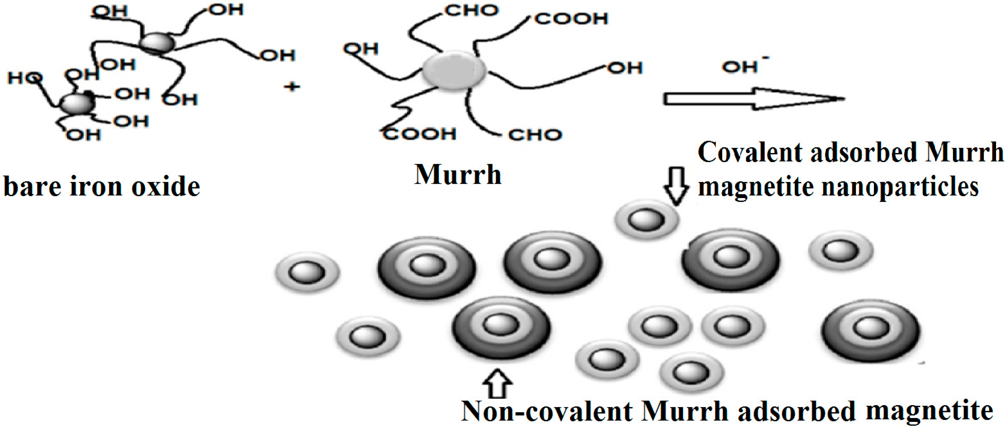
2.1. Production of Capped Iron Oxide Nanoparticles

Several types of iron oxides have been used in the field of magnetic NPs, including Fe3O4 (magnetite), α-Fe2O3 (hematite, weakly ferromagnetic or antiferromagnetic), γ-Fe2O3 (maghemite, ferrimagnetic), FeO (wüstite, antiferromagnetic), ε-Fe2O3 and β-Fe2O3. Among these magnetite and maghemite are popular candidates. The main method used to prepare iron oxide nanoparticles is the co-precipitation method. The main advantage of the co-precipitation process is that a large amount of nanoparticles can be synthesized at the same time, but control of the particle size distribution is limited, because the growth of the crystals is only controlled by kinetic factors. There are two stages during the co-precipitation method: a short burst of nucleation that occurs when the concentration of the species reaches critical supersaturation, followed by a slow growth of the nuclei by diffusion of the solutes to the surface of the crystal [23]. In this respect, these two stages should ideally be separated; i.e., nucleation during the growth period should be avoided to obtain monodisperse iron oxide nanoparticles [23]. The present work aims to prepare magnetite nanoparticles using only one iron compound as starting material, a limited number of additional chemical reagents, and a process that can be carried out under simple reaction conditions, preferably at room temperature, with easy work-up of the products obtained and by which highly pure magnetite can be obtained. This objective has been achieved with a method for preparing coated magnetite nanoparticles comprising: (a) preparing first an aqueous solution containing FeCl3; (b) preparing a second aqueous solution containing potassium iodide; (c) mixing the two solutions; (d) myrrh gum is then added as stabilizing agent; (e) hydrolyzing the mixture at a temperature of 45 °C by adjusting the pH to about 8.5–9 or above to obtain a precipitate; and (f) separating the precipitate from the solution. Thus, magnetite Fe3O4 nanoparticles were prepared according to the following equation [24,25]:
3Fe3+ + I → 2Fe3+ + Fe2+ + 1/2I2
The co-precipitation technique is determined to be the simplest and most efficient way to obtain iron oxide nanoparticles. Iron oxides (either magnetite Fe3O4, hematite α Fe2O3 or maghemite γFe2O3) are usually prepared by aging a stoichiometric mixture of ferrous and ferric salts in aqueous medium. The chemical reaction of Fe3O4 formation may be written as follows:
2Fe3+ + Fe2+ + 8OH → Fe3O4 + 4H2O
It should be expected that at a pH between 8 and 14 that the complete precipitation of Fe3O4 with a stoichiometric ratio of 2:1 (Fe3+/Fe2+) in a non-oxidizing oxygen environment according to the thermodynamics of the reaction in Equation (2) [26].
However, magnetite (Fe3O4) is not very stable and is sensitive to oxidation and is transformed into maghemite (γFe2O3) in the presence of oxygen. It was expected that during the synthesis the magnetite in the dry state would be oxidized to maghemite by air. It is known that ultrafine crystals of magnetite change with time from their characteristic black color to the brown of maghemite, even at room temperature [27]. Moreover, under basic conditions, the oxidation of magnetite involves oxidation-reduction recations that take place on the surface.
It was previously reported that when iodine is added to water, the following reaction takes place [28]:
I2 (1) + H2O (1) → OI (aq) + 2H+ (aq) + I (aq) (K = 2.0 × 10⁻¹³)
The I2 molecules and water molecules react to substances such as hypoiodite (OI) and I. The formation of the hypohalite ion (IO) in neutral aqueous solutions of iodine is negligible. In basic solutions, iodine is converted into iodide and iodate in a two stage reaction [29]:
I2 + 2OH → I + IO + H2O (K = 30)
3OI → 2I + IO3 (K = 1020)
The equilibrium of this reaction can move to either side, depending on the pH of the solution. It was suggested that [30] IO3 is physically adsorbed on iron oxide surfaces. The adsorption on the inner or outer surfaces of iron oxides increased with pH and ionic strength. It was detected that the adsorption of IO3 on iron oxide influenced the crystal growth of magnetite [30]. It was also suggested that the adsorption of iodate on the surfaces of iron oxides would produce more hydroxyl groups which are able to form covalent bonds with different functional groups such as the carboxylic, hydroxyl or amino groups of myrrh.
The addition of capping agents (carboxylate or hydroxyl-carboxylate ions) or organic surface complexing agents (polysaccharides) during the formation of magnetite can help control both the oxidation and the size of the nanoparticles. It was expected that the chelation of these organic ions on the iron oxide surface can either prevent nucleation and thus lead to larger particles or inhibit the growth of the crystal nuclei, leading to small nanoparticles. Myrrh as natural resinous exudate obtained from trees of certain Commiphora species of the Burseraceae family. It consists of a water-soluble gum, alcohol-soluble resins and volatile oil. The gum contains polysaccharides and proteins, while the volatile oil is composed of steroids, sterols and terpenes [31]. Myrrh is a mixture of polysaccharides contains different functional groups can be used as a stabilizing agent for magnetite nanoparticles to avoid their agglomeration in the present work. Thus, stabilization may be attributed to formation of a passivation layer and/or to electrostatic repulsion. The stabilization scheme can be illustrated by Scheme 1.
Scheme 1. Preparation of magnetite capped with myrrh.
Molecules 19 11263 g006
The adsorption on magnetite nanoparticles (MNPs) co-precipitated with myrrh was explained by more complex mechanisms involving intermolecular hydrogen bonding. In this work the covalent coupling of myrrh onto the MNPs, as well as the comparison with MNPs prepared by myrrh adsorption, have been studied. After creating the myrrh shell, it was expected that it would be possible to absorb another layer of myrrh by physical adsorption. It is expected that covalent bonds will be produced from the reaction between the hydroxyls of bare magnetite with the carboxylic, hydroxyl and aldehyde groups of myrrh, as represented in Scheme 1 [32].

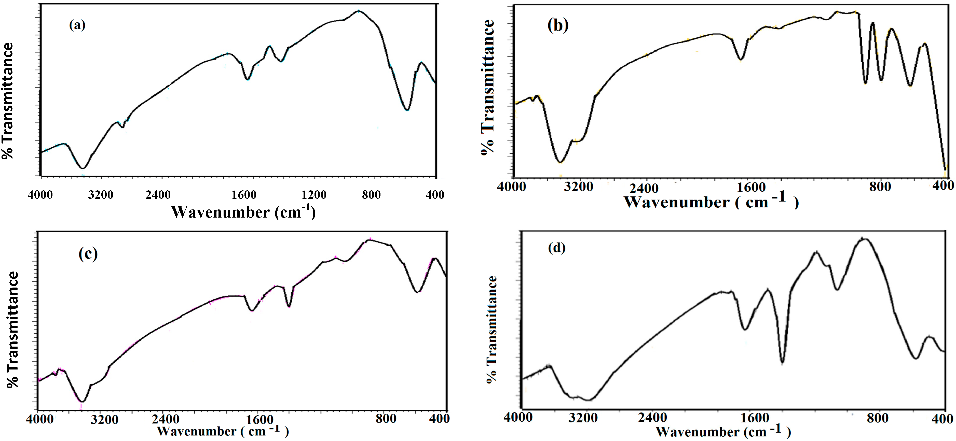
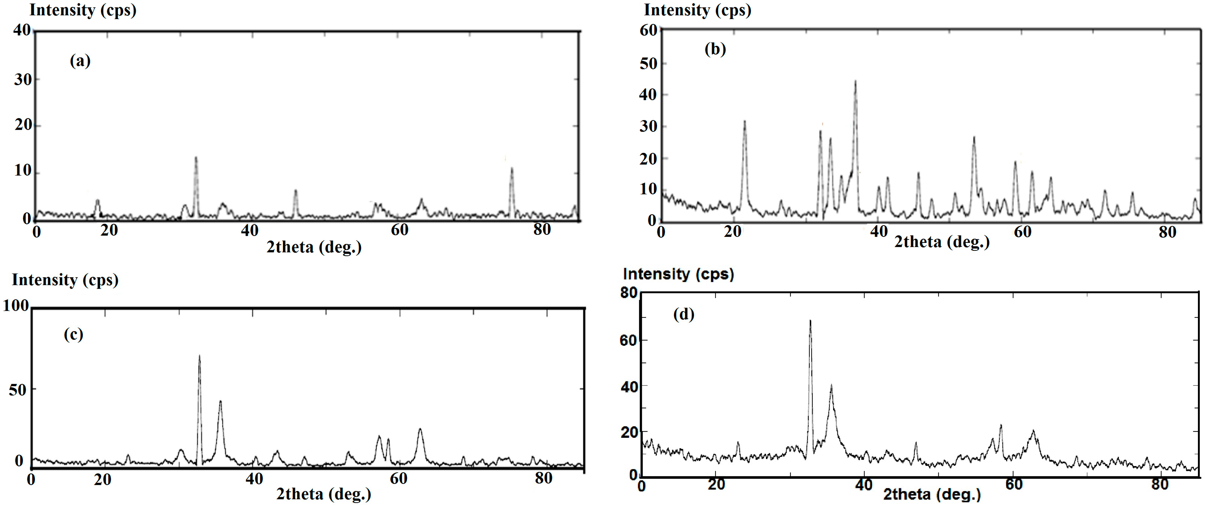
2.2. Characterization of Capped MNPs

FTIR analysis was used to characterize the iron oxide nanoparticles as both magnetite and maghemite or hematite. They can be easily differentiated on symmetry grounds. In this respect the FTIR spectra of iron oxide particles prepared in the presence or absence of iodine, and with myrrh as capping agent in the absence and presence of iodine are presented in Figure 1a–d, respectively.
Figure 1. FTIR spectra of the prepared magnetite (a) absence of iodine; (b) presence of iodine; (c) presence of myrrh; and (d) presence of myrrh and iodine.
Molecules 19 11263 g001
Wavenumbers assigned to the spinel structure are in the 800–400 cm−1 range [33,34,35,36,37]. All IR spectra indicating the presence of magnetite nanoparticles display a peak at around 570 cm−1. The FTIR spectrum of iron oxide in the presence of iodine (Figure 1b), showed new bands at 650 and 750 cm−1 indicating the presence of maghemite [38]. Moreover, the spectrum in Figure 1b indicated the presence of α-Fe2O3 (hematite) nanoparticles, clearly revealed by a large and intense band at 3,450 cm−1 that could be assigned to the structural OH groups. The presence of myrrh was indicated by the disappearance of the hematite and maghemite bands and appearance of broad bands at 3,450, 1,630 and 1,550 cm−1 which are attributed to OH, COOH and C=C stretching and indicated the functionalization of magnetite nanoparticles with the carboxylic or hydroxyl groups of myrrh.
The shift of the C=O vibration (symmetric stretching) of the COOH groups of myrrh to an intense band at about 1630 cm−1 and that of a band at 1050 cm−1 characteristic of CO single bonds for the ferromagnetic phase coated with myrrh reveals the myrrh is chemisorbed onto the nanoparticles as a carboxylate on the magnetite surface. Fe-O stretching of magnetite was also observed at 578 and 628 cm−1. These data indicated that the presence of myrrh prevents the formation of hematite and maghemite to produce stabilized magnetite nanoparticles. By assembling a monolayer of myrrh molecules on the nanoparticle surface an oil-based magnetic fluid is produced. The aqueous-based magnetic fluid is produced by assemblies of bilayers of myrrh on the surface of magnetite. Figure 1c,d of the magnetite/myrrh capped nanoparticles show bands at 2922 and 2852 cm−1 which were attributed to shifted asymmetric and symmetric CH2 stretch bands. This shift was caused by the field of the solid surface and it is an indication that the hydrocarbon chains that surround the particle were in a closed-packed, crystalline state. Furthermore, as we know, myrrh is a mixture of branched polysaccharides and glycoprotein containing numerous functional groups which may be responsible for the linking with magnetite via covalent bonding [31]. It can be concluded that the prepared nanoparticle is stabilized via electrostatic attraction between carboxylate groups of myrrh gum and surface hydroxyl groups of Fe3O4 which is due to the glycoprotein present in gum [39]. Also, the Myrrh has different functional groups usually bind to the surface of the nanocrystals and give rise to a steric hindrance to aggregation [40]. The capping group plays an important role in controlling the particle size. The X-ray diffractograms of four samples of magnetite are represented in Figure 2.
Figure 2. XRD diffractograms of the prepared magnetite (a) absence of iodine; (b) presence of iodine; (c) presence of myrrh; and (d) presence of myrrh and iodine.
Molecules 19 11263 g002
The data showed the presence of both α and γ phases of Fe2O3 (Figure 2b). The existence of maghemite and hematite is considered as topotactic with the [111] and [110] axes of maghemite corresponding to the [001] and [110] axes of hematite [41]. On the other hand, from the expanded view of the XRD profile (Figure 2b), it is found that the (104) and (110) Bragg reflections remain at the same position and shape was well maintained. The data showed a single peak for the tetrahedrally coordinated iron of magnetite (Fe3O4), and maghemite (γ-Fe2O3, whereas the octahedrally coordinated iron of hematite (α-Fe2O3) and the two oxyhydroxides, lepidocrocite (γ-FeOOH) and goethite (α-FeOOH) show a split peak. The data proved that the α-phase of iron oxide nanoparticles (α-Fe2O3, hematite) can be formed during the preparation process. This data indicated that the presence of iodine during the formation of magnetite leads to the formation of hematite and maghemite and hydroxyl groups on the surface of the iron oxide surfaces. There are many different natural structures of iron oxide, such as Fe3O4, α-Fe2O3, γFe2O3, Fe2O3, and β-FeOOH, all of which exhibit magnetic properties. X-ray diffraction patterns of the magnetite nanoparticles (Figure 2) revealed diffraction peaks at (110), (220), (311), (400), (422), and (511), which are the characteristic peaks of the Fe3O4 crystal with a cubic spinel structure. Herein, no peaks were detected which could be assigned to impurities such as γ- or α-ferric oxide. X-ray diffraction data of iron oxide nanoparticles are listed in Table 1. It is noticed that the diffraction data is closer to Fe3O4 than to γ-Fe2O3.
Table 1. Standard XRD data (for 2θ = 20–80°) for Fe3O4, γ-Fe2O3 and the experimental XRD data of Fe3O4 nanoparticles.
Standard Diffraction DataExperimental Data of Fe3O4
Fe3O4γ-Fe2O3
30.130.2829.99
35.4335.6935.51
43.0643.3543.23
53.4153.8753.59
56.9657.4257.12
62.5363.0362.81
73.9774.5674.27
Careful inspection of data illustrated in Figure 2 and listed in Table 1 indicates that only magnetite was formed during the capping of magnetite with myrrh in the presence or absence of iodine. These data indicated that the capping agent forms stable layers and prevents the transformation of magnetite into other iron oxides.
The mean size of Fe3O4 nanocrystallites was estimated using Scherrer’s formula [42]: DS = Kλ/β cos θ, where DS represents the crystallite size, θ represents the position of the Bragg peak and β is the half-width of the diffraction peak. The constant K in the Scherrer’s equation [42] depends on the morphology of the crystallites and K = 1 is usually assumed. The mean size of the crystallites determined from XRD measurements equal 10.3, 6.5, 38.1 and 18.3 ± 0.7 nm for iron oxides prepared in the absence or presence of iodine, presence of myrrh and presence of both myrrh and iodine, respectively. These data indicated that the presence of iodine during the preparation procedure of magnetite reduces the crystal mean size from 10.3 to 6.5 nm and reduces the size from 38.1 to 18.3 nm in the presence of myrrh as capping agent and iodine.

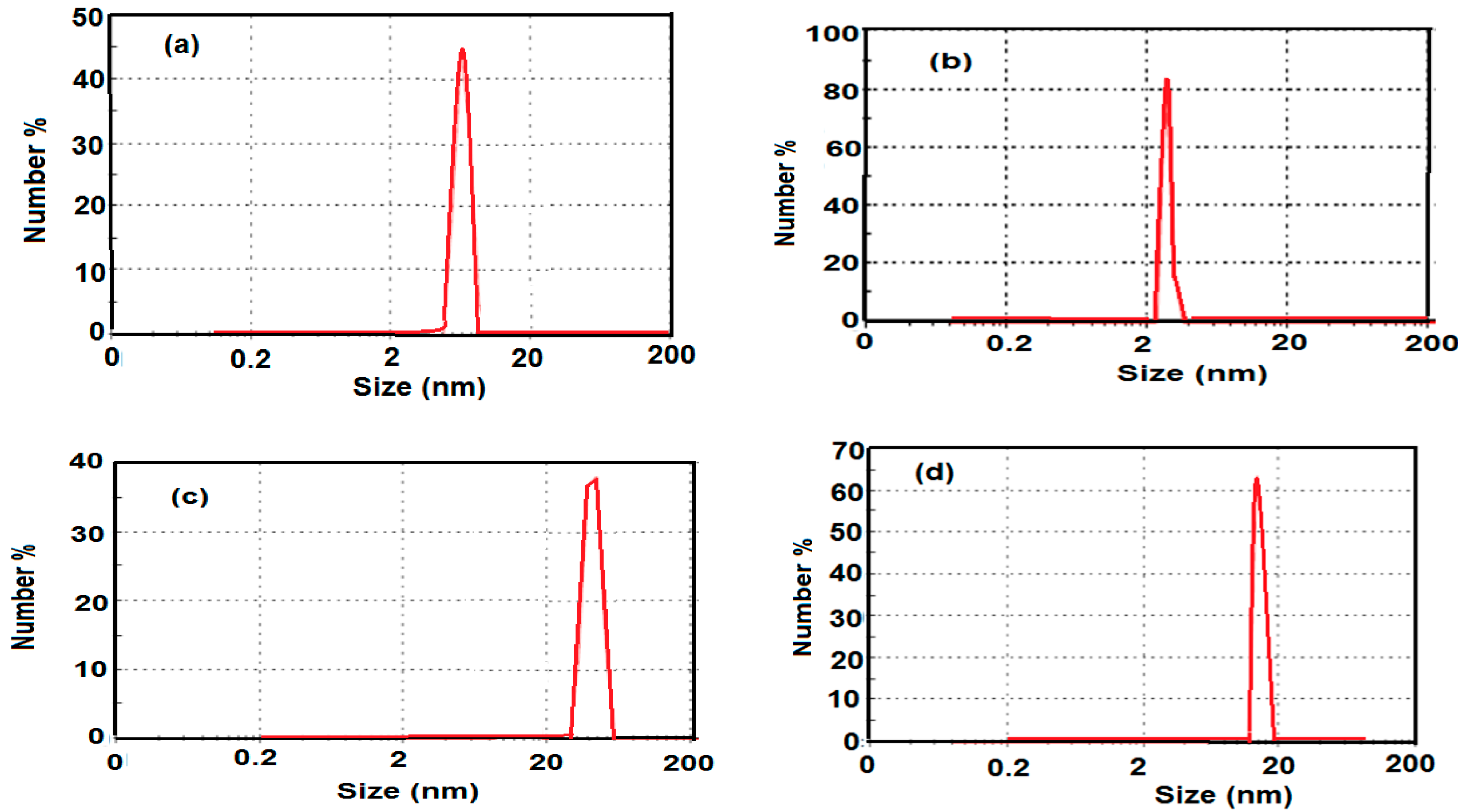
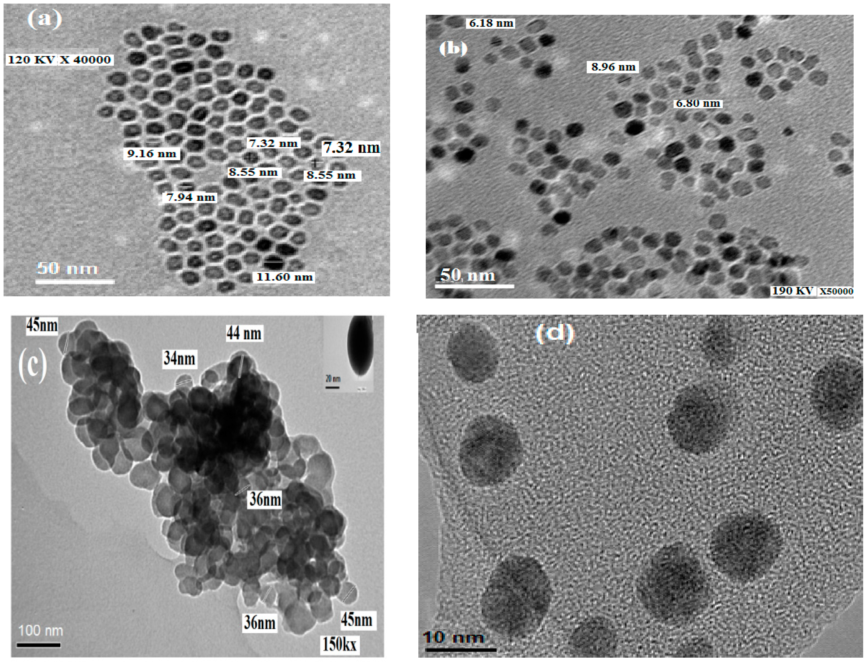
2.3. Morphologies and Particle Size of the Prepared MNPs

The magnetite nanoparticles obtained using myrrh as capping agent can easily be dispersed in aqueous media. The hydrodynamic diameter of iron oxides prepared in the presence or absence of both iodine and myrrh were measured by dynamic light scattering (DLS). The corresponding DLS graphs are presented in Figure 3. The diameter of the iron oxide nanoparticles in the absence and presence of iodine was between 12 and 5 nm, respectively. DLS measurements of capped magnetite nanoparticle in the absence and presence of iodine yielded a hydrodynamic average size of 53 and 18 nm for Fe3O4, respectively. The hydrodynamic diameter of the particles in water was found to increase in the absence of iodine, which can be attributed to the presence of the associated and hydrated layer of magnetite [43]. Dynamic light scattering (DLS) measurements revealed that the nanoparticles prepared in the presence of iodine and myrrh are highly monodisperse in aqueous media. These nanoparticles can also form stable dispersions in PBS buffer solution, which has the same pH value and ionic strength as physiological conditions. This colloid remains stable for three months without noticeable precipitation.
Figure 3. DLS of the prepared magnetite (a) absence of iodine; (b) presence of iodine; (c) presence of myrrh; and (d) presence of myrrh and iodine.
Molecules 19 11263 g003
The nature of the particles without aggregation was confirmed by TEM analysis in physiological buffer. Figure 4 shows transmission electron microscope (TEM) images of myrrh-coated and uncoated magnetite nanoparticles in the presence and absence of iodine. It could be clearly seen that the particles has a good distribution, with a lower percentage of agglomeration in the presence of both iodine and myrrh and the particle size is estimated to be 12 nm. The colloidal stability and distribution increased in the presence of myrrh and iodine with respect to bare Fe3O4 nanoparticles.
It is clearly observed that agglomeration of nanoparticles occurs to some extent. In any case the colloidal stability of magnetite nanoparticles increases due to successful stabilization of the Fe3O4 nanoparticles. The repulsion between the π electrons of double bonds and the carboxylate groups of myrrh prevents full aggregation of nanoparticles. Figure 4a,c show the TEM micrographs of Fe3O4 nanoparticles uncoated and coated with myrrh and in the absence of iodine. The micrographs show the presence of aggregations in contrast to the image obtained using myrrh. This behavior indicated that the iodine enhances the surface modification of magnetite nanoparticles due to an increase in the formation of OH groups which bond with the reactive functional groups of myrrh, as represented in the Scheme 1. It was also observed that the particle size was decreased by incorporation of iodine from 12.1 to 5.8 nm. Moreover, the presence of myrrh and iodine during the preparation of magnetite reduced the particle size from 55 nm to 18 nm and increased the dispersion of the particles as represented in Figure 4. These data indicated that iodine binds so tightly to the particle surface to iron oxide particles that it impedes particle growth. The particle size distributions appear to be quite narrow and the particle shape is reasonably spherical.
Figure 4. TEM micrographs of the prepared magnetite (a) absence of iodine; (b) presence of iodine; (c) presence of myrrh; and (d) presence of myrrh and iodine.
Molecules 19 11263 g004

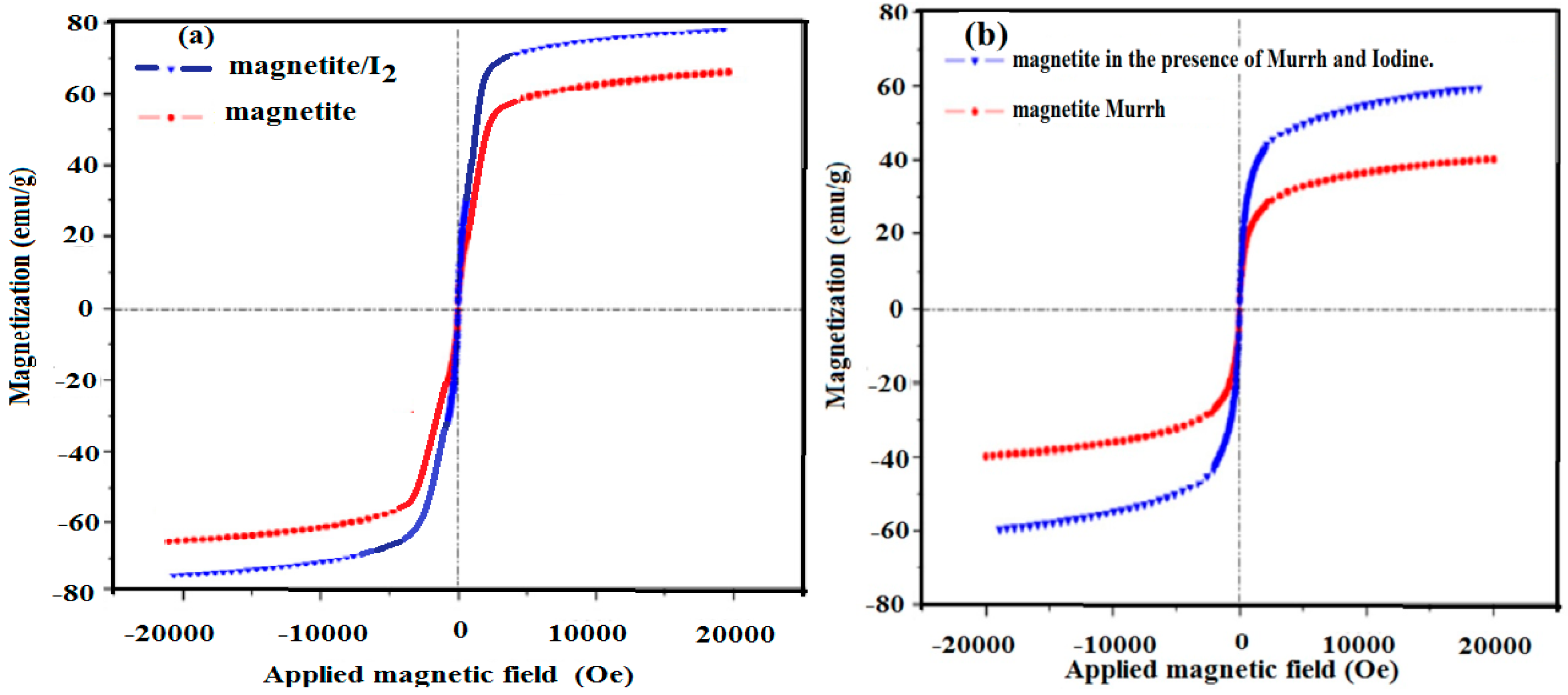
2.4. Magnetic Properties

It is important to study the effect of both particle size and the functionlization of magnetite on its magnetic properties. It is most important to measure the magnetic properties of the core/shell magnetic and superparamagnetism properties for practical applications. In this respect, Figure 5 shows VSM magnetization curves of Fe3O4 NPs prepared in the presence or absence of iodine and myrrh at room temperature. The data indicated that all the prepared magnetite nanoparticles exhibited typical superparamagnetic behavior indicated by not exhibiting hysteresis, remanence and coercivity [44]. It is well known that large saturation magnetization is a measure of the maximum magnetic strength [45]. The large saturation magnetization of iron oxide NPs prepared in the presence of iodine, absence of iodine, the presence of both iodine and myrrh and presence of myrrh were 78.6, 66.3, 59.6 and 39.9 emu/g, which is more than sufficient for magnetic separation with a conventional magnet for use in the environmental applications in water purification from oil or heavy metals. More details on magnetic properties and applications of the prepared magnetic nanoparticles will be discussed in the forthcoming publications. The saturation magnetization of the prepared magnetite particles, which was found to be equal to 78.6 emu/g, can be compared to the theoretical value of 92 emu/g. It was found that the value 84 emu/g was the highest value found in the literature for synthetic magnetite particles [46]. XRD data indicated that the magnetite prepared in the presence of iodine contains Fe3O4 and γ-Fe2O3 but it is more closer to Fe3O4. Moreover, the magnetite surfaces was functionalized with the hydroxyl groups.
Figure 5. Hysteresis loop of the (a) magnetite prepared in the absence of myrrh and (b) presence of myrrh at 298 K.
Molecules 19 11263 g005
The effect of the oxidation on the magnetic properties of magnetite was previously reported [47]. It was found that the oxidation of magnetite can transform magnetite into maghemite. Oxidation caused a decrease in the anisotropic barrier, resulting in less-significant magnetic interactions between particles [47]. In the present work it was noticed that the presence of hydroxyl-functionlized magnetite is the main reason for the increase in the saturation magnetization. The reduced magnetization may be related to the nature of the core or core surface [48]. The magnetization value of the magnetite in the presence of both iodine and myrrh is comparable to magnetite prepared in Gum Karaya [49]. The data show that myrrh as a natural product might have the same potential for applications as the widely used Gum Karaya and chitosan. The magnetization value of the magnetite in the presence of both iodine and myrrh is the same related to reported for magnetite nanoparticles of similar sizes and surface functionalization (60.1–67.9 for 10 nm particles) [50]. A decrease in magnetic properties with particle size has previously been observed [51]. The lower saturation magnetization for the magnetite prepared in the myrrh and absence of iodine than the magnetite prepared in the presence of both iodine and myrrh may be the combined result of its aggregation and the association of myrrh at the particle surface. The data indicated that the existence of the nonmagnetic shell would also lead to a decreased saturation magnetization.

3. Experimental Section

3.1. Materials

Anhydrous ferric chloride, potassium iodide, and ammonium hydroxide (25%) were purchased from Aldrich Chemical Co. (St. Louis, MO, USA) and used as reagent for preparation of magnetite. Phosphate buffer saline (PBS) buffer with pH 7.4 and ionic strength 0.1 was also purchased from Aldrich Chemical Co. (St. Louis, MO, USA). Myrrh gum is a commercial natural product with a yellowish red color extracted from the tree. Myrrh comes from a spiny deciduous thorny tree or shrub which grows as thickets to the height of 15 feet in Saudi Arabia desert regions. The soluble fraction of myrrh was extracted from ethanol/water (1:1 volume %) and used as capping agent after recovery using a vacuum evaporator.

3.2. Preparation of Magnetite Nanoparticles

3.2.1. Preparation of Magnetite Nanoparticles (MNPs)

Aqueous solution of ferric chloride was prepared by dissolving anhydrous FeCl3 (40 g, 0.24 mol) in distilled water (300 mL) to prepare an aqueous solution A. Next, potassium iodide (13.2 g, 0.08 mol) was dissolved in distilled water (50 mL) to prepare an aqueous solution B. The aqueous solutions A and B are then mixed together at room temperature, stirred and allowed to reach equilibrium for one hour while bubbling with pure N2 to keep the mixture oxygen free throughout the preparation procedure. A precipitate formed is filtered out, washed with distilled water, dried at vacuum at 30 °C and weighed to determine the reaction yield (10.1 g, i.e., 86.7% yield). The filtrate (including washings) was then heated to a temperature of 45 °C, and hydrolyzed using 200 mL of 25% ammonia solution which was added dropwise with stirring while bubbling with pure N2 to keep the mixture oxygen free throughout the preparation procedure. Mixing was continued until complete precipitation of black magnetite is achieved. The reaction was continued at the reaction temperature with stirring for 4 h. The precipitate was then left to settle, filtered, washed with distilled water, dried at vacuum at 30 °C (the precipitate should dry without heating) and weighed. The percentage yield of reaction was 90.5%. The same procedure was repeated but the precipitate of the solutions A and B was not filtered from the reaction mixture. The brownish black precipitate formed as the product was washed five times using ethanol and the percentage yield of the reaction was 95.5%.

3.2.2. Preparation of Magnetite Capped with Myrrh

The procedure for preparation of magnetite capped with myrrh was as follows: anhydrous FeCl3 (40 g) was dissolved in distilled water (300 mL) to prepare an aqueous solution A. Next, potassium iodide (13.2 g, 0.08 mol) was dissolved in distilled water (50 mL) to prepare an aqueous solution B. The aqueous solutions A and B were then mixed together at room temperature, stirred and allowed to reach equilibrium for one hour while bubbling with pure N2 to keep the mixture oxygen free throughout the preparation procedure. The precipitate formed is filtered out, washed with distilled water, dried at vacuum at 30 °C and weighed to determine the reaction yield (90% yield). The filtrate (including washings) was then heated to 45 °C. Myrrh (10 g) was solubilized in ethanol-water solvent (100 mL, 1:1 vol%) and added dropwise to the reaction mixture at the same time as 25% ammonia solution (200 mL). The reaction mixture was stirred and bubbled with pure N2 to keep oxygen free throughout the preparation procedure. The reaction was continued at the reaction temperature with stirring for 4 h until complete precipitation of black magnetite was achieved.. The precipitate was then left to settle, filtered, washed with distilled water, dried under vacuum at 30 °C (the precipitate was dried without heating) and weighed. The percentage yield was 95.9%.
The same procedure was repeated without filtration of the reaction products between solutions A and B. The final product was washed with ethanol and air dried to give a reaction yield percentage of 99.5%.

3.3. Characterization of Nanoparticles

FTIR spectra were analyzed with a Nicolet FTIR spectrophotometer (city, state abbrev if US, country) using KBr in the wavenumber range of 4000–500 cm−1 with a resolution accuracy of 4 cm−1. All samples were ground and mixed with KBr and then pressed to form pellets.
X-ray powder diffraction (XRD) patterns were recorded using a D/max 2550 V X-ray diffractometer (X’Pert, Philips, Eindhoven, The Netherlands).
Transmission electron microscopy (TEM) micrographs were taken with a JEOL JEM-2100F (JEOL, Tokyo, Japan). A few drops of magnetite nanoparticle solution were diluted into 1 mL of ethanol, and the resulting ethanol solution was placed onto a carbon coated copper grid and allowed to evaporate. HR-TEM images of the nanocomposites were recorded using a JEM-2100F (JEOL) at an acceleration voltage of 200 kV. All images were acquired at a nominal magnification value of 66,000×. The magnification of the microscope was calibrated with a NIST reference material (gold nanoparticles) which has reference values of the mean particle diameter as determined by TEM. This diameter is traceable to the SI meter as realized at NISTP. Calibration images were recorded at both sessions and combined for the final analysis of the scale factor.
Samples for dynamic light scattering (DLS) were prepared by diluting several drops of the magnetite nanoparticle solution into 2 mL of water under vigorous stirring. The DLS measurements were performed on a Brookhaven Instruments system (Santa Barbara, CA, USA) with a 514.5 nm argon ion laser (model 85 Lexel Laser) as the light source.
Magnetic properties of the prepare magnetite nanomaterials were analyzed using a vibrating sample magnetometer (LDJ9600, VSM, LDJ Electronics Co., Troy, MI, USA).

4. Conclusions

New stable colloid magnetite nanoparticles coated with myrrh were prepared with high purity and yield at low temperature using ferric chloride as precursor. Myrrh bonded on to MNPs with covalent bonds followed by a bilayer adsorption mechanism as shell produced water dispersed MNPs from the reaction between hydroxyl groups on bare magnetite with the carboxylic, hydroxyl and aldehyde groups of myrrh. It was also observed that the particle size was decreased from 12.1 to 5.8 nm by incorporation of iodine. Moreover, the presence of myrrh and iodine during the preparation of magnetite reduced the particle size from 55 nm to 18 nm and increased the dispersion of the particles. These behaviors indicated that iodine binds so tightly to the particle surface to iron oxide particles that it impedes particle growth. The particle size distributions appear to be quite narrow and the particle shape is reasonably spherical.

Acknowledgments

This project was supported by King Saud University, Deanship of Scientific Research, College of Sciences, Research Center.

Author Contributions

A.M.A. suggested the idea and contributed the discussion, A.A.-L. contributed in discussion and S.A.A.-H. Performed the experimental work.

Conflicts of Interest

The authors declare no conflict of interest.

References

  1. Baker, R.R.; Mather, J.G.; Kennaugh, J.H. Magnetic bones in human sinuses. Nature 1983, 301, 79–80. [Google Scholar]
  2. Basavaiah, K.V.; Rao, P.A. One-pot synthesis of superparamagnetic polyaniline microtubes and magnetite nanoparticles via self-assembly method. Curr. Nanosci. 2012, 8, 215–220. [Google Scholar] [CrossRef]
  3. Fidale, L.C.; Nikolajski, M.; Rudolph, T.; Dutz, S.; Schacher, F.H.; Heinze, T. Hybrid Fe3O4@amino cellulose nanoparticles in organic media—Heterogeneous ligands for atom transfer radical polymerizations. J. Colloid. Interface Sci. 2013, 390, 25–33. [Google Scholar] [CrossRef]
  4. Sun, S.; Zheng, H.; Robinson, D.B.; Raoux, S.; Rice, P.M.; Wang, S.X.; Li, G. Monodisperse MFe2O4(M = Fe, Co, Mn) nanoparticles. J. Am. Chem. Soc. 2004, 126, 273–279. [Google Scholar]
  5. Roca, A.G.; Morales, M.P.; O’Grady, K.; Serna, C.J. Structural and magnetic properties of uniform magnetite nanoparticles prepared by high temperature decomposition of organic precursors. Nanotechnology 2006, 17, 2783–2788. [Google Scholar] [CrossRef]
  6. Bourlinos, A.B.; Bakandritsos, A.; Georgakilas, V.; Tzitzios, V.; Petridis, D. Facile synthesis of capped γ-Fe2O3and Fe3O4nanoparticles. J. Mater Sci. 2006, 41, 5250–5256. [Google Scholar] [CrossRef]
  7. Yue, F.J.; Wang, S.; Lin, L.; Ding, H.F.; Wu, D. Effects of ferromagnet-molecule chemical bonding on spin injection in an Fe3O4-molecule granular system. J. Phys. D Appl. Phys. 2012, 45. [Google Scholar] [CrossRef]
  8. Unal, B.; Toprak, M.S.; Durmus, Z.; Sozeri, H.; Baykal, A. Synthesis, structural and conductivity characterization of alginic acid-Fe3O4 nanocomposite. J. Nanopart.Res. 2010, 8, 3039–3048. [Google Scholar]
  9. Chockalingam, A.M.; Babu, H.K.R.; Chittor, R.; Tiwari, J.P. Gum arabic modified Fe3O4 nanoparticles cross linked with collagen for isolation of bacteria. J. Nanobiotechnol. 2010, 8. [Google Scholar] [CrossRef]
  10. Roque, A.C.A.; Bicho, A.; Batalha, I.L.; Cardoso, A.S.; Hussain, A. Biocompatible and bioactive gum Arabic coated iron oxide magnetic nanoparticles. J.Biotechnol. 2009, 144, 313–320. [Google Scholar] [CrossRef]
  11. Kannan, R.; Rahing, V.; Cutler, C.; Pandrapragada, R.; Katti, K.K.; Kattumuri, V.; Robertson, J.D.; Casteel, S.J.; Jurisson, S.; Smith, C.; et al. Nanocompatible chemistry toward fabrication of target-specific gold nanoparticles. J. Am. Chem. Soc. 2006, 128, 11342–11343. [Google Scholar] [CrossRef]
  12. Bandyopadhyaya, R.; Native-Roth, E.; Regev, O.; Yerushalmi-Rozen, R. Stabilization of individual carbon nanotubes in aqueous solutions. Nano Lett. 2002, 2, 25–28. [Google Scholar] [CrossRef]
  13. Park, C.; Lim, K.H.; Kwon, D.; Yoon, T.H. Biocompatible quantum dot nanocolloids stabilized by gum Arabic. Bull. Korean Chem. Soc. 2008, 29, 1277–1279. [Google Scholar] [CrossRef]
  14. Wang, X.; Zhuang, J.; Peng, Q.; Li, Y.D. A general strategy for nanocrystals synthesis. Nature 2005, 437, 121–124. [Google Scholar] [CrossRef]
  15. Nam, J.M.; Thaxton, C.S.; Mirkin, C.A. Nanoparticle-based bio-bar codes for the ultrasensitive detection of proteins. Science 2003, 301, 1884–1886. [Google Scholar] [CrossRef]
  16. Fried, T.; Shemer, G.; Markovich, G. Ordered Two-dimensional arrays of ferrite nanoparticles. Adv. Mater. 2001, 13, 1158–1161. [Google Scholar] [CrossRef]
  17. Yu, M.K.; Jeong, Y.Y.; Park, J.; Park, S.; Kim, J.W.; Min, J.J.; Kim, K.; Jon, S. Drug-loaded superparamagnetic iron oxide nanoparticles for combined cancer imaging and therapy in vivo. Angew. Chem. Int. Ed. 2008, 47, 5362–5365. [Google Scholar] [CrossRef]
  18. Park, J.; An, K.; Hwang, Y.; Park, J.G.; Noh, H.J.; Kim, J.Y.; Park, J.H.; Hwang, N.M.; Hyeon, T. Ultra-large-scale syntheses of monodisperse nanocrystals. Nat. Mater. 2004, 3, 891–895. [Google Scholar] [CrossRef]
  19. Park, S.J.; Kim, S.; Lee, S.; Khim, Z.G.; Char, K.; Hyeon, T. Synthesis and magnetic studies of uniform iron nanorods and nanospheres. J. Am. Chem. Soc. 2000, 122, 8581–8582. [Google Scholar] [CrossRef]
  20. Beketov, I.V.; Safronov, A.P.; Medvedev, A.I.; Alonso, J.; Kurlyandskaya, G.V.; Bhagat, S.M. Iron oxide nanoparticles fabricated by electric explosion of wire: Focus on magnetic nanofluids. AIP Adv. 2012, 2. [Google Scholar] [CrossRef]
  21. Safronov, A.P.; Beketov, I.V.; Komogortsev, S.V.; Kurlyandskaya, G.V.; Medvedev, A.I.; Leiman, D.V.; Larrañaga, A.; Bhagat, S.M. Spherical magnetic nanoparticles fabricated by laser target evaporation. AIP Adv. 2013, 3. [Google Scholar] [CrossRef]
  22. Grossman, J.H.; McNeil, S.E. Nanotechnology in Cancer Medicine. Phys. Today 2012, 65. [Google Scholar] [CrossRef]
  23. Laurent, S.; Forge, D.; Port, M.; Roch, A.; Robic, C.; Vander Elst, L.; Muller, R.N. Magnetic iron oxide nanoparticles: Synthesis, stabilization, vectorization, physicochemical characterizations, and biological applications. Chem. Rev. 2008, 108, 2064–2110. [Google Scholar] [CrossRef]
  24. Atta, A.M.; Dyab, A.K.F. Coated Magnetite Nanoparticles, Method for the Preparation thereof and Their Use. EP 13167616.5, 18 March 2013. [Google Scholar]
  25. Khalil, M.I. Process for Preparing Magnetic (Fe3O4) and Derivatives thereof. EP 2505558 A1, 16 January 2013. [Google Scholar]
  26. Jolivet, J.P.; Chaneac, C.; Tronc, E. Iron oxide chemistry. From molecular clusters to extended solid networks. Chem. Commun. 2004, 5, 481–487. [Google Scholar] [CrossRef]
  27. Cornel, R.M.; Schwertmann, U. The Iron Oxides, Structure, Properties, Reactions and Uses; VCH: Weinheim, Germany, 1996. [Google Scholar]
  28. Madrakian, T.; Afkhami, A.; Zolfigo, M.A.; Ahmadi, M.; Koukabi, N. Application of modified silica coated magnetite nanoparticles for removal of iodine from water samples. Nano-Micro Lett. 2012, 4, 57–63. [Google Scholar]
  29. Cotton, F.A.; Wilkinson, G. Advanced Inorganic Chemistry, 5th ed.; John Wiley & Sons: New York, NY, USA, 1988. [Google Scholar]
  30. Nagata, T.; Fukushi, K. Prediction of iodate adsorption and surface speciation on oxides by surface complexation modeling. Geochim. Cosmochim. Acta 2010, 74, 6000–6013. [Google Scholar] [CrossRef]
  31. Hanuša, L.O.; Řezankab, T.; Dembitsky, V.M.; Moussaieff, A. Myrrh–Commiphora Chemistry. Biomed. Pap. Med. Fac. Univ. Palacky Olomouc. Czech Repub. 2005, 149, 3–28. [Google Scholar] [CrossRef]
  32. Nguyen, T.K.; Thanha, A.; Luke, A.W. Green Functionalisation of nanoparticles for biomedical applications. Nano Today 2010, 5, 213–230. [Google Scholar] [CrossRef]
  33. Tartaj, P.; Morales, M.P.; Veintemillas-Verdaguer, S.; Gonzales-Carreno, T.; Serna, J.C. The preparation of magnetic nanoparticles for applications in biomedicine. J. Phys. D Appl. Phys. 2003, 36. [Google Scholar] [CrossRef]
  34. Morales, M.P.; Pecharroman, C.; Gonzales-Carreno, T. Structural characteristics of uniform γ-Fe2O3particles with different axial (length/width) ratios. J. Solid StateChem. 1994, 108, 158–163. [Google Scholar] [CrossRef]
  35. Belin, T.; Guigue-Millot, N.; Caillot, T.; Aymes, D.; Niepce, J.C. Influence of grain size, oxygen stoichiometry, and synthesis conditions on the γ-Fe2O3 vacancies ordering and lattice parameters. J. Solid State Chem. 2002, 163, 459–465. [Google Scholar] [CrossRef]
  36. Morales, M.P.; Andres-Verges, M.; Veintemillas-Verdaguer, S.; Montero, M.I.; Serna, C.J. Structural effects on the magnetic properties of γ-Fe2O3 nanoparticles. J. Magn. Magn. Mater. 1999, 203, 146–148. [Google Scholar] [CrossRef]
  37. Morales, M.P.; Veintemillas, S.; Montero, M.I.; Serna, C.J.; Roig, A.; Casas, L.I.; Martinez, B.; Sandiumenge, F. Surface and internal spin canting in γ-Fe2O3 nanoparticles. Chem. Mater. 1999, 11, 3058–3064. [Google Scholar] [CrossRef]
  38. Daou, T.J.; Greneche, J.M.; Pourroy, G.; Buathong, S.; Derory, A.; Ulhaq-Bouillet, C.; Donnio, B.; Guillon, D.; Begin-Colin, S. Coupling agent effect on magnetic properties of functionalized magnetite-based nanoparticles. Chem. Mater. 2008, 20, 5869–5875. [Google Scholar] [CrossRef]
  39. Shaswant, B.S.; Dong-Hwang, C. Fast removal of copper ions by gum Arabic modified nano-adsobent. J. Hazard. Mater. 2007, 147, 792–799. [Google Scholar] [CrossRef]
  40. Stolnik, S.; Illum, L.; Davis, S.S. Long circulating microparticulate drug carriers. Adv. Drug Deliver. Rev. 1995, 16, 195–214. [Google Scholar] [CrossRef]
  41. Feitknecht, W.; Mannweiler, U. Der mechanismus der umwandlung von γ-zu-α-eisensesquioxid. Helv.Chim. Acta 1967, 50, 570–581. [Google Scholar] [CrossRef]
  42. Scherrer, P. Bestimmung der Grösse und der inneren Struktur von Kolloidteilchen mittels Röntgensrahlen. Available online: http://www.sajs.co.za/sites/default/files/publications/pdf/Kroon_Sci%20Corr.pdf (accessed on 21 July 2014).
  43. Wan, J.; Cai, W.; Meng, X.; Liu, E. Monodisperse water-soluble magnetite nanoparticles prepared by polyol process for high-performance magnetic resonance imaging. Chem. Commun. 2007, 47, 5004–5006. [Google Scholar]
  44. Zhao, X.; Shi, Y.; Wang, T.; Cai, Y.; Jiang, G. Preparation of silica-magnetite nanoparticle mixed hemimicelle sorbents for extraction of several typical phenolic compounds from environmental water samples. J. Chromatogr. A 2008, 1188, 140–147. [Google Scholar] [CrossRef]
  45. Tan, C.J.; Tong, Y.W. Preparation of superparamagnetic ribonuclease A surface-imprinted submicrometer particles for protein recognition in aqueous media. Anal. Chem. 2007, 79, 299–306. [Google Scholar] [CrossRef]
  46. Dresco, P.A.; Zaitsev, V.S.; Gambino, R.J.; Chu, B. Preparation and properties of magnetite and polymer magnetite nanoparticles. Langmuir 1999, 15, 1945–1951. [Google Scholar] [CrossRef]
  47. Rebodos, R.L.; Vikesland, P.J. Effects of oxidation on the magnetization of nanoparticulate magnetite. Langmuir 2010, 26, 16745–16753. [Google Scholar] [CrossRef]
  48. Burke, N.A.D.; Stöver, H.D.H.; Dawson, F.P. Magnetic nanocomposites: Preparation and characterization of polymer-coated iron nanoparticles. Chem. Mater. 2002, 14, 4752–4761. [Google Scholar] [CrossRef]
  49. Vellora, V.; Padil, T.; Rouha, M.; Herník, M. Hydrocolloid-stabilized magnetite for efficient removal of radioactive phosphates. BioMed Res. Int. 2014, 2014. [Google Scholar] [CrossRef]
  50. Trudel, S.; Jones, C.H.W.; Hill, R.H. Magnetic properties of nanocrystallineiron oxide/amorphous manganese oxide nanocomposite thin films preparedviaphotochemical metal-organic deposition. J. Mater. Chem. 2007, 17, 2206–2218. [Google Scholar] [CrossRef]
  51. Morales, M.P.; Veintemillas-Verdaguer, S.; Serna, C.J. Magnetic properties of uniform γ-Fe2O3nanoparticles smaller than 5nm prepared by laser pyrolysis. J. Mater. Res. 1999, 14, 3066–3072. [Google Scholar] [CrossRef]
  • Sample Availability: Samples of the compounds magnetite nanoparticles are available from the authors.

