Article

Biological Activities and Phytochemical Profiles of Extracts from Different Parts of Bamboo (Phyllostachys pubescens)

1
Department of Agro-environmental Sciences, Faculty of Agriculture, Kyushu University, 6-10-1 Hakozaki, Higashi-ku, Fukuoka, 812-8581, Japan
2
Department of Biological and Environmental Chemistry, Kinki University, Kayanomori 11-6, Iizuka, Fukuoka, 820-8555, Japan
3
Fukuoka Prefecture Institute of Agricultural and Forest Resources, 1438-2, Toyota, Yamamoto-town, Kurume, Fukuoka, 839-0827, Japan
4
International College of Arts and Sciences, Fukuoka Women's University, Fukuoka 813-8529, Japan
5
Department of Materials Science and Chemical Engineering, Kitakyushu National College of Technology, 5-20-1 Shii, Kokuraminami-ku, Kitakyushu, Fukuoka, 802-0985, Japan
*
Author to whom correspondence should be addressed.
Authors contributed equally to this work.
Molecules 2014, 19(6), 8238-8260; https://doi.org/10.3390/molecules19068238
Submission received: 8 April 2014 / Revised: 16 May 2014 / Accepted: 23 May 2014 / Published: 18 June 2014

Abstract

:
Besides being a useful building material, bamboo also is a potential source of bioactive substances. Although some studies have been performed to examine its use in terms of the biological activity, only certain parts of bamboo, especially the leaves or shoots, have been studied. Comprehensive and comparative studies among different parts of bamboo would contribute to a better understanding and application of this knowledge. In this study, the biological activities of ethanol and water extracts from the leaves, branches, outer culm, inner culm, knots, rhizomes and roots of Phyllostachys pubescens, the major species of bamboo in Japan, were comparatively evaluated. The phytochemical profiles of these extracts were tentatively determined by liquid chromatography-mass spectrometry (LC-MS) analysis. The results showed that extracts from different parts of bamboo had different chemical compositions and different antioxidative, antibacterial and antiallergic activities, as well as on on melanin biosynthesis. Outer culm and inner culm were found to be the most important sources of active compounds. 8-C-Glucosylapigenin, luteolin derivatives and chlorogenic acid were the most probable compounds responsible for the anti-allergy activity of these bamboo extracts. Our study suggests the potential use of bamboo as a functional ingredient in cosmetics or other health-related products.

1. Introduction

Bamboo is well known for its extensive use. Besides being used in building construction, its roots and leaves have been used medicinally. Studies have revealed that bamboo leaves have antioxidant, anticancer and antibiotic properties [1,2]. In previous studies, various active compounds, such as flavones, glycosides, phenolic acids, coumarin lactones, anthraquinones and amino acids, have been isolated from the leaves [3,4,5,6,7]. 2, 6-Dimethoxy-p-benzoquinone isolated from the skin of bamboo trees and two chitin-binding peptides (Pp-AMP1 and Pp AMP2) isolated from bamboo shoots were found to have antibiotic activities [8,9]. Stigmasterol and dihydrobrassicasterol isolated from the skin of bamboo shoot showed antibacterial activity [10], as well as tricin and taxifolin [11].
Phyllostachys pubescens (P. pubescens) is the major species of bamboo in Japan, which is widely distributed through the country. In fact, how to stop its further spread is a problem in Japan [12]. Hence, new applications of bamboo in various commercial industries are being explored. Several studies have been performed to demonstrate the use of P. pubescens in terms of its biological activity. These studies have mainly focused on extracts from specific parts of P. pubescens. For example, the antioxidant activity of leaves [13] and shoots [14], antiallegic [15] and anticancer [16] activities of leaves and branches and antibacterial activities of stems [8], shoots [9], and shoot skins [17]. However, comprehensive and comparative studies of extracts from all parts of bamboo using the same extraction solvent have not been done. In this study, P. pubescens was separated into 10 parts, including leaves, branches, outer culm (5 m and 1 m above the ground, respectively), inner culm (5 m and 1 m above the ground, respectively), knots (5 m and 1 m above the ground, respectively), rhizomes and roots (Figure 1). These parts of bamboo were extracted by ethanol and hot water. All resulting extracts were subjected to four assays for bioactivities that are usually of interest to the cosmetics industry. They are melanin synthesis assay, antioxidant assay, antibacterial assay and antiallergic assay. At the same time, the chromatographic profiles of these extracts were determined and their components were partially identified using liquid chromatography-mass spectrometry (LC-MS). Through these tests and analysis, the potential use of bamboo in health-related industries, especially in cosmetics industry was evaluated.

2. Results and Discussion

In the present study, the ethanol and hot water extracts of various parts of P. pubescens (Figure 1) were examined for several biological activities suing the melanin biosynthesis assay (Table 1), antioxidant assay (Table 2), antibacterial assay (Table 3) and immunoglobulin E (IgE) production assay (Table 4). Their phytochemical profiles were also investigated through LC-MS analysis (Table 5, Table 6 and Figures S1–S3).
Figure 1. Parts of P. pubescens used in present study. P. pubescens plants were separated into leaves, branches, outer culm, inner culm, knots, rhizomes and roots. Outer culm, inner culm and knot samples were obtained separately at the height of 5.0 ± 0.3 and 1.0 ± 0.3 meter above ground level.
Molecules 19 08238 g001

2.1. Activity on Melanin Biosynthesis

Table 1 shows the effect of the ethanol extracts and the hot water extracts of P. pubescens on melanin biosynthesis and cell proliferation of B16 melanoma cells. After treating with different concentrations of the extract for 3 days, B16 melanoma cells were examined for cell viability (CV) and melanin content (MC). The cell viability was measured by the classic MTT assay, while the melanin content was determined by the absorbance at 405 nm. One important concept when selecting bioactive extracts that modulate skin pigmentation for cosmetics is that they should have minimal effects on cell proliferation and/or survival.
As shown in Table 1, the ethanol extracts of branches, and outer culm (5 m, 1 m) showed melanin biosynthesis inhibitory activity (Type A) in a dose-dependent manner. The ethanol extract of branches inhibited biosynthesis of melanin at 120 μg/mL (CV was 86.3% and MC was 56.0%). The ethanol extract of the outer culm at 5 m showed activity at 120 and 60 μg/mL (CVs were 86.9% and 98.9%; MCs were 44.4 and 72.2%, respectively). The ethanol extract of the outer culm at 1 m showed activity at 120 and 60 μg/mL (CVs were 124 and 109%; MCs were 49.5 and 79.8%, respectively). On the other hand, the ethanol extracts of the inner culm at both heights (5 m, 1 m), knots at 1 m, rhizomes and roots showed selective melanin biosynthesis-stimulating activity (Type B). The ethanol extract of the inner culm at 5 m stimulated biosynthesis of melanin at 60 μg/mL (CV was 88.5% and MC was 109%). Also, the ethanol extract of the inner culm at 1 m stimulated biosynthesis of melanin at 120, 60 and 20 μg/mL (CVs were 106%, 106% and 103%; MCs were 142%, 151% and 134%, respectively). The ethanol extract of knots at 1 m showed activity at 120 and 60 μg/mL (CVs were 100% and 97.0%; MCs were 133% and 119%, respectively).
Table 1. Effect of the (a) ethanol extracts and (b) the hot water extracts of P. pubescens on melanin biosynthesis and cell proliferation of B16 melanoma cells.
(a)
(a)
PartEthanol extract
120 μg/mL60 μg/mL20 μg/mL
CVMCTypeCVMCTypeCVMCType
Leaf91.2 ± 1.06105 ± 5.52-96.7 ± 8.1099.0 ± 8.53-101 ± 1.98103 ± 1.32-
Branch86.3 ± 1.7156.0 ± 7.90A,C91.0 ± 2.7475.6 ± 3.52-90.2 ± 0.8484.6 ± 2.09-
Outer culm (5 m)86.9 ± 10.144.4 ± 5.64A,C98.9 ± 0.9872.2 ± 1.55A112.9 ± 4.27104 ± 2.34-
Outer culm (1 m)124 ± 9.0849.5 ± 5.38A109 ± 1.2079.8 ± 3.19A124 ± 9.08112 ± 2.97-
Inner culm (5 m)98.8 ± 2.09110 ± 2.67-88.5 ± 10.5109 ± 9.68B,C98.0 ± 1.09106 ± 8.01-
Inner culm (1 m)106 ± 1.80142 ± 2.87B106 ± 5.63151.9 ± 9.59B103 ± 3.32134 ± 3.59B
Knot (5 m)93.6 ± 5.51101 ± 9.90-90.5 ± 1.96104 ± 0.69-93.4 ± 4.86101 ± 3.27-
Knot (1 m)100 ± 6.22133 ± 20.0B97.0 ± 7.14119 ± 2.97B96.8 ± 6.46107 ± 6.67-
Rhizome120 ± 2.97137 ± 19.0-118 ± 1.88144 ± 21.6B114 ± 4.44121 ± 4.2-
Root91.5 ± 2.68118 ± 6.78B88.6 ± 1.2126 ± 5.53B, C104 ± 10.1111 ± 12.7-
(b)
(b)
PartHot water extract
120 μg/mL60 μg/mL20 μg/mL
CVMCTypeCVMCTypeCVMCType
Leaf109 ± 9.06114 ± 7.71-107 ± 8.35108 ± 11.1-109 ± 9.06143.8 ± 2.22B
Branch79.7 ± 10.784.8 ± 8.16C77.3 ± 3.6787.2 ± 1.39C83.1 ± 7.42109 ± 2.73B,C
Outer culm (5 m)121 ± 6.8378.9 ± 6.10A125 ± 3.7693.3 ± 13.0A138 ± 2.2294.6 ± 6.76A
Outer culm (1 m)118 ± 2.81125 ± 16.5-114 ± 2.99104 ± 16.5-116 ± 6.16117 ± 14.8-
Inner culm (5 m)97.5 ± 7.10104 ± 6.62-114 ± 9.85105 ± 15.2-96.2 ± 9.62101 ± 2.82-
Inner culm (1 m)120 ± 5.10113 ± 17.6-110 ± 15.0108 ± 0.28-113 ± 0.76102 ± 1.97-
Knot (5 m)79.1 ± 6.2394.8 ± 5.69C85.8 ± 2.81109 ± 9.61B,C77.7 ± 1.65101 ± 1.86B,C
Knot (1 m)90.1 ± 12.999.5 ± 14.1-103 ± 17.5103 ± 22.0-78.0 ± 8.3096.8 ± 2.91C
Rhizome111 ± 3.1288.9 ± 2.01A123 ± 9.6497.3 ± 15.3A115 ± 7.0393.2 ± 12.6A
Root99.9 ± 2.22114 ± 10.4-94.3 ± 4.94121 ± 9.18B98.2 ± 3.49116 ± 11.2B
Data presented as means ± SD (n = 3). CV, cell viability (%); MC, melanin content (%). Type A (CV-MC ≥ 20): melanin-biosynthesis-inhibitory activity; Type B (MC-CV ≥ 20): melanin-biosynthesis-stimulating activity; Type C (CV ≤ 90%): cytotoxicity. Arbutin (100μg/mL) was used as the positive control for melanin-biosynthesis inhibition. Its CV and MC were 94.7% and 46.5%, respectively. It belongs to the Type A.
It is notable that ethanol extracts of knots from 1 m but not from 5 m showed activity. Also, ethanol extracts of rhizomes (60 μg/mL) and roots (120 and 60 μg/mL) showed melanin-biosynthesis-stimulating activity (CV was 118% and MC was 144% for rhizomes; CVs were 91.5% and 88.6% and MCs were 118% and 126% for roots, respectively). In this assay, DMSO was used to dissolve ethanol extracts, and its final concentration was 0.2%. Under such concentration, DMSO didn’t show cytotoxicity to the B16 melanoma cells (MTT assay, data not shown). Because the results were calculated basing on the comparison with DMSO-treated group, DMSO used in this assay should not affect the results.
Table 2. Antioxidant activity of the ethanol extracts and hot water extracts from P. pubescens.
PartEthanol ExtractHot Water Extract
ORAC
(mgTE/mg)
SOD
Unit (U/μg)
ABTS
IC50 (μg/mL)
ORAC
(mgTE/mg)
SOD
Unit (U/μg)
ABTS
IC50 (μg/mL)
Leaf0.07 ± 0.02ndnd0.37 ± 0.08nd306.7 ± 5.7
Branch0.69 ± 0.044.4 ± 1.0350.6 ± 7.10.84 ± 0.010.6 ± 0.0179.5 ± 3.6
Outer culm (5 m)0.52 ± 0.070.2 ± 0.0nd0.65 ± 0.031.0 ± 0.3113.7 ± 18.2
Outer culm (1 m)0.18 ± 0.010.1 ± 0.0nd0.59 ± 0.050.8 ± 0.1140.1 ± 1.4
Inner culm (5 m)0.72 ± 0.090.9 ± 0.188.5 ± 0.80.29 ± 0.03nd198.3 ± 3.0
Inner culm (1 m)1.35 ± 0.140.2 ± 0.0373.8 ± 3.20.30 ± 0.00nd231.9 ± 4.9
Knot (5 m)0.22 ± 0.00ndnd0.29 ± 0.02nd245.0 ± 4.2
Knot (1 m)0.22 ± 0.00ndnd0.28 ± 0.01nd240.7 ± 1.9
Rhizome0.71 ± 0.020.1 ± 0.0171.5 ± 5.40.31 ± 0.00nd266.7 ± 6.8
Root0.05 ± 0.03ndnd0.54 ± 0.020.2 ± 0.0209.7 ± 7.8
Data presented as means ± SD (n = 3); ORAC, oxygen radical absorbance capacity; SOD, superoxide dismutase; ABTS, 2,2'-azino-bis (3-ethylbenzothiazoline-6-sulphonic acid); ORAC values are expressed as relative Trolox equivalents per milligram; nd, not determined because the value is below the detection limit.
Table 3. Antibacterial activity of the ethanol extracts and the hot water extracts of P. pubescens.
PartEthanol ExtractHot Water Extract
Growth InhibitionMIC/MBC
(μg/mL)
Growth InhibitionMIC/MBC
(μg/mL)
Concentration (μg/mL)Rate (% vs. Control) *Concentration (μg/mL)Rate (% vs. Control) *
Leaf600--60098.1 ± 0.471200/1600
Branch1200--120097.6 ± 1.611400/>1400
Outer culm (5 m)60097.8 ± 11.6400/160060013.7 ± 6.89nd
Outer culm (1 m)600100 ± 0.47400/160060012.1 ± 9.30nd
Inner culm (5 m)600--60099.5 ± 1.68>1600
Inner culm (1 m)600--600--
Knot (5 m)600--60031.2 ± 15.0nd
Knot (1 m)600--600--
Rhizome1200--120044.1 ± 12.9nd
Root1200--120052.4 ± 15.7nd
* Data presented as means ± SD (n = 3). MIC: minimum inhibitory concentration; MBC: minimum bactericidal concentration. -: no antibacterial activity; nd: non-detect. Sorbic acid (200μg/mL) was used as positive control and its inhibition rate was 73.7% ± 10.7%.
Table 4. Anti-allergy activity of the ethanol extracts and the hot water extracts of P. pubescens.
PartIgE Production (%)
Ethanol ExtractHot Water Extract
Leaf97.3 ± 38.957.2 ± 9.28 **
Branch227 ± 95.8103 ± 45.4
Outer culm (5 m)144 ± 27.770.7 ± 13.1 *
Outer culm (1 m)137 ± 10964.1 ± 6.47 **
Inner culm (5 m)110 ± 39.664.1 ± 18.1 *
Inner culm (1 m)93.1 ± 15.066.9 ± 19.8
Knot (5 m)115 ± 61.964.1 ± 10.3 **
Knot (1 m)107 ± 39.873.8 ± 14.8
Rhizome60.6 ± 29.862.8 ± 15.4 *
Root75.1 ± 31.073.7 ± 19.4
Date presented as means ± SD (n = 3). Concentration of each sample is 60 μg/mL. Significant differences between control and each extract were determined by Student’s t-test: * p < 0.05, ** p < 0.01.
The hot water extracts of the outer culm (5 m) and rhizomes showed melanin-biosynthesis-inhibitory activity (Type A behavior) at 120, 60 and 20 μg/mL (CVs were 121%, 125% and 138% and MCs were 78.9, 93.3 and 94.6%, respectively for outer culm at 5 m; CVs were 111%, 123% and 115%, and MCs were 88.9%, 97.3% and 93.2%, for rhizomes). On the other hand, the hot water extracts of leaves, branches, knots at 5 m, and roots showed melanin-biosynthesis-stimulating activity (Type B behavior). The hot water extract of leaves showed activity at 20 μg/mL (CV was 109% and MC was 143%). The hot water extract of branches showed activity at 20 μg/mL (CV was 83.1% and MC was 109%). The latter extract showed relatively strong cytotoxicity at tested concentrations and was classified as type C. The hot water extract of knots at 5 m showed activity at 60 and 20 μg/mL (CVs were 85.8% and 77.7%; MCs were 109% and 101%, respectively). This extract also showed relatively strong cytotoxicity at tested concentrations and was classified as type C (CVs were 79.1%, 85.8% and 77.7%, respectively). The hot water extract of roots showed activity at 60 and 20 μg/mL (CVs were 94.3% and 98.2%; MCs were 121% and 116%, respectively).
The melanin-biosynthesis-inhibition activity of extract prepared from bamboo indicates its potential use as a skin-whitening agent. On the other hand, melanin-biosynthesis-stimulating activity is important for skin tanning agent and hair dyes.

2.2. Antioxidant Activity

Table 2 shows the antioxidant activity of the ethanol extracts and the hot water extracts of P. pubescens. The ethanol extract of the inner culm at 1 m showed the highest ORAC value (1.35 mgTE/mg) in all tested extracts. Other extracts showed ORAC values from 0.07 to 0.84 mgTE/mg. SOD-like activities were detected from several extracts. The ethanol extract of branches showed the strongest SOD-like activity (4.4 U/μg). Also, the ethanol extracts of the outer culm at both heights, inner culm at both heights, and rhizomes and the hot water extracts of branches, outer culm at both heights and roots showed SOD-like activities (0.1 – 1.0 U/μg). The ethanol extract of the inner culm at 5 m showed the strongest ABTS radical decolorization activity in all tested extracts (IC50 = 88.5 μg/mL). The IC50s could be calculated from all hot water extracts. However, among the ethanol extracts, only those of the branches, inner culm at both heights, and rhizomes showed enough activity to calculate their IC50s. The hot water extracts tended to show stronger activity than the ethanol extracts. Skin is a major potential target of oxidative stress. Oxidative stress enhances melanin biosynthesis, damages DNA, and may induce proliferation of melanocytes [18]. Therefore, antioxidants can reduce hyperpigmentation. Considering both the melanin-biosynthesis-inhibiting and antioxidant activities of bamboo extracts, they have potential as skin-whitening agents.
There was no correlation between the intensity of ORAC, SOD and ABTS. This is not a surprising result, because these three assays evaluate the activity throughout quite different mechanisms. The ORAC assay is based on hydrogen atom transfer reactions and the ABTS inhibition rates are based on the electron-transfer ability of the sample’s components. Also, SOD-like activity is based on the antioxidative enzyme-like activity of the sample’s components.

2.3. Antibacterial Activity

Antibacterial activity against Staphylococcus aureus is an important attribute of skin cosmetics, because the proliferation of bacteria causes skin problems such as acne, comedo, papules, cellulitis and allergies [19,20]. Therefore, we also evaluated the antibacterial activity of the extracts from P. pubescens. Table 3 shows the antibacterial activity of the ethanol extracts and the hot water extracts of P. pubescens. The ethanol extracts of the outer culm at both heights and the hot water extracts of leaves, branches and inner culm at 5 m almost completely inhibited the growth of bacteria (growth inhibition rates were 97.8, 100, 98.1, 97.6 and 99.5, respectively). For the part of outer culm at the height of both 5 m and 1 m, the ethanol extracts showed strong antibacterial activity (growth inhibition rates were 97.8% and 100% for 5 and 1 m, respectively), while the hot water extract didn’t show good activity (growth inhibition rates were 13.7% and 12.1%), suggesting that the antibacterial constituents in the outer culm are lipophilic The MIC (minimum inhibitory concentration) and MBC (minimum bactericidal concentration) of active extracts were further determined using higher concentrations. The ethanol extracts of outer culm at both heights of 5 m and 1 m showed the lowest MIC (400 μg/mL). Three extracts showed minimum bacterialcidal effect at 1,600 μg/mL. They are ethanol extracts of outer culm (5 m and 1 m) and hot water extract of leaf. For the hot water extracts that showed weaker activity, the MIC/MBC were not detected because of the low activity of them at the concentrations close to their maximum solubility. Most of hot water extracts showed antibacterial activity at various inhibition rates (12.1%–100%). However, among the ethanol extracts, only the outer culm at 5 and 1 m showed antibacterial activity. The hot water extracts tended to show stronger antibacterial activity than the ethanol extracts. The antibacterial activity of bamboo would be useful in keeping skin healthy.

2.4. Anti-Allergy Activity

Some components in cosmetics cause side effect of allergies, the addiction of ingredients with anti-allergy activity to the cosmetics will be helpful to avoid such side effect.
Table 5. Partial characterization of ethanol extracts of various parts of bamboo by LCMS-IT-TOF.
PartComp. *tR
(min)
UV λ
(nm)
MSMS/MSTentative
Identification
[M+H]+Main Fragments
Leaf17.79254,326595.1222563.2069
385.1727
401.2092
472.1109
325.0804
457.1063
379.0754
Di-C,C-hexosyl
apigenin
29.75254,326583.2920249.1044
331.6180
419.1789
532.1011
-Tricin derivative
310.99254, 326547.0304214.9922
405.2008
316.5860
474.0304
391.0690
260.0637
419.1118
Not identified
413.68254,326639.1865561.3183
427.5450
357,1133
331.0914
331.0833O-hexosyl-O-
deoxyhexosyl
tricin
513.96254,326493.1227235.0158
314.0666
331.0777O-hexosyl tricin
Branch611.01254,326433.1361313.0428
214.9618
283.0601
337.0809
415.0739
162.9025
6-C-glucosyl apigenin
(isovitexin)
Outer culm
(5 m)
74.48254351.0937196.9942
442.0793
253.0631
156.0012
269.3353
315.1813
211.5120
153.9859
Not identified
17.55254,326595.2048401.1621
563.2534
385.1795
511.1788
383.1592
373.1058
318.5544
244.3389
Di-C,C-hexosyl
apigenin
413.65254,326639.1805561.3528,
589.1020
315.0292
173.9611
331.0775
270.0903
415.4247
O-hexosyl-O-
deoxyhexosyl
tricin
Outer culm
(1 m)
17.70254,326595.1777563.2181
385.2050
457.1007
214.9845
325.0885
427.1041
457.0921
379.0553
Di-C,C-hexosyl
apigenin
89.76254,326582.2133249.1086
331.6027
403.1153
371.1527
249.0799
Tricin derivative
912.34254,326549.1879197.1128
384.5780
498.6190
447.1168
495.1613
Not identified
413.57254,326,639.2403561.3504
215.0037
289.1380
401.0721
485.6067
331.0808O-hexosyl-O-
deoxyhexosyl
tricin
Inner culm
(5 m)
17.71254,326595.1699401.0721
215.0030
563.2233
379.1075
427.0928
457.1007
295.0754
379.0791
Di-C,C-hexosyl
apigenin
Inner culm
(1 m)
1010.69254581.1780401.1565
140.0316
214.9463
284.7471
305.0035
219.0867
131.0860
Not identified
1110.92254,326581.2469,215.0359
256.0465
329.6107
155.9239
173.9575Not identified
Knot (5 m)127.77254,326597.1854214.9895
197.0173
256.0255
433.8491
149.0515
165.7342
223.7458
Not identified
Knot (1 m)29.81254,326583.1954249.1038
401.1687
331.5832
237.1147
131.0739
232.1788
231.0688
Tricin derivative
1317.03254,326441.1956354.2400
212.0524
154.9682
265.1592
177.0511
Not identified
Rhizome1415.16254,326323.1311256.0713
181.0395
196.9698
215.0161
240.9718
169.2761Not identified
1515.92254,326353.1676181.0075
156.0012
255.9951
214.9742
177.0655
145.0326
337.1738
Not identified
1616.52254,326411.1811215.0015
206.1046
266.0636
367.1154
235.1485
147.0246
265.1572
177.0882
Not identified
1317.01254, 326441.2149289.0373
197.0155
154.0150
255.9951
265.1526
177.0535
145.0496
Not identified
1718.43254,326455.2147181.0407
214.9568
197.0268
381.6146
266.0714
308.6563
173.9925
124.2863
249.5310
Not identified
1822.73254,326445.1614214.9897
181.0442
197.0006
317.1962
405.2211
283.1105
427.1142
177.9142
362.6228
114.6710
Not identified
Root17.70254,326595.1482196.9985
498.0875
542.1194
325.0225
249.1168
409.1006
457.1415
369.0885
421.0878
439.1011
Di-C,C-hexosyl
apigenin
29.90254,326583.2460249.1353
331.5924
605.1899
360.0969
214.9603
281.0794
403.5934
207.3230
520.5871
286.0984
412.9828
388.1342
Not identified
1911.05245,326579.1288214.9826
247.0254
350.0942
164.0768
411.1128
429.1186
393.1004
349.0813
409.1005
295.0939
Not identified
2012.37245,326549.1083531.1381
197.0006
457.1618
337.1038
382.8247
139.9865
531.1381
197.0006
457.1618
337.1038
382.8247
139.9865
Di-C-glycosyl
apigenin
513.92245,326493.1157295.0853
338.5214
197.0107
475.3166
331.0791
442.3596
244.4431
O-hexosyl tricin
* Compounds that show pseudomolecular ions in mass spectra in both positive and negative ion modes were listed here and indicated in corresponding chromatograms in Figure S1.
Table 6. Partial characterization of water extracts of different parts of bamboo by LCMS-IT-TOF.
PartComp. *tR
(min)
UV λ
(nm)
MSMS/MSTentative
Identification
[M+H]+Main Fragments
Leaf218.36254,278346.0437206.9890
235.0134
173.0079
242.0372
145.0514
292.1701
177.0293
313.7123
Not identified
2212.69254,278355.0828207.0098
146.9837
275.0457
235.0089
185.1625
174.9783
163.2186
Chlorogenic acid
2319.19254,278449.1190207.0091
234.9587
243.0011
177.0516
285.0846
377.4037
299.0534
353.0652
383.0690
339.0555
395.0833
8-C-glucosyl luteolin
(orientin)
2419.79254,278449.0907431.0489
301.1575
206.9786
215.0161
299.0532,
353.0659,
395.0849,
463.4953
329.0699,
383.0960
6-C-glucosyl luteolin
(isoorientin)
2520.86254,278433.1147206.9915
234.0091
174.9798
251.1581
279.0238
177.7364
245.2327
100.8017
8-C-glucosyl apigenin (vitexin)
622.32254,278433.1255175.0077
313.0549
455.0990
168.52856-C-glucosyl apigenin (isoviterxin)
525.95254,278493.1481206.9978
371.0754
159.0127
351.1284
331.0757O-hexosyl
tricin
426.75254,278639.1185191.0127
207.0049
235.0432
253.1692
460.8544
331.0811
315.0479
O-hexosyl-O-
deoxyhexosyl
tricin
Branch218.57254,278346.0656206.9941
234.9992
174.9993
191.0522
248.2391Not identified
116.93254, 278595.1387579.1455
371.1160
249.1300
311.0476
235.0080
207.0031
457.1221
325.0685
427.1048
379.0890
295.0745
Di-C,C-hexosyl apigenin
Outer culm
(5 m)
218.65254, 278346.0495206.9909
235.0080
158.9887
174.9798
193.0579
152.0649
257.2977
172.7825
Not identified
116.90254, 278595.1733579.2583
457.1415
371.0852
249.1032
311.0364
206.9530
379.0866
457.1184
427.1094
325.0682
Di-C,C-hexosyl apigenin
426.62254, 278639.1777557.1886
441.1388
355.1725
175.0428
159.0067
331.0788
315.0678
270.0594
285.0364
O-hexosyl-O-deoxyhexosyl
tricin
Outer culm
(1 m)
218.59254, 278346.0550207.0078
174.9546
235.0164
218.0567
152.0448
202.7962
Not identified
116.89254, 278595.1143579.1475
249.1167
371.1295
207.0176
175.0050
427.0888
379.0915
295.0598
Di-C,C-hexosyl apigenin
2625.86254, 278295.0849207.0089
219.0139
174.9589
147.0010
135.8380
178.2954
Not identified
Inner culm
(5 m)
2716.95254, 278165.0803146.9779-p-courmaric acid
Inner culm
(1 m)
2716.88254, 278165.0865146.9779-p-courmaric acid
Knot
(5 m)
2717.06254, 278165.0806146.9779-p-courmaric acid
Knot
(1 m)
2716.89254, 278165.2503147.0356-p-courmaric acid
Rhizome287.05254,278330.0130206.9530
234.9505
174.9735
266.0403
221.4443
259.8027
104.8472
Not identified
218.66254,278346.0354206.9626
233.9144
174.9779
214.9463
152.0740
174.0595
Not identified
2716.92254,278165.3803146.9779
159.0007
132.8580p-courmaric acid
Root2716.91254,278165.0853146.9894
159.0127
-p-courmaric acid
* Compounds that show pseudomolecular ions in mass spectra in both positive and negative ion modes were listed here and indicated in corresponding chromatograms in Figure S2.
Immunoglobulin E (IgE) is well known as a trigger of allergic reactions [21]. Here, the level of IgE production in Peripheral Blood Lymphocytes (PBL) was used to evaluate anti-allergy activity of the extracts from different parts of bamboo.
Table 4 shows the anti-allergy activity of the ethanol extracts and the hot water extracts of P. pubescens. Compared with the IgE concentration of controls, hot water extracts of leaves, outer culm (5m, 1m), inner culm (5 m), knots (5 m), and rhizomes significantly inhibited the production of IgE in PBL. Among these extracts, leaves showed the strongest anti-allergy activity with an inhibition rate of 42.8%. The inhibition rates of other extracts were 29.3% (outer culm at 5 m), 35.9% (outer culm at 1 m), 35.9% (inner culm at 5 m), 35.9% (knots at 5 m) and 37.2% (rhizomes), respectively. On the other hand, ethanol extracts showed no effect on IgE production in PBL.

2.5. Phytochemical Profile

The chromatographic profiles of each extracts were determined through LCMS analysis (Figures S1 and S2). Very different chromatograms can be seen for the extracts from the leaf, branch, outer culm, inner culm, knot rhizome or root parts, suggesting the ethanol extracts and water extracts from different parts of bamboo have very different chemical compositions. Based on the data from both positive and negative MS and MS/MS spectra, the component of each extract was partially identified referring to the standards or the literature [22,23]. For example, di-C,C-hexosylapigenin (compound 1) was first identified in the ethanol extract of outer culm (1 m) for the presence of a pseudomolecular ion at m/z 595 [M+H]+ and four typical fragment ions of di-C, C-hexosyl-flavones [22,24]. They are m/z 325 [(M+H)-120-150]+, m/z 427 [(M+H)-150-18]+, m/z 457 [(M+H)-120-18]+ and m/z 379 [(M+H)-120-96]+ (Figure S3A). In other extracts, di-C,C-hexosyl apigenin was identified through the pseudomolecular ion, the typical fragment ions and the retention time referring to that in the ethanol extract of outer culm (1 m). Similarly, O-hexosyl-O-deoxyhexosyl tricin (compound 4) was tentatively identified because the presence of a pseudomolecular ion at m/z 639 [M+H]+, the characteristic fragment ion for O-hexosyl-O-deoxyhexosyl derivatives at m/z 331 [(M+H)-162-146]+ [18], fragment ion at m/z 561 [(M+H)-60-18]+ and 357 [(M+H)-120-162]+ (Figure S3B). 6-C-Glucosylapigenin (compound 6) was mainly identified based on the appearance of pseudomolecular ion at m/z 433 [M+H]+ and typical mono-C-glycoside fragment ions at m/z 313 [(M+H)-120]+, m/z 283 [(M+H)-150]+ and m/z 337 [(M+H)-60-18-18]+. The position of the mono-C-glycosylation was indicated by the appearance of fragment at m/z 341 [(M-H)-90] and m/z 323 [(M-H)-90-18] [23,25] (Figure S3C). Chlorogenic acid (compound 22) and p-courmaric acid (compound 27) were identified by their identical retention times, pseudomolecular ions and fragment ions as the corresponding standard compounds. Chlorogenic acid showed a clear psudomolecular at m/z 353 [M-H] and a dominant fragment ion at m/z 191 [(M-H)-162], while p-courmaric acid showed a clear psudomolecular at m/z 163 [M-H]. Because the complex composition of the extracts, only the fractions showing pseudomolecular ions in both positive and negative ion modes were listed in the table and tentatively identified (Table 5 and Table 6). These fractions were indicated in corresponding chromatograms (Figure S1 and S2), functioning as the markers in the characteristic chromatogram of each extract.
The results showed that the glycoside, di-C,C-hexosylapigenin, which existed in the ethanol extracts of leaf, outer culm, inner culm, root and water extracts of leaf and branch (Table 5 and Table 6), is the most common compound in the different parts of bamboo. Besides di-C,C-hexosylapigenin, three other apigenin derivatives, 6-C-glucosylapigenin (compound 6), 8-C-glucosylapigenin (compound 25) and di-C-glycosylapigenin (compound 20) were also found in different extracts. 6-C-Glucosylapigenin was found in the ethanol extract of branch and water extract of leaf, while 8-C-glucosylapigenin was only found in the water extract of leaf and di-C-glycosylapigenin was found in the ethanol extract of root. Another major component found in these extracts was tricin derivatives. O-Hexosyl-O-deoxyhexosyl tricin (compound 4) was found in both ethanol extract and water extract of leaf and outer culm, while O-hexosyltricin (compound 5) was found in the ethanol extracts of leaf and root and the water extract of leaf. Two luteolin derivatives, 6-C-glucosylluteolin (compound 24) and 8-C-glucosylluteolin (compound 23) were also found in the water extract of leaf. In the water extract of outer culm, inner culm, rhizome and root, p-courmaric acid (compound 27) was found.
Although the components of each extract were only partially identified and a quantitative analysis was not done, we tried to find some hints indicating possible active compounds by comparing the results from the LC-MS and activity assays. Apigenin is a naturally occurring flavonoid, which has been reported to possess various activities, including antioxidation [26], antimutagenic [27], anti-inflammation [28], and anticarcinogenic activities [29], and so on. Its derivatives 6-C-glucosyl- apigenin (isovitexin, compound 6) and 8-C-glucosylapigenin (vitexin, compound 25) were found to have anti-diabetic complication activity and anti-Alzheimer’s disease activity [30]. Here, 8-C-glucosylapigenin (compound 25) was only found in the water extract of leaf that showed the strongest anti-allergy activity among all extracts (Table 4), suggesting 8-C-glucosylapigenin had the higher possibility than other three apigenin derivatives to be responsible for the anti-allergy activity. In addition, 8-C-glucosylluteolin (orientin, compound 23), 6-C-glucosylluteolin (isoorientin, compound 24) and chlorogenic acid (compound 22) were also only found in the water extract of leaf (Table 6). Luteolin and luteolin 7-glucoside had been reported to show allergy-preventive activity [31,32]. Chlorogenic acid had a series of biological effects [33] and also had been found to have allergy-preventive activity [34]. Therefore, the most probable compounds responsible for the anti-allergy of bamboo were 8-C-glucosylapigenin, the luteolin derivatives and chlorogenic acid (compound 22). O-Hexosyl-O-deoxyhexosyl tricin (compound 4) mainly appeared in the ethanol extract of outer culm that showed strongest antibacterial and melanin inhibition activity (Table 3 and Table 1), suggesting O-hexosyl-O-deoxyhexosyl tricin was possibly the compound responsible for the antibacterial and melanin inhibition activity, although tricin had no activity against S. aureus [35]. The ethanol extract of inner culm and branch showed best antioxidant activity (Table 2), but we couldn’t identify more compounds from them so far except for 6-C-glucosylapigenin (compound 6) and di-C,C-hexosyl apigenin (compound 1). Apigenin was already known as an antioxidant [36,37]. Further studies are needed to find out the exact active compounds responsible for these bioactivities of bamboo.

3. Experimental

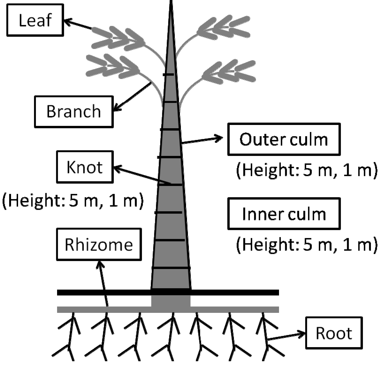
3.1. Plant Materials

Whole plants of 1 or 2-year old P. pubescens were harvested at Kurume, Fukuoka Prefecture, Japan. The average height of the harvested bamboo was 14 m. Then, plants were separated into the following parts: leaves, branches, outer culm, inner culm, knots, rhizomes and roots (Figure 1). At that time, the outer culm, inner culm and knots were obtained separately from heights of 5.0 ± 0.3 and 1.0 ± 0.3 m above ground level. Each part was freeze-dried and milled into powder.
Milled freeze-dried P. pubescens samples were extracted with 99.5% ethanol at room temperature with a shaker at 200 rpm for 48 h and then filtered. The ethanol extracts were concentrated by a rotary evaporator. The yields of ethanol extracts against each dried powder were as follows: leaves, 4.84%; branches, 1.08%; outer culm (5 m), 4.56%; outer culm (1 m), 4.69%; inner culm (5 m), 0.27%; inner culm (1 m), 0.32%; knots (5 m), 1.47%; knots (1 m), 1.55%; rhizomes, 0.45% and roots, 2.63%. To prepare the hot water extracts, P. pubescens samples were extracted with hot water at 120°C for 20 min and the extracted solutions were freeze dried. The yields of hot water extracts were as follows: leaves, 10.4%; branches, 2.67%; outer culm (5 m), 2.96%; outer culm (1 m), 3.69%; inner culm (5 m), 1.60%; inner culm (1 m), 2.21%; knots (5 m), 3.64%; knots (1 m), 4.94%; rhizomes, 2.49% and roots 3.25%.

3.2. Melanin Biosynthesis Assay

This assay was performed as previously described by Arung et al. [38]. The B16 melanoma cells were maintained in EMEM supplemented with 10% (v/v) fetal bovine serum (FBS) and 0.09 mg/mL theophylline. Cells were incubated at 37 °C in a humidified atmosphere of 5% CO2. Cells were placed into a 24-well plate at a density of 1 × 10⁵ cells/mL and incubated for 24 h in medium prior to treatment with extract. After 24 h, the medium was replaced with 998 μL of fresh medium, and 2 μL of ethanol extract dissolved in dimethylsulfoxide (DMSO) or hot water extract dissolved in sterilized water was added. The cells were incubated for an additional 48 h; then the medium was replaced with fresh medium and extract was added again. After 24 h, the remaining adherent cells were used to determine the melanin content and cell viability (see below). To find possible candidates for whitening or tanning agents, we classified the tested extracts into three types (Type A, B, and C). Samples which showed a percentage of melanin content equal to or lower than 20% of cell viability (e.g., CV-MC ≥ 20) were judged as possible whitening agents, and classified as type A. In the other hand, samples which showed a percentage of melanin content equal to or higher than 20% of cell viability (e.g., MC-CV ≥ 20) were judged as possible tanning agents, and classified as type B. Finally, samples showed a percentage of cell viability equal to or lower than 90% were judged to be cytotoxic and classified as type C.

3.2.1. Cell Viability

Cell viability (CV) was determined by use of the microculture tetrazolium technique (MTT) [38]. Culture was initiated, and after incubation, 50 μL of MTT [3-(4,5-dimethylthiazol-2-yl)-2,5-diphenyltetrazolium bromide] in phosphate buffered saline (5 mg/mL) was added to each well. The plates were incubated for 4 h. After removing the medium, formazan crystals were dissolved in 1.0 mL of 0.04 M HCl in isopropanol and the absorbance was measured at 570 nm relative to 630 nm.

3.2.2. Determination of Melanin Content

The melanin content (MC) of cells after treatment with the extract was determined as follows. After removing the medium and washing the cells, the cell pellet was dissolved in 1.0 mL of 1 M NaOH. The crude cell extracts were assayed using a microplate reader (Bio-Tek, Winooski, VT, USA) at 405 nm to determine the melanin content. The results from the samples were analyzed as a percentage of the control culture. Arbutin was used as a positive control.

3.3. Antioxidant Assays

3.3.1. Oxygen Radical Absorbance Capacity Assay

The oxygen radical absorbance capacity (ORAC) assay was performed as described previously by Prior et al. [39]. Data are expressed as milligrams of Trolox equivalent (TE) per milligram of sample extract (mg TE/mg).

3.3.2. Superoxide Dismutase-Like Activity

Superoxide dismutase (SOD)-like activity was evaluated using the SOD Assay Kit-WST (Dojindo Molecular Technologies, Kumamoto, Japan) according to the method described in previous studies [40]. Sample were dissolved in water or ethanol and added to the WST working solutions (200 μL) containing 2-(4-iodophenyl)-3-(4-nitrophenyl)-5-(2, 4-disulfophenyl)-2-H-tetrazolium in 50 mM carbonate buffer (pH 10.2). An enzyme working solution (20 μL) containing xanthine oxidase in the same buffer was added and then incubated for 10 min. The absorbance of each sample was measured at 450 nm in a Tecan Spectra microplate reader (Tecan Japan, Kanagawa, Japan). One unit of SOD-like activity was defined as the amount of extract in 20 μL of sample solution that inhibits the reduction reaction of WST-1 with superoxide anions by 50%. The SOD-like activity (U/mg) of each extract was calculated using the 50% inhibition value (IC50) of the extract.

3.3.3 ABTS Radical Cation Decolorization Assay

The ABTS assay was mostly based on the methods described by Re et al. [41] in which ABTS•+, the oxidant, was generated by persulfate oxidation of ABTS [2,2'-azinobis (3-ethylbenzothiazoline-6-sulfonic acid)]. Specifically, to 5 mL of 7 mM ABTS ammonium aqueous solution, 88 µL of 140 mM potassium peroxydisulfate (K2S2O8) was added, and the resulting mixture was then allowed to stand at room temperature for 12-16 h, yielding a dark blue solution. The mixture was then adjusted by 99.5% ethanol so that it gave an absorbance of 0.7 ± 0.02 units at 734 nm (UVmini-1240, Shimadzu, Kyoto, Japan) to make the working solution. One milliliter of working solution was mixed with 10 µL of extract dissolved in ethanol and shaken well for 10 s; after 4 min of incubation at 30 °C, the absorbance of the reaction mixture was measured at 734 nm.

3.4. Antibacterial Assay

The antibacterial assay was mostly based on the methods described by Tanaka et al. [10]. S. aureus (NBRC 1273) was used for the antibacterial assay. A single colony of the test strain was taken and 5 mL of nutrient broth medium was added to it. This culture was incubated at 37 °C ± 1 °C, 120 rpm for 20 h. It was then added to the bacterial suspension to prepare a bacterial concentration at 105 CFU/mL. The bacterial solution was used for the subsequent antibacterial assay. Each sample was dissolved in DMSO for ethanol extract or sterilized water for hot water extract at maximum concentration. Into each well of a 96-well plate were added 133.5 µL of NB medium, 15 µL of bacteria suspension, and 1.5 µL of solvent with or without each sample. Also, sorbic acid was used as a positive control. The plate was incubated at 37 °C ± 1 °C, 1160 rpm for 18 h. Finally, bacterial growth was measured by a microplate reader at 630 nm (Biotek-ELX800, BioTek). The minimum inhibitory concentration (MIC) is the lowest concentration of an antibacterial agent required to completely inhibit the growth of a particular bacteria, while the minimum bactericidal concentration (MBC) is the lowest concentration of an antibacterial agent required to kill the bacteria. Here, the MIC of active extracts was determined through the antibacterial assay using gradient concentrations. And MBC of them were further determined as follows: a 20 μL aliquot was taken from the wells that treated with extract at higher concentration than its MIC and mixed with 180 μL of fresh medium. Then, 100 μL of the mixture was used to do the subculture on nutrient agar plate. After 24 h incubation at 37 °C ± 1 °C, the colony formation was evaluated. The minimum concentration that leaded to no colony growing on the agar plate was considered as the MBC.

3.5. Immunoglobulin E (IgE) Production Assay

Peripheral blood lymphocytes (PBL) were first separated from heparinized blood of healthy donors using Ficoll-Paque Plus (GE Healthcare, Uppsala, Sweden). And then, PBL cells were cultured in ERDF medium (Kyokuto Pharmaceuticals, Tokyo, Japan) supplemented with 5% FBS, 10% human plasma, 10 ng/mL of recombinant human IL-4 and IL-6 (R&D Systems, USA), 10 μg/mL of muramyl dipeptide (MDP) (Sigma, St.Louis, MO, USA) and 100 ng/mL of the cedar pollen antigen Cry j 1 (Hayashibara Biochemical Laboratories, Okayama, Japan) at the density of 2.0 × 10⁶ cells/mL. 198 μL of such cell suspension and 2 μL of 6mg/ml extract in 10% DMSO solution were added into 96-well plates (final concentration of extract was 60μg/mL). The plate was incubated in a humidified 37°C, 5% CO2 incubator for 10 days. The total IgE concentration in the supernatant was measured by sandwich ELISA (enzyme-linked immunosorbent assay). Briefly, 96-well microplates were coated with anti-human IgE antibody (Biosource, Camarillo, CA, USA). The antibody-coated wells were blocked with 1.0% BSA, following by adding the samples. After washing with PBS containing 0.05% of Tween 20 for three times, biotin-conjugated antihuman IgE antibody (Biosource) and horseradish peroxidase-conjugated streptavidin were added. Finally, a substrate solution [0.1 M citrate buffer (pH 4.0) containing 0.003% of H2O2 and 0.3 mg/mL p-2,2'-azino-bis (3-ethylbenzothiazoline-6-sulfonic acid) diammonium salt] was added. After 15 min, the absorbance was measured at 414 and 490 nm by the microplate reader (iMark, Bio-Rad, Hercules, CA, USA). The relative IgE production was calculated according to the absorbance at 414 nm and 490 nm, and the final inhibition rate was calculated using the following formula: Inhibition rate (%) = (1 − IgE production in treated cells/IgE production in control cells) × 100.

3.6. LCMS Analysis

All extracts were subjected to LCMS analysis using a high-speed liquid chromatography mass spectrometry that combines with iron-trap and time-of-flight technologies (LCMS-IT-TOF, Shimadzu, Tokyo, Japan). The instrument was fitted with an Inertsil ODS-3, 5μm, 1.5 × 150 mm column (GL Science, Tokyo, Japan). The oven temperature was set at 40 °C. A mobile phase composed of solvent A (0.3% acetic acid in water) and B (0.3% acetic acid in acetonitrile or methanol) was employed for the separation. Acetonitrile was used in solvent B for the analysis of ethanol extract, while methanol was used for water extract. The mobile phase was consecutively programmed as follows: 0~60 min, A 90~0%, B 10%~100%; 60~65 min, A 0, B 100%; 65~66 min, A 0%~90%, B 100%~10%; a 10 min post-run was used after each analysis. The total flow rate was 0.15 mL/min. Basing on the previous result of HPLC-PDA analysis, the LC chromatograms of ethanol extracts and water extracts were obtained at UV 254 nm, 326 nm and 254, 278 nm, respectively. The MS instrument was operated using an ESI source in both positive and negative ionization mode with survey scans acquired from m/z 100 to 1000 for both MS and MS/MS. Ionization parameters were as follows: probe voltage, ±4.5 kV; nebulizer gas flow, 1.5 L/min; CDL temperature, 200 °C; heat block temperature, 200 °C.
The samples were dissolved with initial mobile phase (1 mg/mL) and filtered through a 0.45-μm filter. A volume of 5 μL of each sample was injected for the analysis. 8 compounds that had been found in different bamboo species were analyzed and used as a standard. They were catechin (Sigma-Aldrich, Munich, Germany), caffeic acid (Tokyo Chemical Industry, Tokyo, Japan), syringic acid (Tokyo Chemical Industry), chlorogenic acid (Sigma-Aldrich), p-courmaric acid (Sigma-Aldrich), rutin (Wako, Tokyo, Japan), trans-ferulic acid (Tokyo Chemical Industry) and luteolin-7-O-glucoside (EXTRASYNTHESE, Genay, France).

4. Conclusions

In this study, the effect of ethanol and hot water extracts of various parts of bamboo on the melanin biosynthesis regulation (inhibition or stimulation), antioxidation, antibacterial and anti-allergy were comparatively evaluated. We found that the extracts showed different bioactivities in different degrees. For the melanin biosynthesis inhibition, the hot water extracts of outer culm (5 m) and rhizome showed the best activities. For the melanin biosynthesis stimulation, the ethanol extract of inner culm (1 m) showed the strongest activity. For the antioxidant activity, the ethanol extracts of inner culm (1 m), branch and inner culm (5 m) showed the strongest activities. For antibacterial activity against S. aureus, the ethanol extracts of outer culm (5 m and 1 m) showed the strongest activities. The MIC and MBC for both extracts were 400 and 1600 μg/mL, respectively. For anti-allergy activity, the water extract of leaf showed the best IgE inhibition effect. Extracts from the outer culm and inner culm were found to be the most active extracts.
Different parts of bamboo showed different bioactivities, which also varied with the extraction solvent. The difference in chromatographic profile and identified component to some extent explained the different bioactivities of these extracts. The most possible compounds responsible for anti-allergy activity of this bamboo were 8-C-glucosyl apigenin, luteolin derivatives and chlorogenic acid. O-hexosyl-O-deoxyhexosyl tricin was the possible compound responsible for the antibacterial and melanin inhibition activity of bamboo, while apigenin derivatives might be the compounds responsible for the antioxidant activity. This information would be helpful for the further research on the active compounds in bamboo. Taken together, our study provides valuable data to support that bamboo has great potential to be used in the cosmetic industry as well as other health-related industry.

Supplementary Materials

Supplementary materials can be accessed at: https://www.mdpi.com/1420-3049/19/6/8238/s1.

Acknowledgments

The publication was supported in part by the Research Grant for Young Investigators of Faculty of Agriculture, Kyushu University.

Author Contributions

A.T., Q.Z., K.O., H.K. and K.S. designed research; A.T., Q.Z., H.T., H.H., Y.M., R.Y., H.I., A.I. and K.S. performed research and analyzed the data; A.T., Q.Z. and K.S. wrote the paper. All authors read and approved the final manuscript.

Conflicts of Interest

The authors declare no conflict of interest.

References

  1. Lee, H.-O.; Baek, S.-H.; Han, D.-M. Antimicrobial effects of Chamaecyparis obtusa essential oil. Korean J. Appl. Microbiol. Biotechnol. 2001, 29, 253–257. [Google Scholar]
  2. Zhang, Y.; Jiao, J.; Liu, C.; Wu, X.; Zhang, Y. Isolation and purification of four flavone C-glycosides from antioxidant of bamboo leaves by macroporous resin column chromatography and preparative high-performance liquid chromatography. Food Chem. 2008, 107, 1326–1336. [Google Scholar]
  3. Lu, B.-Y.; Zhang, Y.; Wu, X.-Q. Advances in studies on antioxidative activity and cardio-cerebrovascular pharmacology of bamboo-leaf-flavonoids. Linchan Huaxue Yu Gongye 2005, 25, 120–124. [Google Scholar]
  4. Jiao, J.; Zhang, Y.; Liu, C.; Liu, J.; Wu, X. Separation and purification of tricin from an antioxidant product derived from bamboo leaves. J. Agric. Food Chem. 2007, 55, 10086–10092. [Google Scholar] [CrossRef]
  5. Hasegawa, T.; Tanaka, A.; Hosoda, A.; Takano, F.; Ohta, T. Antioxidant C-glycosyl flavones from the leaves of Sasa kurilensis var. gigantea. Phytochemistry 2008, 69, 1419–1424. [Google Scholar] [CrossRef]
  6. Zhang, Y.; Yao, X.; Bao, B. Anti-fatigue activity of a triterpenoid-rich extract from Chinese bamboo shavings (Caulis bamfusae in taeniam). Phytother. Res. 2006, 20, 872–876. [Google Scholar] [CrossRef]
  7. Kurokawa, T.; Itagaki, S.; Yamaji, T.; Nakata, C.; Noda, T.; Hirano, T.; Iseki, K. Antioxidant activity of a novel extract from bamboo grass (AHSS) against ischemia-reperfusion injury in rat small intestine. Biol. Pharm. Bull. 2006, 29, 2301–2303. [Google Scholar] [CrossRef]
  8. Nishina, A.; Hasegawa, K.I.; Uchibori, T.; Seino, H.; Osawa, T. 2,6-dimethoxy-p-benzoquinone as an antibacterial substance in the culm of Phyllostachys heterocycla var. Pubescens, a species of thick-stemmed bamboo. J. Agric. Food Chem. 1991, 39, 266–269. [Google Scholar] [CrossRef]
  9. Fujimura, M.; Ideguchi, M.; Minami, Y.; Watanabe, K.; Tadera, K. Amino acid sequence and antimicrobial activity of chitin-binding peptides, Pp-AMP 1 and Pp-AMP 2, from Japanese bamboo shoots (Phyllostachys pubescens). Biosci. Biotechnol. Biochem. 2005, 69, 642–645. [Google Scholar] [CrossRef]
  10. Tanaka, A.; Shimizu, K.; Kondo, R. Antibacterial compounds from shoot skins of moso bamboo (Phyllostachys pubescens). J. Wood Sci. 2013, 59, 155–159. [Google Scholar] [CrossRef]
  11. Katsuzaki, H.; Sakai, K.; Achiwa, Y.; Imai, K.; Komiya, T. Isolation of antioxidative compounds from bamboo shoots sheath. Nippon Shokuhin Kagaku Kogaku Kaishi 1999, 46, 491–493. [Google Scholar] [CrossRef]
  12. Fujita, S. Ecological and mechanical aspect of bamboo. Biol. Resour. 2012, 6, 2–11. (In Japanese) [Google Scholar]
  13. Guo, X.F.; Yue, Y.D.; Tang, F.; Wang, J.; Yao, X. Antioxidant properties of major flavonoids and subfractions of the extract of Phyllostachys pubescens leaves. J. Food Biochem. 2012, 37, 501–509. [Google Scholar]
  14. Zhang, Z.; Wang, X.; Yu, S.; Zhao, M. Isolation and antioxidant activities of polysaccharides extracted from the shoots of Phyllostachys edulis (Carr.). Int J. Biol Macromol 2011, 49, 454–457. [Google Scholar] [CrossRef]
  15. Higa, J.K.; Panee, J. Bamboo extract reduces interleukin 6 (IL-6) overproduction under lipotoxic conditions through inhibiting the activation of NF-kappaB and AP-1 pathways. Cytokine 2011, 55, 18–23. [Google Scholar] [CrossRef]
  16. Lin, Y.; Collier, A.C.; Liu, W.; Berry, M.J.; Panee, J. The inhibitory effect of bamboo extract on the development of 7,12-dimethylbenz[a]anthracene (DMBA)-induced breast cancer. Phytother Res. 2008, 22, 1440–1445. [Google Scholar] [CrossRef]
  17. Tanaka, A.; Kim, H.J.; Oda, S.; Shimizu, K.; Kondo, R. Antibacterial activity of moso bamboo shoot skin (Phyllostachys pubescens) against Staphylococcus aureus. J. Wood Sci. 2011, 57, 542–544. [Google Scholar] [CrossRef]
  18. Ferreres, F.; Gil-Izquierdo, A.; Andrade, P.B.; Valentao, P.; Tomas-Barberan, F.A. Characterization of C-glycosyl flavones O-glycosylated by liquid chromatography-tandem mass spectrometry. J. Chromatogr. A 2007, 1161, 214–223. [Google Scholar]
  19. Meng, T.X.; Furuta, S.; Fukamizu, S.; Yamamoto, R.; Ishikawa, H.; Arung, E.T.; Shimizu, K.; Kondo, R. Evaluation of biological activities of extracts from the fruiting body of Pleurotus citrinopileatus for skin cosmetics. J. Wood Sci. 2011, 57, 452–458. [Google Scholar] [CrossRef]
  20. Breuer, K.; HÄussler, S.; Kapp, A.; Werfel, T. Staphylococcus aureus: Colonizing features and influence of an antibacterial treatment in adults with atopic dermatitis. Br. J. Dermatol. 2002, 147, 55–61. [Google Scholar] [CrossRef]
  21. Iwamoto, A.; Mitsuda, K.; Inoue, A.; Kato, T.; Inoue, Y.; Kawahara, H. Purification and identification of an IgE suppressor from strawberry in an in vitro immunization system. Cytotechnology 2012, 64, 309–314. [Google Scholar] [CrossRef]
  22. Van Hoyweghen, L.; de Beer, T.; Deforce, D.; Heyerick, A. Phenolic compounds and anti-oxidant capacity of twelve morphologically heterogeneous bamboo species. Phytochem. Anal. 2012, 23, 433–443. [Google Scholar] [CrossRef]
  23. Lv, Z.; Dong, J.; Zhang, B. Rapid identification and detection of flavonoids compounds from bamboo leaves by LC-(ESI)-IT-TOF/MS. BioResources 2012, 7, 1405–1418. [Google Scholar]
  24. Cavaliere, C.; Foglia, P.; Pastorini, E.; Samperi, R.; Lagana, A. Identification and mass spectrometric characterization of glycosylated flavonoids in Triticum durum plants by high-performance liquid chromatography with tandem mass spectrometry. Rapid Commun. Mass Spectrom. 2005, 19, 3143–3158. [Google Scholar]
  25. Ferreres, F.; Silva, B.M.; Andrade, P.B.; Seabra, R.M.; Ferreira, M.A. Approach to the study of C-glycosyl flavones by ion trap HPLC-PAD-ESI/MS/MS: Application to seeds of quince (Cydonia oblonga). Phytochem. Anal. 2003, 14, 352–359. [Google Scholar] [CrossRef]
  26. Huang, C.S.; Lii, C.K.; Lin, A.H.; Yeh, Y.W.; Yao, H.T.; Li, C.C.; Wang, T.S.; Chen, H.W. Protection by chrysin, apigenin, and luteolin against oxidative stress is mediated by the Nrf2-dependent up-regulation of heme oxygenase 1 and glutamate cysteine ligase in rat primary hepatocytes. Arch. Toxicol 2013, 87, 167–178. [Google Scholar] [CrossRef]
  27. Birt, D.F.; Walker, B.; Tibbels, M.G.; Bresnick, E. Anti-mutagenesis and anti-promotion by apigenin, robinetin and indole-3-carbinol. Carcinogenesis 1986, 7, 959–963. [Google Scholar] [CrossRef]
  28. Funakoshi-Tago, M.; Nakamura, K.; Tago, K.; Mashino, T.; Kasahara, T. Anti-inflammatory activity of structurally related flavonoids, Apigenin, Luteolin and Fisetin. Int. Immunopharmacol 2011, 11, 1150–1159. [Google Scholar] [CrossRef]
  29. Czyz, J.; Madeja, Z.; Irmer, U.; Korohoda, W.; Hulser, D.F. Flavonoid apigenin inhibits motility and invasiveness of carcinoma cells in vitro. Int J. Cancer 2005, 114, 12–18. [Google Scholar] [CrossRef]
  30. Choi, J.S.; Islam, M.N.; Ali, M.Y.; Kim, E.J.; Kim, Y.M.; Jung, H.A. Effects of C-glycosylation on anti-diabetic, anti-Alzheimer’s disease and anti-inflammatory potential of apigenin. Food Chem. Toxicol 2014, 64, 27–33. [Google Scholar] [CrossRef]
  31. Iwaoka, E.; Oku, H.; Iinuma, M.; Ishiguro, K. Allergy-preventive effects of the flowers of Impatiens textori. Biol. Pharm. Bull. 2010, 33, 714–716. [Google Scholar] [CrossRef]
  32. Kritas, S.K.; Saggini, A.; Varvara, G.; Murmura, G.; Caraffa, A.; Antinolfi, P.; Toniato, E.; Pantalone, A.; Neri, G.; Frydas, S.; et al. Luteolin inhibits mast cell-mediated allergic inflammation. J. Biol. Regul. Homeost. Agents 2013, 27, 955–959. [Google Scholar]
  33. Li, S.Y.; Chang, C.Q. Biological effects of chlorogenic acid and body health. Wei Sheng Yan Jiu 2005, 34, 762–764. [Google Scholar]
  34. Oku, H.; Ogawa, Y.; Iwaoka, E.; Ishiguro, K. Allergy-preventive effects of chlorogenic acid and iridoid derivatives from flower buds of Lonicera japonica. Biol. Pharm. Bull. 2011, 34, 1330–1333. [Google Scholar] [CrossRef]
  35. Stermitz, F.R.; Beeson, T.D.; Mueller, P.J.; Hsiang, J.; Lewis, K. Staphylococcus aureus MDR efflux pump inhibitors from a Berberis and a Mahonia (sensu strictu) species. Biochem. Syst. Ecol. 2001, 29, 793–798. [Google Scholar]
  36. Shukla, S.; Gupta, S. Apigenin: A promising molecule for cancer prevention. Pharm. Res. 2010, 27, 962–978. [Google Scholar] [CrossRef]
  37. Romanova, D.; Vachalkova, A.; Cipak, L.; Ovesna, Z.; Rauko, P. Study of antioxidant effect of apigenin, luteolin and quercetin by DNA protective method. Neoplasma 2001, 48, 104–107. [Google Scholar]
  38. Arung, E.T.; Shimizu, K.; Kondo, R. Inhibitory effect of artocarpanone from Artocarpus heterophyllus on melanin biosynthesis. Biol. Pharm. Bull. 2006, 29, 1966–1969. [Google Scholar]
  39. Prior, R.L.; Hoang, H.; Gu, L.; Wu, X.; Bacchiocca, M.; Howard, L.; Hampsch-Woodill, M.; Huang, D.; Ou, B.; Jacob, R. Assays for hydrophilic and lipophilic antioxidant capacity (oxygen radical absorbance capacity (ORAC(FL))) of plasma and other biological and food samples. J. Agric. Food Chem. 2003, 51, 3273–3279. [Google Scholar] [CrossRef]
  40. Nakamura, K.; Ogasawara, Y.; Endou, K.; Fujimori, S.; Koyama, M.; Akano, H. Phenolic compounds responsible for the superoxide dismutase-like activity in high-Brix apple vinegar. J. Agric. Food Chem. 2010, 58, 10124–10132. [Google Scholar]
  41. Re, R.; Pellegrini, N.; Proteggente, A.; Pannala, A.; Yang, M.; Rice-Evans, C. Antioxidant activity applying an improved ABTS radical cation decolorization assay. Free Radic Biol. Med. 1999, 26, 1231–1237. [Google Scholar] [CrossRef]
  • Sample Availability: Samples of the compounds are available from the authors.

