Article

Four New Furostanol Saponins from the Rhizomes and Roots of Smilax scobinicaulis and Their Cytotoxicity

1
College of Life Science, Northwest A&F University, Yangling 712100, Shaanxi, China
2
Key Laboratory of Southern Subtropical Plant Diversity, Shenzhen Fairy Lake Botanical Garden, Chinese Academy of Sciences, 160 Xianhu Road, Liantang, Shenzhen 518004, China
3
Shenzhen Boton Flavors & Fragrances Co., Ltd., 19 Daxin Road, Nanshan, Shenzhen 518051, China
*
Author to whom correspondence should be addressed.
Molecules 2014, 19(12), 20975-20987; https://doi.org/10.3390/molecules191220975
Submission received: 15 September 2014 / Revised: 21 November 2014 / Accepted: 2 December 2014 / Published: 15 December 2014
(This article belongs to the Section Natural Products Chemistry)

Abstract

:
Four new furostanol saponins 14, along with two known furostanol saponins 5 and 6 and one known spirostanol saponin 7 were isolated from the rhizomes and roots of Smilax scobinicaulis. The structures of the new saponins were elucidated as 26-O-β-d-glucopyranoside-3β,26-dihydroxy-(25R)-5α-furostan-22-methoxyl-6-one-3-O-α-l-arabinopyranosyl-(1→6)-β-d-glucopyranoside (1), 26-O-β-d-glucopyranoside-3β,26-dihydroxy-(25R)-5α-furostan-22-methoxyl-6-one (2), 26-O-β-d-glucopyranoside-3β,26-dihydroxy-(25R)-5α-furostan-20(22)-en-6-one (3), 26-O-β-d-glucopyranoside-3β,23,26-trihydroxy-(23R, 25R)-5α-furostan-20(22)-en-6-one (4) on the basis of spectroscopic analysis. The isolated saponins were evaluated for cytotoxic activity against two human cancer cell lines including Hela (cervical carcinoma) and SMMC-7221 (hepatocellular carcinoma). Compounds 1 and 7 demonstrated cytotoxicity against the tested cell lines.

1. Introduction

The genus Smilax (family Liliaceae) is mainly distributed in the tropical, subtropical and temperate areas of the world [1]. It is well known that steroidal saponins are abundant in the genus Smilax [2]. Many species of this genus have a long history of use as herbal remedies. Smilax scobinicaulis (C.H.) Wright, commonly known as Hei Ci Ba Qia in Chinese is one of them, which is distributed in Shaanxi, Gansu, Sichuan, Yunnan and other regions of China [3]. The rhizomes and roots of this plant, known as “Jin Gang Teng” and “Wei Ling Xian” in North China, have long been used in folk medicine for the treatment of rheumatic arthritis, lumbago, gout, tumor and inflammatory diseases [1]. Previous phytochemical investigations of this plant led to the isolation of steroidal saponins [4], flavonoids [5] and phenylpropanoids [6]. Bioactivity investigations showed that some of the isolated steroidal saponins had antimicrobial and cytotoxic activity [4,7].
In a continuing search for new steroidal saponins from this plant, a series of steroidal saponins have now been obtained, including four new furostanol saponins 14, two known furostanol saponins 5 and 6 and one known spirostanol saponin 7 (Figure 1), which are reported here as S. scobinicaulis compounds for the first time. All the compounds have been tested for cytotoxicity against Hela and SMMC-7221 human cancer cell lines. In this paper, we report the isolation and cytotoxic activity of these compounds and the structural elucidation of the new compounds.
Figure 1. Chemical structure of compounds 17.
Molecules 19 20975 g001

2. Results and Discussion

The rhizomes and roots of S. scobinicaulis were extracted with ethanol (EtOH). The extract was suspended in water (H2O) and partitioned successively with petroleum ether (PE), ethyl acetate (EtOAc) and n-butanol (n-BuOH). The n-BuOH soluble fraction was subjected to silica gel CC (column chromatography), Sephadex LH-20, reversed phase silica gel CC and semi-preparative High Performance Liquid Chromatography (HPLC) to afford four new furostanol saponins 26-O-β-d-glucopyranoside-3β,26-dihydroxy-(25R)-5α-furostan-22-methoxyl-6-one-3-O-α-l-arabinopyranosyl-(1→6)-β-d-glucopyranoside (1), 26-O-β-d-glucopyranoside-3β,26-dihydroxy-(25R)-5α-furostan-22-methoxyl-6-one (2), 26-O-β-d-glucopyranoside-3β,26-dihydroxy-(25R)-5α-furostan-20(22)-en-6-one (3), 26-O-β-d-glucopyranoside-3β,23,26-trihydroxy-(23R, 25R)-5α-furostan-20(22)-en-6-one (4), along with two known furostanol saponins 26-O-β-d-glucopyranosyl-3β,22ξ,26-trihydroxy-(25R)-5α-furostan-6-one-3-O-α-l-arabinopyranosyl-(1→6)-β-d-glucopyranoside (5) [8], 26-O-β-d-gluco- pyranosyl-3β,26-dihydroxy-(25R)-5α-furostan-20(22)-en-6-one-3-O-α-l-arabinopyranosyl-(1→6)-β-d-glucopyranoside (6) [9] and the known spirostanol saponin sieboldogenin-3-O-α-l-arabino-pyranosyl-(1→6)-β-d-glucopyranoside (7) [10].

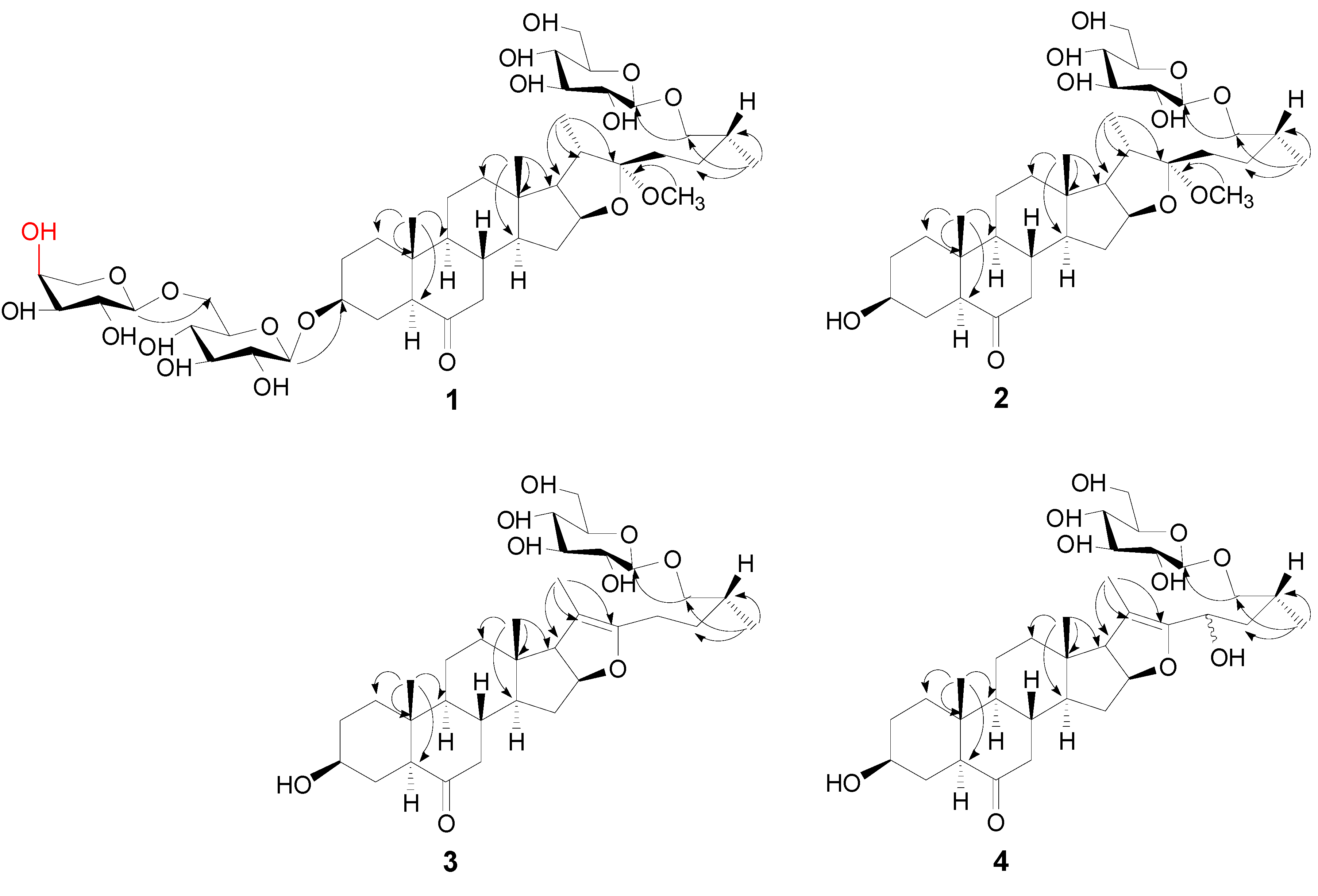
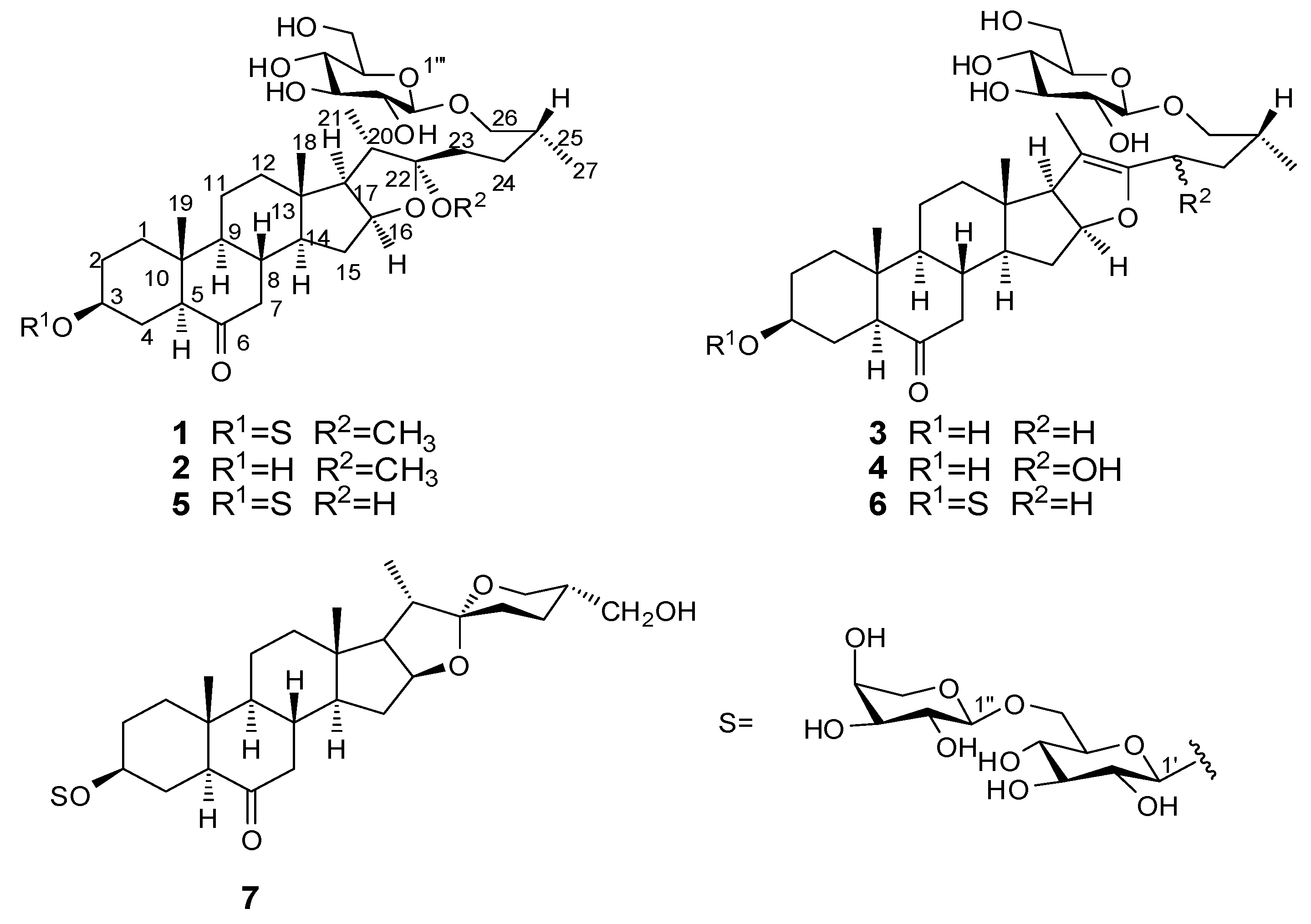
2.1. Structure Elucidation

Compound 1 was isolated as a white amorphous powder and showed a positive reaction (red colour) to the Ehrlich reagent. The molecular formula, C45H74O19, was established from a positive molecular ion peak at m/z 941.4727 [M+Na]+ (calcd. for C45H74NaO19: 941.4717) in the HR-ESI-MS spectrum and supported by the ESI-MS (m/z 941.4 [M+Na]+ and m/z 917.3 [M−H]) spectrum. Its IR spectrum displayed strong absorption bands for hydroxyl groups at 3408 cm−1, for carbonyl group at 1707 cm−1 and absorption bands of alkyl groups at 2927 cm−1. The 1H- and 13C-NMR assignments of 1 were based on the DEPT and 2D-NMR (COSY, HSQC, HMBC and NOESY) experiments and with the positive red colour reaction in Ehrlich’s test suggested 1 was a furostanol saponin. To be specific, The 1H-NMR spectrum of 1 showed four methyl proton signals including two tertiary methyl groups at δH0.86 (3H, s, H-18) and 0.80 (3H, s, H-19) and two secondary methyl groups at δH 1.04 (3H, d, J = 6.0 Hz, H-21) and 0.98 (3H, d, J = 5.0 Hz, H-27), corresponding to C-atom signals at δC 15.5, 12.1, 14.7 and 15.9 in HSQC spectrum, typical steroid methyl signals. Besides, a signal for carbonyl at δC 212.0 was present in the 13C-NMR. A correlation signal between H-7 at δH 2.16 and C-6 at δC 212.0 was observed in the HMBC spectrum (Figure 2), which suggested the carbonyl is located at C-6. Moreover, a methoxyl group at δH 3.17 (3H, s, OCH3) was also observed in the 1H-NMR spectrum. In the HMBC spectrum, the correlations from OCH3 at δH 3.17 to C-22 at δC 112.6 to suggested the OCH3 was linked to the C-22 (Figure 2). The configurations of 1 were mainly determined by NOESY spectrum. A NOESY correlation signal between the H-5 proton at δH 2.40 and the H-9 proton at δH 1.40 was consistent with the 5α configuration. A NOESY cross-peak between H-5 (δH 2.40) and H-3 (δH 3.36) indicated that H-3 was α configuration. In addition, The NOE correlation between H-18 (δH 0.86, 3H, s) and H-20 (δH 2.21, 1H, m) suggested that the C-21 methyl group was α-configuration. Thus, the α-configurations of H-17 and the methoxy at C-22 position were unambiguously deduced based on the strong NOE correlations of H-21/H-17 and H-21/OCH3. The 14α configuration was further confirmed by the NOE correlations of H-14/H-16 and H-16/OCH3 [11]. However, the C-25 configuration of 1 was assigned as 25R based on the observed difference (∆ab = δa − δb = 0.35) of the 1H-NMR chemical shifts of the H2-26 geminal protons, which was in agreement with that of 25R furostane-type steroidal saponins (∆ab < 0.48 for 25R; ∆ab > 0.57 for 25S) [12].
Figure 2. Key HMBC correlations of compounds 14.
Molecules 19 20975 g002
In addition, three anomeric proton signals at δH 4.42 (1H, d, J = 6.5 Hz, glc-1'), 4.34 (1H, d, J = 6.0 Hz, ara-1'') and 4.26 (1H, d, J = 7.0 Hz, glc-1''') existed in the 1H-NMR spectrum and the HSQC showed correlations with anomeric carbon signals at δC 100.9, 103.8 and 103.2, respectively, indicating the presence of three sugar moieties. Combined the 1H-NMR, 13C-NMR (including DEPT) and 2D-NMR (HSQC, HMBC and COSY), two glucopyranosyls and one arabinopyranosyl were found. The relative configurations of the two glucopyranose moieties were all assigned as β-configurations based on their coupling constants (J = 6.5 Hz, Glc-1'; J = 7.0 Hz, Glc-1''') of the anomeric protons. The relative configuration of the arabinopyranose moiety was determined as α-confirguration by the coupling constants (J = 6.0 Hz, Ara-1''). The sugars were determined to be d-glucose and l-arabinose by acid hydrolysis of 1 and then trimethylsilylation and GC analysis on a chiral column in comparison with an authentic sample. Furthermore, the location of the glucopyranosyl was found to be C-26 of the aglycon on the basis of a glycosylation shift of C-26 at δC 74.6 and the HMBC correlation peak between H-1''' of glucopyranosyl at δH 4.26 and C-26 of the aglycone at δC 74.6, which is a structural feature in plant furostanol saponins [11]. The attachments of sugar chain were deduced from the HMBC spectrum, in which the long range correlation between glc-H-1' at δH 4.42 with C-3 at δC 76.8 of the aglycone and ara-H-1'' at δH 4.34 with C-6' at δC 68.2 of the glucopyranosyl were observed (Figure 2), respectively. These results implied that the glucopyranosyl was linked to C-3 of the aglycone and the two sugars were the 1→6 linkage. Thus, the structure of 1 was elucidated as 26-O-β-d-glucopyranoside-3β,26-dihydroxy-(25R)-5α-furostan-22-methoxyl-6-one-3-O-α-l-arabinopyranosyl-(1→6)-β-d-glucopyranoside.
Compound 2, obtained as a white amorphous powder, gave a positive red colour reaction in Ehrlich’s test. The HR-ESI-MS spectrum of 2 showed a positive ion [M+Na]+ at m/z 647.3762, consistent with the molecular formula C34H56O10 (calcd. for C34H56NaO10: 647.3766), which was supported by the ESI-MS (m/z 647.4 [M+Na]+ and m/z 623.4 [M–H]) spectrum. In the IR spectrum, the glycosidic nature of 2 was shown by strong hydroxyl groups absorptions at 3423 cm−1, a carbonyl group at 1713 cm−1 and the alkyl groups at 2927, 2872 and 2853 cm−1 was also displayed in the IR spectrum. Compound 2 was suggested to be a furostanol glycoside with one sugar moiety by Ehrlich’s test and the 1H- and 13C-NMR assignments of 2 based on the DEPT and 2D-NMR (COSY, HSQC, HMBC and NOESY) experiments. The aglycone of 2 was deduced as the same as that of 1 by comparison the 1H- and 13C-NMR spectra of 2 with those of 1. This was further confirmed by comparison Rf values of the aglycones obtained by acid hydrolysis of 1 and 2 on TLC. The only one sugar moiety was assigned to C-26 by a glycosylation shift of C-26 at δC 74.6 and the HMBC correlation between H-1 of the glucopyranosyl at δH 4.26 and C-26 at δC 74.6 (Figure 2). The relative configuration of the glucopyranose moiety was determined as β by the coupling constant (J = 8.0 Hz, Glc-1''') of the anomeric proton. The glucopyranose moiety was determined as d-glucose by acid hydrolysis of 2 and then trimethylsilylation and GC analysis on a chiral column in the same manner. The 25R configuration of 2 was deduced by the difference (∆ab = 0.35) of the chemical shifts of the H2-26 geminal protons. Thus, the structure of 2 was elucidated as 26-O-β-d-glucopyranoside-3β,26-dihydroxy-(25R)-5α-furostan-22α-methoxyl-6-one.
Compound 3 was obtained as a white amorphous powder and positive ion HR-ESI-MS provided an [M+Na]+ ion m/z 615.3510, corresponding to a molecular formula of C33H52O9 (calcd. for C33H52NaO9: 615.3504), which was supported by m/z 615.3 [M+H] + and m/z 591.3 [M–H] in the ESI-MS spectrum. The IR spectrum showed the presence of hydroxyl groups at 3421 cm−1, a carbonyl group at 1697 cm−1 and alkyl groups at 2943 cm−1, 2915 cm−1and 2865 cm−1. It was apparent from the NMR spectroscopic data (1H, 13C, COSY, HSQC, HMBC and NOESY) of 3 that this compound differed from 2 only by the presence of the ∆20,22 double bond. The occurrence of a ∆20,22 double bond was confirmed from the HMBC spectrum which showed significant correlation peaks between the methyl proton signal of CH3-21 with the carbon resonances of C-20 (δC 103.6) and C-22 (δC 151.8) (Figure 2). The sugar moiety also was assigned as β-glucopyranose based on the coupling constant (J = 8.0 Hz, Glc-1''') of the anomeric proton. Its absolute configuration was also a d- as determined by acid hydrolysis of 3 and then trimethylsilylation and GC analysis on a chiral column. The α-configuration of H-17 was determined by the NOE correlations of H-14/H-17. The 25R configuration of 3 was assigned by the observed difference (∆ab = δa − δb = 0.32) of the 1H-NMR chemical shifts of the H2-26 geminal protons (∆ab < 0.48 for 25R; ∆ab > 0.57 for 25S). Therefore, compound 3 was identified as 26-O-β-d-glucopyranoside-3β,26-dihydroxy-(25R)-5α-furostan-20(22)-en-6-one.
Compound 4, a white amorphous powder, was assigned as C33H52O10 on the basis of its HR-ESI-MS data [M+Na]+ m/z 631.3450 (calcd. for C33H52NaO10: 631.3453) and its ESI-MS data m/z 631.55 [M+Na]+, 609.34 [M+H] and 1215.03 [2M−H]. The IR spectrum showed absorptions for hydroxyl groups (3422 cm−1), carbonyl group (1708 cm−1) and alkyl groups (2934 cm−1 and 2870 cm−1). The 1D and 2D NMR spectroscopic data of 4 were in good agreement with those of 3 except for noticeable differences in the δH-23 and δC-23 of the aglycone. The sugar moiety also was determined as β-d-glucopyranose by the coupling constant (J = 8.0 Hz, Glc-1''') of the anomeric proton and GC analysis of hydrolysis followed trimethylsilylation of 4 in comparison with an authentic sample. The configurations of H-17 and the C-25 of 4 was assigned as the same as those of 3 by the NOE correlation of H-14/H-17 and the ∆ab = 0.24 of the two geminal protons of H2-26. In the 1H-NMR spectrum, the proton signal at δH 4.91 (1H, t-like, J = 7.0 Hz, H-23), corresponded C-atom signal at δC 63.6 in the HSQC spectrum showed the presence of the hydroxyl at the C-23. On the basis of this evidence, the structure of 4 was revised as 26-O-β-d-glucopyranoside-3β,23,26-trihydroxy-(25R)-5α-furostan-20(22)-en-6-one. Besides the four new furostanol saponins, three known steroid saponins were isolated from this plant. Their structures were identified by compared the NMR properties with the reported data.

2.2. Cytotoxic Activity

The cytotoxicity of compounds 17 were tested against human cervical carcinoma cells (Hela) and human hepatocellular carcinoma cells (SMMC-7221) by the MTT method. The results revealed that compounds 26 were inactive (IC50 > 100 µM), while compounds 1 and 7 displayed cytotoxicity against Hela carcinoma cell lines with IC50 values of 18.79 ± 1.12 μM and 9.73 ± 1.64 μM, respectively and against SMMC-7221 cancer cell lines with IC50 values of 28.57 ± 1.57 μM and 21.54 ± 1.64 μM, respectively. Cisplatin used as the positive control had the IC50 values of 5.12 ± 0.26 μM (Hela) and 14.23 ± 0.44 μM (SMMC-7221). This investigation indicated that compound 7 which displays a spirostanol saponin structure showed stronger cytotoxic activity, whereas compound 1, with a furostanol saponin structure, displayed moderate cytotoxic activity. Thus, according to our results, the spirostanol saponin skeleton exhibits higher cytotoxic activity than furostanol saponins, which is consistent with the literature [13].

3. Experimental Section

3.1. General

Optical rotations were measured with a Rudolph Research Analytical Autopol III automatic polarimeter (Rudolph Research Analytical, Hackettstown, NJ, USA) with MeOH as solvent. The IR spectra were conducted in KBr on a Bruker Tensor 27 spectrophotometer (Bruker, Billerica, MA, USA). The UV spectra were recorded on a Thermo Scientific Evolution 300 UV-visible spectrophotometer (Thermo Fisher Scientific, Waltham, MA, USA). The 1H- (500 MHz), 13C- (125 MHz) and 2D-NMR spectra were recorded on a Bruker DRX-500 instrument using TMS as internal standard. ESI-MS were collected on a Thermo Fisher LCQ Fleet mass spectrometer instrument (Thermo Fisher Scientific). HR-ESI-MS were carried out on a Bruker Bio-TOF-IIIQ mass spectrometer (Bruker). Absorbance value was measured by a Bio-Tek Epoch Absorbance Microplate Reader to calculate the inhibition rate (Bio-Tek, Winooski, VT, USA).
Semi-preparative HPLC was run with a Waters 1525 pump and a Waters 2489 ultraviolet-visible detector using an Xterra® prep MS C18 column (10 µm, 7.8 × 150 mm) (Waters, Milford, MA, USA). For column chromatography, silica gel (200–300 mesh; Qingdao Haiyang Chemical Co. Ltd, Qingdao, China), Rp-C18 (ODS-A, 50 µm, YMC, Yantai, China), and Sephadex LH-20 (GE Healthcare Bio-Science AB, Fairfield, CT, USA) were used. TLC (5 × 10 cm plates) were performed on GF254 (Qingdao Haiyang Chemical Co. Ltd) plates. Spots were detected after spraying with Ehrlich reagent (1 g 4-dimethylaminobenzaldehyde, in a mixture of 25 mL 37% hydrochloric acid and 75 mL methanol) followed by heating.

3.2. Plant Material

The rhizomes and roots of S. scobinicaulis were collected from Taibai Mountain of Shaanxi Province, China, in October 2010. The plant was identified by Zhenghai Wu of Northwest A&F University, Yangling, Shannxi, China. An authenticated voucher specimen (XB 00045) has been deposited at the Herbarium of Northwest A&F University, Yangling, Shannxi, China.

3.3. Extraction and Isolation

The air-dried powdered rhizomes and roots of S. scobinicaulis (62 kg) was extracted three times with 70% EtOH at room temperature for 48 h to obtain a concentrated extract, which was suspended in H2O and partitioned successively with PE, EtOAc and n-BuOH. The n-BuOH soluble extract (1792 g) was subjected to D101 macroporous adsorption resin and successively eluted with 30% EtOH, 50% EtOH and 70% EtOH to afford three fractions Frs.1–3. Fr.2 (600 g) was subjected to silica gel CC using a stepwise gradient of EtOAc–MeOH–H2O (from 15:1:0.5 to 3:1:0.5) to afford ten sub-fractions Frs. 2a–2j. Fr.2d (9.7 g) was chromatographed on silica gel CC eluting with EtOAc–MeOH (from 17:1 to 3:1) in gradient to yield four sub-fractions Frs.2d1–2d4 based on TLC. Fr.2d2 (1.2 g) was applied to Sephadex LH-20 CC with the eluent of MeOH to yield four sub-fractions Frs.2d2a–2d2d. Fr.2d2a (408 mg) was purified by RP-C18 CC and eluted with 65% MeOH–H2O to give compound 3 (52.7 mg). Fr.2d3 (1.6 g) was applied to Sephadex LH-20 CC with the eluent of MeOH to yield three sub-fractions Frs.2d3a–2d3c. Fr.2d3a (635 mg) was subjected to RP-C18 CC and eluted with 30%–70% MeOH–H2O to result four sub-fractions Frs.2d3a1–2d3a4. Fr.2d3a2 (156 mg) was purified by semi-preparative RP-C18 HPLC with the eluent of 48% MeOH–H2O (215 nm, flow rate: 3 mL/min) to give compound 4 (43.6 mg, tR = 10.4 min). Fr.2d3a3 (238 mg) was purified by semi-preparative RP-C18 HPLC with the eluent 55% MeOH-H2O (215 nm, flow rate: 5 mL/min) to give compound 2 (55.8 mg, tR = 7.9 min). Fr.2i (24.0 g) was chromatographed on Sephadex LH-20 CC with the eluent of MeOH to yield five sub-fractions Frs.2i1–2i5 based on TLC. Fr.2i1 (2.1 g) was applied to RP-C18 CC and eluted with 30%–70% MeOH–H2O to yield four sub-fractions Frs.2i1a–2i1d. Fr.2i1c (1.1 g) was chromatographed on silica gel CC eluting with EtOAc–MeOH–H2O (7:1:0.3–2:1:0.3) in gradient to yield four sub-fractions Frs.2i1c1–2i1c4. Fr.2i1c3 (223 mg) was purified by semi-preparative RP-C18 HPLC with the eluent of 53% MeOH–H2O (215 nm, flow rate: 3 mL/min) to give compound 5 (41.7 mg, tR = 8.7 min) and 1 (42.3 mg, tR = 9.3 min). Fr.2i1d (0.8 g) was chromatographed on silica gel CC eluting with EtOAc–MeOH–H2O (from 15:2:0.3 to 7:2:0.3) in gradient to give compound 7 (43.6 mg) and to yield five sub-fractions Frs.2i1d1-2i1d4. Fr.2i1d3 (339 mg) was purified by semi-preparative RP-C18 HPLC with the eluent of 50% MeOH–H2O (215 nm, flow rate: 2.5 mL/min) to give compound 6 (75.2 mg, tR = 8.3 min).

3.4. New Compound Data

26-O-β-d-Glucopyranoside-3β,26-dihydroxy-(25R)-5α-furostan-22-methoxyl-6-one-3-O-α-l-arabino-pyranosyl-(16)-β-d-glucopyranoside (1). White amorphous powder, mp > 290 °C; [ α ] D 23.4 = −53.348° (c 0.115, CH3OH); IR υmax (KBr): 3408, 2927, 1707 and 1044 cm−1; 1H-NMR and 13C-NMR data see Table 1; ESI-MS: m/z 941.4 [M+Na]+ and 917.3 [M−H]; HR-ESI-MS: m/z 941.4727 [M+Na]+ (Calcd for C45H74NaO19: 941.4717).
Table 1. 1H- and 13C-NMR Data of Compounds 14. (δ in ppm, J in Hz).
NO.1234
δH aδC bδH aδC bδH aδC bδH cδC d
11.80 (m)
1.35 (m)
36.31.79 (m)
1.33 (m)
36.31.80 (m)
1.32 (m)
36.31.64 (m)
1.16 (m)
36.8
21.93 (m)
1.32 (m)
28.61.82 (m)
1.64 (m)
30.11.42 (m)
1.31 (m)
30.11.65 (m)
2.04 (m)
31.6
33.36 (m)76.83.53 (m)69.73.52 (m)69.73.83 (m)69.8
41.43 (m)
2.03 (m)
26.01.41 (m)
1.81 (m)
29.31.43 (m)
1.83 (m)
29.31.90 (m)
2.29 (m)
31.0
52.40 (d, 12.0)56.02.38 (d, 11.0)56.22.37 (dd, 12.5, 2.0)56.22.26 (m)56.8
6212.0212.0212.0209.7
72.16 (t, 13.0)
2.24 (m)
46.12.14 (t, 12.5)
2.25 (dd, 13.0, 4.5)
46.12.15 (m)
2.28 (dd, 13.0, 4.5)
46.22.02 (m)
2.35 (m)
46.8
82.00 (m)37.42.00 (m)37.41.97 (m)37.31.81 (m)37.0
91.40 (m)53.41.40 (m)53.41.36 (m)53.41.15 (m)53.5
1040.840.640.640.7
111.42 (m)
1.70 (m)
21.01.42 (m)
1.68 (m)
21.01.42 m
1.72 m
21.21.52 (m)
1.73 (m)
21.5
121.28 (m)
1.82 (m)
39.11.25 (m)
1.82 (m)
39.21.37 m
1.88 m
39.01.22 (m)39.2
1341.141.143.643.9
141.43 (m)56.01.41 (m)56.01.45 (m)54.61.06 (m)54.6
151.30 (m)
1.95 (m)
31.11.30 (m)
1.94 (m)
31.21.45 (m)
2.15 (m)
33.51.38 (m)
1.97 (m)
33.9
164.40 (m)80.84.41 (q–like, 7.5)80.64.78 (m)84.04.78 (m)84.1
171.80 (m)63.61.79 (m)63.62.56 (d, 10.0)64.02.48 (d, 10.5)64.5
180.86 (s)15.50.85 (s)15.50.72 (s)13.30.66 (s)14.2
190.80 (s)12.10.79 (s)12.10.79 (s)12.00.75 (s)13.0
202.21 (m)39.82.21 (m)39.8103.6104.6
211.04 (d, 6.0)14.71.03 (d, 7.0)14.51.63 (s)10.51.76 (s)11.4
22112.6112.6151.8 154.3
231.63 (m)
1.84 (m)
29.91.62 (m)
1.81 (m)
29.92.16 (m)22.74.91 (t, 7.0)63.6
241.16 (m)
1.62 (m)
27.61.16 (m)
1.61 (m)
27.21.27 (m)
1.65 (m)
30.61.74 (m)
2.38 (m)
39.5
251.76 (m)33.61.75 (m)33.61.78 (m)32.72.44 (m)30.7
263.40 (m)
3.76 (m)
74.63.40 (dd, 9.5, 6.5)
3.75 (dd, 9.5, 6.5)
74.63.41 (dd, 9.5, 6.0)
3.73 (dd, 9.5, 7.0)
74.43.79 (m)
4.03 (m)
75.3
270.98 (d, 5.0)15.90.97 (d, 7.0)16.00.97 (d, 6.5)15.91.17 (d, 6.0)17.6
OCH33.17 (s)46.33.17 (s)46.2
Glc–1'4.42 (d, 6.5)100.9
2'3.19 (m)73.7
3'3.37 (m)76.6
4'3.30 (m)70.4
5'3.45 (m)75.5
6'3.83 (m)
4.09 (d, 11.5)
68.2
Ara–1''4.34 (d, 6.0)103.8
2''3.60 (m)71.0
3''3.54 (m)72.8
4''3.71 (m)68.0
5''3.88 (m)65.2
Glc–1'''4.26 (d, 7.0)103.24.26 (d, 8.0)103.24.25 (d, 8.0)103.14.85 (d, 8.0)104.8
2'''3.37 (m)73.83.20 (t, 8.0)73.83.21 (t, 8.5)73.74.03 (m)75.0
3'''3.73 (m)77.13.28 (m)76.53.27 (m)76.54.22 (m)78.4
4'''3.30 (m)70.43.29 (m)70.33.32 (m)70.34.19 (m)71.6
5'''3.29 (m)76.53.35 (m)76.73.37 (m)76.73.94 (m)78.3
6'''3.67 (m)
3.90 (m)
61.53.68 (dd, 12.0, 5.5)
3.88 (dd, 12.0, 1.5)
61.43.70 (dd, 12.0, 6.0)
3.88 (dd, 12.0, 2.0)
61.44.36 (m)
4.54 (d, 11.0)
62.7
a: Recorded at 500 MHz in CD3OD; b: Recorded at 125 MHz in CD3OD; c: Recorded at 500 MHz in Pyridine-d5; d: Recorded at 100 MHz in Pyridine-d5.
26-O-β-d-Glucopyranoside-3β,26-dihydroxy-(25R)-5α-furostan-22-methoxyl-6-one (2). White amorphous powder, mp > 290 °C; [ α ] D 23.8 = −40.029° (c 0.119, CH3OH); IR υmax (KBr): 3423, 2927, 2872, 2853, 1713 and 1042 cm−1; 1H-NMR and 13C-NMR data see Table 1; ESI-MS: m/z 647.4 [M+Na]+ and 623.4 [M−H]; HR-ESI-MS: m/z 647.3762 [M+Na]+ (Calcd for C34H56NaO10: 647.3766).
26-O-β-d-Glucopyranoside-3β,26-dihydroxy-(25R)-5α-furostan-20(22)-en-6-one (3). White amorphous powder, mp > 290 °C; [ α ] D 24.1 = −6.152° (c 0.115, CH3OH); IR υmax (KBr): 3421, 2943, 2915, 2865, 1697 and 1071 cm−1; 1H-NMR and 13C-NMR data see Table 1; ESI-MS: m/z 615.3 [M+Na]+, 591.3 [M−H] and 1183.5 [2M−H]; HR-ESI-MS: m/z 615.3510 [M+Na]+ (Calcd for C33H52NaO9: 615.3504).
26-O-β-d-Glucopyranoside-3β,23,26-trihydroxy-(23R,25R)-5α-furostan-20(22)-en-6-one (4). White amorphous powder, mp > 290 °C; [α]20.8D = −18.485° (c 0.13, CH3OH); IR υmax (KBr) (cm−1): 3422, 2934, 2870, 1708, and 1077 cm−1; 1H-NMR and 13C-NMR data see Table 1; ESI-MS: m/z 631.55 [M+Na]+, 609.34 [M+H]+ and 1215.03 [2M−H]; HR-ESI-MS: m/z 631.3450 [M+Na]+ (Calcd for C33H52NaO10: 631.3453).
26-O-β-d-Glucopyranosyl-3β,22ξ,26-trihydroxy-(25R)-5α-furostan-6-one-3-O-α-l-arabinopyranosyl-(1→6)-β-d-glucopyranoside (5). A white amorphous powder, molecular formula was C44H72O19. ESI-MS m/z 903.3 [M−1]; 1H-NMR (CD3OD, 500 MHz) δ: 2.41 (1H, d, J = 12.0 Hz, H-5), 1.40 (1H, m, H-9), 1.45 (1H, m, H-14), 4.61 (1H, d, J = 7.5, H-16), 1.83 (1H, m, H-17), 0.85 (3H, s, H-18), 0.80 (3H, s, H-19), 1.04 (3H, d, J = 6.5 Hz, H-21), 3.41 (1H, m, H-26a), 3.75 (1H, m, H-26b), 0.97 (3H, d, J = 6.0 Hz, H-27), 4.42 (1H, d, J = 7.5 Hz, glc-H-1'), 4.10 (1H, d, J = 11.0 Hz, glc-H-6a'), 3.83 (1H, m, glc-H-6b'), 4.35 (1H, d, J = 6.5 Hz, glc-H-1''), 4.26 (1H, d, J = 7.5 Hz, glc-H-1'''); 13C-NMR (CD3OD, 125 MHz) δ: 36.3 (C-1), 28.6 (C-2), 76.7 (C-3), 26.0 (C-4), 56.0 (C-5), 212.0 (C-6), 46.1 (C-7), 37.4 (C-8), 53.4 (C-9), 40.8 (C-10), 21.0 (C-11), 39.2 (C-12), 41.1 (C-13), 56.0 (C-14), 31.2 (C-15), 80.6 (C-16), 62.6 (C-17), 15.5 (C-18), 12.1 (C-19), 39.4 (C-20), 14.4 (C-21), 110.5 (C-22), 35.6 (C-23), 27.2 (C-24), 33.5 (C-25), 74.6 (C-26), 16.0 (C-27), 100.9 (glc-C-1'), 73.7 (glc-C-2'), 76.6 (glc-C-3'), 70.4 (glc-C-4'), 75.5 (glc-C-5'), 68.2 (glc-C-6'), 103.8 (ara-C-1''), 71.0 (ara-C-2''), 72.8 (ara-C-3''), 68.0 (ara-C-4''), 65.2 (ara-C-5''), 103.2 (glc-C-1'''), 73.8 (glc-C-2'''), 77.1 (glc-C-3'''), 70.3 (glc-C-4'''), 76.5 (glc-C-5'''), 61.4 (glc-C-6''').
26-O-β-d-Glucopyranosyl-3β,26-dihydroxy-(25R)-5α-furostan-20(22)-en-6-one-3-O-α-l-arabinopyranosyl-(1→6)-β-d-glucopyranoside (6). A white amorphous powder, molecular formula was C44H70O18. ESI-MS m/z 885.3 [M−1]; 1H-NMR (CD3OD, 500 MHz) δ: 2.41 (1H, d, J = 11.0 Hz, H-5), 2.27 (1H, dd, J = 13.0 and 4.5 Hz, H-7a), 2.19 (1H, m, H-7b), 1.41 (1H, m, H-9), 1.35 (1H, m, H-14), 4.77 (1H, m, H-16), 2.56 (1H, d, J = 10.0, H-17), 0.73 (3H, s, H-18), 0.80 (3H, s, H-19), 1.64 (3H, s, H-21), 3.38 (1H, m, H-26a), 3.72 (1H, m, H-26b), 0.98 (3H, d, J = 6.5 Hz, H-27), 4.43 (1H, d, J = 8.0 Hz, glc-H-1'), 4.10 (1H, d, J = 10.5 Hz, glc-H-6a'), 3.84 (1H, m, glc-H-6b'), 4.35 (1H, d, J = 6.5 Hz, glc-H-1''), 4.26 (1H, d, J = 8.0 Hz, glc-H-1'''); 13C-NMR (CD3OD, 125 MHz) δ: 36.3 (C-1), 28.6 (C-2), 76.7 (C-3), 26.0 (C-4), 56.0 (C-5), 212.0 (C-6), 46.3 (C-7), 37.3 (C-8), 53.4 (C-9), 40.8 (C-10), 21.2 (C-11), 39.0 (C-12), 43.7 (C-13), 54.6 (C-14), 33.5 (C-15), 84.0 (C-16), 64.0 (C-17), 13.3 (C-18), 12.1 (C-19), 103.6 (C-20), 10.5 (C-21), 151.8 (C-22), 22.7 (C-23), 30.6 (C-24), 32.7 (C-25), 74.4 (C-26), 16.0 (C-27), 100.9 (glc-C-1'), 73.7 (glc-C-2'), 76.5 (glc-C-3'), 70.3 (glc-C-4'), 75.4 (glc-C-5'), 68.2 (glc-C-6'), 103.8 (ara-C-1''), 71.0 (ara-C-2''), 72.8 (ara-C-3''), 68.1 (ara-C-4''), 65.3 (ara-C-5''), 103.1 (glc-C-1'''), 73.7 (glc-C-2'''), 77.1 (glc-C-3'''), 70.1 (glc-C-4'''), 76.5 (glc-C-5'''), 61.4 (glc-C-6''').
Sieboldogenin-3-O-α-l-arabinopyranosyl-(1→6)-β-d-glucopyranoside (7). A white amorphous powder, molecular formula was C38H60O14. 1H-NMR (DMSO, 500 MHz) δ: 2.33 (1H, d, J = 12.0 Hz, H-5), 1.30 (1H, m, H-9), 1.32 (1H, m, H-14), 4.31 (1H, q-like, J = 7.5, H-16), 0.73 (3H, s, H-18), 0.67 (3H, s, H-19), 0.92 (3H, d, J = 6.5 Hz, H-21), 3.41 (1H, m, H-26a), 3.51 (1H, m, H-26b), 3.73 (1H, d, J = 10.0 Hz, H-27a), 3.49 (1H, m, H-27b), 4.23 (1H, d, J = 7.5 Hz, glc-H-1'), 3.89 (1H, d, J = 11.0 Hz, glc-H-6a'), 3.49 (1H, m, glc-H-6b'), 4.21 (1H, d, J = 5.0 Hz, glc-H-1''); 13C-NMR (DMSO, 125 MHz) δ: 36.5 (C-1), 29.1 (C-2), 76.1 (C-3), 26.6 (C-4), 55.7 (C-5), 210.4 (C-6), 46.4 (C-7), 37.2 (C-8), 53.0 (C-9), 40.8 (C-10), 21.0 (C-11), 39.4 (C-12), 40.9 (C-13), 56.0 (C-14), 31.6 (C-15), 80.6 (C-16), 62.1 (C-17), 16.6 (C-18), 13.3 (C-19), 42.0 (C-20), 15.0 (C-21), 109.4 (C-22), 26.7 (C-23), 21.3 (C-24), 35.2 (C-25), 60.5 (C-26), 60.3 (C-27), 100.9 (glc-C-1'), 73.9 (glc-C-2'), 77.1 (glc-C-3'), 70.7 (glc-C-4'), 75.9 (glc-C-5'), 68.7 (glc-C-6'), 103.9 (ara-C-1''), 71.0 (ara-C-2''), 73.0 (ara-C-3''), 67.7 (ara-C-4''), 65.2 (ara-C-5'').

3.5. Acid Hydrolysis and Sugar Analysis

Compounds 14 (30 mg each) in 1 M HCl (MeOH–H2O, 1:1) were heated under reflux for 8 h. After removal of the solvent, the residue was partitioned between CHCl3 and H2O. The water layer was neutralized with 5% NaOH and desalted (Sephadex LH-20, MeOH). The desalted segment was found to contain d-glucose and l-arabinose by TCL comparison with authentic sample (EtOAc–MeOH–H2O–AcOH = 6.5:2.0:1.5:1.5) and by measurement of their optical rotation ([α]20 D + 60°, c = 0.15, H2O; [ α ] D 20 = +118°, c = 0.15, H2O) for compound 1, and was only found to contain d-glucose for compounds 24.

3.6. GC Analysis of the Sugar Moieties in 14

Compounds 14 (each 4 mg) in 1 M HCl (5 mL, dioxane–H2O 1:1, v/v) were heated at 95 °C for 6 h, respectively. The reaction mixtures were evaporated in vacuo. The residues were dissolved in water and then extracted with CHCl3 for three times, respectively. After evaporating the aqueous phases to dryness in vacuo, the residues were dissolved in pyridine (5 mL) and 1-(trimethylsilyl)-imidazole (0.5 mL) at room temperature for 30 min. The reaction mixtures were dried with a stream of N2. The residues were partitioned between CHCl3 and H2O. The organic layers were subjected to GC analysis using an l-Chirasil-Val column (0.32 mm × 25 m) [14]. Temperature of the injector and detector was 200 °C. A temperature gradient system was used for the oven; the initial was temperature was maintained at 100 °C for 1 min and then increasing up to 180 °C at a rate of 5 °C/min. Peaks of the hydrolysate of 1 were detected at 8.92 and 9.80 min (l-arabinose) and 14.71 min (d-glucose). However, respectively detecting only one peak at 14.73, 14.72 and 14.72 min in the hydrolysate of 2, 3 and 4, suggested that all the sugar moieties of 2, 3 and 4 are d-glucose. Retention times for authentic samples of d-glucose and l-arabinose (Sigma Aldrich, St. Louis, MO, USA) after being treated in the same manner with 1-(trimethylsilyl)-imidazole in pyridine were detected at 8.90 and 9.78 min (l-arabinose), 14.71 min (d-glucose).

3.7. Cytotoxicity Assay

The cytotoxic activities of compounds 17 were performed against human cervical carcinoma cells (Hela) and human hepatocellular carcinoma cells (SMMC-7221) by MTT [3-(4,5-dimethylthiazol-2-yl)-2,5-diphenyltetrazolium bromide] assay [15], with cisplatin as positive control. Cell lines were maintained in Dulbecco’s Modified Eagle Medium (DMEM) medium containing 10% fetal bovine serum and were cultivated in humidified incubator at 5% CO2 and 37 °C. The human cancer cells in the log phase of their growth cycle (2.5 × 10⁴ cells/mL) were added to each well of the 96-well plates (100 µL/well) and were cultivated for 16 h. The test compounds were then added and the cells were further incubated for 48 h. After, 20 µL of MTT solution (5 mg/mL in PBS) were added to each well and the cells were incubated for addtitional 4 h. The supernatant was discarded and after washing with PBS, DMSO (100 µL/well) was added to dissolve the formazan crystals. The optical density (OD) was measured by enzyme immunoassay instrument at 570 nm. Each assay was done in triplicate.

4. Conclusions

Four new furostanol saponins 14 were isolated and identified from the rhizomes and roots of Smilax scobinicaulis. Besides, two known furostanol saponins 56 and one known spirostanol saponin 7 are described for the first time in this plant. Among the isolated saponins, compounds 1 and 7 displayed cytotoxicity against the Hela and SMMC-7221 cancer cell lines. The discovery of these new bioactive compounds further expands our knowledge of the structural diversity of the bioactive products produced by the plant Smilax scobinicaulis, and lays a chemical foundation for its pharmacological application.

Supplementary Materials

Supplementary materials can be accessed at: https://www.mdpi.com/1420-3049/19/12/20975/s1.

Acknowledgments

This research was supported by the National Natural Science Foundation of China (31070540).

Author Contributions

In this paper, the extraction and isolation were accomplished by Jing Xu, Qi Wang, Yingli Cao and Miao Sun; Shixiu Feng and Jing Xu carried out the structural elucidations; Jing Xu also participated in the MTT assay and analyzed the results; Jing Xu was in charge of writing the manuscript and Shixiu Feng help revising the manuscript; As corresponding author Cunli Zhang organized the study.

Conflicts of Interest

The authors declare no conflict of interest.

References

  1. Zhang, C.L.; Gao, J.M.; Zhu, W. Steroidal saponins from the rhizomes and roots of Smilax scobinicaulis. Phytochem. Lett. 2012, 5, 49–52. [Google Scholar] [CrossRef]
  2. Challinor, V.L.; Parsons, P.G.; Chap, S.; White, E.F.; Blanchfield, J.T.; Lehmann, R.P.; de Voss, J.J. Steroidal saponins from the roots of Smilax sp.: Structure and bioactivity. Steroids 2012, 77, 504–511. [Google Scholar] [CrossRef]
  3. Liu, J.Y.; Fu, J.X.; Gao, J.M.; Qiu, M.H. A new furostanol glycoside from the rhizomes of Smilax scobinicaulis. J. Northwest Sci.-Tech. Univ. Agric. For. 2002, 30, 222–224. [Google Scholar]
  4. Zhang, C.L.; Feng, S.X.; Zhang, L.X.; Ren, Z.J. A new cytotoxic steroidal saponin from the rhizomes and roots of Smilax scobinicaulis. Nat. Prod. Res. 2013, 27, 1255–1260. [Google Scholar] [CrossRef] [PubMed]
  5. Xi, P.Z.; Wen, L.S.; Ma, Y.; Shao, Y.T.; Guan, H.; Zhang, C.L. Study on chemical constituents of root of Smilax scobinicaulis. Chin. Pharm. J. 2011, 46, 980–983. [Google Scholar]
  6. Wang, P.; Xu, J.; Wang, Q.; Feng, S.X.; Chen, T.; Zhang, C.L. Phenylpropanoids and diphenylethene compounds from roots and rhizomes of Smilax scobinicaulis. Chin. J. Chin. Mater. Med. 2013, 38, 1531–1535. [Google Scholar]
  7. Zhang, C.L.; Zhu, W.; Li, X.M.; Su, B.F.; Yan, X.Y. Antisepsis activity of the rhizomes of Smilax scobinicaulis. Sci. Silvae Sin. 2006, 42, 69–73. [Google Scholar] [CrossRef]
  8. Kubo, S.; Mimaki, Y.; Sashida, Y.; Nikaido, T.; Ohmoto, T. Steroidal saponins from the rhizomes of Smilax Sieboldii. Phytochemistry 1992, 31, 2445–2450. [Google Scholar] [CrossRef] [PubMed]
  9. Peng, J.P.; Yao, X.S.; Tezuka, Y.; Kikuchi, T. Furostanol glycosides from bulbs of Allium Chinense. Phytochemistry 1996, 41, 283–285. [Google Scholar] [CrossRef] [PubMed]
  10. Woo, M.H.; Do, J.C.; Son, K.H. Five new spirostanol glycosides from the subterranean parts of Smilax sieboloii. J. Nat. Prod. 1992, 55, 1129–1135. [Google Scholar] [CrossRef]
  11. Matsuo, Y.; Watanabe, K.; Mimaki, Y. New Steroidal Glycosides from Rhizomes of Clintonia udensis. Biosci. Biotechnol. Biochem. 2008, 72, 1714–1721. [Google Scholar] [CrossRef] [PubMed]
  12. Liu, C.X.; Guo, Z.Y.; Xue, Y.H.; Cheng, J.; Huang, N.Y.; Zhou, Y.; Cheng, F.; Zou, K. Five new furostanol saponins from the rhizomes of Tupistra chinensis. Fitoterapia 2012, 83, 323–328. [Google Scholar] [CrossRef] [PubMed]
  13. Shen, S.; Li, G.Y.; Huang, J.; Chen, C.J.; Ren, B.; Lu, G.; Tan, Y.; Zhang, J.X.; Li, X.; Wang, J.H. Steroidal saponins from Fritillaria pallidiflora Schrenk. Fitoterapia 2012, 83, 785–794. [Google Scholar] [CrossRef] [PubMed]
  14. Marino, S.D.; Borbone, N.; Iorizzi, M.; Esposito, G.; McClintock, J.B.; Zollo, F. Bioactive asterosaponins from the starfish Luidia quinaria and Psilaster cassiope. Isolation and structure characterization by two-dimensional NMR spectroscopy. J. Nat. Prod. 2003, 66, 515–519. [Google Scholar]
  15. Carmichael, J.; DeGraff, W.G.; Gazdar, A.F.; Minna, J.D.; Mitchell, J.B. Evaluation of a Tetrazolium-based Semiautomated Colorimetric Assay: Assessment of Chemosensitivity Testing. Cancer Res. 1987, 47, 936–942. [Google Scholar] [PubMed]
  • Sample Availability: Samples of the compounds 17 are available from the authors.

