Article

Two New Coumarins from Talaromyces flavus

1
Institute of Traditional Chinese Medicine and Natural Products, College of Pharmacy, Jinan University, Guangzhou 510632, China
2
Research Center of Natural Resources of Chinese Medicinal Materials and Ethnic Medicine, Jiangxi University of Traditional Chinese Medicine, Nanchang 330004, China
3
State Key Laboratory of Mycology, Institute of Microbiology, Chinese Academy of Sciences, Beijing 100190, China
*
Authors to whom correspondence should be addressed.
These authors contributed equally to this work.
Molecules 2014, 19(12), 20880-20887; https://doi.org/10.3390/molecules191220880
Submission received: 22 October 2014 / Revised: 28 November 2014 / Accepted: 9 December 2014 / Published: 12 December 2014
(This article belongs to the Section Natural Products Chemistry)

Abstract

:
Two new coumarins, talacoumarins A (1) and B (2), were isolated from the ethyl acetate extract of the wetland soil-derived fungus Talaromyces flavus BYD07-13. Their structures were elucidated by spectroscopic data (NMR, MS) analyses. The absolute configuration of C-12 in 1 was assigned using the modified Mosher’s method, whereas that of C-12 in 2 was deduced via the circular dichroism data of its corresponding [Rh2(OCOCF3)4] complex. Compounds 1 and 2 were evaluated for their anti-Aβ42 aggregation, cytotoxic, and antimicrobial activities. The results showed that the two compounds had moderate anti-Aβ42 aggregation activity, and this is the first report on the Aβ42 inhibitory aggregation activity of coumarins.

1. Introduction

Fungi of the genus Talaromyces have been reported to produce a series of bioactive compounds [1,2,3,4,5]. Metabolites produced by T. flavus have also been reviewed [5]. In our searching for bioactive secondary metabolites from fungi, we have previously isolated a series of polyesters [6], as well as one sequiterpene [7] from the wetland derived fungus Talaromyces flavus BYD07-13, which was collected from a soil sample in Baiyangdian (Hebei Province, China). Ongoing chemical study on this fungus has now resulted in the isolation and identification of two new coumarins, named talacoumarins A (1) and B (2) (Figure 1). In this paper, we describe the isolation, structure elucidation, as well as the anti-Aβ42 aggregation, cytotoxic, and antimicrobial activities of 1 and 2.
Figure 1. Chemical structures of compounds 1 and 2.
Molecules 19 20880 g001

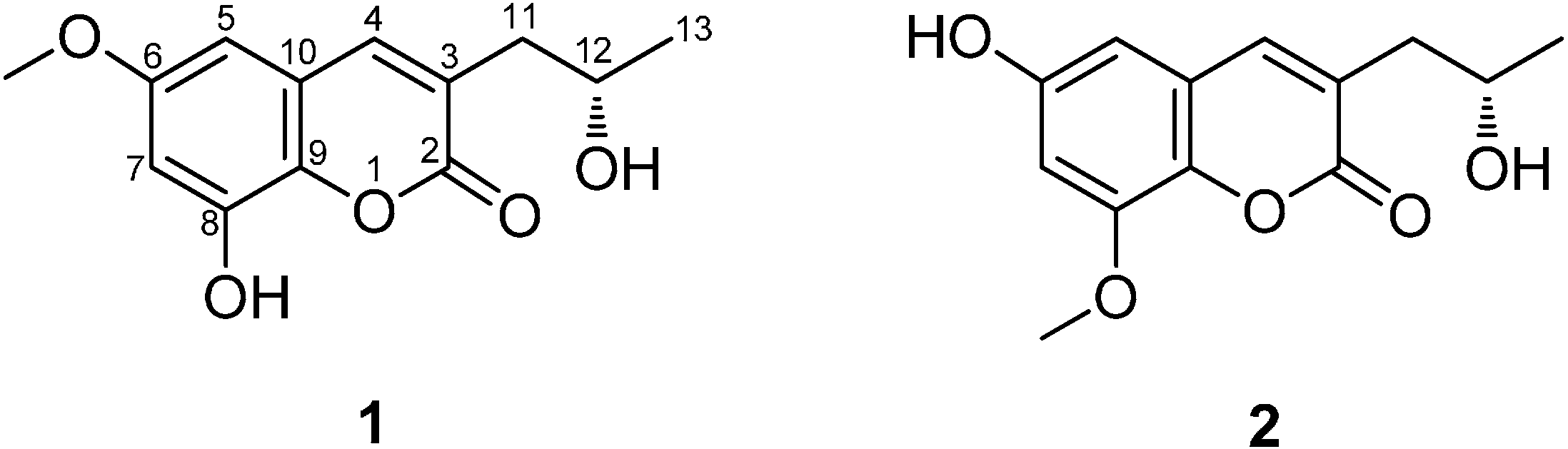
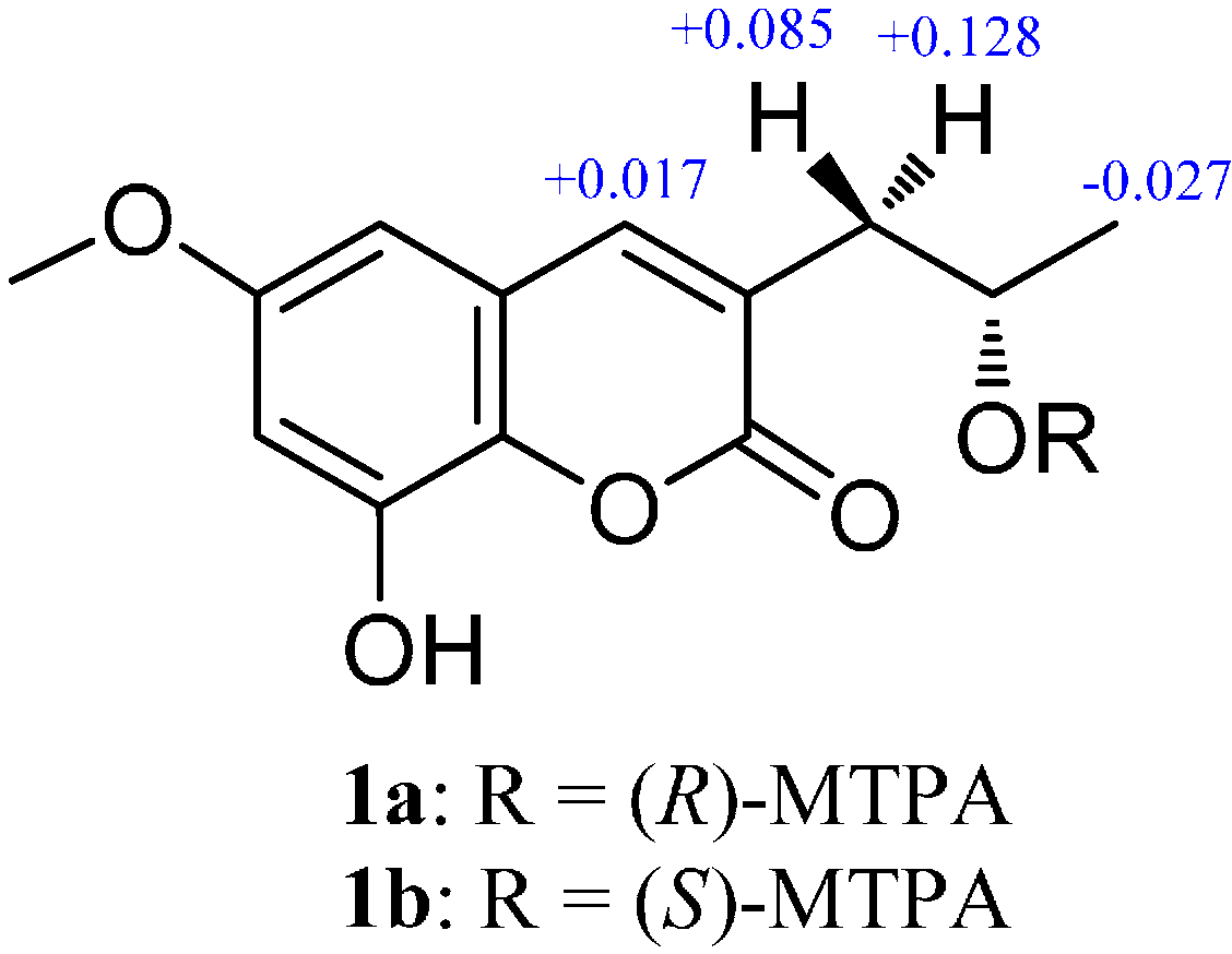
2. Results and Discussion

Compound 1 was isolated as a yellow amorphous powder, and its molecular formula was determined as C13H14O5 on the basis of the HR-ESI-MS (m/z 273.0740, calcd. 273.0739 [M+Na]+). The UV spectrum showed an absorption band with λmax 208, 257, and 294 nm, characteristic of coumarins [8]. The IR spectrum of 1 displayed absorption bands for hydroxyl (3261 cm−1), carbonyl (1684 cm−1) and aromatic (1592 and 1497 cm−1) groups. The 1H-NMR spectrum (Table 1) of 1 exhibited a pair of meta-positioned aromatic protons at δH 6.63 and 6.61 (each, 1H, d, J = 2.7 Hz), an olefinic proton at δH 7.72 (1H, s). It also displayed the signals of one methoxyl at δH 3.74 (3H, s), one methyl at δH 1.10 (3H, d, J = 6.2 Hz), an oxygenated methyne proton at δH 3.92 (1H, m), one methylene protons at δH 2.54 (1H, dd, J = 13.5, 4.9 Hz), 2.45 (1H, dd, J = 13.5, 7.8 Hz), as well as a phenolic hydroxyl at δH 10.20 (1H, s), and an alcoholic hydroxyl at δH 4.62 (1H, d, J = 4.5 Hz). The 13C-NMR spectrum (Table 1) combined with DEPT 135 spectrum displayed 13 resonances for an ester carbonyl carbon (δC 161.0), eight aromatic/olefinic carbons (δC 155.6, 145.1, 141.1, 136.4, 126.5, 120.2, 104.9, 100.1), one oxymethine carbon (δC 64.2), one methoxyl group (δC 55.4), one methylene carbon (δC 40.3), and one methyl group (δC 23.4).
The 1H-1H COSY correlations between H-12 and Ha,b-11/H3-13/12-OH, in combination with the HMBC correlations from Ha,b-11 to C-2/C-3/C-4, H-4 to C-2/C-9/C-10, H-5 to C-4/C-6/C-7/C-9, H-7 to C-9, and H3-13 to C-11/C-12, indicating the presence of 3-propyl-6,8-dioxy coumarin moiety in 1 (Figure 2). Moreover, the methoxyl group was located at C-6 by the HMBC correlation from 6-OCH3 to C-6. Considering the 13C-NMR chemical shifts of C-8 (δC 145.1) and C-12 (δC 64.2), as well as molecular formula, the two carbons should be connected with hydroxyl groups. Thus, the structure of 1 was fully elucidated to be as indicated in Figure 1, and it was named talacoumarin A.
Table 1. 13C- (100 MHz) and 1H- (400 MHz) NMR data for compounds 1 and 2 in DMSO-d6 (δ in ppm).
Position12
δCδH (J in Hz)δCδH (J in Hz)
1
2161.0160.9
3126.5126.6
4141.17.72 (s)140.97.69 (s)
5100.16.63 (d, 2.7)102.86.51 (d, 2.3)
6155.6153.9
7104.96.61 (d, 2.7)102.46.66 (d, 2.3)
8145.1147.0
9136.4135.7
10120.2119.9
1140.32.45 (dd, 13.5, 7.8), Ha40.22.44 (dd, 13.6, 7.7), Ha
2.54 (dd, 13.5, 4.9), Hb2.52 (overlapped), Hb
1264.23.92 (m)64.23.92 (m)
1323.41.10 (d, 6.2)23.41.09 (d, 6.1)
8-OH10.20 (s)
12-OH4.62 (d, 4.5)
6-OCH355.43.74 (s)
8-OCH355.93.84 (s)
Figure 2. Selected 1H-1H COSY and HMBC correlations of 1 and 2.
Molecules 19 20880 g002
The absolute configuration at C-12 was determined to be S by comparison of its optical rotation value with that of (S)-orthosporin (1: [ α ] D 20 +56.0 (c 0.5, CH3OH); (S)-orthosporin: [ α ] D 22 +61.8 (c 1.0, CH3OH) [9]). As confirmation, the absolute configuration at C-12 was established by the modified Mosher’s method [10]. The values of the (R)- and (S)-MTPA esters 1a and 1b also indicated the S configuration for C-12 (Figure 3).
Compound 2 possessed the same molecular formula and UV absorption characteristic as that of 1, suggesting 2 may be a coumarin isomer of 1. The NMR spectroscopic data (Table 1) suggested 2 was very similar to 1, except for the position of the methoxy group, which was located at C-8 for 2 instead of at C-6 based on the HMBC correlations from 8-OCH3 to C-8, from H-5 to C-7/C-9, and from H-7 to C-8/C-9 (Figure 2). With the aid of the 1H-1H COSY, HSQC, and HMBC correlations, the planar structure of 2 was established and all the 1H- and 13C-NMR signals were assigned.
Figure 3. Δδ values (in ppm) = δS − δR obtained for (S)-(1a) and (R)-MTPA (1b) esters.
Molecules 19 20880 g003
The optical rotation of 2 ( [ α ] D 20 +55.6 (c 0.5, CH3OH)) was consistent with that of 1, which suggested that 2 had the same configuration. The absolute configuration at C-12 was determined on the basis of the circular dichroism of the complex formed in situ with [Rh2(OCOCF3)4] [11,12], with the inherent contribution of the ligand subtracted. Upon addition of [Rh2(OCOCF3)4] to a solution of 2 in CH2Cl2, a metal complex was formed, acting as an auxiliary chromophore. It has been demonstrated that the sign of the E band (at ca. 350 nm) can be used to correlate the absolute configuration of a secondary and tertiary alcohol by applying the bulkiness rule. In this case, the Rh complex of 2 displayed a positive E band (Figure 4), correlating with a 12S absolute configuration. Hence, the structure of 2 was established as shown in Figure 1 and named to be talacoumarin B.
Figure 4. The CD spectrum of the Rh complex of 2 with the inherent CD spectrum subtracted.
Molecules 19 20880 g004
So far, natural products from fungi with the 3-alkyl-6,8-dioxycoumarin scaffold are relatively rare, and only eight such compounds have been reported [8,13,14,15]. The inhibitory activities against Aβ42 aggregation of compounds 1 and 2, along with that of the crude extract, were tested by a thioflavin T (ThT) assay [16] with epigallocatechin gallate (EGCG) as the positive control. Compounds 1 and 2 showed moderate anti-Aβ42 aggregation activities, with relative inhibitory rates of (49.33 ± 3.16)% and (44.99 ± 3.64)% [the positive control EGCG had a relative inhibitory rate of (67.23 ± 2.51)%] at the concentration of 100 μM, while the crude extract has no activity. This represents the first report of the anti-Aβ42 aggregation activity of coumarins. Compounds 1, 2, and the crude extract were also evaluated for the cytotoxicity against five human tumor cell lines (HL-60, SMMC-7721, A-549, MCF-7, and SW480) and the antimicrobial activity against Escherichia coil, Staphylococcus aureus, Candida albicans, and Aspergillus niger. However, none of them showed any cytotoxic (IC50 > 40 μM) or antimicrobial activities (MIC > 1.0 mg/mL).

3. Experimental Section

3.1. General Procedures

Optical rotations were measured using a JASCO P-1020 polarimeter (JASCO Corporation, Tokyo, Japan). The IR spectra (KBr) were recorded on a JASCO FT/IR-480 plus Fourier transform infrared spectrometer (JASCO Corporation). The UV spectra were recorded in CH3OH using a JASCO V-550 UV/Vis spectrophotometer (JASCO Corporation). 1H- (400 MHz), 13C- (100 MHz), DEPT 135 (100 MHz), and 2D (1H-1H COSY, HSQC, and HMBC) NMR spectra were recorded in DMSO-d6 on a Bruker AV 400 spectrometer using solvent signals (DMSO-d6: δH 2.50/δC 39.5) as an internal standard (Bruker Corporation, Fallanden, Switzerland). HR-ESI-MS were measured on a Waters Synapt G2 TOF mass spectrometer (Waters Corporation, Manchester, UK). Column chromatographies (CCs) were carried out on silica gel (200–300 mesh, Marine Chemical Group Corporation, Qingdao, China), and ODS (60–80 µm, YMC, Tokyo, Japan). Silica gel GF254 (Marine Chemical Group Corporation) was used for analytical TLC. The analytical HPLC was performed on a Shimadzu HPLC system equipped with a LC-20AB pump, and a SPD-20A diode array detector (Shimadzu, Kyoto, Japan), using a Phenomenex Gemini C18 column (5 μm, 4.6 mm × 250 mm, Phenomenex Inc., Torrance, CA, USA). The Preparative HPLC was performed on Shimadzu LC-6AD system equipped with a LC-6AD pump, and a SPD-M20A detector (Shimadzu), using an RP-18 column (5 μm, 21.2 mm × 250 mm, Gemini, Phenomenex Inc.; detector set at 220 and 254 nm).

3.2. Fungal Material and Culture

The strain of Talaromyces flavus (No.BYD07-13) was identified on the basis of the morphological characters and gene sequence analyses. The ITS, beta-tubulin, and calmodulin sequences of the strain have been deposited at GenBank as KF917583, KF917584, and KF917585, respectively. It was deposited in the culture collection at the Institute of Traditional Chinese Medicine and Natural Products, College of Pharmacy, Jinan University, Guangzhou, China. The fermentation of No.BYD07-13 was described in our previous paper [6].

3.3. Extraction and Isolation

The fermented material was extracted three times with EtOAc (3 × 6.0 L), and the organic solvent was evaporated to dryness under vacuum to afford the crude extract (46.2 g), which was dissolved in 90% v/v aqueous CH3OH (500 mL) and partitioned against an equal volume of cyclohexane. The aqueous CH3OH layer was evaporated to dryness under reduced pressure to give an aqueous CH3OH extract (w, 28.1 g), which was subjected to ODS column chromatography (CC) using a CH3OH-H2O gradient elution (30:70, 50:50, 70:30, and 100:0, v/v) to give four fractions (w1 to w4). The fraction w1 (574 mg) was further separated by ODS CC using a CH3OH/H2O gradient elution (30:70, 40:60, and 50:50, v/v, each 0.5 L) to give three subfractions (w1a to w1c). The subfraction w1a (351 mg) was purified by preparative HPLC eluting with CH3CN/H2O (v/v 15:85, flow rate: 8 mL/min) to afford compounds 1 (37.9 mg, tR 21.2 min) and 2 (5.0 mg, tR 16.1 min).

3.4. Spectroscopic Data

Talacoumarin A (1). Yellow amorphous powder, [ α ] D 20 +56.0 (c 0.5, CH3OH); UV (CH3OH) λmax (log ε) 208 (4.65), 257 (4.07), 294 (4.27) nm; IR (KBr): νmax 3261, 2962, 2920, 2853, 1684, 1592, 1510, 1454, 1392, 1351, 1203, 1164, 1109, 1053, 855, 840 cm−1; 1H- (DMSO-d6) and 13C-NMR (DMSO-d6) data, see Table 1. HR-ESI-MS: m/z 273.0740 ([M+Na]+, C13H14O5Na; calcd. 273.0739).
Talacoumarin B (2). Yellow amorphous powder, [ α ] D 20 +55.6 (c 0.5, CH3OH); UV (CH3OH) λmax (log ε) 208 (4.65), 257 (4.07), 294 (4.27) nm; IR (KBr): νmax 3261, 2962, 2920, 2853, 1684, 1592, 1510, 1454, 1392, 1351, 1292, 1203, 1109, 1053, 855, 841 cm−1; 1H-NMR (DMSO-d6) and 13C-NMR (DMSO-d6) data, see Table 1. HR-ESI-MS: m/z 273.0737 ([M+Na]+, C13H14O5Na; calcd. 273.0739).

3.5. Preparation of (S)- and (R)-MTPA Esters of 1 (1a and 1b)

A solution of 1 (0.5 mg) in pyridine-d5 (0.5 mL) was treated with (S)-MTPA chloride (8 µL) under an atmosphere of nitrogen in an NMR tube. The mixture was stirred at room temperature for 20 h to obtain the (R)-MTPA ester (1b). The same procedure was used to prepare the (S)-MTPA ester (1a) with (R)-MTPA chloride.

3.6. Absolute Configuration of the Secondary Alcohol in 2

According to a published procedure [11,12], 2 (0.5 mg) was dissolved in a dry solution of the stock [Rh2(OCOCF3)4] complex (1.0 mg) in CH2Cl2 (200 μL). The first CD spectrum was recorded immediately after mixing, and its time evolution was monitored until stationary (about 10 min after mixing). The inherent CD was subtracted. The observed sign of the E band at ca. 350 nm in the induced CD spectrum was correlated to the absolute configuration of the C-12 secondary alcohol moiety.

3.7. ThT Fluorescence Assay

The ThT fluorescene assay was performed as described in our previous paper [16], with EGCG as the positive control. Biological activity was determined as relative inhibitory activity (Vi) for each sample according to the formula: Vi = 100 − [(Fi − Fb)/F0] × 100, where Fi is the fluorescence value of the sample, Fb is its bank value, and F0 is the fluorescence value for free aggregation of a sample of Aβ42 incubated in the same buffer/HFIP/DMSO system and in absence of inhibitors.

3.8. Cytotoxicity Assay

Cytotoxic activity was tested against five human cell lines (HL-60, SMMC-7721, A-549, MCF-7, and SW480) using the MTT method as described in our previous paper [6]. Cisplatin and paclitaxel were used as the positive controls.

3.9. Antimicrobial Assay

The antimicrobial activity against E. coil, S. aureus, C. albicans, and A. niger were evaluated by an agar dilution method, which described in our previous paper [7]. Tobramycin was used as the positive control.

4. Conclusions

Our investigation on the metabolites of the wetland soil-derived fungus T. flavus BYD07-13 resulted in the isolation of two new coumarins, named talacoumarins A (1) and B (2). Compounds 1 and 2 both have the 3-alkyl-6,8-dioxycoumarin moiety, which is relatively rare in fungal metabolites. The absolute configurations of 1 and 2 were determined by the modified Mosher’s method and the CD analysis of the in situ formed [Rh2(OCOCF3)4] complex. It is noteworthy that the absolute configuration of branched chain alcohol of 3-alkyl-6,8-dioxycoumarin was determined for the first time. Compounds 1 and 2 showed moderate anti-Aβ42 aggregation activity, making this the first report on the Aβ42 inhibitory aggregation activity of coumarins.

Acknowledgments

This work was financially supported by grants from the Ministry of Science and Technology of China (2012ZX09301002-003001006), the National Natural Science Foundation of China (81422054, 81373306), the Guangdong Natural Science Funds for Distinguished Young Scholar (S2013050014287), Guangdong Province Universities and Colleges Pearl River Scholar Funded Scheme (Hao Gao, 2014).

Author Contributions

XSY, HG, YY and JWH designed research; JWH, DPQ, RQK and XZL performed research and analyzed the data; JWH and YY wrote the paper. All authors read and approved the final manuscript.

Conflicts of Interest

The authors declare no conflict of interest.

References

  1. Bara, R.; Zerfass, I.; Aly, A.H.; Goldbach-Gecke, H.; Raghavan, V.; Sass, P.; Mándi, A.; Wray, V.; Polavarapu, P.L.; Pretsch, A.; et al. Atropisomeric dihydroanthracenones as inhibitors of multriesistant Staphylococcus aureus. J. Med. Chem. 2013, 56, 3257–3272. [Google Scholar] [CrossRef] [PubMed]
  2. Bara, R.; Aly, A.H.; Wray, V.; Lin, W.H.; Proksch, P.; Debbab, A. Talaromins A and B, new cyclic peptides from the endophytic fungus Talaromyces wortmannii. Tetrahedron Lett. 2013, 54, 1686–1689. [Google Scholar] [CrossRef]
  3. Guo, J.P.; Zhu, C.Y.; Zhang, C.P.; Chu, Y.S.; Wang, Y.L.; Zhang, J.X.; Wu, D.K.; Zhang, K.Q.; Niu, X.M. Thermolides, potent nematocidal PKS-NRPS hybrid metabolites from thermophilic fungus Talaromyces thermophilus. J. Am. Chem. Soc. 2012, 34, 20306–20309. [Google Scholar] [CrossRef]
  4. Li, H.X.; Huang, H.B.; Shao, C.L.; Huang, H.R.; Jiang, J.Y.; Zhu, X.; Liu, Y.Y.; Liu, L.; Lu, Y.J.; Li, M.F.; et al. Cytotoxic norsesquiterpene peroxides from the endophytic fungus Talaromyces flavus isolated from the mangrove plant Sonneratia apetala. J. Nat. Prod. 2011, 74, 1230–1235. [Google Scholar] [CrossRef] [PubMed]
  5. Proksa, B. Talaromyces flavus and its metabolites. Chem. Pap. 2010, 64, 696–714. [Google Scholar] [CrossRef]
  6. He, J.W.; Mu, Z.Q.; Gao, H.; Chen, G.D.; Zhao, Q.; Hu, D.; Sun, J.Z.; Li, X.X.; Li, Y.; Liu, X.Z.; et al. New polyesters from Talaromyces flavus. Tetrahedron 2014, 70, 4425–4430. [Google Scholar] [CrossRef]
  7. He, J.W.; Liang, H.X.; Gao, H.; Kuang, R.Q.; Chen, G.D.; Hu, D.; Wang, C.X.; Liu, X.Z.; Li, Y.; Yao, X.S. Talaflavuterpenoid A, a new nardosinane-type sesquiterpene from Talaromyces flavus. J. Asian Nat. Prod. Res. 2014, 16, 1029–1034. [Google Scholar] [CrossRef] [PubMed]
  8. Xu, J.; Kjer, J.; Sendker, J.; Wray, V.; Guan, H.S.; Edrada, R.; Müller, W.E.G.; Bayer, M.; Lin, W.H.; Wu, J.; et al. Cytosporones, coumarins, and an alkaloid from the endophytic fungus Pestalotiopsis sp. isolated from the Chinese mangrove plant Rhizophora mucronata. Bioorg. Med. Chem. 2009, 17, 7362–7367. [Google Scholar] [CrossRef] [PubMed]
  9. Ichihara, A.; Hashtmoto, M.; Hirat, T.; Takeda, I.; Sasamura, Y.; Sakamura, S.; Sato, R.; Tajimi, A. Structure, synthesis, and stereochemistry of (+)-orthosporin, a phytotoxic metabolite of Rhynchosporium orthosorum. Chem. Lett. 1989, 18, 1495–1498. [Google Scholar] [CrossRef]
  10. Dale, J.A.; Mosher, H.S. Nuclear magnetic resonance enantiomer regents. Configurational correlations via nuclear magnetic resonance chemical shifts of diastereomeric mandelate, O-methylmandelate, and alpha-methoxy-alpha-trifluoromethylphenylacetate (MTPA) esters. J. Am. Chem. Soc. 1973, 95, 512–519. [Google Scholar]
  11. Gerards, M.; Snatzke, G. Circular dichroism, XCIII determination of the absolute configuration of alcohols, olefins, epoxides, and ethers from the CD of their “in situ” complexes with [Rh2(O2CCF3)4]. Tetrahedron: Asymmetry 1990, 1, 221–236. [Google Scholar] [CrossRef]
  12. Frelek, J.; Szczepek, W.J. [Rh2(OCOCF3)4] as an auxiliary chromophore in chiroptical studies on steroidal alcohols. Tetrahedron: Asymmetry 1999, 10, 1507–1520. [Google Scholar] [CrossRef]
  13. Ayer, W.A.; Racok, J.S. The metabolites of Talaromyces flavus: Part 1. Metabolites of the organic extracts. Can. J. Chem. 1990, 68, 2085–2094. [Google Scholar] [CrossRef]
  14. Stierle, A.A.; Stierle, D.B.; Kemp, K. Nover sesquiterpenoid matrix metalloproterinase-3 inhibitors from an acid mine waste extremophile. J. Nat. Prod. 2004, 67, 1392–1395. [Google Scholar] [CrossRef] [PubMed]
  15. Teles, H.L.; Sordi, R.; Silva, G.H.; Castro-Gamboa, I.; Bolzani, V.D.S.; Pfenning, L.H.; Abreu, L.M.D.; Costa-Neto, C.M.; Young, M.C.M.; Araújo, Ȃ.R. Aromatic compounds produced by Periconia atropurpurea, an endophytic fungus associated with Xylopia aromatica. Phytochemistry 2006, 67, 2686–2690. [Google Scholar] [CrossRef] [PubMed]
  16. Zheng, Q.C.; Chen, G.D.; Kong, M.Z.; Li, G.Q.; Cui, J.Y.; Li, X.X.; Wu, Z.Y.; Guo, L.D.; Cen, Y.Z.; Zheng, Y.Z.; et al. Nodulisporisteriods A and B, the first 3,4-seco-4-methyl-progesteroids from Nodulisporium sp. Steroids 2013, 78, 896–901. [Google Scholar] [CrossRef] [PubMed]
  • Sample Availability: Not available.

