Article

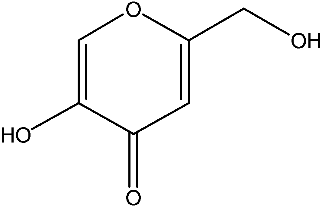
Augmenting the Antifungal Activity of an Oxidizing Agent with Kojic Acid: Control of Penicillium Strains Infecting Crops

Foodborne Toxin Detection and Prevention Research Unit, Western Regional Research Center, USDA-ARS, 800 Buchanan St., Albany, CA 94710, USA
*
Author to whom correspondence should be addressed.
Molecules 2014, 19(11), 18448-18464; https://doi.org/10.3390/molecules191118448
Submission received: 24 September 2014 / Revised: 4 November 2014 / Accepted: 5 November 2014 / Published: 12 November 2014
(This article belongs to the Section Natural Products Chemistry)

Abstract

:
Oxidative treatment is one of the strategies for preventing Penicillium contamination in crops/foods. The antifungal efficacy of hydrogen peroxide (H2O2; oxidant) was investigated in Penicillium strains by using kojic acid (KA) as a chemosensitizing agent, which can enhance the susceptibility of pathogens to antifungal agents. Co-application of KA with H2O2 (chemosensitization) resulted in the enhancement of antifungal activity of either compound, when compared to the independent application of each agent alone. Of note, heat enhanced the activity of H2O2 to a greater extent during chemosensitization, whereby the minimum inhibitory or minimum fungicidal concentrations of H2O2 was decreased up to 4 or 13 fold, respectively, at 35–45 °C (heat), when compared to that at 28 °C (normal growth temperature). However, heat didn’t increase the antifungal activity of KA, indicating specificity exists between heat and types of antifungals applied. The effect of chemosensitization was also strain-specific, where P. expansum (both parental and fludioxonil-resistant mutants) or P. italicum 983 exhibited relatively higher susceptibility to the chemosensitization, comparing to other Penicillium strains tested. Collectively, chemosensitization can serve as a potent antifungal strategy to lower effective dosages of toxic antifungal substances, such as H2O2. This can lead to coincidental lowering of environmental and health risks.

Graphical Abstract

1. Introduction

Kojic acid (5-hydroxy-2-(hydroxymethyl)-4H-pyran-4-one, KA, Figure 1), is a natural compound produced by certain filamentous fungi (Aspergillus, Penicillium) or Acetobacter ([1,2] and references therein). KA is widely used as a food additive, as a depigmenting/skin-whitening agent (via inhibition of tyrosinase, a key enzyme involved in melanogenesis in melanoma and melanocytes), as an antitumor or anti-leishmanial agent, etc. [3,4,5,6]. KA can inhibit bacterial/fungal infection [7,8], where KA functions as an enhancer of host immunity [9,10]. For instance, KA stimulates phagocytosis, induces the generation of reactive oxygen species in macrophages, and potentiates phytohemagglutinin-based proliferation of lymphocytes [9,10]. KA also showed a fungistatic antifungal activity against Cryptococcus neoformans, a causative agent triggering human cryptococcosis, in which KA inhibited melanin biosynthesis required for fungal infectivity [11].
Certain Aspergillus strains, such as A. flavus or A. parasiticus, produce hepato-carcinogenic aflatoxins (AFs). Recently, the use of atoxigenic (i.e., AF non-producing) Aspergillus strains as biocontrol agents in agricultural environments [12] has prompted further investigation into their mode of action. It was found that although the atoxigenic strains do not produce AFs, as expected, they do still produce KA [13], which can act as an antagonizing agent against other co-infecting microbes in crops [7]. Therefore, the secondary metabolite KA produced by microorganisms could function as a “biotic” stressor to co-infecting pathogens [7].
Figure 1. Structure of kojic acid (KA).
Molecules 19 18448 g001
The filamentous fungi in the genus Penicillium are frequently involved in food contamination or postharvest decay. For example, P. expansum is the main producer of the mycotoxin patulin (PAT) that triggers harmful effects on human/animal health [14]. Apples and apple products are the main sources of PAT contamination. PAT is a potential endocrine disruptor, and modulates hormone production [14]. Meanwhile, other Penicillium strains, such as P. digitatum and P. italicum are the most devastating citrus pathogens, causing significant economic losses to the industry during post-harvest [15]. Therefore, development of strategies for early intervention of mycotoxin production or fungal contamination in crops or foods is urgently needed.
Various environmental factors, such as fluctuation of temperature, humidity or pH, etc., can be sources of “abiotic” stress to fungal pathogens. Accordingly, abiotic stresses artificially applied could adversely affect the growth of fungi, resulting in the reduction of fungal contamination or postharvest decay. Heat or oxidative treatment is one of the strategies to prevent contamination by Penicillium in foods or crops. For example, immersion of pear fruit in heated water (55 °C) controlled P. expansum-triggered fruit decay, which also allowed the delay of fruit ripening during storage [16]. Likewise, combined application of oxidizing compounds, such as H2O2 and sodium hypochlorite (NaClO), resulted in synergistic antifungal effect on P. expansum, rendering effective control of postharvest decay in fresh fruits [17].
Co-application of certain types of compounds with conventional antimicrobial drugs/fungicides can enhance the effectiveness of drugs/fungicides through a mechanism termed “chemosensitization” [18,19,20,21]. For example, the combination of the antifungal drug fluconazole (FLC) with various non-antifungal agents, such as traditional Chinese plant extracts, inhibitors of cell signaling (e.g., calcineurin) or heat shock protein 90, etc., increased the susceptibility of the yeast pathogen Candida albicans to FLC [20]. Therefore, chemosensitization strategy could lead to (1) lowering dosages of commercial, toxic drugs or antifungal agents required for effective control of pathogens and (2) controlling pathogen resistance to antifungal drugs/agents [22,23,24].
In this study, antifungal chemosensitization is investigated for effective control of pathogenic strains of Penicillium (See Table 1) by co-applying KA with H2O2, which mimics host reactive oxygen species. Levels of compound interactions, at moderate (28 °C) to high temperatures (35, 45 °C), are determined and compared according to the method outlined by the Clinical Laboratory Standards Institute (CLSI) M38-A [25]. Results demonstrate that KA chemosensitizes Penicillium strains to H2O2, lowering the effective dosages of H2O2 required for control of Penicillium. The potency of H2O2 is greater when Penicillium strains are treated with heat (35–45 °C).
Table 1. Penicillium strains used in this study.
Penicillium StrainsStrain CharacteristicsSource/Reference
Group A (P. expansum):
P. expansum W1Plant pathogen (Parental strain)[26]
P. expansum FR2Plant pathogen, Fludioxonil resistant mutant derived from P. expansum W1[26]
P. expansum W2Plant pathogen (Parental strain)[26]
P. expansum FR3Plant pathogen, Fludioxonil resistant mutant derived from P. expansum W2[26]
Group B (Other Penicillium strains):
P. glabrum 766Plant pathogenNRRL a
P. chrysogenum 824Fleming’s penicillin-producing strainNRRL
P. griseofulvum 2159Plant pathogenNRRL
Group C (Citrus pathogens):
P. digitatum 786Plant pathogenNRRL
P. italicum 983Plant pathogenNRRL
a: NRRL, National Center for Agricultural Utilization and Research, USDA-ARS, Peoria, IL, USA.

2. Results and Discussion

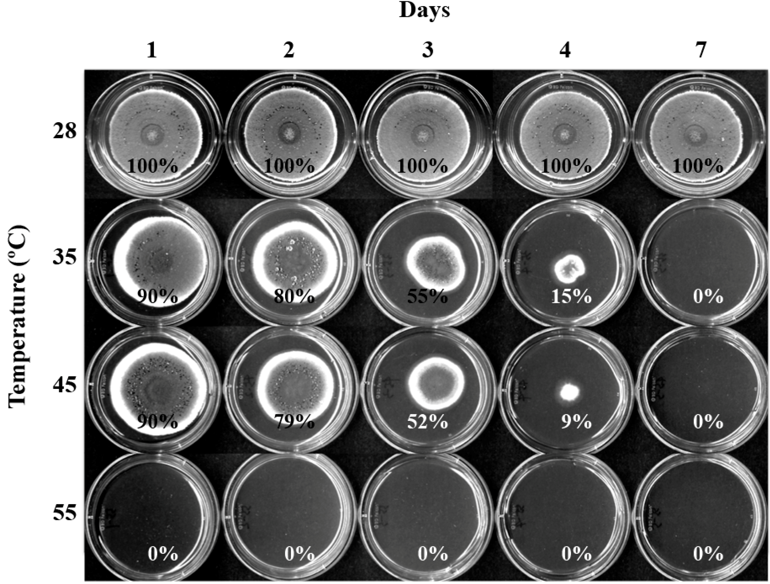
2.1. Susceptibility of P. expansum W1 to High Temperatures

Heat response of Penicillium was tested using P. expansum W1 (Parental, wild type strain) as a representative strain. P. expansum W1 was cultivated on potato dextrose agar (PDA) at high temperatures (35, 45, 55 °C) (1, 2, 3, 4 d), and was then transferred to 28 °C (moderate, normal growth temperature) for growth recovery (for up to 7 d; see Experimental section). For control, W1 was cultivated at 28, 35, 45 and 55 °C for 7 d.
As shown in Figure 2, P. expansum W1 did not grow (namely, no sign of germination) on PDA when it was cultivated solely at 35, 45 or 55 °C for 7 d. However, P. expansum W1 treated with heat (35, 45 °C) for 1–4 d could recover growth after cells were transferred to 28 °C (grown up to 7 d). W1 cultivated at 55 °C could not recover growth even after 1 day-heat treatment at 55 °C. Based on this result, moderate (28 °C) to high (35, 45 °C) temperatures, except 55 °C, were chosen for further antifungal investigation in this study.
Figure 2. Fungal plate bioassay showing sensitivity of P. expansum W1 to high temperatures (35, 45, 55 °C). The % values shown are relative growth rate of W1 compared to that at 28 °C (positive control). SD < 5%.
Molecules 19 18448 g002

2.2. Susceptibility of Penicillium Strains to Chemosensitization

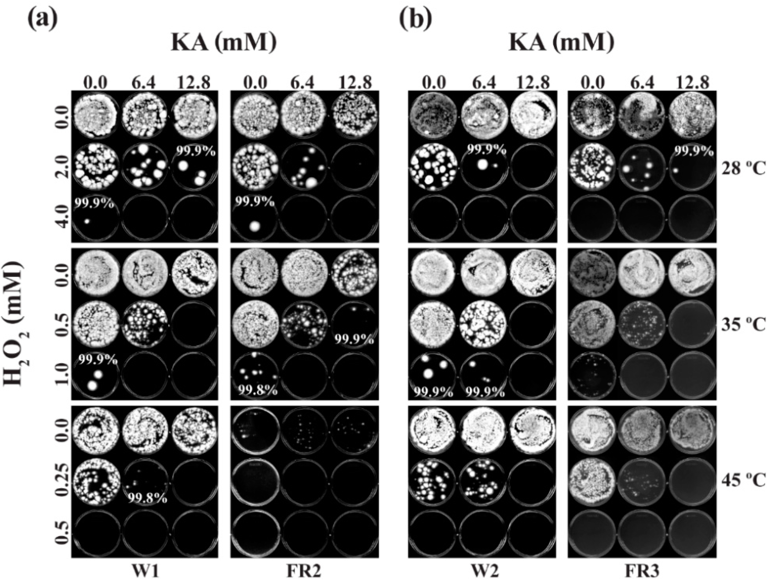
2.2.1. Effect of Co-Application of KA with H2O2 on Fungal Growth: at 24 h

At 24 h of CLSI-based fungal cultivation (at 28, 35, 45 °C), Penicillium strains didn’t visibly grow in RPMI liquid medium (microtiter plates), and hence, the values of Minimum Inhibitory Concentrations (MICs)/Fractional Inhibitory Concentration Indices (FICIs) could not be determined (Supplementary Tables S1 and S2), whereas, Minimum Fungicidal Concentrations (MFCs)/Fractional Fungicidal Concentration Indices (FFCIs) could be determined when the liquid cultures (200 μL/micotiter plate well) from the respective temperature were transferred onto PDA and cultivated for additional 48 h (at 28 °C; see Experimental section) except P. digitatum 786, which was unable to grow on PDA if cultured at 45 °C in microtiter plates (namely, heat sensitive). The values of FFCIs ranged from “additive” (0.5 < FFCI ≤ 1) to “neutral” (1 < FFCI ≤ 2) [27] (Supplementary Table S1 (Average MFC/FFCI values of Group A, B, C strains), Supplementary Table S2 (MFC/FFCI values of individual strains in Group A, B, C)). Despite the absence of calculated “synergistic” interaction, as determined by “additive” or “neutral” interactions during chemosensitization (Supplementary Table S1), there was enhanced antifungal activity of H2O2 and also KA in most Penicillium strains examined at 24 h, which was reflected in lowered MFCs of each compound when combined.
In general, the higher the temperature, the lower the concentration of H2O2 required for achieving ≥99.9% killing of Penicillium strains (viz. enhanced antifungal activity of H2O2 by heat). For example, the average MFCs (MFCAlone) of H2O2 in Group A were 7.0, 2.0 or 0.6 mM at 28, 35 or 45 °C, respectively, thus exhibiting the highest or lowest MFCAlone value at 28 or 45 °C, respectively. When H2O2 was co-applied with KA (chemosensitization), the average MFCs of H2O2 were lowered further, resulting in MFCCombined values of 4.0, 1.0 or 0.3 mM at 28, 35 or 45 °C, respectively. Therefore, the values of MFCCombined (chemosensitization) were 1.7 to 2.0 times lower comparing to that of MFCAlone (independent application). Of note, there was up to 13-fold decrease in MFCCombined in Group A at 45 °C (MFC: 0.3 mM) comparing to that at 28 °C (MFC: 4.0 mM).
The effect of chemosensitization was also identified in Group B, where co-application of H2O2 with KA lowered the MFC values of H2O2. Consequently, chemosensitization resulted in 1.2 to 1.8 times lower MFC values (MFCCombined) of H2O2 comparing to the independent application of H2O2, alone. The only exception is P. griseofulvum 2159, which showed indifference to chemosensitization (namely, no change in MFC values with KA + H2O2; FFCI value = 2.0). Comparing to Group A, the average MFCs of H2O2 for Group B were relatively higher, viz. 1.8 to 4.0 times higher for MFCAlone and 2.0 to 4.7 times higher for MFCCombined, respectively (Supplementary Table S1). Therefore, results indicated that Group B strains were less susceptible to H2O2 than the Group A.
In Group C, two citrus pathogens were examined for their responses to H2O2/KA. The MFC values of H2O2 in P. italicum 983 were 2, 1 or 0.125 mM for MFCAlone (independent application) and 1, 1 or 0.0625 mM for MFCCombined (chemosensitization) at 28, 35 or 45 °C, respectively (Supplementary Table S2). Hence, P. italicum 983 exhibited similar trends of H2O2 sensitivity to Group A or B strains, where (1) heat enhanced H2O2 activity and (2) chemosensitization enhanced the antifungal activity of H2O2 (Exception: no change in MFCCombined at 35 °C). However, tests with P. digitatum 786, the other citrus pathogen, showed that while the MFCs (MFCAlone) of H2O2 were lowered from 4 to 1 mM when temperature was increased from 28 to 35 °C (namely, heat enhancement of H2O2 activity), this strain was unable to grow at 45 °C (viz. higher heat sensitivity than P. italicum 983). Moreover, P. digitatum 786 showed indifference to KA-mediated chemosensitization, resulting in no change in MFC values when KA was co-applied with H2O2 (Supplementary Table S2; FFCI = 2.0). Therefore, two citrus pathogens exhibited different responses to the antifungal treatments. Noteworthy is that P. digitatum was previously shown to possess a capability to suppress a defense-related H2O2 production in host tissue [28]. Collectively, results indicated that the effect of antifungal chemosensitization is strain-specific.
Regarding the chemosensitizing agent KA, MFC values of KA were also lowered in most strains when KA was co-applied with H2O2, where the level of average MFCCombined was 1.3 to 2.3 times lower than MFCAlone depending on types of strains (Supplementary Table S1; See Supplementary Table S2 for exceptions, where FFCI = 2.0). However, unlike in H2O2, heat did not enhance the activity of KA, suggesting specificity also exists between heat and types of antifungal compounds applied for the enhancement of antifungal activity.
Altogether, at 24 h, KA chemosensitizes most Penicillium strains tested, where co-application of KA with H2O2 resulted in increased antifungal activity of either agents. Furthermore, heat (35, 45 °C) enhanced the activity of H2O2, while that of KA was vastly unaffected by heat treatment. Regarding the stability of H2O2, H2O2 is sensitive to light, pH and/or heat. Especially, heat can induce chemical decomposition of H2O2 into H2O and O2. The O2 generated can be used for mitochondrial respiration in fungi, while it also means increase of pressure. Therefore, O2 level and pressure as well as the level of mitochondrial respiration may be of interest in the future chemosensitization study.

2.2.2. Effect of Co-Application of KA with H2O2 on Fungal Growth: at 48 h

At 48 h of CLSI-based fungal cultivation, both MICs and MFCs (thus FICI and FFCI values, accordingly) could be determined depending on types of Penicillium strains or growth temperatures. For instance, MICs/FICIs could be measured in Group A or C at 28 °C (in microtiter plates), while those values could not be determined at 35 or 45 °C due to incapability of their growth at the higher temperatures. Whereas, MICs/FICIs could be determined in Group B at both 28 and 35 °C (45 °C: no growth), thus showing relatively higher tolerance of Group B to heat (35 °C) comparing to Group A or C in the liquid culture (Table 2, Supplementary Table S3).
The MIC values, namely MICAlone or MICCombined, of H2O2 or KA were lowered after chemosensitization, as follows: (1) 2.2 or 3.5 times lowered for H2O2 or KA, respectively, in Group A (28 °C), (2) 1.5 or 1.7 times lowered for H2O2 and 1.4 or 2.1 times lowered for KA at 28 or 35 °C, respectively, in Group B, and (3) 2.0 times lowered for both H2O2 and KA in Group C (28 °C). In all Groups, FICI values ranged from “additive” (0.5 < FICI ≤ 1) to “neutral” (1 < FICI ≤ 2) [27]. Of note, in Group B, there was up to 4-fold decrease in MICCombined when temperature was increased from 28 °C (MIC: 5.3 mM) to 35 °C (MIC: 1.3 mM). Despite the absence of calculated “synergistic” interaction, as determined by “additive” or “neutral” interactions (Table 2), there was enhanced antifungal activity of H2O2 and KA in most Penicillium strains examined at 48 h, which was reflected in lowered MICs of each compound when combined.
The values of MFCs/FFCIs could also be determined on PDA for most strains (Group A, B or C) at 28, 35 and 45 °C. Exceptions are P. glabrum 766, P. chrysogenum 824, P. italicum 983 and P. digitatum 786, which were unable to grow at 45 °C. Noteworthy is that, when compared to 24 h (See above; Supplementary Tables S1 and S2), 48 h of cultivation of Penicillium strains, in general, required lower concentration of H2O2 to achieve ≥99% fungal death. For example, when Group A strains were co-treated with 12.8 mM of KA (chemosensitization) for 48 h, ≥99.9% fungal death was achieved with 2.0, 0.5 or 0.2 mM of H2O2 at 28, 35 or 45 °C, respectively (Table 2; See also Figure 3), while similar level of fungal death was achieved with 4.0, 1.0 or 0.3 mM of H2O2 at 28, 35 or 45 °C, respectively, at 24 h (Supplementary Table S1). Therefore, when compared to 24 h, 1.5 to 2-fold less concentrations of H2O2 were required to achieve ≥99.9% fungal death at 48 h during chemosensitization. Similar trends were also observed in Group B and C (The only exception was MFCs in Group C at 28 °C, where MFCAlone or MFCCombined was similar to or slightly higher than that of 24 h).
Table 2. Antifungal chemosensitization of kojic acid (KA; mM) to hydrogen peroxide (H2O2; mM) at different temperatures tested against Penicillium strains. Summary of CLSI-based microdilution bioassays (Average MIC/FICI and MFC/FFCI values of Group A, B, C strains at 48 h) a.
Group A (P. expansum W1, FR2, W2, FR3)
28 °C
CompoundsMIC AloneMIC CombinedFICIMFC AloneMFC CombinedFFCI
MeanKA25.67.20.725.6 b10.40.9
H2O24.01.84.02.0
t-test KA-p <0.005--p <0.005 c-
H2O2-p <0.005--p <0.005-
35 °C
MeanKA///25.612.80.8
H2O2// 1.50.5
t-test KA-/--p <0.005-
H2O2-/--p <0.05-
45 °C
MeanKA///25.616.00.6
H2O2// 0.40.2
t-test KA-/--p <0.05-
H2O2-/--P, insignificant-
Group B (P. glabrum 766, P. chrysogenum 824, P. griseofulvum 2159)
28 °C
MeanKA25.617.61.425.613.91.1
H2O28.05.38.04.7
t-test KA-P, insignificant--P, insignificant-
H2O2-P, insignificant--P, insignificant-
35 °C
MeanKA25.611.71.025.619.21.4
H2O22.31.34.02.7
t-testKA-P, insignificant P, insignificant--P, insignificant-
H2O2-P, insignificant-
45 °C
MeanKA//////, ND d
H2O2// //
t-testKA-/--/-
H2O2-/--/-
Group C (P. italicum 983, P. digitatum 786)
28 °C
MeanKA25.612.81.025.625.62.0
H2O23.01.53.03.0
t-testKA-ND e--ND e-
H2O2-ND e--ND e-
35 °C
MeanKA///25.619.21.4
H2O2//0.60.4
t-test KA-/--ND e-
H2O2/-ND e-
45 °C
MeanKA//////, ND f
H2O2////
t-test KA-/--/-
H2O2/-/-
a MIC: Minimum inhibitory concentration, MFC: Minimum fungicidal concentration, FICI: Fractional Inhibitory Concentration Indices, FFCI: Fractional Fungicidal Concentration Indices; b KA was tested up to 12.8 mM. For calculation purpose, 25.6 mM (doubling of 12.8 mM) was used; c Student’s t-test for paired data (combined, i.e., chemosensitization) was vs. mean MIC or MFC of each compound (alone, i.e., no chemosensitization) determined in strains; d ND, Not determined (No cell growth except P. griseofulvum 2159, for which FFCI is neutral); e ND, Not determined (Few data); f ND, Not determined (No growth of Group C strains).
Figure 3. Chemosensitization (KA + H2O2) test in P. expansum wild type and fludioxonil-resistant mutants at moderate (28 °C) to high (35, 45 °C) temperatures. Results shown here are determination of MFCs of antifungal agents (48 h). (a) W1 and FR2; (b) W2 and FR3.
Molecules 19 18448 g003
As observed in 24 h, the higher the temperature, the lower the concentration of H2O2 needed for achieving ≥99.9% killing of Penicillium strains at 48 h. For example, the average MFCAlone values of H2O2 in Group A at 48 h were 4.0, 1.5 or 0.4 mM at 28, 35 or 45 °C, respectively, indicating 10 times lower concentration of H2O2 (45 °C) was required to achieve ≥99.9% fungal death, when compared to 28 °C. With the chemosensitization, MFCCombined values of H2O2 were lowered further to 2.0, 0.5 or 0.2 mM at 28, 35 or 45 °C, respectively, thus showing that 10 times less H2O2 was needed to achieve ≥99.9% fungal death at 45 °C, when compared to 28 °C. Similar trends in the heat enhancement of H2O2 activity were also found in Groups B and C during chemosensitization. As observed in 24 h, the average MFCs of H2O2 in Group B (both MFCAlone and MFCCombined) were relatively higher than that in Group A (namely, 2.0 to 5.4 times higher), thus confirming further that Group B was less susceptible to H2O2 than Group A.
Interestingly, although Group B exhibited higher tolerance to heat (at 35 °C) comparing to Group A or C (See above; microtiter plate liquid bioassay), two strains in Group B, P. glabrum 766 and P. chrysogenum 824, were unable to recover their growth on PDA at the elevated temperature, viz. 45 °C (thus cannot determine their MFCs/FFCIs). However, Group A strains could still recover their growth at 45 °C. Thus, results indicated differential range of optimum growth temperatures for each fungus tested.
Regarding the chemosensitizing agent KA, MFCs of KA were also lowered in most strains when KA was co-applied with H2O2, where the level of MFCCombined of KA (chemosensitization) was 1.3 to 2.4 times lower than MFCAlone (independent application), depending on types of strains (See Supplementary Table S3 for exceptions, where FICI or FFCI = 2.0). However, at 45 °C, MIC/MFC values of either H2O2 or KA in Group B or C couldn’t be determined since most strains in these Groups were unable to grow.
Collectively, at 48 h, KA chemosensitizes most Penicillium strains tested, where co-application of KA with H2O2 resulted in increased antifungal activity of either agents. As observed in 24 h, heat (35, 45 °C) enhanced the activity of H2O2, while that of KA was vastly unaffected by heat treatment. Therefore, results proved further that specificity exists between heat and types of antifungals applied. Also, effect of chemosensitization was strain-specific, where P. expansum (both parental and fludioxonil-resistant mutant strains) or P. italicum 983 exhibited relatively higher susceptibility to H2O2, comparing to other Penicillium strains. Considering KA can be oxidized by H2O2 under heating conditions, the oxidized KA might also possess an antifungal property. Determination of the precise amount of KA by using HPLC, etc., warrants future study.

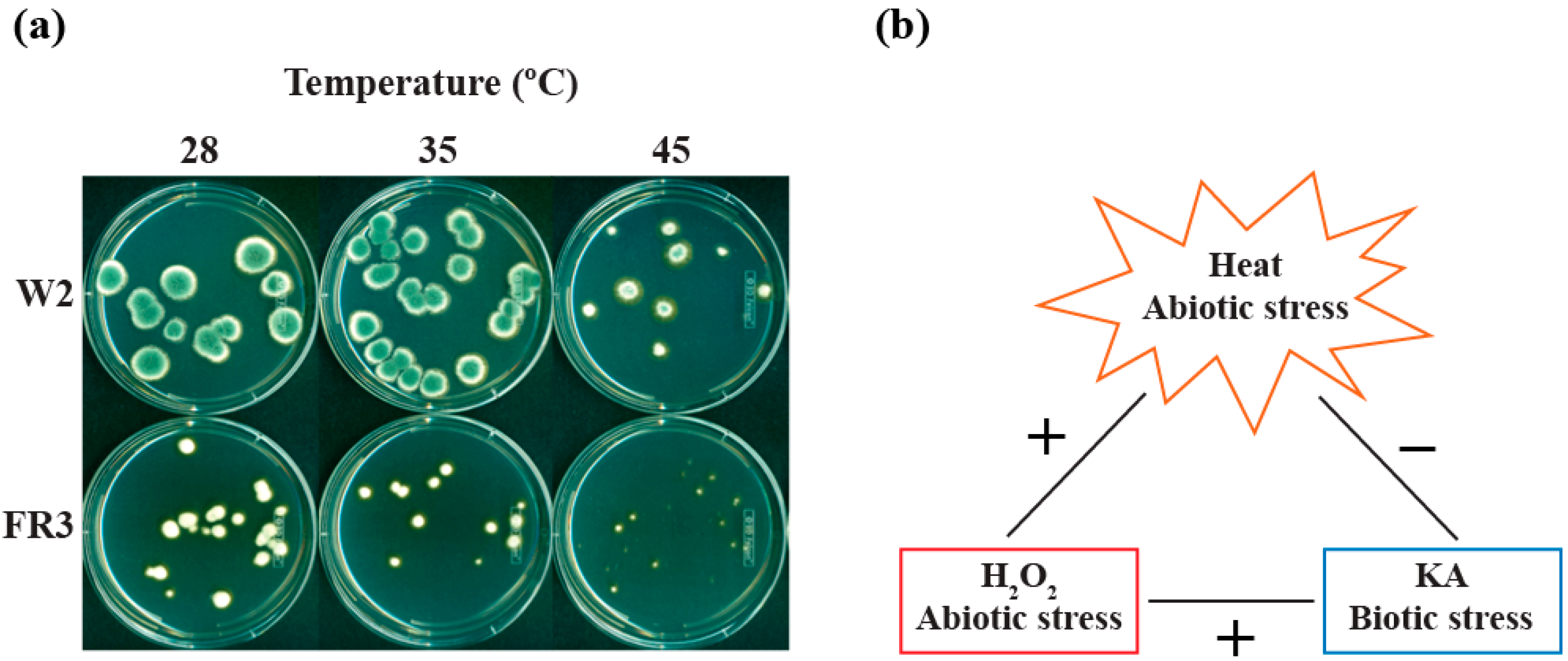
2.2.3. Effect of High Temperatures on the Growth of Fludioxonil-Resistant Mutants

The fludioxonil-resistant mutant P. expansum FR2 showed hypersensitivity to heat (45 °C) comparing to other P. expansum strains (W1, W2, FR3). As shown in Figure 3, P. expansum FR2 barely grew at 45 °C, where very tiny colonies appeared on PDA even without H2O2 or KA treatment. If FR2 was treated with H2O2 (as low as 0.25 mM) at 45 °C, colony growth was completely inhibited (namely, no signs of germination). Whereas, the impact of KA on the growth of FR2 at 45 °C was negligible (See Figure 3). Therefore, results indicated that fludioxonil resistance and heat sensitivity in FR2 are co-segregating traits, where the mutation responsible for fungicide resistance might also affect cellular fitness and/or mitochondrial function [29] in the presence of heat stress. Of note, a similar interrelationship between fungicide resistance and fungal sensitivity to high temperatures was previously identified in another fungal plant pathogen, Monilinia fructicola [30].
The other fludioxonil-resistant mutant, P. expansum FR3, did not exhibit similar type of heat sensitivity as observed in FR2 (Figure 3). However, in a parallel study, the colony growth of FR3 was noticeably reduced compared to that of P. expansum W2 (parental strain) (Figure 4a). For instance, both P. expansum W2 and FR3 exhibited gradual reduction in colony growth on PDA when the temperature was increased from 28 °C to 45 °C (Figure 4a). However, the colony size of P. expansum FR3 mutant was much smaller than that of W2 (parental), even at the normal growth temperature (28 °C). Moreover, the color of FR3 colonies remained pale at all conditions, while that of W2 was blue, indicating cellular physiology or a process, such as secondary metabolism responsible for pigment development, in FR3 is also impaired. Similar type of interrelationship between fungicide resistance and reduced fungal growth was previously identified in other fungi [31].
Altogether, results from two fludioxonil resistant mutants (FR2, FR3) showed that one or more traits are co-segregated with fungicide resistance. Previous study showed that certain fungi with mutations in genes responsible for signal transduction of environmental stress, such as Mitogen-Activated Protein Kinase (MAPK) signaling pathway, could develop fludioxonil resistance [32]. Coinciding with the fludioxonil resistance was the increased sensitivity of this MAPK mutant to high osmotic stress, thus exhibiting co-segregation of two different traits. Alternatively, results suggested that vulnerable or susceptible targets for fungal control could be identifiable in fungicide resistant mutants, such as heat/stress sensitivity or impaired growth, etc., as determined in this study. Precise characterization of the links between heat susceptibility/reduced growth and fludioxonil resistance, identified in FR2 and FR3, warrants future study. Summary of agent interactions, between biotic and abiotic stressors, is described in Figure 4b.
Figure 4. (a) Reduced growth of P. expansum FR3, a fludioxonil-resistant mutant, comparing to W2, a parental strain; (b) Diagram showing the interaction characteristics between heat, KA and H2O2. +: positive interaction (i.e., enhancement of antifungal activities when combined), −: no interaction (i.e., no enhancement of antifungal activities when combined).
Molecules 19 18448 g004

3. Experimental Section

3.1. Fungal Strains and Culture Conditions

Fungal strains used in this study are summarized in Table 1. Penicillium strains were cultured on potato dextrose agar (PDA) at 28 °C, except when otherwise noted in the text (see also below).

3.2. Chemicals

Antifungal compounds (kojic acid (KA) (Figure 1), hydrogen peroxide (H2O2)) were procured from Sigma Co. (St. Louis, MO, USA). KA was dissolved in dimethylsulfoxide (DMSO; absolute DMSO amount: <2% in media) before incorporation into culture media. Throughout this study, control plates (No treatment) contained DMSO at levels equivalent to that of cohorts receiving antifungal agents, within the same set of experiments. H2O2 was diluted in sterile water before incorporation into culture media.

3.3. Antifungal Bioassay

3.3.1. Growth Recovery Test in Penicillium Strain Treated with High Temperatures: Agar Plate-Bioassay

Agar plate-based bioassay was performed to evaluate susceptibility of Penicillium expansum W1 to high temperatures. First, fungal conidia (5 × 10³) were spotted on PDA (triplicate PDA plates per each temperature), and were initially incubated at three different temperatures (35, 45, 55 °C). Triplicate PDA plates were then removed from each temperature (35, 45 or 55 °C) at day 1, 2, 3, and 4, and were transferred to 28 °C for additional 6, 5, 4, and 3 d of growth, respectively, resulting in a total of 7 d of incubation for each treatment (e.g., 1 d growth at 45 °C + 6 d growth at 28 °C = Total 7 d growth, 2 d growth at 45 °C + 5 d growth at 28 °C = Total 7 d growth, 3 d growth at 45 °C + 4 d growth at 28 °C = Total 7 d growth, 4 d growth at 45 °C + 3 d growth at 28 °C = Total 7 d growth). For controls, Penicillium strains were grown solely at 28 °C or respective temperature (35, 45, 55 °C) for 7 d. The level of growth recovery at 28 °C was evaluated based on fungal radial growth, which was compared to that determined at 28 °C.

3.3.2. Microtiter Plate (Microdilution) Liquid Bioassay

To determine the precise level of chemosensitizing activity of KA (0.1, 0.2, 0.4, 0.8, 1.6, 3.2, 6.4, 12.8 mM) to H2O2 (0.25, 0.5, 1, 2, 4, 8, 16 mM) in the strains of Penicillium, checkerboard bioassays (triplicate) (0.4 × 10⁴–5 × 10⁴ CFU/mL) were performed in microtiter wells (at 28, 35, 45 °C) using a broth microdilution method (in RPMI 1640 medium; Sigma Co., St. Louis, MO, USA), according to protocols outlined by the Clinical and Laboratory Standards Institute (CLSI) M38-A [25]. RPMI 1640 medium was supplemented with 0.03% L-glutamine and buffered with 0.165 mM 3-(N-morpholino) propanesulfonic acid. Minimum Inhibitory Concentrations (MICs), lowest concentration of agents showing no visible fungal growth in microtiter wells (200 μL per well), were assessed after 48 h. Minimum Fungicidal Concentrations (MFCs), lowest concentration of agents showing ≥99.9% fungal death, were determined following completion of MIC assays (28, 35, 45 °C) by spreading entire volumes of microtiter wells (200 μL) onto individual PDA plates, and culturing for another 48 h (at 28 °C). Compound interactions, Fractional Inhibitory Concentration Indices (FICIs) and Fractional Fungicidal Concentration Indices (FFCI), were calculated as follows: FICI or FFCI = (MIC or MFC of compound A in combination with compound B/MIC or MFC of compound A, alone) + (MIC or MFC of compound B in combination with compound A/MIC or MFC of compound B, alone). Levels and types of compound interactions between antifungal agents (H2O2 and KA) were defined as: synergistic (FICI ≤ 0.5), additive (0.5 < FICI ≤ 1), neutral (1 < FICI ≤ 2) or antagonistic (2 < FICI) [27]. If preferred, the Odds’ [33] methodology can be substituted in parallel calculations of compound interactions.

3.4. Statistical Analysis

Statistical analysis (student’s t-test) was performed based on “Statistics to use” [34], where p < 0.05 was considered significant.

4. Conclusions

In this study, levels of interactions between biotic (KA) and abiotic (H2O2) stressors were determined for the enhancement of antifungal efficacy as follows: With chemosensitization (KA + H2O2), (1) MIC or MFC of KA or H2O2 was lowered during chemosensitization depending on types of strains or culture conditions (See Table 3 for summary). Since KA induces the generation of reactive oxygen species in cells, such as macrophages (during phagocytosis) [9,10], elevated oxidative stress occurred during KA-mediated chemosensitization in the presence of H2O2 (which also mimics host reactive oxygen species) may be the possible mechanism of enhanced activity of the combinational treatment (KA + H2O2).
Table 3. Responses of Penicillium strains to chemosensitization (Summary). Data shown are comparison of MICs or MFCs of H2O2 (antifungal oxidant) or KA (chemosensitizing agent) treated alone or in combination at different temperatures (MICs at 24 h are not determined due to no growth of all strains examined).
H2O2KA
StrainsTreatment28 °C35 °C45 °C28 °C35 °C45 °C
MICs, 48 h
Group AAlone4.0ND aND a25.6ND aND a
Combined1.8ND aND a7.2ND aND a
Group BAlone8.02.3ND a25.625.6ND a
Combined5.31.3ND a17.611.7ND a
Group CAlone3.0ND aND a25.6ND aND a
Combined1.5ND aND a12.8ND aND a
MFCs, 24 h
Group AAlone7.02.00.625.625.625.6
Combined4.01.00.311.212.811.2
Group BAlone14.78.01.125.625.625.6
Combined8.04.70.913.917.117.1
Group CAlone3.01.0ND b25.625.6ND b
Combined2.51.0ND b19.225.6ND b
MFCs, 48 h
Group AAlone4.01.50.425.625.625.6
Combined2.00.50.210.412.816.0
Group BAlone8.04.0ND c25.625.6ND c
Combined4.72.7ND c13.919.2ND c
Group CAlone3.00.6ND d25.625.6ND d
Combined3.00.4ND d25.619.2ND d
a ND: Not determined (No growth of strains); b ND: Not determined (No growth of P. digitatum 786); c ND: Not determined (No cell growth except P. griseofulvum 2159, for which FFCI is neutral); d ND: Not determined (No growth of Group C strains).
Considering KA affects the cellular nitric oxide metabolism [35], it is possible that nitrosative stress, such as peroxynitrite, a toxic free radical, may also be linked to the chemosensitization; (2) Most P. expansum strains (Group A) tested were sensitive to KA-mediated chemosensitization. In Group B, P. glabrum 766 was sensitive to chemosensitization in almost all conditions tested, while P. griseofulvum 2159 was mostly insensitive to chemosensitization. P. chrysogenum 824 was sensitive to chemosensitization at 24 h, while it was insensitive at 48 h; (3) In Group C, the citrus pathogens, the effect of chemosensitization was dependent upon cultivation time and temperatures, where P. digitatum 786 showed less sensitivity to the chemosensitization comparing to P. italicum 983. Altogether, the effectiveness of KA-mediated chemosensitization with H2O2 was fungal strain-specific. We speculate that different levels of antioxidant defense efficiency, metabolism or responses to nitrosative stress, etc., in different Penicillium strains (i.e., Groups A, B and C) may trigger the differential responsiveness of fungi to the chemosensitization. The fact that KA is produced by different Penicillium strains ([1] and references therein) may also explain why several Penicillium strains are not that much sensitive to KA and H2O2. Comparison of the production of KA by each analyzed Penicillium strain warrants future study; (4) Comparing to 24 h, 48 h of cultivation of Penicillium strains, in general, required lower concentration of H2O2 to achieve ≥99% fungal death. Results indicated that H2O2 effectively damaged the cellular integrity of Penicillium strains, which may result in the inhibition of cell division cycle as well as antioxidant defense and metabolism in fungi.
The effect of temperatures during chemosensitization is determined as follows: (1) The higher the temperature, the lower the concentration of H2O2 required for achieving ≥99.9% fungal death, indicating heat and H2O2 synergize each other for the enhancement of antifungal activity. However, heat did not enhance the activity of KA, indicating specificity also exists between heat and types of antifungal compounds applied for the antifungal efficacy; (2) P. expansum FR2, fludioxonil-resistant mutant, showed hypersensitivity to heat (45 °C), while the colony growth of P. expansum FR3, the other fludioxonil-resistant mutant, was severely reduced comparing to its parental strain at all temperatures tested. Results indicated that susceptible/sensitive targets for fungal control might be identifiable in such mutants, which is coincided with the mutation involved in fungicide resistance.
In conclusion, KA, a safe, natural compound, possesses a potential to serve as an antifungal chemosensitizing agent in combination with oxidative stressor(s). This potential appears to be greatest with P. expansum strains or P. italicum 983. Chemosensitization can lower effective dosages of toxic antifungal substances, such as H2O2, leading to coincidental lowering of environmental and health risks. Antifungal efficacy of H2O2 was greater when Penicillium strains were treated with heat (35 °C, 45 °C). Considering that much higher temperatures (e.g., 55 °C as shown in the Introduction section) are conventionally used for fungal control in crops, the temperatures defined as heat in this study (35, 45 °C), especially during chemosensitization, are markedly lower, and thus could reduce crop damage associated with heat treatment. The use of safe chemosensitizing agents, such as KA, that debilitate fungal pathogens may be a viable approach to control agro/food fungal pathogens.

Supplementary Materials

Supplementary materials can be accessed at: https://www.mdpi.com/1420-3049/19/11/18448/s1.

Acknowledgments

We thank Chang-Lin Xiao, Department of Plant Pathology, Washington State University, Wenatchee, WA, USA, for providing us the wild type and fludioxonil-resistant P. expansum strains. This research was conducted under USDA-ARS CRIS Project 5325-42000-037-00D.

Author Contributions

Jong H. Kim designed and performed research including data analysis and interpretation, literature search, and wrote the manuscript. Kathleen L. Chan performed antifungal assays and prepared the figures.

Conflicts of Interest

The authors declare no conflict of interest.

References

  1. Liu, X.; Xia, W.; Jiang, Q.; Xu, Y.; Yu, P. Synthesis, characterization, and antimicrobial activity of kojic acid grafted chitosan oligosaccharide. J. Agric. Food Chem. 2014, 62, 297–303. [Google Scholar] [CrossRef] [PubMed]
  2. Rodrigues, A.P.D.; Farias, L.H.S.; Carvalho, A.S.C.; Santos, A.S.; do Nascimento, J.L.M.; Silva, E.O. A novel function for kojic acid, a secondary metabolite from Aspergillus fungi, as antileishmanial agent. PLoS One 2014, 9, e91259. [Google Scholar] [CrossRef] [PubMed]
  3. Bentley, R. From miso, sake and shoyu to cosmetics: A century of science for kojic acid. Nat. Prod. Rep. 2006, 23, 1046–1062. [Google Scholar] [CrossRef] [PubMed]
  4. Chang, T.S. An updated review of tyrosinase inhibitors. Int. J. Mol. Sci. 2009, 10, 2440–2475. [Google Scholar] [CrossRef] [PubMed]
  5. Lajis, A.F.; Hamid, M.; Ariff, A.B. Depigmenting effect of kojic acid esters in hyperpigmented B16F1 melanoma cells. J. Biomed. Biotechnol. 2012, 2012. [Google Scholar] [CrossRef] [PubMed]
  6. Leyden, J.J.; Shergill, B.; Micali, G.; Downie, J.; Wallo, W. Natural options for the management of hyperpigmentation. J. Eur. Acad. Dermatol. Venereol. 2011, 25, 1140–1145. [Google Scholar] [CrossRef] [PubMed]
  7. Bracarense, A.A.; Takahashi, J.A. Modulation of antimicrobial metabolites production by the fungus Aspergillus parasiticus. Braz. J. Microbiol. 2014, 45, 313–321. [Google Scholar] [CrossRef] [PubMed]
  8. Novotny, L.; Rauko, P.; Abdel-Hamid, M.; Vachalkova, A. Kojic acid—A new leading molecule for a preparation of compounds with an anti-neoplastic potential. Neoplasma 1999, 46, 89–92. [Google Scholar] [PubMed]
  9. Niwa, Y.; Akamatsu, H. Kojic acid scavenges free radicals while potentiating leukocyte functions including free radical generation. Inflammation 1991, 15, 303–315. [Google Scholar] [CrossRef] [PubMed]
  10. Rodrigues, A.P.; Carvalho, A.S.; Santos, A.S.; Alves, C.N.; do Nascimento, J.L.; Silva, E.O. Kojic acid, a secondary metabolite from Aspergillus sp., acts as an inducer of macrophage activation. Cell Biol. Int. 2011, 35, 335–343. [Google Scholar] [CrossRef] [PubMed]
  11. Chee, H.Y.; Lee, E.H. Fungistatic activity of kojic acid against human pathogenic fungi and inhibition of melanin production in Cryptococcus neoformans. Mycobiology 2003, 31, 248–250. [Google Scholar] [CrossRef]
  12. Ehrlich, K.C.; Cotty, P.J. An isolate of Aspergillus flavus used to reduce aflatoxin contamination in cottonseed has a defective polyketide synthase gene. Appl. Microbiol. Biotechnol. 2004, 65, 473–478. [Google Scholar] [CrossRef] [PubMed]
  13. Kim, J.H.; Mahoney, N.; Chan, K.L.; Campbell, B.C.; Haff, R.P.; Stanker, L.H. Use of benzo analogs to enhance antimycotic activity of kresoxim methyl for control of aflatoxigenic fungal pathogens. Front. Microbiol. 2014, 5, 87. [Google Scholar] [CrossRef] [PubMed]
  14. Frizzell, C.; Elliott, C.T.; Connolly, L. Effects of the mycotoxin patulin at the level of nuclear receptor transcriptional activity and steroidogenesis in vitro. Toxicol. Lett. 2014, 229, 366–373. [Google Scholar] [CrossRef] [PubMed]
  15. Vilanova, L.; Viñas, I.; Torres, R.; Usall, J.; Jauset, A.M.; Teixido, N. Infection capacities in the orange-pathogen relationship: Compatible (Penicillium digitatum) and incompatible (Penicillium expansum) interactions. Food Microbiol. 2012, 29, 56–66. [Google Scholar] [CrossRef] [PubMed]
  16. Dore, A.; Molinu, M.G.; Venditti, T.; D’Hallewin, G. Immersion of “Coscia” pear fruit in water at 55 degrees C for 60 sec controls Penicillium expansum decay and delays ripening during short storage. Commun. Agric. Appl. Biol. Sci. 2010, 75, 741–745. [Google Scholar] [PubMed]
  17. Cerioni, L.; Lazarte Mde, L.; Villegas, J.M.; Rodriguez-Montelongo, L.; Volentini, S.I. Inhibition of Penicillium expansum by an oxidative treatment. Food Microbiol. 2013, 33, 298–301. [Google Scholar] [CrossRef] [PubMed]
  18. Campbell, B.C.; Chan, K.L.; Kim, J.H. Chemosensitization as a means to augment commercial antifungal agents. Front. Microbiol. 2012, 3, 79. [Google Scholar] [CrossRef] [PubMed]
  19. Lavigne, J.P.; Brunel, J.M.; Chevalier, J.; Pages, J.M. Squalamine, an original chemosensitizer to combat antibiotic-resistant gram-negative bacteria. J. Antimicrob. Chemother. 2010, 65, 799–801. [Google Scholar] [CrossRef] [PubMed]
  20. Liu, S.; Hou, Y.; Chen, X.; Gao, Y.; Li, H.; Sun, S. Combination of fluconazole with non-antifungal agents: A promising approach to cope with resistant Candida albicans infections and insight into new antifungal agent discovery. Int. J. Antimicrob. Agents 2014, 43, 395–402. [Google Scholar] [CrossRef] [PubMed]
  21. Niimi, K.; Harding, D.R.; Parshot, R.; King, A.; Lun, D.J.; Decottignies, A.; Niimi, M.; Lin, S.; Cannon, R.D.; Goffeau, A.; et al. Chemosensitization of fluconazole resistance in Saccharomyces cerevisiae and pathogenic fungi by a D-octapeptide derivative. Antimicrob. Agents Chemother. 2004, 48, 1256–1271. [Google Scholar] [CrossRef]
  22. Kim, J.H.; Chan, K.L.; Mahoney, N.; Campbell, B.C. Antifungal activity of redox-active benzaldehydes that target cellular antioxidation. Ann. Clin. Microbiol. Antimicrob. 2011, 10, 23. [Google Scholar] [CrossRef] [PubMed]
  23. Musiol, R.; Mrozek-Wilczkiewicz, A.; Polanski, J. Synergy against fungal pathogens: Working together is better than working alone. Curr. Med. Chem. 2014, 21, 870–893. [Google Scholar] [CrossRef] [PubMed]
  24. Veri, A.; Cowen, L.E. Progress and prospects for targeting Hsp90 to treat fungal infections. Parasitology 2014, 141, 1127–1137. [Google Scholar] [CrossRef] [PubMed]
  25. Clinical and Laboratory Standards Institute (CLSI). Reference Method for Broth dilution Antifungal Susceptibility Testing of Filamentous Fungi: Approved Standard, 2nd ed.; CLSI document M38-A2; Clinical and Laboratory Standards Institute: Wayne, PA, USA, 2008; Volume 28. [Google Scholar]
  26. Li, H.X.; Xiao, C.L. Characterization of fludioxonil-resistant and pyrimethanil-resistant phenotypes of Penicillium expansum from apple. Phytopathology 2008, 98, 427–435. [Google Scholar] [CrossRef] [PubMed]
  27. Isenberg, H.D. Clinical Microbiology Procedures Handbook, 1 ed.; American Society for Microbiology: Washington, DC, USA, 1992. [Google Scholar]
  28. Macarisin, D.; Cohen, L.; Eick, A.; Rafael, G.; Belausov, E.; Wisniewski, M.; Droby, S. Penicillium digitatum suppresses production of hydrogen peroxide in host tissue during infection of citrus fruit. Phytopathology 2007, 97, 1491–1500. [Google Scholar] [CrossRef] [PubMed]
  29. Chatre, L.; Ricchetti, M. Are mitochondria the Achilles’ heel of the Kingdom Fungi? Curr. Opin. Microbiol. 2014, 20, 49–54. [Google Scholar] [CrossRef] [PubMed]
  30. Ma, Z.; Yoshimura, M.A.; Michailides, T.J. Identification and characterization of benzimidazole resistance in Monilinia fructicola from stone fruit orchards in California. Appl. Environ. Microbiol. 2003, 69, 7145–7152. [Google Scholar] [CrossRef] [PubMed]
  31. Jayasinghe, C.K.; Fernando, T.H. Growth at different temperatures and on fungicide amended media: Two characteristics to distinguish Colletotrichum species pathogenic to rubber. Mycopathologia 1998, 143, 93–95. [Google Scholar] [CrossRef] [PubMed]
  32. Kojima, K.; Takano, Y.; Yoshimi, A.; Tanaka, C.; Kikuchi, T.; Okuno, T. Fungicide activity through activation of a fungal signalling pathway. Mol. Microbiol. 2004, 53, 1785–1796. [Google Scholar] [CrossRef] [PubMed]
  33. Odds, F.C. Synergy, antagonism, and what the chequerboard puts between them. J. Antimicrob. Chemother. 2003, 52. [Google Scholar] [CrossRef] [PubMed]
  34. Kirkman, T.W. Statistics to Use. Available online: http://www.physics.csbsju.edu/stats/ (accessed on 16 September 2014).
  35. Dung, T.T.; Kim, S.C.; Yoo, B.C.; Sung, G.H.; Yang, W.S.; Kim, H.G.; Park, J.G.; Rhee, M.H.; Park, K.W.; Yoon, K.; et al. (5-Hydroxy-4-oxo-4H-pyran-2-yl)methyl 6-hydroxynaphthalene-2-carboxylate, a kojic acid derivative, inhibits inflammatory mediator production via the suppression of Syk/Src and NF-κB activation. Int. Immunopharmacol. 2014, 20, 37–45. [Google Scholar] [CrossRef]
  • Sample Availability: Samples of the compounds, such as kojic acid and hydrogen peroxide, are available from the vendor described in this manuscript.

