Article

New Lignans from the Leaves and Stems of Kadsura philippinensis

1
School of Pharmacy, College of Medicine, National Taiwan University, 1, Sec. 1, Jen-Ai Rd., Taipei 100, Taiwan
2
Department of Life Sciences, National Cheng Kung University, No. 1 University Road, Tainan 701, Taiwan
3
Graduate Institute of Natural Products, School of Pharmacy, Kaohsiung Medical University, Shih-Chuan 1st Road, Kaohsiung 807, Taiwan
4
National Research Institute of Chinese Medicine, Taipei 112, Taiwan
5
Department of Chemistry, National Sun Yat-sen University, 70, Lien-hai Road, Kaohsiung 804, Taiwan
6
Graduate Institute of Ecology and Evolutionary Biology, China Medical University, Taichung 40402, Taiwan
*
Author to whom correspondence should be addressed.
Molecules 2013, 18(6), 6573-6583; https://doi.org/10.3390/molecules18066573
Submission received: 13 May 2013 / Revised: 29 May 2013 / Accepted: 30 May 2013 / Published: 4 June 2013
(This article belongs to the Section Natural Products Chemistry)

Abstract

:
Three novel C19 homolignans, taiwankadsurins D (1), E (2) and F (4), and two new C18 lignans kadsuphilins N (3) and O (5) were isolated from the aerial parts of Taiwanese medicinal plant Kadsura philippinensis. The structures of compounds 15 were determined by spectroscopic analyses, especially 2D NMR techniques. The structure of compound 5 was further confirmed by X-ray crystallographic analysis. Compounds 1 and 2 have a 3,4-{1'-[(Z)-2''-methoxy-2''-oxoethylidene]}-pentano(2,3-dihydrobenzo[b]furano)-3-(2'''-methoxycarbonyl-2'''-hydroxy-2''',3'-epoxide) skeleton.

Graphical Abstract

1. Introduction

Kadsura belongs to the family Schisandraceae and it is only distributed in eastern and southern Asia [1]. Species of Kadsura were used in Chinese folk medicine for the treatment of cold, rheumatoid arthritis and gastroenteritis and as an anodyne to relieve pain [2]. The major constituents of Kadsura plants were reported to be bioactive lignans, which possess antitumor, antiviral and anti-hepatitic activities [3,4,5,6,7,8]. K. philippinensis Elm. is an evergreen vine, mainly distributed at low altitude onremote islands of Taiwan such as Green Island [9]. Our previous phytochemical studies on the EtOAc extracts of K. philippinensis resulted in the isolation of two novel triterpene dilactones and many lignans [10,11,12,13,14,15,16,17]. In this paper, we report the isolation and structure elucidation of three new C19 homolignans, named taiwankadsurins D-F, and two new C18 lignans, designated kadsuphilins N and O.

2. Results and Discussion
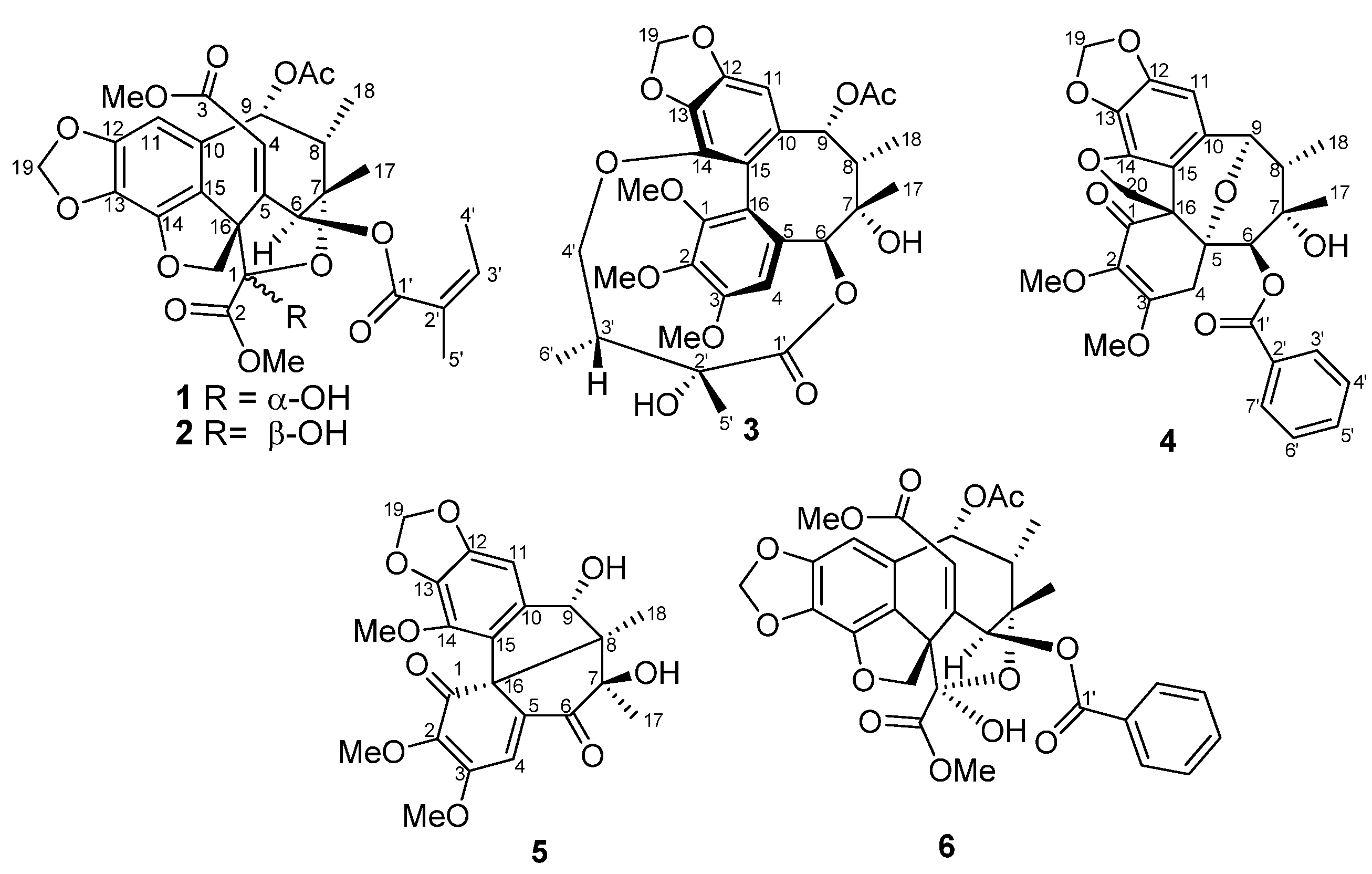
The leaves and stems of K. philippinensis were extracted with mixture of CH2Cl2 and acetone, then suspended in H2O and extracted with EtOAc. The EtOAc-soluble part was subjected to extensive chromatography including flash column, normal and reversed-phase HPLC, furnishing compounds 15 (Figure 1).
Figure 1. Chemical structures of compounds 16.
Molecules 18 06573 g001
Taiwankadsurin D (1), ( [ α ] D 25 +57°, CH2Cl2) had a molecular formula C29H32O13, as derived from its HREIMS at m/z 611.1735 ([M+Na]+, calcd 611.1741) indicating 14 degrees of unsaturation. The UV absorption (273, 225 nm) and IR bands (1,731, 1,721 and 1,628 cm−1) indicated a benzyl and α,β, unsaturated ester functionalities. The 1H-NMR of 1 exhibited two methoxyl singlets (δ 3.93, 3.59), an acetyl singlet (δ 2.13), two methyl singlets (δ 1.31, 1.99), two methyl doublets (δ 1.36, J = 6.9 Hz; δ 2.05, J = 7.2 Hz), two oxymethylene protons (δ 5.00, 4.53, each d, J = 10.2 Hz) and two dioxymethylene protons (δ 5.97, 5.98, each s-like). According to 13C-NMR and DEPT spectra, compound 1 had total 29 signals including seven methyl, two methylene, six methine and fourteen quaternary carbons. Moreover, 1H-NMR spectroscopic data of 1 showed characteristic signals of H-4 (δ 5.99), H-6 (δ 6.28) and H-9 (δ 6.55), and 13C-NMR data of C-1 (δ 97.5 s), C-2 (δ 171.0 s) and C-3 (δ 165.4 s) similar to those of taiwankadsurin A (6), suggesting that compound 1 is an analogue of the latter [10]. However, a benzoyl group in 6 was missing and replaced with an angeloyl group at C-6 in 1. Further HMBC correlations (Figure 2) of H-11/C-12, C-13, C-15 and H-20/C-14, C-15, C-16, confirmed that compound 1 possessed a dihydrobenzofuran system. The ethylidene-octane ring was also deduced from the HMBC correlations of H-9/C-7,C-10,C-11,C-15; Me-18/C-7,C-8,C-9; Me-17/C-6,C-7,C-8 and H-6/C-4,C-5, C-7, C-8. The acetyl and angeloyl groups attaching at C-9 and C-6 respectively, were resulted from the HMBC correlations of H-9 (δ 6.55) with the acetyl carbonyl, and H-6 (δ 6.28) with the angeloyl carbonyl. Furthermore, methoxyl groups (δΗ 3.93, δΗ 3.59) attaching at carbonyls C-2 (δC 171.0) and C-3 (δC 165.4) were deduced from their mutual HMBC correlations.
It was noted that the dioxygenated tertiary carbon C-1 connected to C-7 through an ether bridge to account for the last degree of unsaturation. The relative configuration of 1 was determined by the NOESY experiment and by comparing the NMR data of 1 with those of taiwankadsurin A (Figure 2). Assuming that H-9 was β-oriented due to quite similar NMR spectra of 1 and taiwankadsurin A [10], thus, cross peaks between H-4, H-9 and Me-5', and correlation between H-9 and H-8, rather than Me-18 suggested that H-8 and 6-O-angeloyl group should be positioned on the β−face of the molecule. On the other hand, correlation between Me-18(eq) and Me-17(eq) accounted for the α−disposition of the ether ring between C-1 and C-7. In addition, NOESY correlation between H-6 and the methoxyl protons at C-2 indicated that H-6 and the hydroxyl group attached at C-1 are α−oriented. On the basis of above findings, the relative configuration of 1 was assigned as 1R*, 6S*, 7S*, 8S*, 9R*, 16S*.
Figure 2. Selected HMBC (arrow) and NOESY (double headed arrow) correlations of 1.
Molecules 18 06573 g002
Taiwankadsurin E (2) is an isomer of 1 as inferred from the identical molecular weight in HRMS, similar UV and IR absorptions and NMR data. The 1H-NMR spectrum (Table 1) of 2 had the same characteristic peaks with 1 except that H-6 was downfield shifted to δΗ 6.91, while the methoxyl protons at C-2 was upfield shifted to δΗ 3.61. Detail analysis of HMBC correlations of 2 revealed that the locations of angeloyl, acetyl and methoxyl groups were the same as 1. The configuration of 2 was established from NOESY experiment, in which most of the cross peaks were identical to those of 1. However, the correlation between H-6 and the methoxy at C-2 was missing in 2. Therefore, the structure of 2 was established, being an 1-epimer of 1.
Table 1. 1H-NMR data (CDCl3) of compounds 15 a,b.
Position1 a2 b3 a4 b5 a
45.99, brs6.06, d (2.4)6.84, s3.08, d (18.4 )7.34, s
3.17, d (18.4 )
66.28, d (2.7)6.69, d (2.4)5.76, s5.42, s
82.23, m2.23, m1.97, m2.00, m
96.55, d (2.7)6.63, d (2.8)5.48, s4.82, brs4.87, d (12.9)
116.45, s6.44, s6.47, s6.28, s6.71, s
171.31, s1.34, s1.37, s0.97, s1.31, s
181.04, d (6.9)1.02, d (6.8)1.30, d (6.9)1.36, d (7.6)0.98, s
195.97, s5.93, s5.94, s5.83, s5.90, d (1.5)
5.98, s5.94, s6.03, s5.98, s5.91, d (1.5)
204.53, d (10.2)4.59, d (10.0) 4.30, d (9.6)
5.00, d (10.2)4.98, d (10.0) 4.43, d (9.6)
OMe-1 3.46, s
OMe-23.93, s3.57, s3.84, s3.66, s3.77, s
OMe-33.59, s3.58, s3.92, s4.07, s4.11, s
OMe-14 3.81, s
OAc2.13, s2.13, s1.49, s
1'
2'
3'6.28, overlap6.23, q (7.2)1.92, m7.32, m
4'2.05, d (7.2)2.06, d (7.2)3.63, dd (5.0, 8.0)7.35, m
4.16, dd (5.0, 8.0)
5'1.99, s2.00, s1.23, s7.55, d (7.2)
6' 0.96,d (7.2)7.35, m
7' 7.32, m
OH-9 4.28, d (12.9)
a recorded at 300 MHz. b recorded at 400 MHz.
Kadsuphilin N (3), ( [ α ] D 26 −2.4, CH2Cl2), had a molecular formula of C30H36O12 as deduced from a pseudo-molecular ion [M+Na]+ at m/z 611.2107 in the HRESIMS. The UV absorption bands at 212, 259 and 292 nm suggested that 1 possessed a biphenyl chromophore. The IR absorption indicated the presence of hydroxyl (3,479 cm−1) and carbonyl (1,738 cm−1) groups. The 13C-NMR spectroscopic data and DEPT analysis revealed that compound 3 contains 30 carbons, including ten quaternary sp2 carbons (δC 121.2, 121.7, 130.8, 132.8, 137.5, 139.0, 141.8, 148.6, 151.5 and 152.3), two ester carbons (δC 168.7 and 172.4), two quaternary sp3 oxygen-bearing carbons (δC 73.8 and 76.6), two oxygen-bearing methylene carbons (δC 72.4 and 101.5), two sp2 methine carbons (δC 102.5 and 111.0), two sp3 methine carbons (δC 42.4 and 44.1), two oxygen-bearing sp3 methine carbons (δC 83.9 and 86.6), and eight methyl groups (δC 12.8, 17.8, 20.3, 21.4, 28.4, 56.2, 60.5 and 60.7). The HMBC correlations of H-11/C-9, C-10, C-12, C-13, C-15; H-9/C-10, C-15; H-4/C-2, C-3, C-5, C-6, C-16; H-6/C-5, C-16; Me-17/C-6, C-7, C-8; Me-18/C-7, C-8, C-9 implied that compound 3 indeed possessed a schizandrin type dibenzocyclooctadiene system [16]. Moreover, HMBC correlations of H2-19/C-12, C-13; OMe-1/C-1; OMe-2/C-2; OMe-3/C-3 and H-9/ acetyl carbonyl assigned the methylenedioxy group and three methoxyl groups attached to the aromatic ring and an acetyl group at C-9. In addition, the ester linkage could be proved by correlations of H-6/C-1'; Me-5'/C-1', C-2', C-3'; Me-6'/C-2', C-3', C-4' and H-4'/C-14. From the above interpretation, the structure of 3 could be established as 9-acetylgomisin D. The configuration was determined by CD spectrum and NOESY experiment. The strong positive Cotton effect at 229 nm and the negative Cotton effect at 245 nm assigned the S-configuration of the biphenyl system [18]. The NOESY correlations of H-9/H-8, H-11, H-8/Me-17 and H-6/H-4, Me-17(eq) revealed that the cyclooctadiene ring had a twist-boat-chair form and H-8, H-9 and Me-17 were β-oriented while H-6 and Me-18 were α-configuration (Figure 3). The correlations of H-3'/Me-5' and the NMR data were in good agreement with the configuration of ester linkage that was also present in gomisin D [19].
Figure 3. Selected HMBC (arrow) and NOESY (double headed arrow) correlations of 3.
Molecules 18 06573 g003
Taiwankadsurin F (4) was isolated as a pale yellow amorphous solid. The molecular formula C29H28O10 was deduced from a pseudo-molecular ion at m/z 559.1580 [M+Na]+ in HRESIMS. The UV spectrum showed absorptions at λmax 255, 220 nm and IR bands at νmax 3,510, 1,735, 1,725 cm−1 suggested that compound 4 contained phenyl, benzoyl, α,β-unsaturated ketone and hydroxyl functionalities. The 1H and 13C-NMR spectroscopic data (Table 1 , Table 2) revealed that 4 possessed a substituted cyclohex-2-enone moiety and a spirodihydrobenzofuran ring in a homolignan skeleton similar to kadsuphilol G [14]. The difference could be the angeloyloxy side chain, which was substituted with a benzoyloxy group. This scaffold was supported from HMBC correlations of H-19/ C-12, C-13; H-11/ C-9, C-12, C-13, C-15; Me-18/C-7, C-8, C-9; Me-17/C-6, C-7, C-8; H-4/C-2, C-3, C-5, C-16 and H-20/C-1, C-14, C-15. Moreover, the key HMBC correlations of H-6/ benzoyl carbonyl (δC 165.4) and H-9/ C-5 assigned the benzoyl group at C-6. It was found that an ether linkage appeared between C-5 and C-9 due to calculation of double bond equivalence. The relative configuration of 4 was determined by comparing the coupling constants of 4 with those of kadsuphilol G and NOESY experiments. Thus a twist-boat-chair configuration was elucidated on the basis of CD observation, in which a positive Cotton effect was found at 216 nm and a negative one at 249 nm. The NOESY correlations of H-11/H-8 /H-9 and H-8/ Me-17 suggested that H-8, H-9 Me-17 were all in β-face while Me-18 was α-oriented (Figure 4). Because compound 4 had a TBC-S configuration, the oxygen bridge could be assigned as α-disposed. On the basis of the above interpretation, the structure of compound 4 was established and the name taiwankadsurin F was given.
Table 2. 13C-NMR data (CDCl3) of compounds 15 a,b.
Position1 a2 b3 a4 b5 a
197.5, C97.6, C151.5,C193.0, C196.3, C
2171.0, C170.1, C141.8,C132.5, C140.6, C
3165.4, C165.5, C152.3, C157.4, C159.0, C
4117.2, CH118.3, CH111.0, CH40.7, CH2123.4, CH
5150.5, C149.8, C130.8, C77.6, C143.7, C
672.5, CH72.5, CH86.6 , CH77.3, CH200.7, C
779.2, C78.4, C73.8, C72.5, C80.7, C
845.4, CH45.5, CH44.1, CH43.7, CH60.4, C
970.3, CH70.2, CH83.9, CH77.3, CH75.7, CH
10127.9, C127.8, C132.8,C127.9, C144.5, C
1198.7, CH99.9, CH102.5, CH95.9, CH100.4, CH
12150.4, C149.8, C148.6, C151.3, C151.2, C
13129.1, C128.9, C137.5, C129.5, C136.4, C
14144.9, C142.6, C139.0, C140.9, C139.5, C
15118.0, C120.5, C121.2, C121.3, C125.7, C
1657.0, C58.6, C121.7, C56.9, C69.5, C
1728.2, CH328.5, CH328.4, CH323.1, CH319.9, CH3
188.9, CH38.5, CH317.8, CH315.3, CH315.1, CH3
19101.8, CH2101.5, CH2101.5, CH2101.3, CH2101.7, CH2
2080.4, CH278.5, CH2 78.2, CH2
OMe-1 60.5, CH3
OMe-253.6, CH354.0, CH360.7, CH360.7, CH360.2, CH3
OMe-351.8, CH351.7, CH356.2, CH358.9, CH358.3, CH3
OMe-14 59.4, CH3
OAc168.9, C168.9, C168.7, C
21.0, CH320.3, CH320.3, CH3
1'166.0, C166.1, C172.4, C165.4, C
2'126.3, C126.5, C76.6, C129.5, C
3'142.3, CH141.7, CH42.4, CH128.3, CH
4'16.0, CH315.9, CH372.4, CH2129.7, CH
5'20.4, CH321.0, CH321.4, CH3133.9, CH
6' 12.8, CH3129.7, CH
7' 128.3, CH
a recorded at 75 MHz. b recorded at 100 MHz.
Figure 4. Selected HMBC (arrow) and NOESY (double headed arrow) correlations of 4.
Molecules 18 06573 g004
Kadsuphilin O (5) was obtained as pale yellow crystals, with molecular formula C22H22O9 as determined by HRESIMS (12 degrees of unsaturation). IR absorption bands at 3,420, 1,716 and 1,620 cm−1 indicated the presence of hydroxyl, carbonyl and aromatic moieties. The 1H-NMR data (Table 1) and HMQC spectrum showed characteristic signals for two aromatic (δH 6.71, 7.34), one methoxyene-dioxy (δH 5.90, 5.91 as an AB quartet), one oxygen-bearing methine (δH 4.87), two tert-methyl (δH 0.98, 1.31) and three methoxyl (δH 3.77, 3.81 and 4.11) protons. A methine doublet at δH 4.28 (J = 12.9 Hz) revealed the presence of a hydroxy due to no correlation was found in HMQC. 13C-NMR data and DEPT spectra revealed that compound 5 contained five pairs of double bonds (δC 100.4, 123.4, 125.7, 136.4, 139.5, 140.6, 143.7, 144.5, 151.2, 159.0), two ketone carbonyl carbons (δC 196.3 and 200.7) and three quaternary carbons (δC 60.4, 69.5, 80.7), one oxygenated methine carbon (δC 75.7), a methylenedioxy carbon (δC 101.7), two methyl carbons (δC 15.1 and 19.9) and three methoxyl carbons (δC 58.3, 59.4, 60.2). Thus compound 5 possessed five ring systems after deduction of seven double bonds. In the HMBC spectrum, correlations of H-11/C-9, C-12, C-13, C-14, C-15; H-19/C-12, C-13; H-4/C-2, C-3, C-5, C-6, C-16; Me-17/C-6, C-7, C-8; Me-18/C-7, C-8; H-9/C-7, C-15 and C-9-OH/C-9 suggested a dibenzocyclo-octadiene framework with a ketone substituted at the C-6 position. Furthermore, the linkage between C-8 and C-16 was deduced by the correlations of H-9 and Me-18 with C-16, and the remaining ketone group could be assigned to the C-1 position. This finding was further confirmed by comparing the NMR data with those of heteroclitin G [20]. The relative configuration of 5 was determined by NOESY correlation and CD. The CD spectrum of 5 was similar to that of kadsutherin C [21]. The negative Cotton effect at 240 nm and the positive Cotton effect at 218 nm accounted for S-configuration for the biphenyl skeleton. Assuming that the H-9 of 5 was in a β-orientation similar to heteroclitin G, the NOESY correlations of HO-9/Me-18 and Me-18/Me-17 indicated that they were on the α-face and OH-7 was β-oriented. Therefore, the configuration of the bipentacyclic ring was established. The structure of 5 was finally confirmed by a single-crystal X-ray diffraction analysis, from which a perspective drawing of 5 is provided in Figure 5.
Figure 5. Selected HMBC correlations and X-ray perspective drawing of 5.
Molecules 18 06573 g005

3. Experimental

3.1. General

Melting points were measured on a Büchi melting point B-540 apparatus and are uncorrected. Optical rotations were recorded on a JASCO DIP-1000 polarimeter. IR and UV spectra were measured on HORIBA FT-720 and U-3210 spectrophotometers, respectively. The 1H- and 13C-NMR, COSY, HMQC, HMBC, and NOESY spectra were recorded respectively on a Bruker FT-300 spectrometer (300 MHz for 1H and 75 MHz for 13C) or on a Bruker AVANCE 400 (400 MHz for 1H and 100 MHz for 13C) using TMS as an internal standard. The chemical shifts were given in δ values (ppm) and coupling constants in Hz. Low-resolution FABMS were recorded on a VG Quattro 5022 mass spectrometer, and HREIMS were measured on a JEOL JMS-SX 102 spectrometer. Silica gel 60 (Merck) was used for column chromatography (CC), and precoated silica gel plates (Merck, Kieselgel 60 F-254, 1 mm) were used for preparative TLC.

3.2. Plant Material

The leaves and stems of K. philippinensis were collected at Green Island, Taiwan, in November, 2002. A voucher sample (specimen code: TP 93-2) was deposited at the School of Pharmacy, College of Medicine, National Taiwan University, Taipei, Taiwan.

3.3. Extraction and Isolation

K. philippinensis was extracted with mixture of CH2Cl2 and acetone and partitioned between EtOAc and H2O (1:1). The EtOAc-soluble part was subjected to Si gel column chromatography (n-hexane/EtOAc, 1:0 to 0:1), and after monitoring by 1H-NMR, the middle fraction (fr. 21) was further eluted on LH-20 (MeOH) to give five subfractions (fr.21-1~5). Fr.21-5 was chromatographed on a flash column (Si gel, n-hexane/EtOAc, 15:1-0:1) and further separated by normal phase HPLC (n-hexane/CH2Cl2/MeOH, 35:65:1) to furnish taiwankadsurins D (1, 13 mg) and E (2, 2 mg). Kadsuphilin N (3, 14 mg) was isolated from fr.21-2, which was chromatographed on a flash column (n-hexane/ acetone/EtOAc, 15:1:1 to 1:1:1) and further purified with normal phase HPLC (n-hexane/CH2Cl2/ MeOH, 30:70:1) and reverse phase HPLC (MeOH/H2O, 65:35) alternatively. Fraction fr.21-4 was separated on a Si gel column (n-hexane/EtOAc, 25:1 to 0:1) and a reverse phase HPLC (MeOH/H2O, 65:35) column to yield taiwankadsurin F (4, 4 mg) and kadsuphilin O (5, 7 mg).

3.4. Spectroscopic Data

Taiwankadsurin D (1). [ α ] D 26 +57 ° (c 0.5, CH2Cl2); UV λmax (MeOH) 225, 273 nm; CD (MeOH, c = 0.2) nm (ε) 222 (−1.30), 254 (+1.27); IR (neat) νmax 3,450, 2,938, 1,731, 1,721, 1,628 cm−1; 1H-NMR and 13C-NMR (CDCl3, 300/75 MHz) see Table 1 , Table 2, respectively; HRESIMS m/z 611.1735 (calcd for C29H32O13Na, 611.1741).
Taiwankadsurin E (2). [ α ] D 26 −11° (c 0.2, CH2Cl2); UV λmax (MeOH) 233, 276 nm; CD (MeOH, c = 0.2) nm (ε) 228 (−0.78), 247 (+0.27); IR (neat) νmax 3,457, 1,728, 1,717 cm−1; 1H-NMR and 13C-NMR (CDCl3, 400/100 MHz) see Table 1 , Table 2, respectively; HRESIMS m/z 611.1737 (calcd for C29H32O13Na, 611.1741).
Kadsuphilin N (3). [ α ] D 25 −2.4° (c 1.3, CH2Cl2); UV λmax (MeOH) 212, 259, 292 nm; CD (MeOH, c = 0.16) nm (ε) 229 (+33.56), 245 (−2.97), 293 (−5.54); IR (neat) νmax 3,479, 1,738, 1,624, 1,594 cm−1; 1H-NMR and 13C-NMR (CDCl3, 300/75 MHz) see Table 1 , Table 2, respectively; HRESIMS m/z 611.2107 (calcd for C30H36O12Na, 611.2104).
TaiwankadsurinF (4). [ α ] D 25 −13.2° (c 0.6, CH2Cl2); UV λmax (MeOH) 220, 255 nm; CD (MeOH, c = 0.3) nm (ε) 216 (+17.34), 249 (−8.77), 290 (−1.53); IR (neat) νmax 3,510, 1,735, 1,725, 1,660, 1,580 cm−1; 1H-NMR and 13C-NMR (CDCl3, 400/100 MHz), see Table 1 , Table 2, respectively; HRESIMS m/z 559.1573 (calcd for C29H28O10Na, 559.1580).
Kadsuphilin O (5). [ α ] D 25 −8.0° (c 0.6, CH2Cl2); MP 167 °C; UV λmax (MeOH) 215, 246, 283 nm; CD (MeOH, c = 0.22) nm (ε) 218 (+7.66), 240 (−28.11), 282 (−6.75); IR (neat) νmax 3,420, 1,716, 1,620 cm−1; 1H-NMR (CDCl3, 300 MHz) and 13C-NMR (CDCl3, 75 MHz), see Table 1 and Table 2, respectively; HRESIMS m/z 453.1158 (calcd for C22H22O9Na, 453.1161). Crystal data: C22H22O9, M = 430.40, trigonal system, space group P21, a = 10.706(2), b = 8.218(2), c = 10.9345(9) Å, V = 960.1(3) Å3, Z = 2, d = 1.489 Mg/cm3. A crystal of dimensions 0.60 × 0.60 × 0.20 mm was used for measurements on a RIGAKU AFC7S diffractometer with a graphite monochromator (ω-2θscans, 2θmax= 52.0°), Mo Kα radiation. The total number of independent reflections measured was 2,134, of which 2026 were observed (|F|2 ≥ 2σ|F|2). The crystal structure was solved by the direct method SHELX-86 [22] and expanded using difference Fourier techniques, refined by the program SHEXTL-97 [23] and full-matrix least-squares calculations. Final indices: Rf = 0.030, Rw = 0.0784, w = 1/[σ2 (Fo2) + (0.070P)2 + 0.1457P], where P = (Fo2 + 2 Fc2)/3). Copies of the deposited crystal data (CCDC 829589) can be obtained, free of charge, from the Cambridge Crystallographic Data Centre, 12 Union Road, Cambridge CB2 1EZ, UK; fax: +44 (0) 1223 336033 or E-Mail: [email protected].

4. Conclusions

Phytochemical investigation of the aerial part of Taiwanese Kadsura philippinensis has resulted in isolation of five new lignans 15, including three novel C19 homolignans, designated taiwankadsurins D, E and F. Their structures have been established by spectroscopic analyses, especially 2D NMR techniques. In addition, the structure of compound 5 was further confirmed by X-ray crystallographic analysis.

Acknowledgments

This work was supported from the National Science Council of the Republic of China (Grant NSC-98-2320-B-002-027-MY3).

Conflicts of Interest

The authors declare no conflict of interest.

References

  1. Ookawa, N.; Ikeya, Y.; Taguchi, H.; Yosioka. The Constituents of Kadsura Japonica DUNAL. I. The Structures of Three New Lignans, Acetyl-, Angeloyl- and Caproyl-binankadsurin A. Chem. Pharm. Bull. 1981, 29, 123–127. [Google Scholar] [CrossRef]
  2. Hu, X.; Zhang, W.K.; Zhu, Q.S. Zhong Hua Ben Cao; (in Chinese). Shanghai Scientific & Techincal Publishers: Shanghai, China, 1999; Volume 2, pp. 912–913. [Google Scholar]
  3. Charlton, J.L. Antiviral Activity of Lignans. J. Nat. Prod. 1998, 61, 1447–1451. [Google Scholar] [CrossRef]
  4. Chang, J.B.; Reiner, J.; Xie, J.G. Progress on the chemistry of dibenzocyclooctadiene lignans. Chem. Rev. 2005, 105, 4581–4609. [Google Scholar] [CrossRef]
  5. Wu, M.D.; Huang, R.L.; KuoYang, L.M.; Hung, C.C.; Ong, C.W.; Kuo, Y.H. The Anti-HBsAg (human type B hepatitis, surface antigen) and anti-hbeag (human type B hepatitis, e antigen) C18 dibenzocyclooctadiene lignans from Kadsura matsudai and Schizandra arisanensi. Chem. Pharm. Bull. 2003, 51, 1233–1236. [Google Scholar] [CrossRef]
  6. Li, L.N. Biologically active components from traditional Chinese medicines. Pure Appl. Chem. 1998, 70, 547–554. [Google Scholar] [CrossRef]
  7. Chen, D.F.; Zhang, S.X.; Kozuka, M.; Sun, Q.Z.; Feng, J.; Wang, Q.; Mukainaka, T.; Nobukuni, Y.; Tokuda, H.; Nishino, H.; et al. Interiotherins C and D, two new lignans from Kadsura interior and antitumor-promoting effects of related neolignans on Epstein−Barr virus activation. J. Nat. Prod. 2002, 65, 1242–1245. [Google Scholar] [CrossRef]
  8. Chen, D.F.; Zhang, S.X.; Wang, H.K.; Zhang, S.Y.; Sun, Q.Z.; Cosentino, L.M.; Lee, K.H. Novel Anti-HIV Lancilactone C and Related Triterpenes from Kadsura lancilimba. J. Nat. Prod. 1999, 62, 94–97. [Google Scholar] [CrossRef]
  9. Li, H.L.; Chaw, S.M. Schisandraceae-Flora of Taiwan; Epoch: Taipei, Taiwan, 1996; Volume 2, p. 425. [Google Scholar]
  10. Shen, Y.C.; Lin, Y.C.; Kuo, Y.H.; Cheng, Y.B.; Liaw, C.C. Taiwankadsulins A, B and C, three new C-19 Homolignans from Kadsura philippinensis. Org. Lett. 2005, 7, 5297–5300. [Google Scholar] [CrossRef]
  11. Shen, Y.C.; Lin, Y.C.; Chiang, M.Y.; Yeh, S.F.; Cheng, Y.B.; Liaw, C.C. Kadsuphilactones A and B, Two New Triterpene Dilactones from Kadsura philippinensis. Org. Lett. 2005, 7, 3307–3310. [Google Scholar] [CrossRef]
  12. Shen, Y.C.; Liaw, C.C.; Cheng, Y.B.; Ahmed, A.F.; Lai, M.C.; Liou, S.S.; Wu, T.S.; Kuo, Y.H.; Lin, Y.C.; Khalil, A.T. C-18 Dibenzocyclooctadiene Lignans from Kadsura philippinensis. J. Nat. Prod. 2006, 69, 963–966. [Google Scholar] [CrossRef]
  13. Shen, Y.C.; Lin, Y.C.; Ahmed, A.F.; Cheng, Y.B.; Chen, C.T.; Liaw, C.C.; Kuo, Y.H. Four new nonaoxygenated C18 dibenzocylcooctadiene lignans from Kadsura philippinensis. Chem. Pharm. Bull. 2007, 55, 280–283. [Google Scholar] [CrossRef]
  14. Shen, Y.C.; Cheng, Y.B.; Lan, T.W.; Liaw, C.C.; Liou, S.S.; Kuo, Y.H.; Khalil, A.T. Kadsuphilols A-H, new oxygenated lignans from Kadsura philippinensis. J. Nat. Prod. 2007, 70, 1139–1145. [Google Scholar] [CrossRef]
  15. Shen, Y.C.; Lin, Y.C.; Cheng, Y.B.; Chang, C.J.; Lan, T.W.; Liou, S.S.; Chien, C.T.; Liaw, C.C.; Khalil, A.T. New Oxygenated Lignans from Kadsura philippinensis. Helv. Chim. Acta. 2008, 91, 483–494. [Google Scholar] [CrossRef]
  16. Shen, Y.C.; Lin, Y.C.; Cheng, Y.B.; Chiang, M.Y.; Liou, S.S.; Khalil, A.T. Dibenzocyclooctadiene lignans from Kadsura philippinensis. Phytochemistry 2009, 70, 114–120. [Google Scholar] [CrossRef]
  17. Cheng, Y.B.; Lin, Y.C.; Khalil, A.K.; Liou, S.S.; Lee, G.C.; Kuo, Y.H.; Shen, Y.C. Seven new lignan esters from Kadsura philippinensis. Helv. Chim. Acta 2011, 94, 148–158. [Google Scholar] [CrossRef]
  18. Liu, J.S.; Li, L. Schisandrins L–O and acetyl schisandrin L from Kadsura coccinea. Phytochemistry 1993, 32, 1293–1296. [Google Scholar] [CrossRef]
  19. Ikeya, Y.; Taguchi, H.; Yosioka, I.; Iitaka, Y.; Kobayashi, H. The constituents of schizandra chinensis baill. ii. the structure of a new lignan, gomisin d. Chem. Pharm. Bull. 1979, 27, 1395–1401. [Google Scholar] [CrossRef]
  20. Yang, X.W.; Miyashiro, H.; Hattori, M.; Namba, T.; Tezuka, Y.; Kikuchi, T.; Chen, D.F.; Xu, G.J.; Hori, T.; Extine, M.; et al. Isolation of novel lignans, heteroclitins F and G, from the Stems of Kadsura heteroclita, and anti-lipid peroxidative actions of heteroclitins A–G and related compounds in the in vitro Rat Liver Homogenate System. Chem. Pharm. Bull. 1992, 40, 1510–1516. [Google Scholar] [CrossRef]
  21. Lu, Y.; Chen, D.F. Kadsutherins A–C: Three new dibenzocyclooctane lignans from the stems of Kadsura species. Helv. Chim. Acta 2006, 89, 895–901. [Google Scholar] [CrossRef]
  22. Sheldrick, G.M. SHELXS-86. Program for the solution of crystal structures; University of Göttingen: Göttingen, Germany, 1985. [Google Scholar]
  23. Sheldrick, G.M. SHELXS-97. Program for the solution of crystal structures.; University of Göttingen: Göttingen, Germany, 1997. [Google Scholar]
  • Sample Availability: Samples of the compounds may not be available from the authors.

