Article

In Vitro Action of Flavonoids in the Canine Malignant Histiocytic Cell Line DH82

Biotechnology Unit, University of Ribeirão Preto, Ribeirão Preto 14096-900, SP, Brazil
*
Author to whom correspondence should be addressed.
Molecules 2013, 18(12), 15448-15463; https://doi.org/10.3390/molecules181215448
Submission received: 28 October 2013 / Revised: 17 November 2013 / Accepted: 18 November 2013 / Published: 12 December 2013
(This article belongs to the Section Natural Products Chemistry)

Abstract

:
Cancer is commonly diagnosed in dogs over the age of 10 and is a leading cause of death due to the lack of effective drugs. Flavonoids possess antioxidant, anti-inflammatory and anticarcinogenic properties and have been studied as chemopreventive agents in human cancer therapy. However, the literature on dogs is sparse. In this study, we analyzed the effect of nine flavonoids on cell viability, DNA damage and topoisomerase IIa/IIb gene expression in a canine tumor cell line (DH82). Apigenin, luteolin, trans-chalcone and 4-methoxychalcone showed the highest degree of cytotoxicity in the absence of considerable DNA damage, whereas genistein exhibited low cytotoxicity but induced a high level of DNA damage. These five flavonoids inhibited topoisomerase IIa and IIb gene expression to variable extents and with variable specificity. Genistein exerted a lower inhibitory effect on the two topoisomerases than luteolin and apigenin. trans-Chalcone and 4-methoxychalcone exerted greater inhibition of topoisomerase IIa expression than topoisomerase IIb. The differences in the effects between genistein and luteolin and apigenin might be explained by the position of ring B, whereas the more specific effect of chalcones on topoisomerase IIa might be due to their open chain structure.

1. Introduction

Dogs have accompanied their owners in the increase in life expectancy as a result of advances in veterinary practice, pet food and veterinary medicine. Unfortunately, as seen in humans, the incidence of cancer has increased among dogs, especially among older animals. It is estimated that one in every 3–4 dogs will develop some form of cancer during their lives, twice as much as in humans [1,2,3]. Despite advances in oncology that have permitted successful treatment, cancer continues to be a leading cause of death in humans and animals. In this respect, alternative approaches are needed to change this scenario and the use of dietary flavonoids as chemopreventive and chemotherapeutic agents is gaining attention for human cancers, but is still overlooked for dogs [1,4].
Flavonoids are a diverse class of polyphenolic compounds produced by plants, which can be divided into three main groups: flavones, flavanones (2,3-dihydroflavones), and isoflavones. These groups differ in structure and ring substitutions [5]. Flavonoids have been indicated as important components of the human diet that contribute to the prevention of heart disease, neurodegenerative diseases, diabetes, and cancer [6,7,8]. The main property of flavonoids is their antioxidant effect, which is due to their ability to chelate metal ions and to sequester and inactivate free radicals [9,10]. However, studies using cell and animal models indicate that flavonoids can also act as inhibitors or inducers of a variety of cellular processes, including the inhibition of growth factor signaling pathways [11,12] and enzyme activity [13,14] and induction of tumor suppressor genes [15,16,17], apoptosis [18], and DNA damage [19,20,21,22], in addition to promoting alterations in gene expression by epigenetic mechanisms [23,24,25]. Moreover, in the presence of metals such as copper and iron, flavonoids increase the formation of free radicals and act as pro-oxidants, causing DNA oxidation, with consequent genotoxic and mutagenic effects including DNA double-strand breaks [26]. This property can be exploited against tumor cells since these cells contain a higher concentration of intracellular copper, especially bound to DNA, thus increasing the genotoxic effect of flavonoids [27].
Flavonoids have also been reported to inhibit topoisomerase II [28], a ubiquitous enzyme with two isoforms in mammals, topoisomerase IIa and IIb. These enzymes promote transient DNA breaks during the processes of chromosome segregation, transcription, and DNA replication.
The availability of canine tumor cells is an alternative to test new antitumor drugs; however, apart from genistein which has been shown to induce apoptosis in canine lymphoma cell lines, there are few in vitro studies supporting the use of flavonoids as chemopreventive or chemotherapeutic agents in dogs [1,29]. The DH82 cell line was established from the neoplastic progenitor cells of a dog with canine malignant histiocytosis [30]. This disease has a poor prognosis in dogs and is characterized by a rapid clinical progression which often results in death. Chemotherapy is a therapeutic alternative, but success rates are still low [31]. Therefore, we investigated the effects of nine flavonoids on the cell viability, DNA damage and topoisomerase IIa and IIb gene expression in DH82 cells in order to increase the knowledge of the effects of flavonoids in this canine tumor cell line.
Figure 1. Inhibition of the viability of DH82 cells induced by flavonoids and topoisomerase after 24, 48 and 72 h of treatment. (A) Quercetin; (B) myricetin; (C) fisetin; (D) epigallocatechin gallate (EGCG); (E) genistein; (F) luteolin; (G) apigenin; (H) trans-chalcone; (I) 4-methoxychalcone; (J) etoposide; (L) merbarone. Symbols represent the mean of at least three independent experiments and the error bar indicates the standard deviation. Different letters indicate statistically significant results (p < 0.05).
Molecules 18 15448 g001

2. Results and Discussion

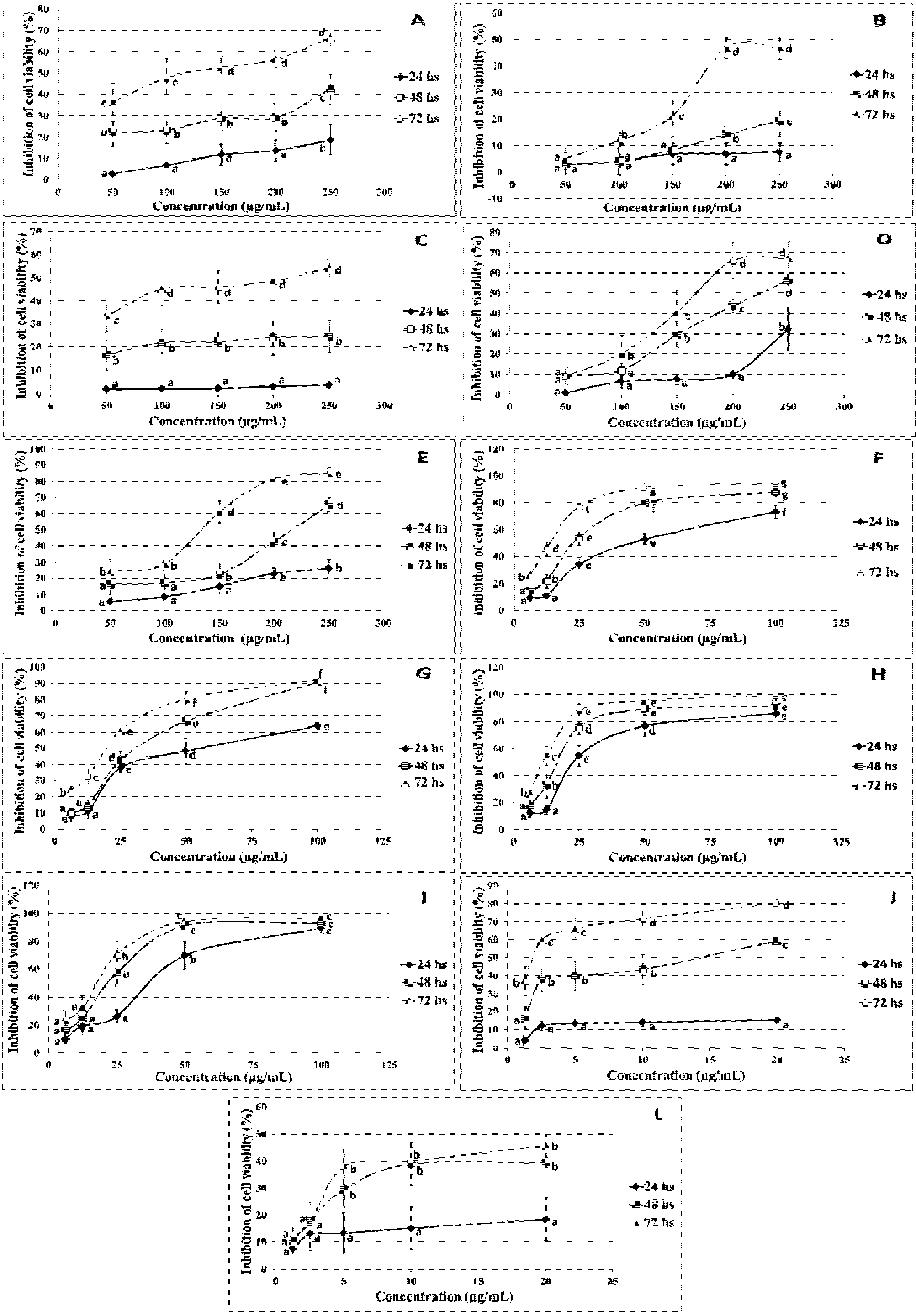
2.1. Cytotoxic Effect of Flavonoids

Studies have shown that flavonoids exert cytotoxic activity against cancer cells, a property that can be explored for the development of new cancer therapies [32]. In an attempt to extend these therapies to dogs, we evaluated the cytotoxic effects of flavonoids in the canine malignant histiocytosis cell line DH82. The MTT assay was used to evaluate the viability of cells treated with different concentrations of the flavonoids and topoisomerase inhibitors for 24, 48 and 72 h. As can be seen in Figure 1, all substances reduced cell viability in a dose-dependent fashion, but with marked differences. Luteolin, apigenin, trans-chalcone and 4-methoxychalcone significantly reduced cell viability at lower concentrations within 24 h of treatment, whereas the other flavonoids were less effective after the same period of treatment.
The IC50 values obtained for the different flavonoids could be divided into two groups (Table 1). Quercetin, genistein, epigallocatechin gallate (EGCG), myricetin, and fisetin exhibited IC50 values higher than 200 µM after 72 h of treatment, or 600 µM after 24 h of treatment. Lower IC50 values were observed for apigenin, luteolin, trans-chalcone and 4-methoxychalcone, ranging from 52.8 µM at 72 h of treatment (luteolin) to 165.4 µM at 24 h (apigenin). trans-Chalcone was the most effective flavonoid after 24 h of treatment (IC50: 129.2 µM), whereas luteolin was the most effective flavonoid after 48 and 72 h of treatment (IC50: 87.7 and 52.8 µM, respectively). The IC50 values obtained for luteolin, apigenin, trans-chalcone and 4-methoxychalcone were comparable to that of the topoisomerase inhibitor merbarone. Although quercetin, genistein, fisetin, EGCG and myricetin showed low cytotoxicity in the DH82 cell line, several studies have demonstrated their antiproliferative activity against different cell lines [33,34,35]. This reflects the cell-specific effect of flavonoids which is influenced by the capacity of each cell type to absorb, inactivate or metabolize these substances to more toxic metabolites [36,37,38].
Table 1. IC50 values of flavonoids and topoisomerase inhibitors in DH82 cells.
Flavonoids and DrugsIC50 (µM)
24 h48 h72 h
Luteolin156.287.752.8
Apigenin165.4124.379.5
4-Methoxychalcone154.092.776.4
trans-Chalcone129.289.359.5
Quercetin>992.5>661.7>302.2
Genistein>1110>740.1>370
EGCG>654.5>436.6>218.2
Myricetin>942.7>628.5>628.5
Fisetin>1048>1048>349.5
Etoposide95.519.13.6
Merbarone205.577.865.3

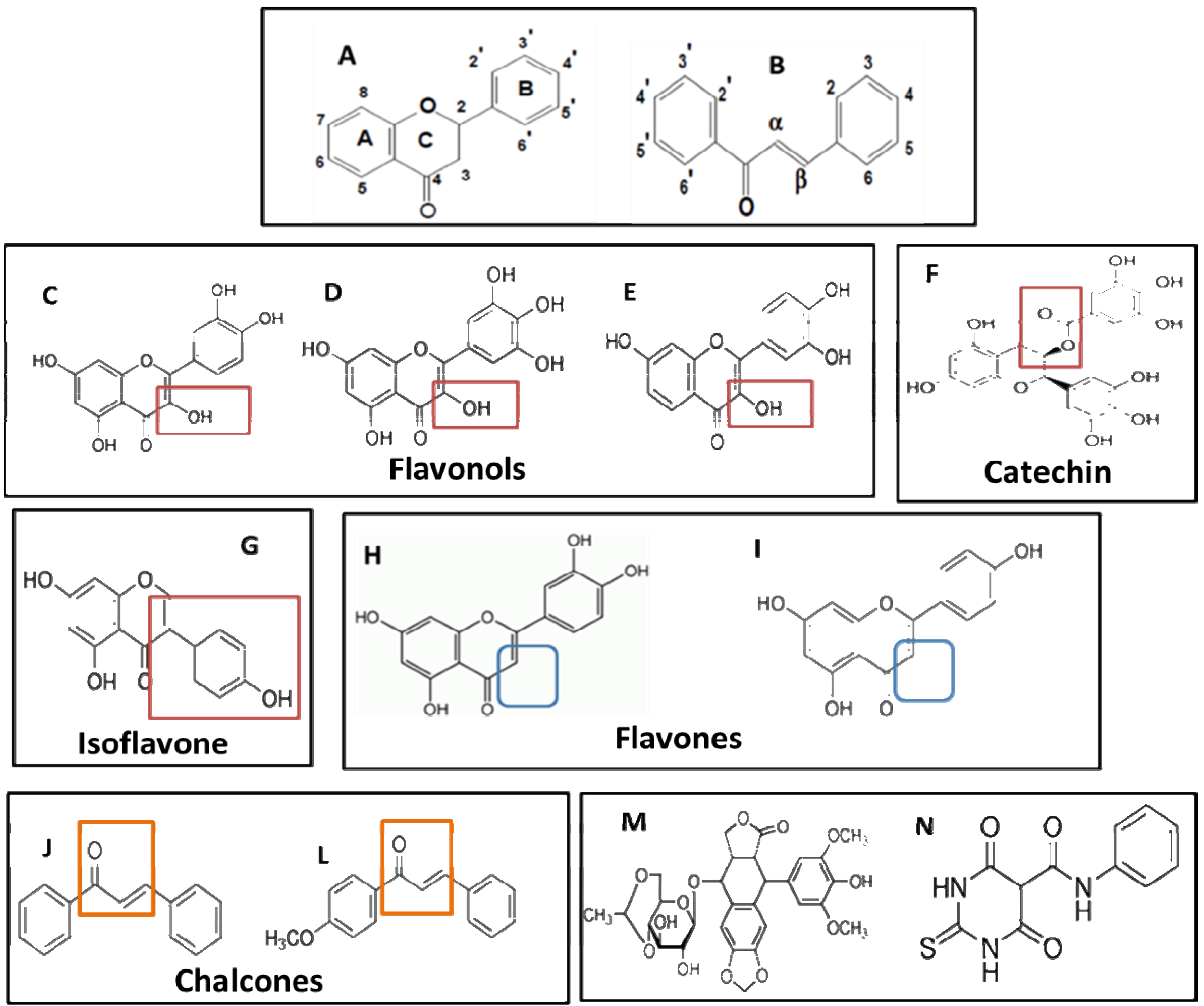
2.2. Analysis of the Relationship between Structure and Cytotoxic Effect

The biological activity of flavonoids has been suggested to be correlated with the position, number and substitution of the hydroxyl group in the A and B rings [39]. In this study, the differences in the cytotoxic effect and IC50 values of the flavonoids permitted a preliminary attempt to establish a structure-cytotoxic effect relationship. In all treatments, the IC50 values of the two flavonols luteolin and apigenin were comparable to that of the topoisomerase inhibitor merbarone, whereas the flavones quercetin, myricetin and fisetin exhibited values higher than 200 µM. The main difference between flavones and flavonols is the absence of a 3-hydroxyl group at position 3 of the C ring. In genistein, the B ring is attached to this position, whereas in EGCG an extra ring (galloyl-D-ring) is attached to it. Luteolin differs from apigenin by the presence of a 3-hydroxyl at position 3’ of the B ring, but has only a slightly superior cytotoxic effect (Table 2 and Figure 2). Therefore, a group attached to position 3 of the C ring seems to be the most important structural feature for the cytotoxic effect of these flavonoids in the DH82 cell line.
Table 2. Structural differences (number and position of substituents) between the nine flavonoids tested in this study.
FlavonoidsClassSubstituentsTotal OHTotal COH3
Chain AChain BChain C
Cytotoxic compounds
trans-ChalconeChalcone00
4-MethoxychalconeChalconeCOH3-4'01
LuteolinFlavoneOH-5,7OH-3',4'O-4; anel-240
ApigeninFlavoneOH-5,7OH-4'O-4; anel-230
Low cytotoxicity compounds
QuercetinFlavonolOH-,5,7OH-3',4'O-4; OH-3; anel-250
MyricetinFlavonolOH-,5,7OH-3',4',5'O-4; OH-3; anel-260
FisetinFlavonolOH-7OH-4',5'O-4; OH-3; anel-240
EGCGCatechinOH-5,7OH-3',4',5'galato-3; anel-280
GenisteinIsoflavoneOH-5,7OH-4'O-4; anel-330
The two chalcones tested also exhibited lower IC50 values, which were comparable to that of the topoisomerase IIa inhibitor merbarone. After 24 h of treatment, the order of cytotoxicity based on the IC50 values of the most effective flavonoids was trans-chalcone > 4-methoxychalcone > luteolin > apigenin, while at 48 and 72 h the order was luteolin > trans-chalcone > 4-methoxychalcone > apigenin (Table 1). The main difference of chalcones compared to flavones and to the other flavonoids tested is the absence of the heterocyclic C ring (Figure 2). Again, the C ring seems to play a key role in the cytotoxic effects observed. trans-Chalcone and 4-methoxychalcone differ in the position of the double bond in the open C ring and in the presence of a methoxy group attached to position 4 of the A ring in 4-methoxychalcone (Figure 2). These differences may explain the lower IC50 value observed for trans-chalcone.
Figure 2. Chemical structure of flavonoids and topoisomerase inhibitors. (A) General structure of flavonoids; (B) general structure of chalcones; (C) quercetin; (D) myricetin; (E) fisetin; (F) epigallocatechin gallate (EGCG); (G) genistein; (H) luteolin; (I) apigenin; (J) trans-chalcone; (L) 4-methoxychalcone; (M) etoposide; (N) merbarone.
Molecules 18 15448 g002
The presence of a catechol group (3',4'-OH) in the B ring is considered a prerequisite for cytotoxic activity [40]. The positive influence of dihydroxylation of carbons 3 and 6 has also been demonstrated [41]. However, the present results indicate that a substituent at position 3 of the C ring reduced the cytotoxicity of the flavonoids tested. A reduction in cytotoxic activity when a hydroxyl group is present at position 3 of the C ring has been demonstrated [36]. This feature is found in quercetin, myricetin and fisetin, which exhibited very low cytotoxicity against the DH82 cell line. Luteolin and apigenin lack a substituent at position 3 of the C ring and were more cytotoxic than the former. Moreover, trans-chalcone and 4-methoxychalcone have an open C ring and both lack a hydroxyl group in their structure. We postulate that the absence of the hydroxyl group near the ketone in ring C is an important feature for the cytotoxic activity of flavonoids, whereas the presence of substituents in the A and B rings has a modulatory effect on cytotoxicity.

2.3. Genotoxic Effect

We also investigated the induction of DNA damage by flavonoids in the DH82 cells using the comet assay in order to determine whether the cytotoxicity of the flavonoids studied is directly related to genotoxicity. Most flavonoids showed a DNA damage index (DDI) of 1 or lower (Table 3), indicating low genotoxicity in DH82 cells. Only genistein exerted a significant dose- and time-dependent genotoxic effect, with a DDI of up to 2.79 (Table 3). The formation of comets with defined tails (long and intense) is strongly associated with the stabilization of topoisomerase-DNA cleavage complexes [42]. This is the mechanism of action of etoposide (positive control), thus explaining the high genotoxicity of this topoisomerase inhibitor (DDI 3.22 ± 0.065 after 24 h) when compared to merbarone (DDI 1.14 ± 0.088 after 24 h), which does not stabilize topoisomerase II-DNA cleavage complexes. It was therefore suggested that merbarone induces less DNA damage than etoposide [43], a fact confirmed in the present study using DH82 cells (Table 3). Greater stabilization of the DNA-topoisomerase cleavage complex is a major determinant of DNA damage, since it increases the chance that the complex collides with the replication and transcription machinery [44]. Genotoxic effects mediated by this mechanism have been reported for several flavonoids [45,46,47], including those tested here [45,46,47]. However, the only flavonoid that induced significant DNA damage in the present study was genistein (Table 3). In contrast, this flavonoid did not induce significant cytotoxicity at the same concentration or after the same time of treatment. Similarly, etoposide, a topoisomerase inhibitor, induced low cytotoxicity (Figure 1), but high DNA damage (Table 3), when the cells were treated with a concentration of 2.5 µg/mL for 24 h. Similar results have been reported in other studies [42]. Quercetin, EGCG, myricetin and fisetin did not induce significant cytotoxicity or genotoxicity, suggesting low specificity of these flavonoids for DH82 cells. Luteolin and apigenin also did not induce DNA damage, but were moderately cytotoxic, indicating that low genotoxicity is not related to low specificity for DH82 cells. The two chalcones tested were cytotoxic, but did not induce elevated levels of DNA damage. There are few studies investigating the biological effects of these substances and it is unlikely that their cytotoxic effects are due to genotoxicity induced by topoisomerase poisoning. Topoisomerase II poisoning can occur by redox-independent and redox-dependent mechanisms. In the first mechanism, flavonoids with a closed chain require the formation of a pseudo-ring between the ketone group of the C ring and a close hydroxyl (OH at position 5). In redox-dependent poisoning, the presence of a pyrogallol (OH at positions 3', 4' and 5') is necessary, whose oxidation leads to the formation of hydrogen peroxide, inducing the formation of topoisomerase-DNA complexes [44,48]. Chalcones have an open C ring and those tested in this study do not contain any hydroxyl group (Figure 2 and Table 2), supporting the possibility that they do not induce DNA damage as demonstrated by the comet assay, especially not through topoisomerase poisoning.
Table 3. Genotoxicity of flavonoids and topoisomerase inhibitors in DH82 cells.
Genotoxicity
SubstancesConcentration (µg/mL)DNA Damage Index: 0–4
24 h 6 h
Control (0.5% DMSO)0.36 ± 0.070 a0.4 ± 0.061 a
Etoposide2.53.22 ± 0.065 h2.5 ± 0.115 f
Merbarone2.51.14 ± 0.088 d0.79 ± 0.052 c
Genistein2002.79 ± 0.067 g1.84 ± 0.17 e
1001.80 ± 0.083 e1.15 ± 0.105 d
501.10 ± 0.065 d0.83 ± 0.095 c
Quercetin2000.65 ± 0.094 b-
EGCG2001.03 ± 0.031 d-
Myricetin2000.88 ± 0.055 c-
Fisetin2000.83 ± 0.032 c-
Luteolin250.74 ± 0.066 b-
12.50.68 ± 0.075 b-
trans-Chalcone250.64 ± 0.051 b0.51 ± 0.079 a
12.50.57 ± 0.043 a0.50 ± 0.039 a
4-Methoxychalcone250.73 ± 0.085 b0.48 ± 0.062 a
12.50.66 ± 0.074 b0.47 ± 0.056 a
Apigenin250.80 ± 0.0630.66 ± 0.096 b
12.50.74 ± 0.052 b0.57 ± 0.107 a
DNA damage index: 0 (no DNA damage) to 4 (total DNA damage). The results are expressed as the mean ± standard error of at least three independent experiments. Different letters indicate statistically significant results (p < 0.05).

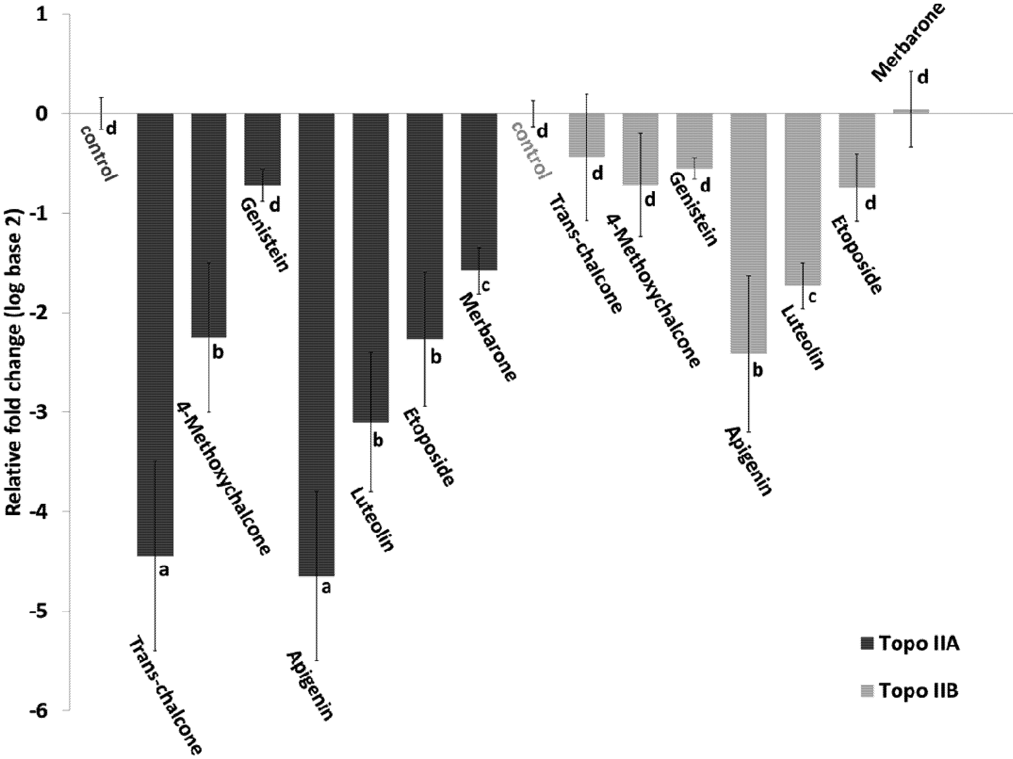
2.4. Effect of Flavonoids on the Expression of Topoisomerase II

The effect of trans-chalcone, 4-methoxychalcone, luteolin, apigenin, genistein and the two topoisomerase inhibitors on the expression of the topoisomerase IIa and IIb genes in DH82 cells was evaluated by quantitative RT-PCR. Genistein was chosen for analysis because it was the most genotoxic flavonoid, while the other flavonoids were the most cytotoxic. As shown in Figure 3, except for merbarone, all flavonoids and etoposide inhibited the two topoisomerases to variable extents (negative fold change). The repression of topoisomerase IIa was more marked than that of topoisomerase IIb. This finding was more evident for trans-chalcone and 4-methoxychalcone, which inhibited topoisomerase IIa ten and three times more than topoisomerase IIb, respectively. The highest levels of repression of the IIa isoform were −4.65 and −4.45 seen in the treatments with apigenin and trans-chalcone, respectively. Apigenin exerted the strongest inhibitory effect on the IIb isoform, followed by luteolin.
The cytotoxicity and genotoxicity results might be explained by the repression or expression of specific pathways targeted by the different flavonoids studied. In this respect, many flavonoids induce cell cycle arrest related to topoisomerase II inhibition [35,49,50]. One example is the inhibition of topoisomerase IIa mRNA expression by genistein in HeLa cells, which has been shown to be related to interruption of the cell cycle and induction of apoptosis [51]. In this study, we determined whether flavonoids with significant cytotoxic and genotoxic activity influence DNA topoisomerase IIa and IIb gene expression at the transcriptional level. Luteolin, apigenin, trans-chalcone, and 4-methoxy-chalcone inhibited mRNA expression of both topoisomerases II and, more specifically, of the IIa isoform. The repression of topoisomerase IIa induces cell death by mitotic failure as a result of incorrect segregation of the chromosomes to the cell poles during mitosis [52].
Figure 3. Effect of flavonoids and topoisomerase inhibitors on the expression of topoisomerase IIa and IIb genes. The cells were treated with flavonoids at a concentration of 12.5 µg/mL for 6 h. The concentration of etoposide and merbarone was 2.5 µg/mL. The results are expressed as the mean ± standard error of at least three independent experiments. Different letters indicate statistically significant results (p < 0.1).
Molecules 18 15448 g003
The more marked inhibition of the IIa isoform may be advantageous for exploring chalcones in cancer therapies, since the IIb isoform seems to play a key role in the induction of secondary neoplasias [19,53] and in the cardiotoxic effect of drugs such as doxorubicin [54]. Moreover, the inhibition of topoisomerase IIb may compromise DNA repair in neuronal tissue [55,56]. Another fact is that catalytic inhibitors are more effective in cells expressing topoisomerase II at low levels [57]. Therefore, the sensitivity of cancer cell lines to catalytic inhibitors could be increased by combining them with chalcones, thus reducing the use of topoisomerase poisons and, consequently, side effects such as secondary neoplasias.

3. Experimental

3.1. Materials

The topoisomerase inhibitors merbarone, etoposide, quercetin, trans-chalcone, and 4-methoxychalcone were purchased from Sigma-Aldrich (St. Louis, MO, USA). All other flavonoids were purchased from Cayman Chemical (Ann Arbor, MI, USA). (3-(4,5-Dimethylthiazol-2-yl)-2,5-diphenyltetrazolium bromide (MTT), dimethyl sulfoxide (DMSO), Triton X-100, Dulbecco’s modified Eagle’s medium (DMEM), fetal bovine serum, and DNase I were purchased from Sigma-Aldrich. Normal and low-melting point agarose were purchased from USB (Cleveland, OH, USA). NaCl, Tris, NaOH and EDTA were purchased from Synth (Diadema, SP, Brazil). The Illustra RNAspin Mini RNA Isolation kit was purchased from GE Healthcare (Piscataway, NJ, USA). The TaqMan® Gene Expression assay and High-Capacity cDNA Reverse Transcription kit were purchased from Applied Biosystems (Foster City, CA, USA).

3.2. Cell Culture

The canine malignant histiocytic cell line DH82 (ATCC number: CRL-10389) [30] was cultured in DMEM supplemented with 10% fetal bovine serum and incubated in a humidified incubator in a 5% CO2 atmosphere at 37 °C. In the different assays, DMSO was used as a solvent of the chemical compounds at an atoxic concentration for the cells (0.5% or less).

3.3. Cytotoxicity Assay

The cytotoxicity of the different chemicals was analyzed by the MTT assay [58]. The cells were seeded in 96-well plates at a concentration of 2 × 10⁵ cells/well and cultured for 24 h in a 5% CO2 atmosphere at 37 °C. After this period, the chemicals diluted in fresh medium to five different concentrations were added to the wells and the cells were cultured under the same conditions for 24, 48 and 72 h. The medium with the substances was changed at intervals of 24 h. All cell treatments were carried out in triplicate. After each treatment period, the medium was replaced with fresh medium without chemicals, 20 µL MTT solution (5 mg/mL) was added to each well, and the plates were incubated for an additional 4 h under the same conditions. The plates were centrifuged at 3500 × g for 5 min, the supernatant was discarded, and 200 µL DMSO was added to dissolve the formed formazan crystals. Absorbance was read in a Thermoplate reader at a wavelength of 550 nm. The results were plotted as the percentage of inhibition of cell viability (ICV) calculated using the following formula:
ICV (%) = [1 − (average absorbance of experimental group/average absorbance of cells treated with 0.5% DMSO control group)] × 100

3.4. DNA Damage Assay

DNA damage was evaluated by the single-cell gel electrophoresis assay (comet assay) [59]. The cells were cultured in 24-well plates at a concentration of 2 × 10⁵ cells/well (5% CO2 atmosphere, 37 °C) in the presence of the chemicals at a concentration that permitted ~70% cell viability, for periods of 6 and 24 h. Etoposide and merbarone (2.5 µg/mL) were used as positive controls and 0.5% DMSO as solvent control. After the treatments, the cells were transferred to a microtube and centrifuged at 350 × g for 5 min at 4 °C. The supernatant was discarded and the cell pellet was resuspended in 200 µL low-melting point agarose (0.5% in PBS). The agarose/cell suspension was pipetted onto microscope slides previously coated with low-melting point agarose (1.5% in PBS). The agarose/cell suspension was covered with coverslips and the slides were incubated for 10 min at 4 °C. Three slides were prepared per treatment. After removal of the coverslips, the slides were immersed in lysis buffer (2.5 M NaCl, 100 mM EDTA, 10 mM Tris, 1% Triton X-100, and 10% DMSO) for 20 h at 4 °C in the dark. After rinsing with electrophoresis solution (10 M NaOH, 200 mM EDTA, pH 13) for 20 min at 4 °C in the dark, the slides were submitted to electrophoresis at 0.8 volts/cm for 20 min at 4 °C in the dark. Next, the slides were removed from the electrophoresis chamber and rinsed with neutralization buffer (0.4 M Tris, pH 7.5) for 15 min, air dried at room temperature, and fixed with absolute ethanol for 3 min. The slides were stained with 40 µL of an ethidium bromide solution (0.02 mg/mL) and visualized under a fluorescent microscope. One hundred comets/slide were randomly selected and DNA damage was visually scored based on the length and intensity of the tail into five classes ranging from 0 (no DNA damage) to 4 (maximal DNA damage) [60]. The DNA damage index (DDI) was calculated using the formula:
DDI = (No. of cells in class 0 × 0 + No. of cells in class 1 × 1 + No. of cells in class 2 × 2 + No. of cells in class 3 × 3 + No. of cells in class 4 × 4)/No. of comets analyzed

3.5. Quantification of Topoisomerase IIa and Topoisomerase IIb Messenger RNA (mRNA)

The cells were cultured in 24-well plates at a concentration of 2 × 10⁶ cells/well in a 5% CO2 atmosphere at 37 °C. After adhesion, the cells were treated for 6 h with the chemicals at a concentration that permitted ~70% cell viability. Next, mRNA was extracted from the cells and purified using the Illustra RNAspin Mini RNA Isolation kit (GE Healthcare). After treatment with DNAse I (Sigma-Aldrich), 1 µg mRNA was used for synthesis of cDNA using the High-Capacity cDNA Reverse Transcription kit (Applied Biosystems). Messenger RNA was quantified in a total volume of 20 μL using the TaqMan® Gene Expression Assay (Applied Biosystems) according to manufacturer instructions. The reaction mixture contained 1 µL 20X TaqMan® Gene Expression Assay (TaqMan® Gene Expression Assay IDs are shown in Table 1), 10 µL 2X TaqMan® Gene Expression Master Mix, 1 µL cDNA, and 8 µL RNase-free water. A negative control was included for each gene and amplification reactions were performed in triplicate in an Mx3005P real-time thermocycler (Stratagene, La Jolla, CA, USA), with one hold at 95 °C for 10 min and 40 cycles of 95 °C for 15 s and 60 °C for 1 min. The fluorescence data were collected at the end of the annealing/elongation step. Data were analyzed and plotted using the MxPro software (Stratagene). The RPL32 gene was used as a reference for normalization of the results and fold-differences in gene expression were calculated relative to control samples (cells treated with 0.5% DMSO) using the ΔΔCT method.

3.6. Statistical Analysis

The data were analyzed by two-way analysis of variance (ANOVA), followed by the Scott-Knott test (p < 0.05 or p < 0.1). The IC50 values were calculated by nonlinear regression analysis from the ICV-concentration data. The results of ICV, DDI and fold-change in gene expression are plotted as the mean ± standard error of at least three biological replicates.

4. Conclusions

Although chemotherapy for canine cancer is available, treatment is often palliative and euthanasia is still necessary in most cases. Even at low doses, the commonly used drug doxorubicin can cause cardiomyopathies [61]. Therefore, the search for novel anticancer drugs is also mandatory for dogs. The results of this study raise the possibility of exploring flavonoids for anticancer therapies in dogs, as well as in humans, in view of their lower toxicity when compared to other drugs.

Acknowledgments

This study was supported by FAPESP (Grant No. 10/18122-7). We thank the staff of the Biotechnology Unit, UNAERP, for general support, and K. Markendorf for English revision.

Conflicts of Interest

The authors declare no conflict of interest.

References

  1. Baek, S.J.; McEntee, M.F.; Legendre, A.M. Review paper: Cancer chemopreventive compounds and canine cancer. Vet. Pathol. 2009, 46, 576–588. [Google Scholar] [CrossRef]
  2. Dobson, J.M.; Samuel, S.; Milstein, H.; Rogers, K.; Wood, J.L.N. Canine neoplasia in the UK: Estimates of incidence rates from a population of insured dogs. J. Small Anim. Pract. 2002, 43, 240–246. [Google Scholar] [CrossRef]
  3. Merlo, D.F.; Rossi, L.; Pellegrino, C.; Ceppi, M.; Cardellino, U.; Capurro, C.; Ratto, A.; Sambucco, P.L.; Sestito, V.; Tanara, G.; et al. Cancer incidence in pet dogs: Findings of the animal tumor registry of genoa, italy. J. Vet. Intern. Med. 2008, 22, 976–984. [Google Scholar] [CrossRef]
  4. Patterson, S.L.; Maresso, K.C.; Hawk, E. Cancer chemoprevention: Successes and failures. Clin. Chem. 2013, 59, 94–101. [Google Scholar] [CrossRef]
  5. Beecher, G.R. Overview of dietary flavonoids: Nomenclature, occurrence and intake. J. Nutr. 2003, 133, 3248S–3254S. [Google Scholar]
  6. De Kok, T.M.; de Waard, P.; Wilms, L.C.; van Breda, S.G. Antioxidative and antigenotoxic properties of vegetables and dietary phytochemicals: The value of genomics biomarkers in molecular epidemiology. Mol. Nutr. Food Res. 2010, 54, 208–217. [Google Scholar] [CrossRef]
  7. Christen, P.; Cuendet, M. Plants as a source of therapeutic and health products. Chimia 2012, 66, 320–323. [Google Scholar] [CrossRef]
  8. Russo, M.; Spagnuolo, C.; Tedesco, I.; Russo, G.L. Phytochemicals in cancer prevention and therapy: Truth or dare? Toxins 2010, 2, 517–551. [Google Scholar] [CrossRef]
  9. Procházková, D.; Boušová, I.; Wilhelmová, N. Antioxidant and prooxidant properties of flavonoids. Fitoterapia 2011, 82, 513–523. [Google Scholar] [CrossRef]
  10. Pietta, P.G. Flavonoids as antioxidants. J. Nat. Prod. 2000, 63, 1035–1042. [Google Scholar] [CrossRef]
  11. Hasebe, Y.; Egawa, K.; Yamazaki, Y.; Kunimoto, S.; Hirai, Y.; Ida, Y.; Nose, K. Specific inhibition of hypoxia-inducible factor (hif)-1 alpha activation and of vascular endothelial growth factor (vegf) production by flavonoids. Biol. Pharm. Bull. 2003, 26, 1379–1383. [Google Scholar] [CrossRef]
  12. Schindler, R.; Mentlein, R. Flavonoids and vitamin e reduce the release of the angiogenic peptide vascular endothelial growth factor from human tumor cells. J. Nutr. 2006, 136, 1477–1482. [Google Scholar]
  13. Hodnick, W.F.; Bohmont, C.W.; Capps, C.; Pardini, R.S. Inhibition of the mitochondrial nadh-oxidase (nadh-coenzyme q oxido-reductase) enzyme system by flavonoids: A structure-activity study. Biochem. Pharmacol. 1987, 36, 2873–2874. [Google Scholar] [CrossRef]
  14. Loizzo, M.R.; Said, A.; Tundis, R.; Rashed, K.; Statti, G.A.; Hufner, A.; Menichini, F. Inhibition of angiotensin converting enzyme (ace) by flavonoids isolated from ailanthus excelsa (roxb) (simaroubaceae). Phytother. Res. PTR 2007, 21, 32–36. [Google Scholar] [CrossRef]
  15. Bermudez-Soto, M.J.; Larrosa, M.; Garcia-Cantalejo, J.M.; Espin, J.C.; Tomas-Barberan, F.A.; Garcia-Conesa, M.T. Up-regulation of tumor suppressor carcinoembryonic antigen-related cell adhesion molecule 1 in human colon cancer caco-2 cells following repetitive exposure to dietary levels of a polyphenol-rich chokeberry juice. J. Nutr. Biochem. 2007, 18, 259–271. [Google Scholar] [CrossRef]
  16. Whitsett, T.G., Jr.; Lamartiniere, C.A. Genistein and resveratrol: Mammary cancer chemoprevention and mechanisms of action in the rat. Expert Rev. Anticancer Ther. 2006, 6, 1699–1706. [Google Scholar] [CrossRef]
  17. Fini, L.; Selgrad, M.; Fogliano, V.; Graziani, G.; Romano, M.; Hotchkiss, E.; Daoud, Y.A.; de Vol, E.B.; Boland, C.R.; Ricciardiello, L. Annurca apple polyphenols have potent demethylating activity and can reactivate silenced tumor suppressor genes in colorectal cancer cells. J. Nutr. 2007, 137, 2622–2628. [Google Scholar]
  18. Fresco, P.; Borges, F.; Marques, M.P.; Diniz, C. The anticancer properties of dietary polyphenols and its relation with apoptosis. Curr. Pharm. Des. 2010, 16, 114–134. [Google Scholar] [CrossRef]
  19. Azarova, A.M.; Lin, R.K.; Tsai, Y.C.; Liu, L.F.; Lin, C.P.; Lyu, Y.L. Genistein induces topoisomerase iibeta- and proteasome-mediated DNA sequence rearrangements: Implications in infant leukemia. Biochem. Biophys. Res. Commun. 2010, 399, 66–71. [Google Scholar] [CrossRef]
  20. Barjesteh van Waalwijk van Doorn-Khosrovani, S.; Janssen, J.; Maas, L.M.; Godschalk, R.W.; Nijhuis, J.G.; van Schooten, F.J. Dietary flavonoids induce mll translocations in primary human cd34+ cells. Carcinogenesis 2007, 28, 1703–1709. [Google Scholar] [CrossRef]
  21. Lin, C.N.; Chen, H.L.; Yen, M.H. Flavonoids with DNA strand-scission activity from rhus javanica var. Roxburghiana. Fitoterapia 2008, 79, 32–36. [Google Scholar] [CrossRef]
  22. Varela-Barca, F.N.; Agnez-Lima, L.F.; de Medeiros, S.R. Base excision repair pathway is involved in the repair of lesions generated by flavonoid-enriched fractions of pepper tree (schinus terebinthifolius, raddi) stem bark. Environ. Mol. Mutagen. 2007, 48, 672–681. [Google Scholar] [CrossRef]
  23. Gilbert, E.R.; Liu, D. Flavonoids influence epigenetic-modifying enzyme activity: Structure—function relationships and the therapeutic potential for cancer. Curr. Med. Chem. 2010, 17, 1756–1768. [Google Scholar] [CrossRef]
  24. Landis-Piwowar, K.R.; Dou, Q.P. Polyphenols: Biological activities, molecular targets, and the effect of methylation. Curr. Mol. Pharmacol. 2008, 1, 233–243. [Google Scholar]
  25. Li, Y.; Tollefsbol, T.O. Impact on DNA methylation in cancer prevention and therapy by bioactive dietary components. Curr. Med. Chem. 2010, 17, 2141–2151. [Google Scholar] [CrossRef]
  26. Hadi, S.M.; Bhat, S.H.; Azmi, A.S.; Hanif, S.; Shamim, U.; Ullah, M.F. Oxidative breakage of cellular DNA by plant polyphenols: A putative mechanism for anticancer properties. Semin. Cancer Biol. 2007, 17, 370–376. [Google Scholar] [CrossRef]
  27. Shamim, U.; Hanif, S.; Ullah, M.F.; Azmi, A.S.; Bhat, S.H.; Hadi, S.M. Plant polyphenols mobilize nuclear copper in human peripheral lymphocytes leading to oxidatively generated DNA breakage: Implications for an anticancer mechanism. Free Radic. Res. 2008, 42, 764–772. [Google Scholar] [CrossRef]
  28. Nitiss, J.L. Targeting DNA topoisomerase ii in cancer chemotherapy. Nat. Rev. Cancer 2009, 9, 338–350. [Google Scholar] [CrossRef]
  29. Jamadar-Shroff, V.; Papich, M.G.; Suter, S.E. Soy-derived isoflavones inhibit the growth of canine lymphoid cell lines. Clin. Cancer Res. Off. J. Am. Assoc. Cancer Res. 2009, 15, 1269–1276. [Google Scholar] [CrossRef]
  30. Wellman, M.; Krakowka, S.; Jacobs, R.; Kociba, G. A macrophage-monocyte cell line from a dog with malignant histiocytosis. In Vitro Cell Dev. Biol. 1988, 24, 223–229. [Google Scholar] [CrossRef]
  31. Fulmer, A.K.; Mauldin, G.E. Canine histiocytic neoplasia: An overview. Can. Vet. J. La Revue Vet. Can. 2007, 48, 1041–1043, 1046–1050. [Google Scholar]
  32. Chahar, M.K.; Sharma, N.; Dobhal, M.P.; Joshi, Y.C. Flavonoids: A versatile source of anticancer drugs. Pharmacogn. Rev. 2011, 5, 1–12. [Google Scholar] [CrossRef]
  33. Kobayashi, T.; Nakata, T.; Kuzumaki, T. Effect of flavonoids on cell cycle progression in prostate cancer cells. Cancer Lett. 2002, 176, 17–23. [Google Scholar] [CrossRef]
  34. Matsuo, M.; Sasaki, N.; Saga, K.; Kaneko, T. Cytotoxicity of flavonoids toward cultured normal human cells. Biol. Pharm. Bull. 2005, 28, 253–259. [Google Scholar] [CrossRef]
  35. Yadegarynia, S.; Pham, A.; Ng, A.; Nguyen, D.; Lialiutska, T.; Bortolazzo, A.; Sivryuk, V.; Bremer, M.; White, J.B. Profiling flavonoid cytotoxicity in human breast cancer cell lines: Determination of structure-function relationships. Nat. Prod. Commun. 2012, 7, 1295–1304. [Google Scholar]
  36. Boots, A.W.; Li, H.; Schins, R.P.; Duffin, R.; Heemskerk, J.W.; Bast, A.; Haenen, G.R. The quercetin paradox. Toxicol. Appl. Pharmacol. 2007, 222, 89–96. [Google Scholar] [CrossRef]
  37. Araujo, K.C.; de, M.B.C.E.M.; Pazini, F.; Valadares, M.C.; de Oliveira, V. Bioconversion of quercetin and rutin and the cytotoxicity activities of the transformed products. Food Chem. Toxicol. Int. J. Publ. Bri. Ind. Biol. Res. Assoc. 2013, 51, 93–96. [Google Scholar]
  38. Metodiewa, D.; Jaiswal, A.K.; Cenas, N.; Dickancaite, E.; Segura-Aguilar, J. Quercetin may act as a cytotoxic prooxidant after its metabolic activation to semiquinone and quinoidal product. Free Radic. Biol. Med. 1999, 26, 107–116. [Google Scholar] [CrossRef]
  39. Plochmann, K.; Korte, G.; Koutsilieri, E.; Richling, E.; Riederer, P.; Rethwilm, A.; Schreier, P.; Scheller, C. Structure-activity relationships of flavonoid-induced cytotoxicity on human leukemia cells. Arch. Biochem. Biophys. 2007, 460, 1–9. [Google Scholar] [CrossRef]
  40. Kawaii, S.; Tomono, Y.; Katase, E.; Ogawa, K.; Yano, M. Antiproliferative activity of flavonoids on several cancer cell lines. Biosci. Biotechnol. Biochem. 1999, 63, 896–899. [Google Scholar] [CrossRef]
  41. Chang, H.; Mi, M.; Ling, W.; Zhu, J.; Zhang, Q.; Wei, N.; Zhou, Y.; Tang, Y.; Yuan, J. Structurally related cytotoxic effects of flavonoids on human cancer cells in vitro. Arch. Pharmacal Res. 2008, 31, 1137–1144. [Google Scholar] [CrossRef]
  42. Kang, K.; Oh, S.H.; Yun, J.H.; Jho, E.H.; Kang, J.H.; Batsuren, D.; Tunsag, J.; Park, K.H.; Kim, M.; Nho, C.W. A novel topoisomerase inhibitor, daurinol, suppresses growth of hct116 cells with low hematological toxicity compared to etoposide. Neoplasia 2011, 13, 1043–1057. [Google Scholar]
  43. Fortune, J.M.; Osheroff, N. Merbarone inhibits the catalytic activity of human topoisomerase iialpha by blocking DNA cleavage. J. Biol. Chem. 1998, 273, 17643–17650. [Google Scholar] [CrossRef]
  44. Bandele, O.J.; Osheroff, N. The efficacy of topoisomerase ii-targeted anticancer agents reflects the persistence of drug-induced cleavage complexes in cells. Biochemistry 2008, 47, 11900–11908. [Google Scholar] [CrossRef]
  45. Bandele, O.J.; Osheroff, N. Bioflavonoids as poisons of human topoisomerase ii alpha and ii beta. Biochemistry 2007, 46, 6097–6108. [Google Scholar] [CrossRef]
  46. Chowdhury, A.R.; Sharma, S.; Mandal, S.; Goswami, A.; Mukhopadhyay, S.; Majumder, H.K. Luteolin, an emerging anti-cancer flavonoid, poisons eukaryotic DNA topoisomerase i. Biochem. J. 2002, 366, 653–661. [Google Scholar] [CrossRef]
  47. Lanoue, L.; Green, K.K.; Kwik-Uribe, C.; Keen, C.L. Dietary factors and the risk for acute infant leukemia: Evaluating the effects of cocoa-derived flavanols on DNA topoisomerase activity. Exp. Biol. Med. 2010, 235, 77–89. [Google Scholar] [CrossRef]
  48. Lopez-Lazaro, M.; Calderon-Montano, J.M.; Burgos-Moron, E.; Austin, C.A. Green tea constituents (−)-epigallocatechin-3-gallate (egcg) and gallic acid induce topoisomerase i- and topoisomerase ii-DNA complexes in cells mediated by pyrogallol-induced hydrogen peroxide. Mutagenesis 2011, 26, 489–498. [Google Scholar] [CrossRef]
  49. O’Prey, J.; Brown, J.; Fleming, J.; Harrison, P.R. Effects of dietary flavonoids on major signal transduction pathways in human epithelial cells. Biochem. Pharmacol. 2003, 66, 2075–2088. [Google Scholar] [CrossRef]
  50. Ramos, S. Cancer chemoprevention and chemotherapy: Dietary polyphenols and signalling pathways. Mol. Nutr. Food Res. 2008, 52, 507–526. [Google Scholar] [CrossRef]
  51. Zhou, N.; Yan, Y.; Li, W.; Wang, Y.; Zheng, L.; Han, S.; Yan, Y.; Li, Y. Genistein inhibition of topoisomerase iialpha expression participated by sp1 and sp3 in hela cell. Int. J. Mol. Sci. 2009, 10, 3255–3268. [Google Scholar] [CrossRef]
  52. Deweese, J.E.; Osheroff, N. The DNA cleavage reaction of topoisomerase ii: Wolf in sheep’s clothing. Nucleic Acids Res. 2009, 37, 738–748. [Google Scholar] [CrossRef]
  53. Azarova, A.M.; Lyu, Y.L.; Lin, C.P.; Tsai, Y.C.; Lau, J.Y.; Wang, J.C.; Liu, L.F. Roles of DNA topoisomerase ii isozymes in chemotherapy and secondary malignancies. Proc. Natl. Acad. Sci. USA 2007, 104, 11014–11019. [Google Scholar] [CrossRef]
  54. Lyu, Y.L.; Kerrigan, J.E.; Lin, C.P.; Azarova, A.M.; Tsai, Y.C.; Ban, Y.; Liu, L.F. Topoisomerase iibeta mediated DNA double-strand breaks: Implications in doxorubicin cardiotoxicity and prevention by dexrazoxane. Cancer Res. 2007, 67, 8839–8846. [Google Scholar] [CrossRef]
  55. Mandraju, R.; Chekuri, A.; Bhaskar, C.; Duning, K.; Kremerskothen, J.; Kondapi, A.K. Topoisomerase iibeta associates with ku70 and parp-1 during double strand break repair of DNA in neurons. Arch. Biochem. Biophys. 2011, 516, 128–137. [Google Scholar] [CrossRef]
  56. Mandraju, R.K.; Kannapiran, P.; Kondapi, A.K. Distinct roles of topoisomerase ii isoforms: DNA damage accelerating alpha, double strand break repair promoting beta. Arch. Biochem. Biophys. 2008, 470, 27–34. [Google Scholar] [CrossRef]
  57. Larsen, A.K.; Escargueil, A.E.; Skladanowski, A. Catalytic topoisomerase ii inhibitors in cancer therapy. Pharmacol. Ther. 2003, 99, 167–181. [Google Scholar] [CrossRef]
  58. Mosmann, T. Rapid colorimetric assay for cellular growth and survival: Application to proliferation and cytotoxicity assays. J. Immunol. Methods 1983, 65, 55–63. [Google Scholar] [CrossRef]
  59. McKelvey-Martin, V.J.; Green, M.H.L.; Schmezer, P.; Pool-Zobel, B.L.; de Méo, M.P.; Collins, A. The single cell gel electrophoresis assay (comet assay): A european review. Mutat. Res./Fundam. Mol. Mech. Mutagen. 1993, 288, 47–63. [Google Scholar] [CrossRef]
  60. Collins, A.R.; Ma, A.G.; Duthie, S.J. The kinetics of repair of oxidative DNA damage (strand breaks and oxidised pyrimidines) in human cells. Mutat. Res. 1995, 336, 69–77. [Google Scholar] [CrossRef]
  61. Silva, C.E.V.; Camacho, A.A. Alterações ecocardiográficas em cães sob tratamento prolongado com doxorrubicina. Arq. Bras. Med. Vet. Zootec. 2005, 57, 300–306. [Google Scholar] [CrossRef]
  • Sample Availability: Samples of the compounds are available from the authors.

