Article

Steroidal and Phenolic Glycosides from the Bulbs of Lilium pumilum DC and Their Potential Na+/K+ ATPase Inhibitory Activity

1
Chemistry Science and Technology School, Zhanjiang Normal University, 29 Cunjin Road, Zhanjiang 524048, China
2
Development Center for New Materials Engineering Technology in Universities of Guangdong, Zhanjiang Normal University, 29 Cunjin Road, Zhanjiang 524048, China
3
Department of Pharmacy, Shaoyang Medical College Level Speciaity School, 18 Baoqing Road, Shaoyang 422000, China
*
Author to whom correspondence should be addressed.
Molecules 2012, 17(9), 10494-10502; https://doi.org/10.3390/molecules170910494
Submission received: 2 July 2012 / Revised: 25 August 2012 / Accepted: 29 August 2012 / Published: 3 September 2012
(This article belongs to the Section Natural Products Chemistry)

Abstract

:
A new steroidal saponin, named pumilum A (1), and a new phenolic glycoside, threo-1-(4′-hydroxy-2′-methoxyphenyl)-2-(2′′,4′′-dihydroxyphenyl)-1,3-propanediol-4′-O-β-D-glucopyranoside (7) were isolated from the methanolic extract of the bulbs of Lilium pumilum DC, along with five known steroidal saponins. Their chemical structures were elucidated on the basis of detailed spectroscopic analysis, including 1D and 2D NMR techniques and chemical methods. In addition, the inhibitory activity of all the isolates on Na+/K+ ATPase was evaluated.

1. Introduction

“Bǎi-hé”, a Chinese crude drug, is prepared from the bulbs of the Lilium species and is regularly used nowadays in China as a sedative, antitussive and anti-inflammatory agent [1,2,3,4]. Previously, phenolic glycosides, steroidal saponins and steroidal alkaloids have been isolated from the bulbs of the Lilium species [5,6,7,8,9,10,11,12]. However, to date, a survey of the literature showed that no chemical work has been done on Lilium pumilum DC.
Our studies indicated that the methanolic extract of the bulbs of Lilium pumilum DC showed significant Na+/K+ ATPase inhibition activity (IC50 value: 0.5 × 10⁻⁵ M). To further investigate the constituents and screen the bioactive constituents from its bulbs, a phytochemical study was performed that resulted in the isolation of new compounds 1 and 7, along with five known steroidal saponins. In the present article, we describe the structural elucidation of new compounds 1 and 7, together with the Na+/K+ ATPase inhibitory activity of all these compounds 17 (Figure 1).
Figure 1. Structures of compounds 17 isolated from the bulbs of Lilium pumilum DC.
Molecules 17 10494 g001

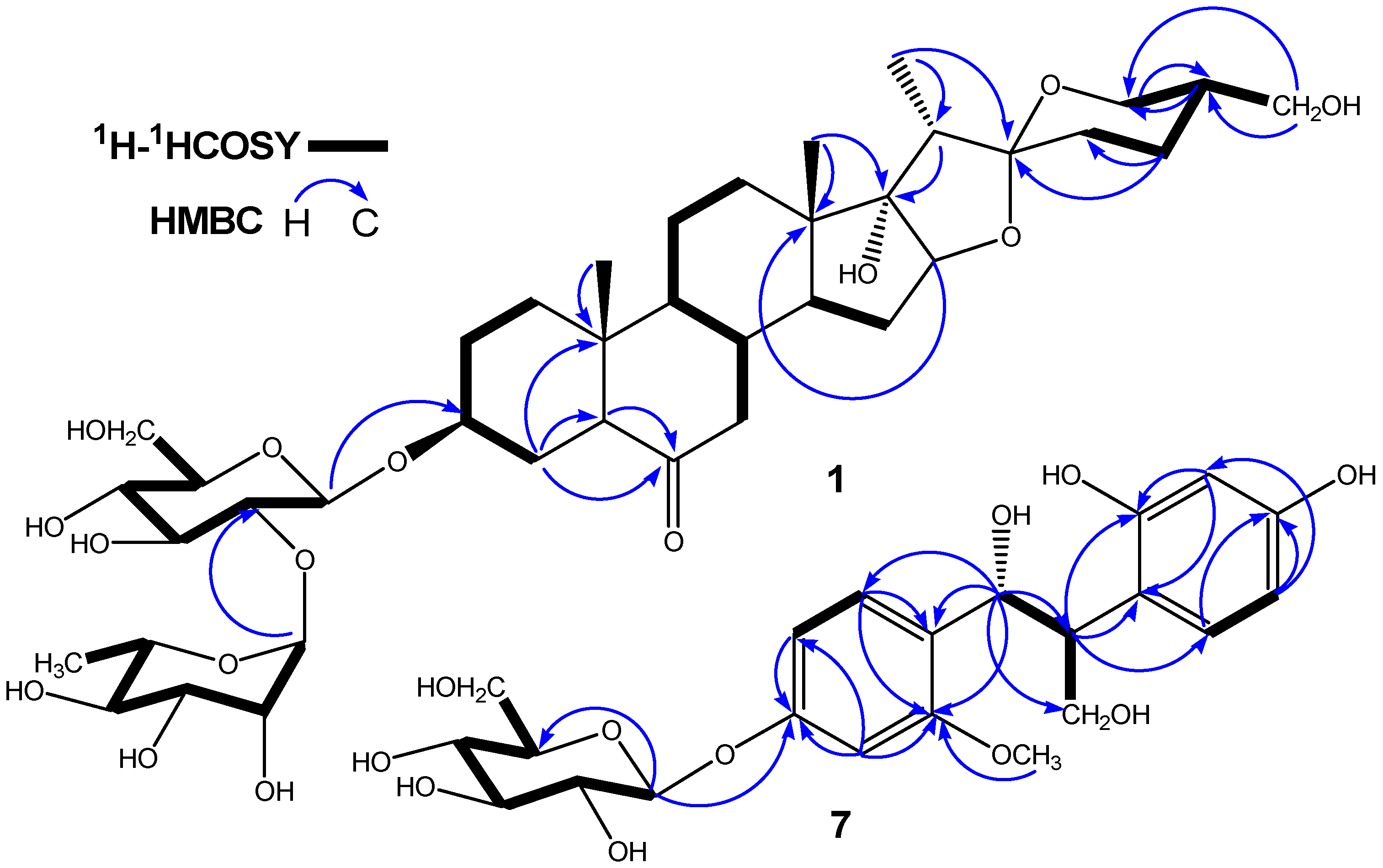
2. Results and Discussion

The methanolic extract from the bulbs of Lilium pumilum DC was successively subjected to column chromatography over silica gel, ODS, Diaion HP-20 and semipreparative HPLC to afford six steroidal saponins and a new phenolic glycoside. All compounds were obtained from this plant for the first time.
Compound 1 had the molecular formula C39H62O15, as deduced from the positive-ion HR-ESI-MS (m/z 793.6704 [M+Na]+) and 13C-NMR spectrum. The glycosidic nature of 1 was indicated by the strong absorption bands at 3435 cm−1 and 1045 cm−1 in the IR spectrum. The typical absorptions at 975, 915, 895, and 860 cm−1 in the IR spectrum suggested that 1 was a spirostanol saponin [13,14]. The intensity of the absorptions (895 > 915) indicated that the absolute configuration of C-25 was R. The 1H-NMR spectrum contained signals for two oxygenated methylene proton signals at δ 4.58 (2H, d, J = 7.8 Hz, H-26), 3.74 (1H, dd, J = 10.6, 5.5 Hz, H-27a) and 3.67 (1H, dd, J = 10.7, 7.2 Hz, H-27b), two anomeric proton signals at δ 6.13 (1H, br s, H-1″) and 5.11 (1H, d, J = 7.2 Hz, H-1′), as well as four methyl proton signals at δ 0.82 (3H, s), 0.91 (3H, s), 1.21 (3H, d, J = 7.0 Hz), and 1.81 (3H, d, J = 6.8 Hz). The signal at δ 1.81 was due to the methyl group of rhamnose (Table 1). The 13C-NMR and DEPT spectrum showed a quaternary carbon signal at δ 109.7, which is the characteristic C-22 of a spirostanol skeleton [15,16]. The presence of a carbonyl group in 1 was established from the IR (1707 cm−1) and 13C-NMR spectra (δ 209.4). The existence of D-glucose and L-rhamnose were determined by hydrolysis of 1 with 1 M HCl and GC analysis. The J values of the anomeric proton signals indicated the β- and α-configuration at the anomeric centers of D-glucose and L-rhamnose, respectively. These findings showed that 1 was a (25R)-spirostanol diglycoside.
Table 1. 13C and 1H-NMR data of 1 in pyridine-d5 (400 MHz for H, 100 MHz for C).
NO.CHNO.CH
136.81.56 (1H, m)2044.72.21 (1H, q, J = 7.2 Hz)
1.08 (1H, m)219.71.21 (3H, d, J = 6.8 Hz)
229.31.84 (1H, m)22109.7
1.37 (1H, m)2331.51.92 (1H, m)
376.03.99 (1H, m) 1.45 (1H, m)
426.42.21 (1H, m)2424.12.17 (1H, m)
1.75 (1H, m) 1.38 (1H, m)
556.42.26 (1H, m)2539.22.03 (1H, m)
6209.4 2664.04.58 (2H, d, J = 7.8 Hz)
746.92.54 (1H, m)2764.53.74 (1H, dd, J = 11, 5.5 Hz)
2.42 (1H, m) 3.67 (1H, dd, J = 11, 7.2 Hz)
838.11.83 (1H, m)Glc
953.71.37 (1H, m)1′99.25.11 (1H, d, J = 7.2 Hz)
1040.8 2′79.54.22 (1H, m)
1121.21.34 (1H, m)3′78.44.30 (1H, m)
1.57 (1H, m)4′72.14.15 (1H, m)
1232.02.13 (1H, m)5′78.23.86 (1H, m)
1.49 (1H, m)6′62.84.33 (1H, m)
1345.8 4.49 (1H, m)
1453.22.17 (1H, m)Rha
1532.02.17 (1H, m)1″102.46.13 (1H, br s)
1.46 (1H, m)2″72.64.68 (1H, m)
1689.84.42 (1H, m)3″72.94.37 (1H, m)
1789.9 4″74.24.39 (1H, m)
1817.30.82 (3H, s)5″69.54.81 (1H, m)
1913.20.91 (3H, s)6″18.71.81 (3H, d, J = 6.8 Hz)
Comparison of the 1H and 13C-NMR spectra of 1 with those of 2 indicated that 1 contained a same sugar chain as 2, the difference between these two saponins only appearing in the F-ring of their aglycons. The 1H and 13C-NMR spectra of 1 clearly displayed that C-27, which was presented as a methyl in 2 [1H-NMR: δ 0.71 (3H, d, J = 5.4 Hz); 13C-NMR: δ 17.3 (Me)], was modified to a hydroxymethyl in 1 [1H-NMR: δ 3.74 (1H, dd, J = 11, 5.5 Hz) and 3.67 (1H, dd, J = 11, 7.2 Hz); 13C-NMR: δ 64.5 (-CH2OH)]. The chemical shift values of C/H-27 and detailed 2D-NMR analysis of COSY, ROESY and HMBC correlations also implied that the aglycone moiety of 1 had one more hydroxyl group attached at C-27 (Figure 2 and Figure 3). On the basis of the above evidence, the structure of 1 was formulated as (25R)-3β,17α,27-triol-spirostan-6-one 3-O-α-L-rhamnopyranosyl-(1→2)-β-D-gluco- pyranoside, and it was named pumilum A.
Figure 2. Key HMBC and 1H-1H COSY correlations of 1 and 7.
Molecules 17 10494 g002
Figure 3. Selected ROESY correlations of 1.
Molecules 17 10494 g003
Compound 7 had the molecular formula C22H28O11, deduced from the negative-ion HR-ESI-MS (m/z 467.1936 [M−H]) and 13C-NMR spectrum, which exhibited 16 carbon signals due to the aglycone, in addition to six carbon signals of a β-D-glucopyranosyl group. The 1H and 13C-NMR spectra of 7 showed the presence of two aromatic ABX systems [δ (H) 7.55 (1H, d, J = 8.4 Hz, H-6′), 7.16 (1H, d, J = 8.0 Hz, H-6′′), 7.04 (1H, dd, J = 2.0, 8.4 Hz, H-5′), 7.08 (1H, d, J = 2.0 Hz, H-3′ ), 6.69 (1H, d, J = 2.0 Hz, H-3′′), and 6.51 (1H, dd, J = 2.0, 8.0 Hz, H-5′′); δ (C) 161.8 (C-4′′), 161.5 (C-2′′), 159.9 (C-4′), 157.3 (C-2′), 132.8 (C-6′), 125.6 (C-6′′), 119.9 (C-1′′), 114.8 (C-1′), 111.2 (C-5′), 106.7 (C-5′′), 105.8 (C-3′), and 97.4 (C-3′′)] (Table 2), suggesting the presence of two 1, 2, 4-trisubstituted phenyls in 7. The detailed 2D-NMR analysis of 1H-1H COSY, HMQC, and HMBC correlations also suggested that the aglycone moiety of 7 had two 1, 2, 4-trisubstituted phenyls (Figure 2). In the aliphatic region, the 1H-NMR spectra of 7 were completely identical to those of 1, 2-bis(4-hydroxy-3-methoxyphenyl)-1,3-propandiol 4′-O-β-D-glucopyranoside [17], ABMX-type signals were clearly observed [including an oxygen-bearing methylene at δ 4.27 (2H, dd, J = 9.8, 5.0 Hz, H-3), an oxygen-bearing methine at δ 5.56 (1H, d, J = 7.4 Hz, H-1), and a methine at δ 3.50 (1H, td, J = 7.4, 5.0 Hz, H-2)]. In combination with the presence of three aliphatic carbon signals [δ 66.8 (C-3), 78.9 (C-1), and 44.9 (C-2)] in the 13C-NMR spectrum of 7 supported the presence of the aliphatic 1,3-propanediol chain. Additionally, the 1H and 13C-NMR spectra of 7 displayed the presence of one glucopyranosyl unit [δ (H) 5.71 (1H, d, J = 7.6 Hz, H-1′′′), δ (C) 102.4 (C-1′′′)], as well as a methoxyl group [δ (H) 3.66 (3H, s), δ (C) 55.6]. The HMBC spectrum of 7 exhibited long-range correlations from H-1 (δ 5.56) to C-2 (δ 44.9), C-3 (δ 66.8), C-1′ (δ 114.8), C-2′ (δ 157.3), C-6′ (δ 132.8) and C-1′′ (δ 119.9); and from H-2 (δ 3.50) to C-1 (δ 78.9), C-3 (δ 66.8), C-1′ (δ 114.8), C-1′′ (δ 119.9), C-2′′ (δ 161.5), and C-6′′ (δ 125.6), respectively, indicated the locations of the two phenyls linked to the aliphatic chain, 1,3-propanediol. In addition, the HMBC spectra of 7 revealed the methoxyl group (δ 3.67) was coupled to aromatic carbon C-4′′ (δ 161.8), suggesting that the methoxyl groups was linked to C-4′′ in 7 (Figure 2). Thus, the aglycone of 7 was believed to be 1-(4′-hydroxy-2′-methoxyphenyl)-2-(2′′,4′′-dihydroxyphenyl)-1, 3-propanediol.
Table 2. 13C and 1H-NMR data of 7 in pyridine-d5 (500 MHz for H, 125 MHz for C).
NO.CHNO.CH
178.95.56 (1H, d, J = 7.4 Hz)3′′97.4 6.69 (1H, d, J = 2.0 Hz)
244.93.50 (1H, td, J = 7.4, 5.0 Hz)4′′161.8
366.84.27 (2H, dd, J = 9.8, 5.0 Hz)5′′106.7 6.51 (1H, dd, J = 2.0, 8.0 Hz)
1′114.8 6′′125.6 7.16 (1H, d, J = 8.0 Hz)
2′157.3 1′′′102.4 5.71 (1H, d, J = 7.6 Hz)
3′105.37.08 (1H, d, J = 2.0 Hz)2′′′74.9 4.27 a
4′159.9 3′′′79.0 4.11 (1H, m)
5′111.27.04 (1H, dd, J = 2.0, 8.4 Hz)4′′′71.2 4.26 a
6′132.87.55 (1H, d, J = 8.4 Hz)5′′′78.4 4.43 (1H, m)
1′′119.9 6′′′62.54.31 (1H, m)
2′′161.5 2′-CH3O55.63.67 (3H, s)
a Overlapped signals.
The J value (7.6 Hz) of the anomeric proton implied the β-configuration of the glucose moiety. The β-D-glucopyranosyl group was attached to the C-4′ position of the aglycone, which was supported by a long-range correlation between the anomeric proton signal (δ 5.71) of the β-D-glucopyranosyl group and the C-4′ signal (δ 159.9) in the HMBC spectrum of 7. The relative stereochemistry of C-1 and C-2 of 7 was determined to be threo by comparing the coupling constant H-1 and H-2 (J = 7.4 Hz) with those of the erythro and threo isomers [18,19]. Therefore, 7 was assigned as threo-1-(4′-hydroxy-2′-methoxyphenyl)-2-(2′′, 4′′-dihydroxyphenyl)-1, 3-propanediol-4′-O-β-D-glucopyranoside.
The structures of the other isolated components, namely (25R)-3β,17α-diol-5α-spirostan-6-one 3-O-α-L-rhamnopyranosyl-(1→2)-β-D-glucopyranoside (2), (25R)-3β-hydroxyl-5α-spirostan-6-one 3-O-α-L-rhamnopyranosyl-(1→2)-β-D-glucopyranoside (3), (25R)-3β-hydroxy-5α-spirostan-6-one 3-O-α-L arabinopyranosyl-(1→6)-β-D-glucopyranoside (4), dioscin (5), and (25R)-5α-spirostan-3β,17α-diol 3-O-β-D-xylopyranosyl-(1→4)-[α-L-arabinopyranosyl-(1→6)]-β-D-glucopyranoside (6) were determined by comparison to the 1H and 13C-NMR spectral data in the literature [2,20,21,22,23].
The inhibitory activity of 17 on Na+/K+ ATPase was assayed. Ouabain was used as a positive control (IC50 value: 0.1 × 10⁻⁵ M). Saponins 3 and 4 were found to inhibit sensitive Na+/K+ ATPase with the IC50 values of 7.3 × 10⁻⁵ and 9.1 × 10⁻⁶ M, respectively, while the others were inactive [IC50 > 1.0 × 10⁻⁴ M]. Thus, this research suggested that the saponins from the methanolic extract may be primary Na+/K+ ATPase inhibitors. The weak Na+/K+ ATPase inhibitory activity of the initial extract (compounds 17) is thus presumably due to other as yet unidentified compounds. One possible reason for this is that compounds with highly Na+/K+ ATPase inhibitory activity are low in the BuOH-soluble fraction of the methanolic extract from the bulbs of Lilium pumilum DC. Another possible reason is compounds with increased activity may be found in other extraction fractions (ethyl acetate extract, chloroform extract and petroleum ether extract) of the methanolic extract from the bulbs of this medicine plant. Therefore, more chemical work needs to be done in the future.

3. Experimental

3.1. General

Specific rotation measurements were recorded on a Perkin-Elmer 242 MC polarimeter. UV spectra were recorded on a Hewlett-Packard HP-845 UV-VIS spectrophotometer. IR spectra were recorded on a Nicolet 470 spectrometer and MS on a Varian MAT-212 mass spectrometer and a Shimadzu GC-MS model QP2010 Plus spectrophotometer, respectively. NMR spectra were recorded on a Bruker AM-400 spectrameter (400 MHz for 1H-NMR) or a Bruker DRX-500 (500 MHz for 1H-NMR) spectrameter using standard Bruker pulse programs. Chemical shifts are given as δ values with reference to tetramethylsilane (TMS) as internal standard. Column chromatography separations were carried out on silica gel (200–300 mesh, Qingdao Haiyang Chemical Co. Ltd, Qingdao, China), ODS (50 mesh, AA12S50, YMC), Diaion HP-20 (Pharmacia, Peapack, NJ, USA) and Sephadex LH-20 (Pharmacia, Peapack, NJ, USA). Ouabain sensitive dog kidney Na+/K+ ATPase was obtained from Sigma (Shanghai, China). All other chemicals used were of biochemical reagent grade.

3.2. Plant Material

The bulbs of Lilium pumilum DC were collected in Lanzhou, Gansu Province of China in September 2010, and were identified by one of the authors (Chun-Yan Fu of Department of Pharmacy, Shaoyang Medical College Level Speciaity School, Shaoyang). A voucher specimen (NO. 20100908) has been deposited in the authors’ laboratory.

3.3. Extraction and Isolation

Fresh bulbs of Lilium pumilum DC (5 kg) were cut into pieces and extracted with methanol (3 × 8 L, 3h each) under reflux. The solvent was removed at reduced pressure to give a viscous residue (328 g). The entire crude extract was suspended in H2O (3 L), and extracted with n-BuOH five times (3 L each) to give an n-BuOH extract (206 g). This extract was then fractionated by silica gel column chromatography (column: 90 cm × 9 cm) using a mobile phase composed of CHCl3-MeOH (9:1–6:1–4:1–2:1–1:1, v/v), and finally with MeOH alone to afford six fractions [Fr.1 (98 mg), Fr.2 (11.2 g), Fr.3 (37.8 g), Fr.4 (11.1 g), Fr.5 (14.3 g), and Fr.6 (51.6 g)]. Fr.2 (11.2 g) was chromatographed on silica gel (column: 70cm × 4 cm) eluting with CHCl3-MeOH-H2O (40:10:1) and ODS silica gel with MeOH-H2O (4:1) and MeCN-H2O (1:1) to furnish 3 (14.5 mg), 4 (16.8 mg), 5 (16.3 mg), and 6 (20.7 mg). Fr.1 (98 mg) was passed through a Diaion HP-20 column with a gradient mixture of MeOH-H2O and the MeOH eluate fraction was subjected to silica gel column eluting with CHCl3-MeOH-H2O (40:10:1; 30:10:1) and ODS silica gel with CH3CN-H2O (1:1–5:7–1:2) to give 2 (15.6 mg) and 7 (17.2 mg) as the pure compounds, and 1 with a few impurities. Final purification of 1 (19.4 mg) was accomplished by semipreparative HPLC. The method was an isocratic gradient of 70% methanol in H2O over 15 min, and then followed by 95% methanol for 5 min (flow rate: 3 mL/min).

3.4. Characterization of Pumilum A (1)

Obtained as a white amorphous powder, Molecules 17 10494 i001: −88.7° (c 0.048, MeOH); UV λmax (MeOH): 279 nm; IR vmax (KBr): 3435, 2945, 2880, 1707, 1455, 1375, 1245, 1205, 1155, 1080, 1045, 985, 975, 915, 895, 860, 805, 710 cm−1. HR-ESI-MS m/z 793.6907 (C39H62O15Na [M+Na]+, Cal. 793.6904). 1H-NMR and 13C-NMR (pyridine d5) data see Table 1.

3.5. Characterization of Threo-1-(4′-hydroxy-2′-methoxyphenyl)-2-(2′′,4′′-dihydroxy-phenyl)-1,3-propanediol-4′-O-β-D-glucopyranoside (7)

Obtained as an amorphous powder. Molecules 17 10494 i001: −76.3° (c 0.5, MeOH). UV (MeOH) λmax: 282 nm. IR vmax (KBr): 3433, 3391, 1630, 1601, 1495, 1470, 1441, 1386, 1345, 1253, 1202, 1172, 1143, 1041, 955, 860, 826 cm−1. HR-ESI-MS m/z 467.1936 ([M−H], Calcd. for C22H27O11, 467.1903). 1H-NMR and 13C-NMR (pyridine d5) data see Table 2.

3.6. Acid Hydrolysis of Compound 1

A solution (2.5 mg) of 1 in 1 M HCl (1 mL) was heated at 100 °C for 2 h under an N2 atmosphere. After cooling, the solution was removed by blowing with N2. The residue was dissolved in a solution of 1-(trimethylsilyl) imidazole (0.5 mg) in pyridine (1.0 mL), and stirred at 60 °C for 5 min. After removal of the solvent with a stream of N2, the residue was partitioned between H2O and CH2Cl2 (1:1 v/v). The CH2Cl2 fraction was analyzed by GC using a L-Chirasil-Val column (0.32 mm × 25 m). The temperature of the injector and detector were 200 °C. A temperature gradient system was used for the oven, starting at 100 °C for 1 min and increasing up to 180 °C at a rate of 5 °C/min. The peaks of the hydrolysates of 1 were confirmed by comparison of retention times of authentic samples D-glucose, L-rhamnose treated with 1-(trimethylsilyl) imidazole [24].

3.7. Assay of Na+/K+ ATPase Activity

The Na+/K+ ATPase activity was assayed according to the reported method with some modification [25]. The reaction mixture composed of 50 mM Tris-HCl (pH 7.3, 37 °C), 3 mM ATP, 4 mM Mg2+, 130 mM Na+, 20 mM K+, and 0.02 units of Na+/K+ ATPase, with or without test compound dissolved in dimethyl sulfoxide (DMSO), was incubated for 20 min at 37 °C. The concentration of DMSO in the reaction mixture was held at 5%. The reaction was terminated by addition of 50% CCl3COOH. The released inorganic phosphate was determined by the following method. To the test solution was added 0.5% sodium dodecyl sulfate, 0.1% 2,4-diaminophenol 2 M HCl in 1% Na2SO3 and 1% ammonium heptamolybdate in 1 M H2SO4. After 25 min, the absorbance at 660 nm was recorded.

4. Conclusions

Two new compounds, pumilum A (1) and threo-1-(4′-hydroxy-2′-methoxyphenyl)-2-(2′′,4′′-dihydroxyphenyl)-1,3-propanediol-4′-O-β-D-glucopyranoside (7) were isolated from the the bulbs of Lilium pumilum DC, along with five known steroidal saponins. Compounds 3 and 4 exhibited weak inhibitory activity on Na+/K+ ATPase with the IC50 values of 7.3 × 10⁻⁵ and 9.1 × 10⁻⁶ M, respectively.

Supplementary Materials

Supplementary materials can be accessed at: https://www.mdpi.com/1420-3049/17/9/10494/s1.

Acknowledgements

This study was supported by the Scientific Research Fund of Hunan Education Department (10C0294) and Research Foundation for Advanced Talents, Zhanjiang Normal University (ZL1009).

References

  1. Mimaki, Y.; Sashida, Y. Steroidal saponins and alkaloids from the bulbs of Lilium brownie var. colchesteri. Chem. Pharm. Bull. 1990, 38, 3055–3059. [Google Scholar] [CrossRef]
  2. Mimaki, Y.; Ishibashi, N.; Ori, K.; Sashida, Y. Steroidal glycosides from the bulbs of Lilium dauricum. Phytochemistry 1992, 31, 1753–1758. [Google Scholar] [CrossRef]
  3. Jiang Su New Medical College, Dictionary of Traditional Chinese Crude Drugs; Scientific Technologic: Shanghai, China, 1977; Volume 1, pp. 856–858.
  4. Munafo, J.P., Jr.; Ramanathan, A.; Jimenez, L.S.; Gianfagna, T.J. Isolation and structural determination of steroidal glycosides from the bulbs of Easter Lily (Lilium longiflorum Thunb.). Agric. Food Chem. 2010, 58, 8806–8813. [Google Scholar]
  5. Mimaki, Y.; Sashida, Y.; Shimomura, H. Shimomura, H. Lipid and steroidal constituents of Lilium auratum var. Platyphyllum and L. tenuifolium. Phytochemistry 1989, 28, 3453–3458. [Google Scholar] [CrossRef]
  6. Shimomura, H.; Sashida, Y.; Mimaki, Y. Steroidal saponins, pardarinoside A–G from the bulbs of Lilium pardarinum. Phytochemistry 1989, 28, 3163–3170. [Google Scholar] [CrossRef]
  7. Mimaki, Y.; Sashida, Y. Steroidal and phenolic constituents of Lilium speciosum. Phytochemistry 1991, 30, 937–940. [Google Scholar] [CrossRef]
  8. Satou, T.; Mimaki, Y.; Kuroda, M.; Sashida, Y.; Hatakeyama, Y. A pyrroline glucoside ester and steroidal saponins from Lilium Martagon. Phytochemistry 1996, 41, 1225–1230. [Google Scholar] [CrossRef]
  9. Francisa, J.A.; Rumbeihab, W.; Naira, M.G. Constituents in Easter lily flowers with medicinal activity. Life Sci. 2004, 76, 671–683. [Google Scholar] [CrossRef]
  10. Hiroko, S.; Yutaka, S.; Yoshihiro, M. Phenolic glycerides from Lilium auratum. Phytochemistry 1987, 26, 844–845. [Google Scholar] [CrossRef]
  11. Ori, K.; Mimaki, Y.; Mito, K.; Sashida, Y.; Nikaido, T.; Ohmoto, T.; Masuko, A. Jatropham derivatives and steroidal saponins from the bulbs of Lilium hansonii. Phytochemistry 1992, 31, 2767–2775. [Google Scholar]
  12. Nakano, K.; Nishizawa, K.; Murakami, K.; Takaishi, Y.; Tomimatsu, T. Steroidal alkaloids glucosides from Lilium cordatum. Phytochemistry 1986, 26, 301–303. [Google Scholar]
  13. Mskhiladze, L.; Legault, J.; Lavoie, S.; Mshvildadze, V.; Kuchukhidze, J.; Elias, R.; Pichette, A. Cytotoxic steroidal saponins from the flowers of Allium leucanthum. Molecules 2008, 13, 2925–2934. [Google Scholar] [CrossRef]
  14. Jones, R.N.; Katzenellenbogen, E.; Dobriner, K. The infrared absorption spectra of the steroid sapogenins. J. Am. Chem. Soc. 1953, 75, 158–166. [Google Scholar]
  15. Li, X.C.; Wang, D.Z.; Yang, C.R. Steroidal saponins from chlorophytum malayense. Phytochemistry 1990, 29, 3893–3898. [Google Scholar] [CrossRef]
  16. Li, X.C.; Yang, C.R.; Ichikawa, M.; Matsuura, H.; Kasai, R.; Yamasaki, K. Steroidal saponins from polygonatum kingianum. Phytochemistry 1992, 31, 3559–3563. [Google Scholar] [CrossRef]
  17. Warashina, T.; Nagatani, Y.; Noro, T. Constituents from the bark of Tabebuia impetiginosa. Phytochemistry 2006, 54, 14–20. [Google Scholar]
  18. Yoshikawa, K.; Mimura, N.; Arihara, S. Isolation and absolute structures of enantiomeric 1,2-bis(4-hydroxy-3-methoxyphenyl)-l,3-propanediol 1-O-glucosides from the bark of Hovenia trichocarpa. J. Nat. Prod. 1998, 61, 1137–1139. [Google Scholar] [CrossRef]
  19. Hsiao, J.J.; Chiang, H.C. Lignans from the wood of Araliabipinnata. Phytochemistry 1995, 39, 899–902. [Google Scholar] [CrossRef]
  20. Abbas, F.A. Steroidal saponins from Solanum unguiculatum (A.) rich. Sci. Pharm. 2001, 69, 219–234. [Google Scholar]
  21. Jia, Z.J.; Ju, Y. Steroidal saponins from Smilax lebrunii. Phytochemistry 1992, 31, 3173–3175. [Google Scholar] [CrossRef]
  22. Zhang, Y.; Li, H.Z.; Zhang, Y.J.; Jacob, M.R.; Khan, S.I.; Li, X.C.; Yang, C.R. Atropurosides A-G, new steroidal saponins from Smilacina atropurpurea. Steroids 2006, 71, 712–719. [Google Scholar] [CrossRef]
  23. Zhou, L.B.; Chen, D.F. Steroidal saponins from the roots of Asparagus filicinus. Steroids 2008, 73, 83–87. [Google Scholar] [CrossRef]
  24. Fu, W.W.; Fu, J.N.; Zhang, W.M.; Sun, L.X.; Pei, Y.H.; Liu, P. Platycoside O, a new triterpenoidsaponin from the roots of Platycodon grandiflorum. Molecules 2011, 16, 4371–4378. [Google Scholar] [CrossRef]
  25. Mimaki, Y.; Satou, T.; Kuroda, M.; Sashida, Y.; Hatakeyama, Y. New steroidal constituents from the bulbs of Lilium candidum. Chem. Pharm. Bull. 1998, 46, 1829–1832. [Google Scholar] [CrossRef]
  • Sample Availability: Contact the corresponding author.

