Article

A Minor Dihydropyran Apocarotenoid from Mated Cultures of Blakeslea trispora

1
Departamento de Química Orgánica, Facultad de Ciencias, Universidad de Granada, Avda. Fuente Nueva, s/n, 18071 Granada, Spain
2
Departamento de Ingeniería Química, Química Fisíca y Químíca Orgánica, Facultad de Ciencias Experimentales, Universidad de Huelva, Campus el Carmen, s/n, 21071 Huelva, Spain
*
Author to whom correspondence should be addressed.
Molecules 2012, 17(11), 12553-12559; https://doi.org/10.3390/molecules171112553
Submission received: 8 October 2012 / Revised: 15 October 2012 / Accepted: 22 October 2012 / Published: 24 October 2012
(This article belongs to the Section Natural Products Chemistry)

Abstract

:
The heterocyclic C15 apocarotenoid 1 was isolated from mated cultures of the strains F986 (+) and F921 (−) of Blakeslea trispora. This new compound formed during sexual interaction is a minor constituent of the culture media and its structure was elucidated by spectroscopic data, including 2D-NMR. A plausible biosynthetic pathway involving a double degradation of β-carotene, followed by several oxidations of the resulting monocyclofarnesane C15 fragment is proposed.

1. Introduction

Blakeslea trispora (syn. Choanephora trispora, Mucoromycotina, Mucorales, Choanephoraceae) is used for the industrial preparation of β-carotene, a natural pigment antioxidant and pro-vitamin A with many applications in the food, pharmaceutical, and cosmetic industries [1,2,3,4]. The wild-type strains of this fungus belong to either the (+) or the (–) sex, and many pairs of opposite sex strains, cultured together (“mated” cultures”) increase their β-carotene content and spark the morphological program of the sexual cycle. These physiological effects were attributed to the action of apocarotenoids such as trisporic acid C (1, Figure 1) and similar compounds present in mated cultures of Blakeslea [5,6,7,8,9,10]. The culture media of Blakeslea contains apocarotenoids belonging to the following three families: “trisporoids” with 18 carbons [5,11,12,13,14,15,16], “cyclofarnesoids” with 15 carbons [16,17,18,19,20] and those featuring seven carbons [16]. As a follow-up to our study about production and identification of bioactive apocarotenoids from Blakeslea trispora, in this paper we report the isolation and structural elucidation of the minor cyclofarnesane apocarotenoid 2 from mated cultures of B. trispora. Additionally a plausible biosynthetic pathway justifying its formation during sexual interaction is presented.
Figure 1. Trisporic C acid (1).
Molecules 17 12553 g001

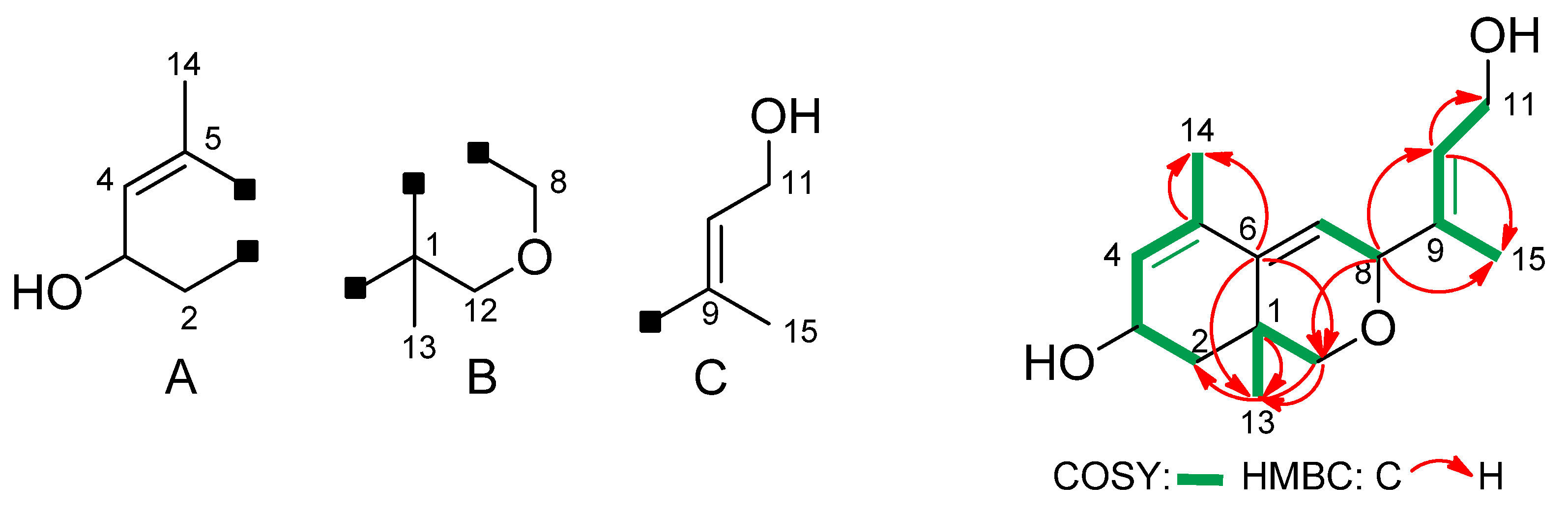
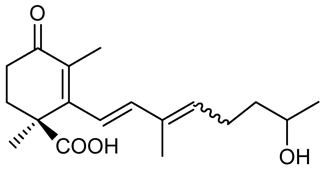
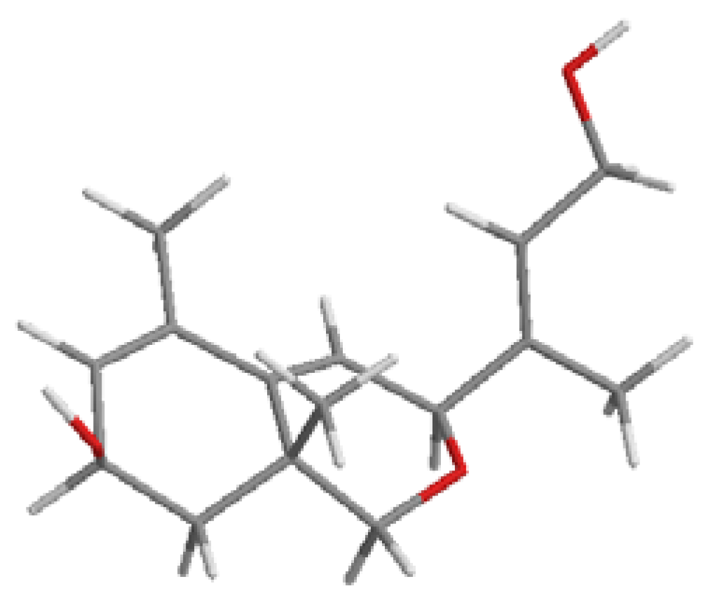
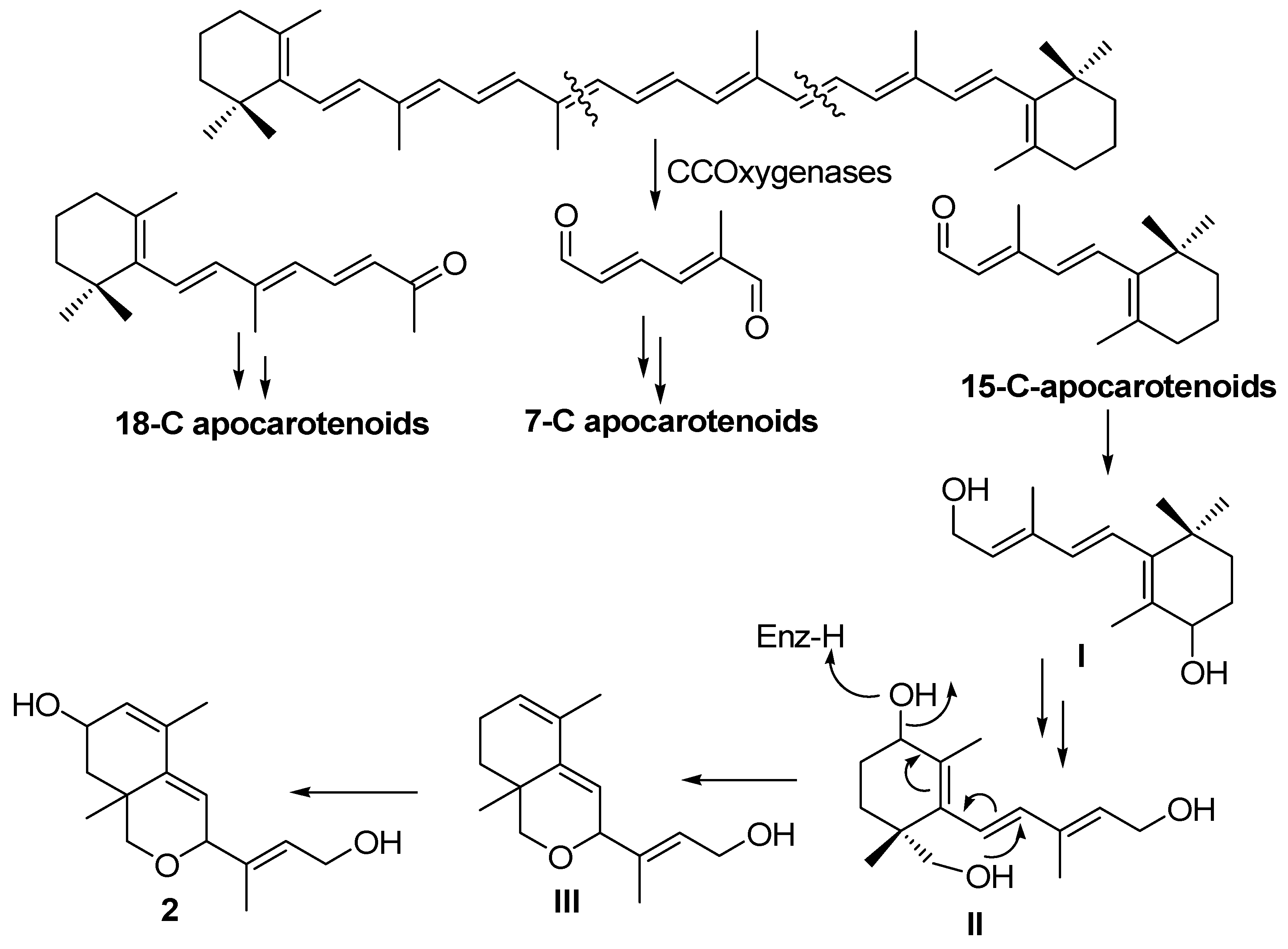
2. Results and Discussion
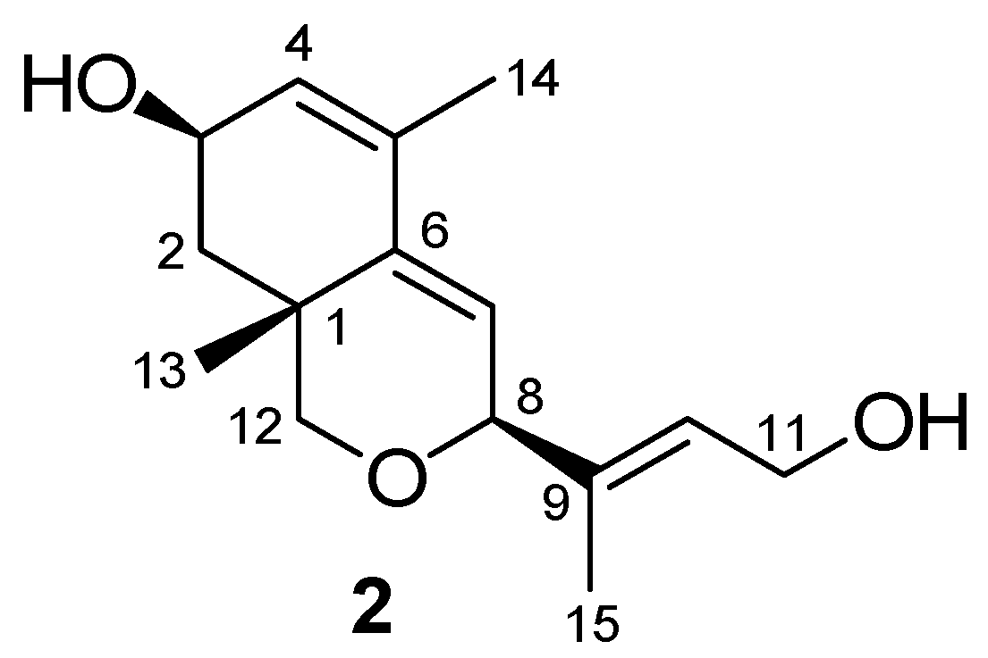
Sexually (+) and sexually (−)B. trispora strains F921 and F986 were cultured together for three days on agar medium. Following semi-preparative normal phase HPLC separation, a careful search for the neutral metabolites in the agar extracts has permitted the isolation of a few mg of compound 2 (relative concentration: 4 mg in 1 L of medium). It is important to point out that compound 2 is not present in single cultures meaning that the product was produced during sexual interaction. This compound was isolated as a colourless syrup and high-resolution mass spectrum (FAB+) showed a molecular ion [M+Na]+ at m/z 273.1464, corresponding to a molecular formula C15H22O3 (five degrees of unsaturation) and its IR spectrum exhibited an absorption band corresponding to a hydroxyl group (3417 cm−1). The 13C-NMR and HSQC spectra revealed 15 carbon signals, including three methyl groups, three methylene groups (two oxygenated), five methyne groups (two oxygenated and three sp2) and four quaternary carbons (three sp2). These data establish the presence of three double bonds, two rings (one oxygenated) and two hydroxyl groups in the structure of 2. Some of the COSY and HMBC correlations depicted in Figure 2 established the presence of frameworks A–C in its structure. Connectivity among these frameworks was deduced from the HMBC correlations (Figure 2, Table 1).
Figure 2. Key COSY and HMBC correlations for apocarotenoid 2.
Molecules 17 12553 g002
Table 1. Mono- and bi-dimensional NMR data for compound 2.
C/HδHδCCOSYHSQCHMBC
1 32.1
2a1.65–1.42 m38.4H3, H2bC2C12a, C13
2b1.65–1.42 mH3, H2a
4.34 br s64.4H4, H2a, H2bC3
45.72 d (4.6)125.8H3, H14C4
5 133.0
6 140.9
75.65 d (3.3)120.5H8C7
84.64 br s76.5H7C8
9 138.0
105.54 t (6.4)127.3H11, H15C10C8, C15
114.24 d (6.4)59.5H10C11C9, C10
12a3.32 d (10.7)70.5H12b, H13C12C8, C6, C13
12b3.36 d (10.7)H12a, H13
131.35 s24.6H12a, H12bC13C12, C6, C2a, C1
141.86 s19.4H4C14C6, C5, C4
151.77 s14.9H10C15C8, C9, C10
J in Hz in parentheses.
These and all other data allowed us to establish the structure of 2 as 8,12-epoxy-1,6-cyclofarnesa-4,6,9-triene-3,11-diol (Figure 3), a new C15 apocarotenoid.
Figure 3. Apocarotenoid 2.
Molecules 17 12553 g003
The side chain double bond compound possesses the E stereochemistry as indicated by the 13C-NMR chemical shifts of C11 (δC 59.5) and C15 (δC 14.9). Also, relative syn stereochemistry between the secondary hydroxyl, the methyl group at C13 and the side chain is proposed based on the following considerations: first, one of the two fused rings, cyclohexene and oxacyclohexene, adopt the most favorable semi-chair conformation depicted in Figure 4 due to the presence of the C13 angular methyl. Secondly, the multiplicity of the H8 (br s) and H3 (br s) in the 1H-NMR spectrim involves pseudoaxial and pseudoequatorial dispositions, respectively.
Apocarotenoid 2 is the first compound in Blakeslea trispora and the second of all the Mucoromycotina fungi that contains a dihydropyran framework. This framework is reminiscent of the azaphylones, fungal metabolites with a polyketide origin [21].
Figure 4. Conformation of apocarotenoid 2.
Molecules 17 12553 g004
Considering its origin (i.e., sexual interaction of B. trispora) and its structural framework related to that of C15 apocarotenoids the following biosynthetic pathway is proposed for the formation of 2 (Scheme 1).
Scheme 1. Biosynthetic pathway of apocarotenoid 2.
Molecules 17 12553 g005
The pathway begins with the double asymmetric β-carotene degradation catalyzed by carotene cleavage oxygenases giving rise to the three fragment precursors of the three families of apocarotenoids (18-C apocarotenoids, 7-C apocarotenoids and 15-C apocarotenoids) [16]. This type of carotene degradation is stimulated by the sexual interaction of opposite sex strains [9,22]. The apocarotenoid 2 comes from the 15 carbons fragment I, which undergoes reduction of the aldehyde group to a primary alcohol and then undergoes two hydroxylation processes (mediated probably by cytochrome-P450 dependent enzymes) at C4 and C13 giving rise to the intermediate II. At this point a heterocyclization process starting from primary hydroxyl at C13 with a shift of a secondary hydroxyl at C4 by means of a SN2' like reaction leads to III. Then a new hydroxylation at position C3 on III gives rise to metabolite 2. This biosynthetic pathway suggests the existence of specific hydroxylating enzymes in each Mucoromicotina sp. acting at specific positions on each apocarotenoid, and may support the hypothesis of the existence of different sexual signals for each species.

3. Experimental

3.1. General

NMR spectra (1H- and 13C-) were recorded with a Varian Direct-Drive 500 (1H 500 MHz/13C 125 MHz) spectrometer. For high-resolution MS we used an Autospec-Q VG-Analytical (Fisons) mass spectrometer. For semi-preparative normal-phase HPLC the neutral extracts was dissolved in t-BuOMe (at 20 g dry extract/L). Aliquots (0.5 mL) were injected onto a column (10 × 250 mm; 5 µm silica particles; Agilent) with a 15 mm refillable guard pre-column filled with the same material placed in a Series 1100 liquid chromatograph (Agilent). The column was eluted at room temperature at a flow rate of 2 mL/min for 25 min with t-BuOMe and monitored with a refractometer.

3.2. Strains and Culture Conditions

Strains F986 and F921 are wild-type (+) and (−) strains of Blakeslea (Choanephora) trispora, respectively, and were obtained from VKM (All-Russian Collection of Microorganisms, Moscow, Russia). Plates containing 25 mL minimal agar medium [23] were inoculated with 5 × 10³ spores of each sex and incubated in the dark at 30 °C for three days.

3.3. Extraction and Fractionation of Apocarotenoids.

The initial extracts for apocarotenoid analyses were obtained by freezing (−20 °C for at least 2 h) and thawing (22 °C for 1 h) the media and centrifuging the liquid (4,000 × g, 15 min). Neutral extracts were obtained by adjusting the initial extracts to pH 8.0 with KOH and extracting three times with EtOAc. Acid extracts were obtained by adjusting the remaining aqueous phase to pH 2.0 with HCl and extracting with EtOAc. Water was removed by mixing with anhydrous Na2SO4 and filtering; the organic solvent was removed by evaporation under low pressure. For the sake of chemical stability, all procedures were carried out under dim light. An initial extract of 500 mL (from 1 L of medium of mated cultures F921 × F986) yielded 114 mg of neutral extract. This neutral extract was fractionated by semi-preparative HPLC. The fraction (16.7 < RT < 17.1 min) contained 2 (4 mg).
(1R,3R,8S,E)-8,12-Epoxy-1,6-cyclofarnesa-4,6,9-triene-3,11-diol (2): Colourless syrup. [α]D +20.1 (c = 1, CHCl3). HRMS (FAB), m/z: 273.1464 ([M+Na]+; calcd. for C15H22O3Na, 273.1467). IR (film) νmax: 3417, 2964, 2923, 2857, 1654, 1458, 1407, 1110, 1032 cm−1. 1H-NMR (CDCl3, Me4Si): see Table 1. 13C-NMR (CDCl3, Me4Si): see Table 1.

4. Conclusions

The sexual interaction of strains F986 (+) and F921 (−) of B. trispora produces known apocarotenoids, in addition to small amounts of a heterocyclic cyclofarnesane whose novel structure corresponds to (3S,7R,8aR)-3-((E)-4-hydroxybut-2-en-2-yl)-5,8a-dimethyl-3,7,8,8a-tetrahydro-1H-isochromen-7-ol. Biogenetically this apocarotenoid derives from after successive transformations (reduction, regiospecific hydroxylations and heterocyclization) of a 15 carbons fragment produced in a double asymmetric β-carotene degradation. The presence of specific apocarotenoids in each Mucoromycotina species reinforces the hypothesis of the existence of different sexual signals.

Supplementary Materials

Supplementary materials can be accessed at: https://www.mdpi.com/1420-3049/17/11/12553/s1.

Acknowledgments

This research was financed by Junta de Andalucía (Grants FQM 340, CVI 910, and P08-CVI-03901) and the Spanish Government (Grant CTQ 2010-16818, subprogram BQ).

References

  1. Ciegler, A. Microbial carotenogenesis. Adv. Appl. Microbiol. 1965, 7, 1–34. [Google Scholar] [CrossRef]
  2. Avalos, J.; Cerdá-Olmedo, E. Fungal carotenoid production. In Handbook of Fungal Biotechnology; Arora, D.K., Ed.; Marcel Dekker, Inc.: New York, NY, USA, 2004; pp. 367–378. [Google Scholar]
  3. Bhosale, P. Environmental and cultural stimulants in the production of carotenoids from microorganisms. Appl. Microbiol. Biotechnol. 2004, 63, 351–361. [Google Scholar] [CrossRef]
  4. Namitha, K.K.; Negi, P.S. Chemistry and biotechnology of carotenoids. Crit. Rev. Food Sci. Nutr. 2010, 50, 728–760. [Google Scholar] [CrossRef]
  5. Caglioti, L.; Cainelli, G.; Camerino, B.; Mondelli, R.; Prieto, A.; Quilico, A.; Salvatori, T.; Selva, A. The structure of trisporic C acid. Tetrahedron Suppl. 1966, 7, 175–187. [Google Scholar]
  6. Austin, D.J.; Bu’Lock, J.D.; Drake, D. The Biosynthesis of trisporic acids from β-carotene via retinal and trisporol. Experientia 1970, 26, 348–349. [Google Scholar] [CrossRef]
  7. Gooday, G.W. Fungal sex hormones. Ann. Rev. Biochem. 1974, 43, 35–49. [Google Scholar] [CrossRef]
  8. Nieuwenhuis, M.; van den Ende, H. Sex specificity of hormone synthesis in Mucor. mucedo. Arch. Microbiol. 1975, 102, 167–169. [Google Scholar] [CrossRef]
  9. Schachtschabel, D.; David, A.; Menzel, K.-D.; Schimek, C.; Wöstemeyer, J.; Boland, W. Cooperative biosynthesis of trisporoids by the (+) and (−) mating types of the Zygomycete Blakeslea. trispora. ChemBioChem 2008, 9, 3004–3012. [Google Scholar] [CrossRef]
  10. Walter, M.H.; Strack, D. Carotenoids and their cleavage products: Biosynthesis and functions. Nat. Prod. Rep. 2011, 28, 663–692. [Google Scholar] [CrossRef]
  11. Austin, D.J.; Bu’Lock, J.D.; Gooday, G.W. Trisporic acids: Sexual hormones from Mucor. mucedo and Blakeslea. trispora. Nature 1969, 223, 1178–1179. [Google Scholar]
  12. Cainelli, G.; Grasselli, P.; Selva, A. Structure of trisporic acid B. Chim. Ind. 1967, 49, 628–629. [Google Scholar]
  13. Sutter, R.P.; Dadok, J.; Bothner-By, A.A.; Smith, R.R.; Mishra, P.K. Cultures of separates mating types of Blakeslea. trispora make D and E forms of trisporic acids. Biochemistry 1989, 28, 4060–4066. [Google Scholar]
  14. Bu’Lock, J.D.; Jones, B.E.; Winskill, N. The apocarotenoid system of sex hormones and prohormones in Mucorales. Pure Appl. Chem. 1976, 47, 191–202. [Google Scholar] [CrossRef]
  15. Sutter, R.P.; Whitaker, J.P. Zygophore-stimulating precursors (pheromones) of trisporic acids active in (−)-Phycomyces. blakesleeanus. Acid-catalyzed anhydro derivatives of methyl 4-dihydrotrisporate-C and 4-dihydrotrisporate-C. J. Biol. Chem. 1981, 256, 2334–2341. [Google Scholar]
  16. Barrero, A.F.; Herrador, M.M.; Arteaga, P.; Gil, J.; González, J.-A.; Alcalde, E.; Cerdá-Olmedo, E. New apocarotenoids and β-carotene cleavage in Blakeslea. trispora. Org. Biomol. Chem. 2011, 9, 7190–7195. [Google Scholar] [CrossRef]
  17. Sutter, R.P.; Zawodny, P.D. Apotrisporin: A major metabolite of Blakeslea. trispora. Exp. Mycol. 1984, 8, 89–92. [Google Scholar] [CrossRef]
  18. Sutter, R.P. A new sesquiterpenoid isolated from Phycomyces. blakesleeanus and Blakeslea. trispora. Exp. Mycol. 1986, 10, 256–258. [Google Scholar] [CrossRef]
  19. Cainelli, G.; Camerino, B.; Grasselli, P.; Mondelli, R.; Morrocchi, S.; Prieto, A.; Quilico, A.; Selva, A. Structure del trisporone e dell’anhidrotrisporone. Chim. Ind. 1967, 49, 748–751. [Google Scholar]
  20. Polaino, S.; González-Delgado, J.; Arteaga, P.; Herrador, M.M.; Barrero, A.F.; Cerdá-Olmedo, E. Apocarotenoids in the sexual interaction of Phycomyces. blakesleeanus. Org. Biomol. Chem. 2012, 10, 3002–3009. [Google Scholar]
  21. Somoza, A.D.; Lee, K.-H.; Chiang, Y.-M.; Oakley, B.R.; Wang, C.C.C. Reengineering an azaphilone biosynthesis pathway in Aspergillus. nidulans to create lipoxygenase inhibitors. Org. Lett. 2012, 14, 972–975. [Google Scholar]
  22. Polaino, S.; Herrador, M.M.; Cerdá-Olmedo, E.; Barrero, A.F. Splitting of β-carotene in the sexual interaction of Phycomyces. Biomol. Chem. 2010, 8, 4229–4231. [Google Scholar] [CrossRef]
  23. Cerdá-Olmedo, E. Standard growth conditions and variations. In Phycomyces; Cerdá-Olmedo, E., Lipson, E.D., Eds.; Cold Spring Harbor: New York, NY, USA, 1987; pp. 337–339. [Google Scholar]
  • Sample Availability: Samples of the compounds are available from the authors.

