Article

Discovery and Validation of SIRT2 Inhibitors Based on Tenovin-6: Use of a 1H-NMR Method to Assess Deacetylase Activity

by
Lisa Pirrie
1,
Anna R. McCarthy
1,
Louise L. Major
1,
Vaida Morkūnaitė
2,
Asta Zubrienė
2,
Daumantas Matulis
2,
Sonia Lain
3,4,
Tomas Lebl
1 and
Nicholas J. Westwood
1,*
1
School of Chemistry and Biomedical Sciences Research Complex, University of St Andrews and EaStCHEM, North Haugh, St Andrews, Fife KY16 9ST, UK
2
Department of Biothermodynamics and Drug Design, Institute of Biotechnology, Vilnius University, Graiciuno 8LT-02241, Vilnius, Lithuania
3
Department of Microbiology, Tumor and Cell Biology, Karolinska Institutet, Stockholm SE-17177, Sweden
4
Centre for Oncology & Molecular Medicine, University of Dundee, Ninewells Hospital & Medical School, Dundee DD1 9SY, UK
*
Author to whom correspondence should be addressed.
Molecules 2012, 17(10), 12206-12224; https://doi.org/10.3390/molecules171012206
Submission received: 14 September 2012 / Revised: 21 September 2012 / Accepted: 15 October 2012 / Published: 18 October 2012
(This article belongs to the Special Issue Chemical Genetics)

Abstract

:
The search for potent and selective sirtuin inhibitors continues as chemical tools of this type are of use in helping to assign the function of this interesting class of deacetylases. Here we describe SAR studies starting from the unselective sirtuin inhibitor tenovin-6. These studies identify a sub-micromolar inhibitor that has increased selectivity for SIRT2 over SIRT1 compared to tenovin-6. In addition, a 1H-NMR-based method is developed and used to validate further this class of sirtuin inhibitors. A thermal shift analysis of SIRT2 in the presence of tenovin-6, -43, a control tenovin and the known SIRT2 inhibitor AGK2 is also presented.

1. Introduction

The identification and optimisation of sirtuin inhibitors has received considerable attention in recent years including our own contributions to the optimisation of cambinol and the discovery of the tenovins [1,2,3,4,5,6,7,8,9]. The sirtuins, of which there are seven human isoforms SIRT1-SIRT7, belong to the class III family of histone deacetylases (HDACs) [10].The major role of these enzymes is as NAD+-dependent deacetylases of histone and non-histone substrates although some members of this protein family possess ADP ribosylase activity, and more recently SIRT5 has been shown to possess NAD+ dependant demalonylase and desuccinylase activities [11]. SIRT1 has a broad set of substrates including the important tumour suppressor p53 and inhibition of SIRT1 has therefore been linked to anti-cancer therapy [12]. A very recent report describes the use of tenovin-6 in combination with imatinib (a BCR-ABL kinase inhibitor) in a mouse model of chronic myeloid leukemia (CML). Treatment with both compounds has been shown to result in significant loss of CML stem cells, a result that cannot be achieved by the use of imatinib alone since CML stem cells do not require BCR-ABL to replicate [13,14,15]. The authors propose that this enhanced killing of the stem cells results from the inhibition of SIRT1 by tenovin-6. Tenovin-6 was also shown to be effective in a cell culture model for CML acquired resistance where treatment of cells with tenovin-6 blocked the acquisition of BCR-ABL mutations. When used in combination with imatinib high levels of apoptosis were observed [13,14,15]. Inhibition of SIRT2, whose known substrates include tubulin and histones H3 and H4, has been studied in the context of neurodegenerative diseases [16,17]. For example AGK2, a SIRT2 selective inhibitor (IC50 = 3.5 µM), has already been used to provide novel insights into aspects of both Parkinson’s and Huntington’s disease [18,19]. Whilst the use of AGK2 provides an important approach to studying SIRT2 function, the development of additional chemical tools may also prove beneficial. Here we report on the discovery of a sub-micromolar inhibitor of SIRT2 that has been validated using a novel 1H-NMR-based method.

2. Results and Discussion
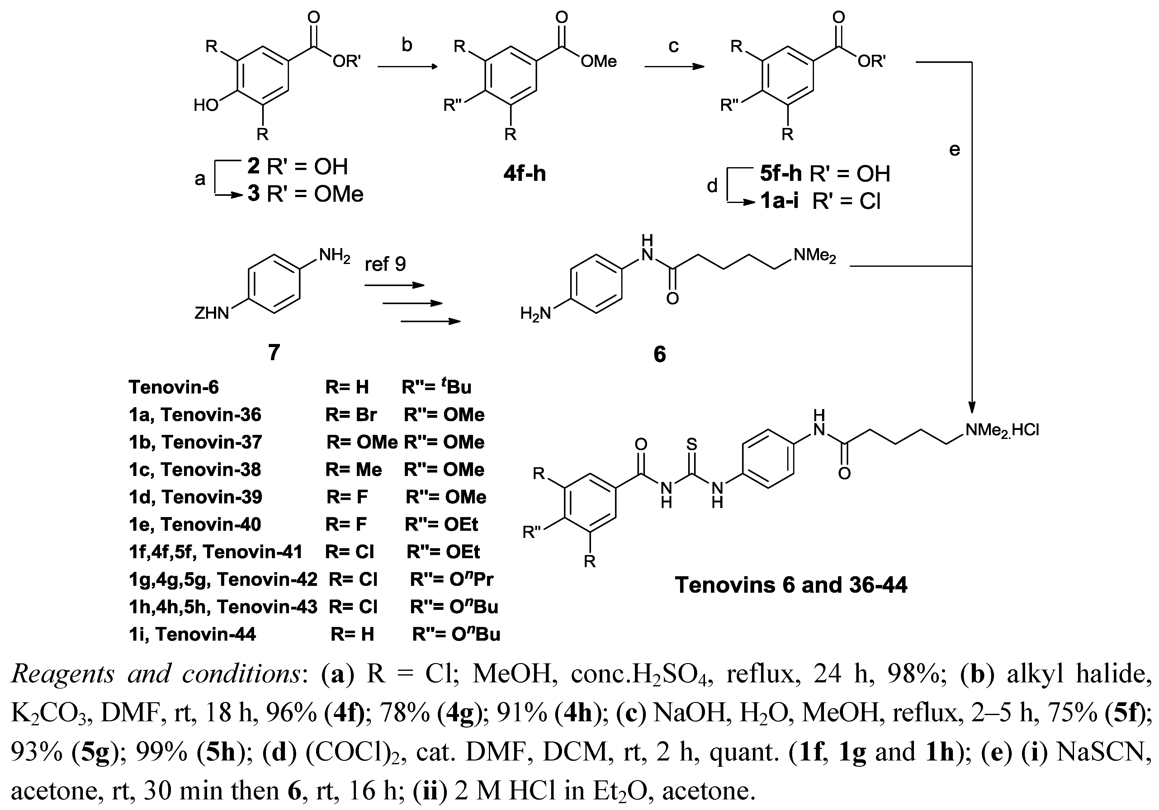
2.1. Chemistry

Initial studies generated structure activity relationships for tenovin-6 (Scheme 1 and Table 1). Previously we have shown that modification of the substituents in the N-benzoyl ring of tenovin-6 led to more active analogues and it was therefore decided to prepare additional analogues substituted in this region to tune selectivity towards SIRT2 over SIRT1 [9]. In brief, the synthesis of the novel tenovin-6 analogues followed our published route (Scheme 1) [9]. Whilst several of the acid chlorides 1 were available (compounds 1ae,i), the acid chlorides 1fh (see Scheme 1 for substituents) required for the synthesis of tenovins 41–43 were synthesised in four steps from the corresponding 4-hydroxycarboxylic acid 2 (R = Cl) as follows: esterification of the carboxylic acid 2 using standard conditions was followed by O-alkylation of the corresponding ester 3 (R = Cl) using the required alkyl halides to give 4fh. Hydrolysis of the ester functionality in 4fh followed by reaction of the resulting acids 5fh with oxalyl chloride afforded the required acid chlorides 1fh in high overall yields. The acid chlorides 1ai were then coupled with amine 6, synthesised in four steps from p-phenylenediamine (7), to give the required tenovin-6 analogues [9].
Scheme 1. Synthesis of tenovin analogues.
Molecules 17 12206 g007
Table 1. Activity of novel tenovin analogues against SIRT1 and SIRT2.
TenovinRR′′SIRT1IC50SIRT2IC50Selectivity Factor
% at 60 µm a% at 60 μM a
6HtBu73.9 ± 121b94.9 ± 110 b2.1
36BrOMe66.2 ± 251.6 ± 282.7 ± 116.0 ± 33.2
37OMeOMe12.5 ± 1n.d.18.1 ± 1n.d.n.d.
38MeOMe34.2 ± 365.4 ± 154.7 ± 144.9 ± 11.5
39FOMe79.5 ± 447.2 ± 181.2 ± 518.0 ± 32.6
40FOEt72.0 ± 435.3 ± 389.1 ± 117.5 ± 22.0
41ClOEt79.4 ± 128.5 ± 181.5 ± 212.9 ± 12.2
42ClOnPr81.0 ± 323.5 ± 296.1 ± 14.7 ± 35.0
43ClOnBu89.2 ± 221.5 ± 199.6 ± 30.8 ± 0.426.9
44HOnBu87.7 ± 136.5 ± 588.2 ± 27.2 ± 15.1
a % inhibition measured at 60 μM concentration of inhibitor ± SE (standard error, n = 2); b see reference 8; n.d. not determined.

2.2. In Vitro Inhibition of SIRT1 and SIRT2

The assessment of a compound’s ability to inhibit SIRT2 function in vitro is frequently carried out within the sirtuin community using a commercially available assay [20]. This assay, which relies on the deacetylation of a fluorescently labelled acetylated peptide substrate (Figure 1), was initially used here (Table 1).
Figure 1. Commercially available sirtuin assay uses a fluorescently labelled peptide substrate containing an N-acetylated lysine residue [20]. Removal of the N-acetyl group is coupled with conversion of NAD+ to nicotinamide (in red), 2′-O-acetyl ADP-ribose (in blue) and the deacetylated substrate. Subsequent reaction with trypsin releases the quenched fluorophore.
Molecules 17 12206 g001
The 3,5-dibromo-4-methoxy-substituted analogue, tenovin-36 showed reduced activity against SIRT2 compared with tenovin-6 but was even less active against SIRT1 (c.f. Table 1, entries 1 and 2). It was decided to explore this substitution pattern further. Replacement of the 3,5-halogen atoms in tenovin-36 by electron-donating substituents, OMe or Me (tenovins-37 and -38) led to a detrimental effect on both potency and selectivity. Returning to 3,5-dihalo-substituted analogues, tenovin-39 showed a similar activity profile to tenovin-36 (c.f. entries 2 and 5). Increasing the size of the alkyl chain in the 4-alkoxy-substituent from OMe in tenovin-39 to OEt in tenovin-40 led to little improvement. On moving back to chloro-substituents in the OEt series, the activity and selectivity was retained and it was demonstrated that further increases in the alkoxy chain length led to a significant increase in activity towards SIRT2, while the SIRT1 activity was almost unchanged (tenovins 41-43, c.f. entries 7–9). Tenovin-43 is, to the best of our knowledge, one of the most potent SIRT2 inhibitors reported to date [21,22]. Removal of the chloro substituents present in tenovin-43 to give tenovin-44 had a detrimental effect on activity.

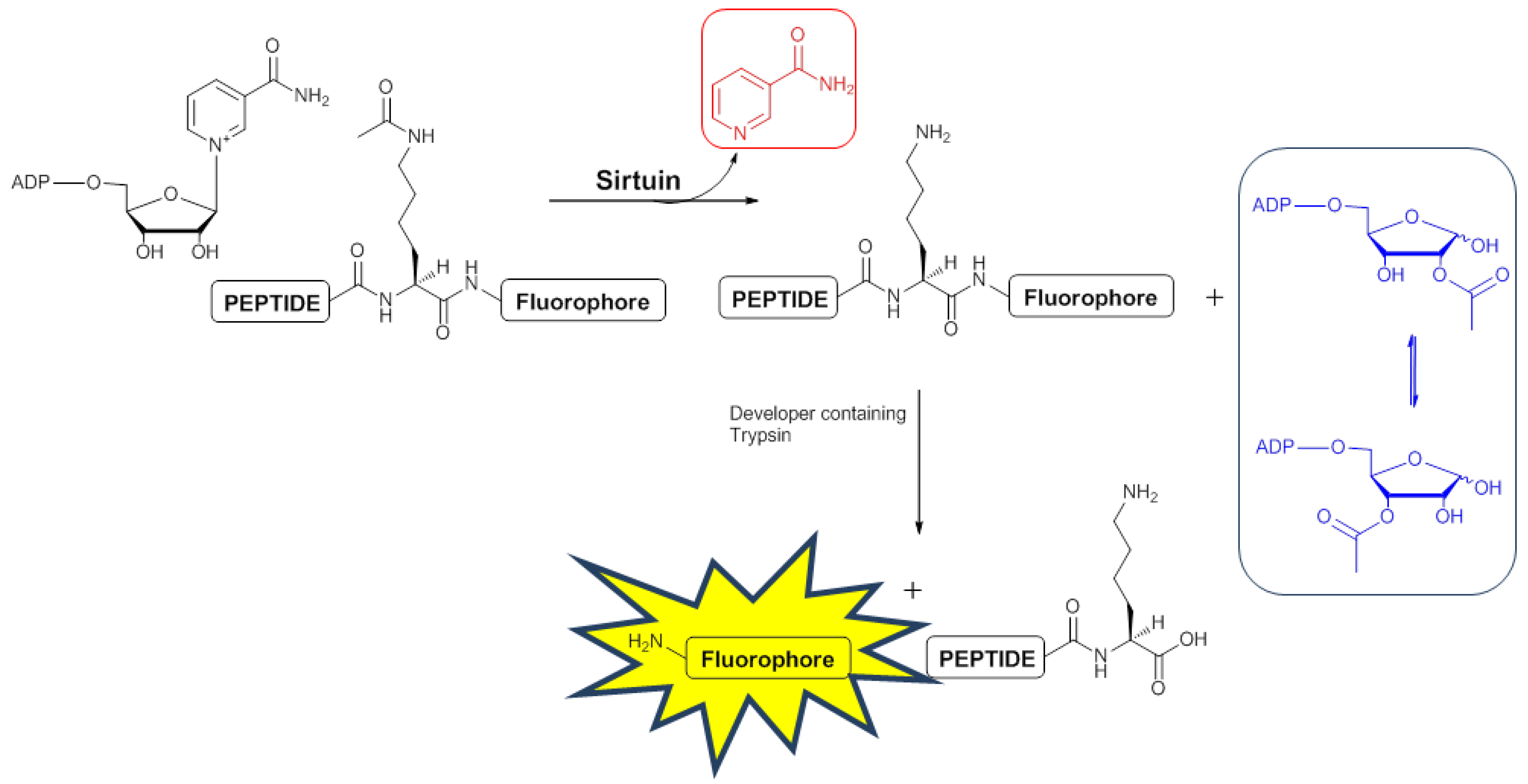
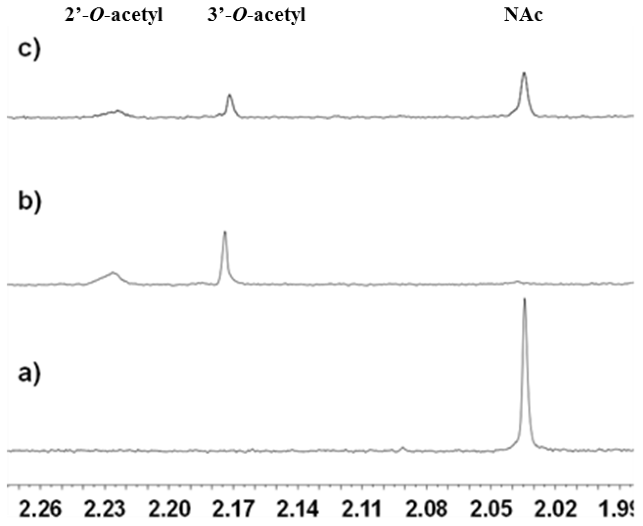
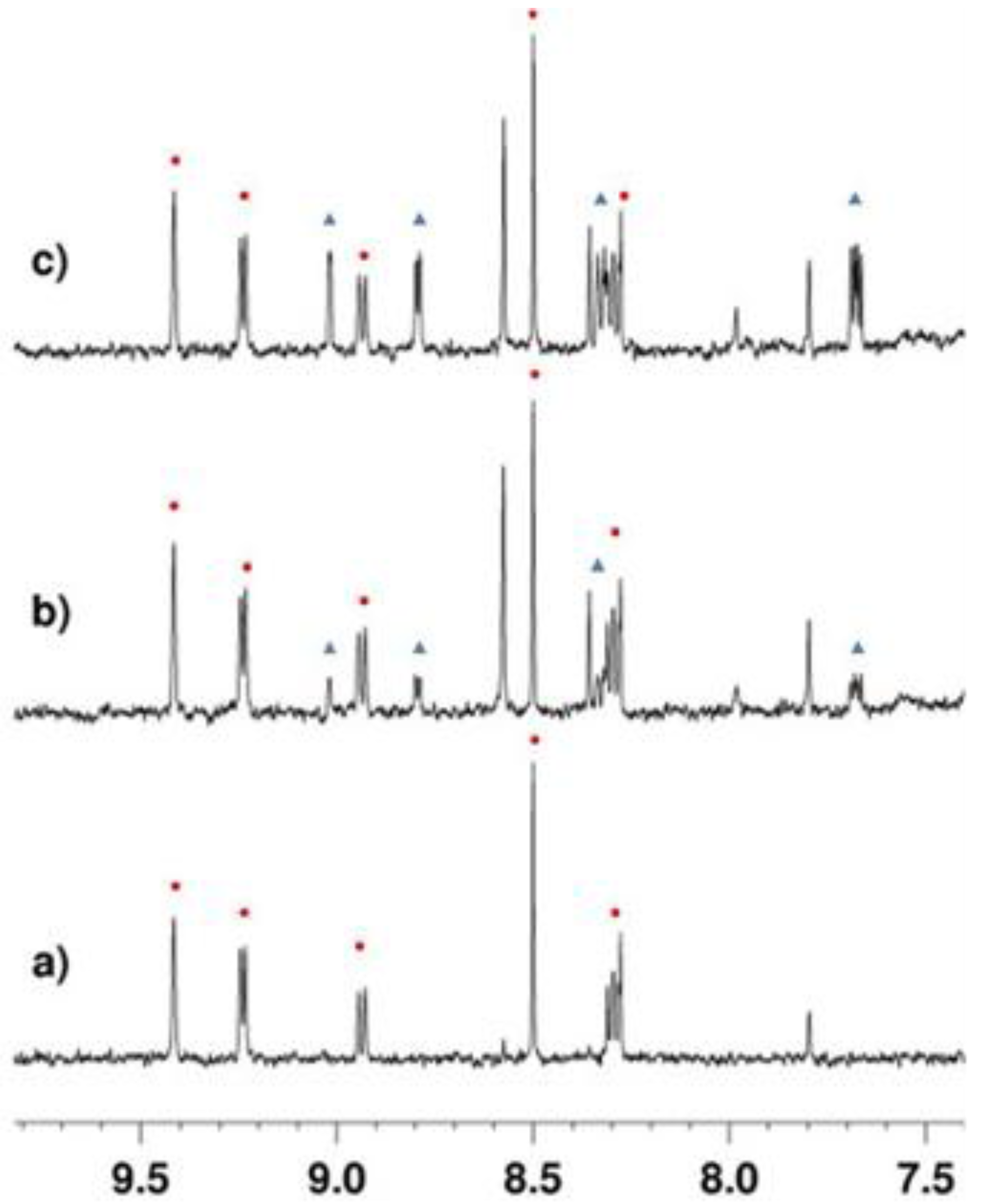
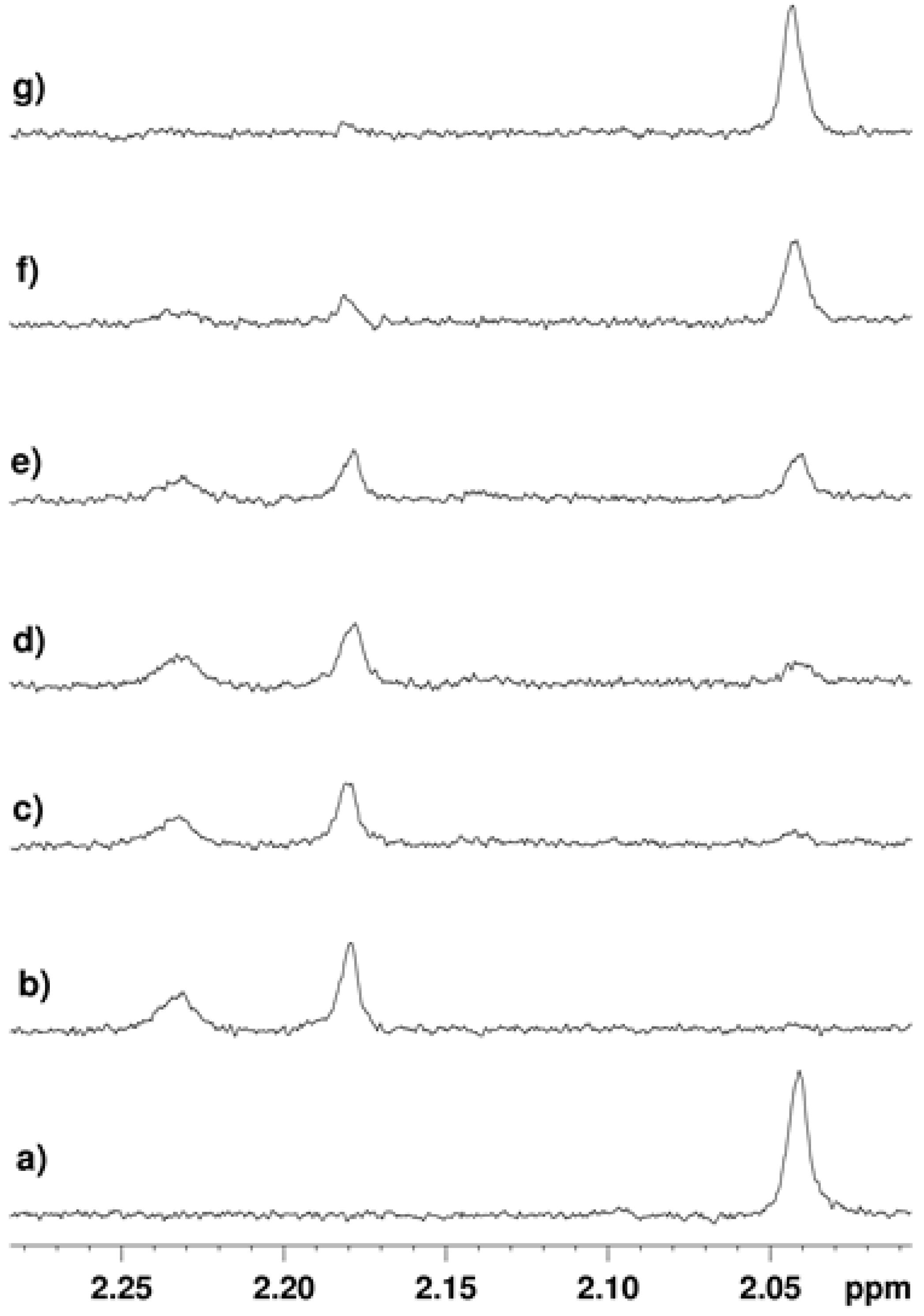
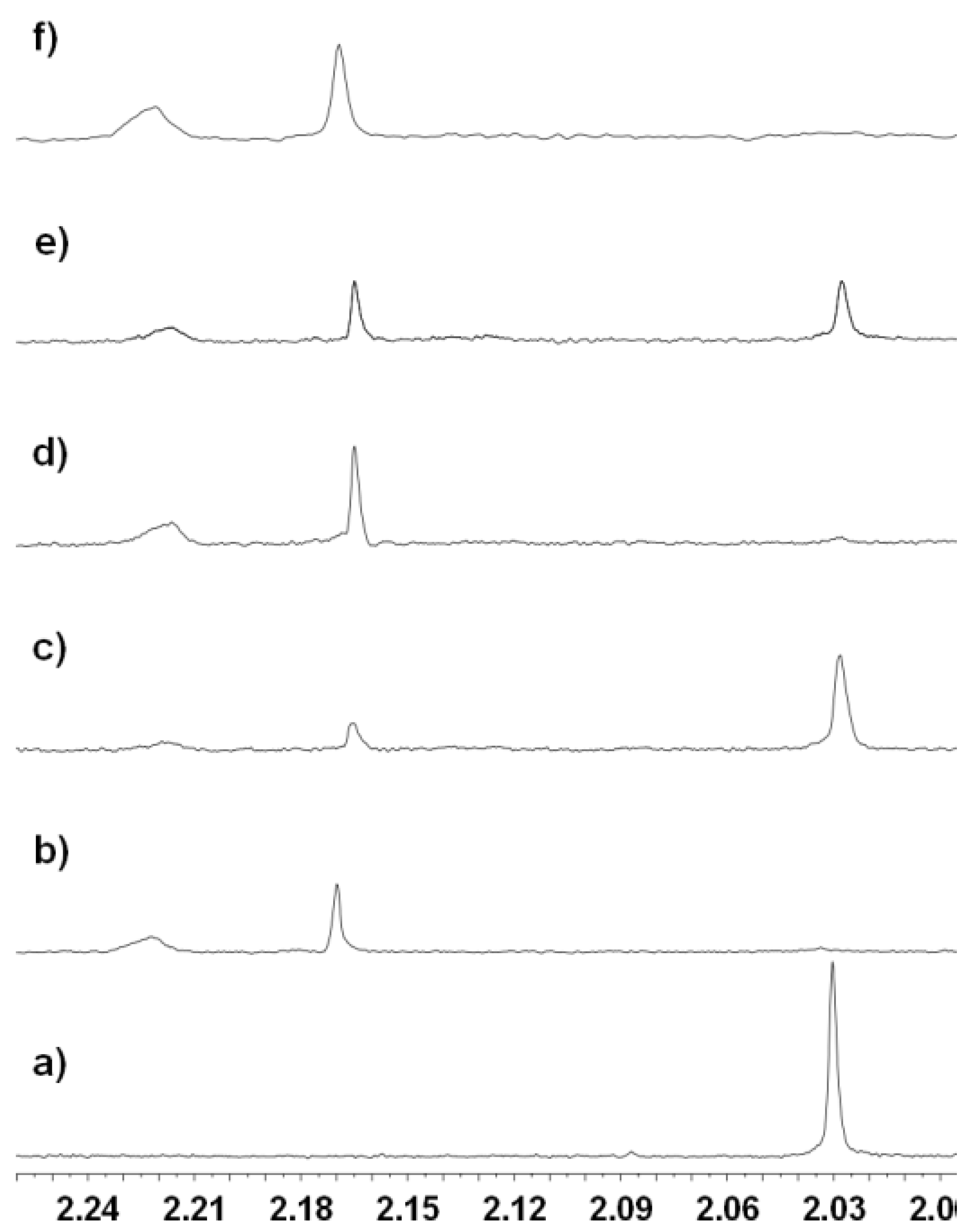
2.3. Development of a 1H-NMR Assay for Deacetylase Activity

With the increase in the use of high-throughput screening as a starting point for chemical tool and drug discovery projects, attention has turned to methods by which false positives can arise from screening campaigns. For example, stabilisers of the luciferase enzyme and compounds that quench the fluorescence in neuraminidase assays have recently been shown to produce misleading results in HT assays [23,24]. In addition there has been some challenges reported with the commercial sirtuin assay used here [25]. With this in mind, we decided to generate an alternative method to validate our hits using 1H-NMR methods. This approach is appealing because it would use a non-fluorescently labelled substrate to enable the direct monitoring of the deacetylation of the substrate to give the product peptide. It would also allow the production of additional reaction products such as nicotinamide or 2'/3'-O-acetyl ADP-ribose to be monitored giving multiple readouts.

2.3.1. Deacetylation of a Histone H4 Peptide by SIRT2

As described above, histone H4 is a known substrate of SIRT2 and it was therefore decided to use the 11 amino acid-containing peptide GLGKGGAK(Ac)RHR based on H4 as the SIRT2 substrate [16]. A C-terminal His-tagged version of the human sirt2 gene corresponding to residues 50 to 356 was cloned into a pET32a vector and the protein overexpressed using BL21(DE3) cells. Standard purification methods were used to access untagged SIRT2 in large quantities. Encouragingly, SIRT2-mediated deacetylation of the H4 substrate was observed by 1H-NMR (Figure 2). When the H4 substrate was characterised by 1H-NMR a peak at 2.03 ppm assigned to the acetyl methyl group was observed (Figure 2a). Band-selective 1H, 13C-HMBC experiments confirmed the assignment was correct (See Figures S2 and S3).
Figure 2. 1D 1H-NMR with double solvent suppression (for both H2O and Tris buffer) recorded at 37 °C. Sample contained 1 mM NAD+, 200 µM peptide, 10 µM SIRT2 in buffer (pH 8). (a) Before addition of enzyme. (b) After addition of enzyme and 15 mins. incubation. (c) Sample containing 200 μM tenovin-6 after incubation with enzyme for 15 min. Throughout this work, the H4 substrate was used at a final concentration of 200 μM as this enabled monitoring using a reasonable number of scans.
Molecules 17 12206 g002
Reaction of the H4 substrate with SIRT2 for 15 min at 37 °C followed by reanalysis led to the spectrum shown in Figure 2b in which the signal assigned to the lysine N-acetyl group in the substrate had disappeared (c.f. Figure 2a). The new signals at 2.17 ppm and 2.22 ppm (Figure 2b,c) were assigned as the acetyl groups in the O-acetylated ADP-ribose products formed from NAD+ (Figure 1) as determined by doping of authentic material into the reaction mixture (Figures S4 and S5). Doping experiments using an authentic sample of the deacetylated peptide (Figure S6) also confirmed that the H4 substrate was deacetylated by SIRT2. Finally, the observation of a new set of signals in the aromatic region of the spectrum were explained by the formation of nicotinamide from NAD+ (blue triangles in Figure 3b), consistent with the expected reaction. Doping of the final reaction mixture with authentic nicotinamide confirmed that it was formed in the SIRT2-catalysed reaction (Figure 3c).
Figure 3. 1D 1H-NMR with double solvent suppression (H2O and Tris buffer) recorded at 37 °C. (a) before addition of enzyme; (b) after incubation with SIRT2 for 20 mins at 37 °C. (c) after addition of authentic nicotinamide. Circles: signals from NAD+; Triangles signals from nicotinamide.
Molecules 17 12206 g003

2.3.2. Inhibition of the Deacetylase Reaction of SIRT2 by the Tenovins

Having shown that deacetylation of the H4 substrate could be monitored by 1H-NMR, it was decided to assess whether tenovin-6 (Scheme 1) could inhibit the reaction. When the free base of tenovin-6 was used (final concentration 200 μM) a significant reduction in the amount of substrate that was deacetylated by SIRT2 in 15 min was observed (c.f. Figure 2b,c) consistent with SIRT2 inhibition by tenovin-6. More detailed studies across a 0–500 µM final concentration range of tenovin-6 showed that the degree of inhibition observed was dose-dependent (Figure 4). The degree of inhibition was quantified by integration of the N-acetyl methyl group of the starting peptide with respect to the satellite peak of the TRIS buffer, the concentration of which was constant for all samples (Table S1). From this data an IC50 of 139.2 ± 9.5 µM was calculated for tenovin-6.
Figure 4. 1H-NMR spectrum obtained: (a) before addition of SIRT2; (b) 8 min after addition of SIRT2; the N-acetyl signal had disappeared consistent with substrate turnover by SIRT2. (cg) 8 min after addition of enzyme with tenovin-6 at a final concentration of: (c) 25 µM; (d) 50 µM. (e) 100 µM; (f) 200 µM; (g) 500 µM.
Molecules 17 12206 g004
The effect of the new SIRT2 selective inhibitor, tenovin-43 was then assessed. Inhibition of SIRT2 by tenovin-43 was observed, after a 20 min incubation period (c.f. the 8 min incubation period used to generate the data in Figure 4). A large signal corresponding to the N-acetyl in the H4 substrate was observed when tenovin-43 was present at a final concentration of 200 μM [compare Figure 5a (no enzyme) 5b (+SIRT2) and 5c (+SIRT2 and tenovin-43)]. Inhibition was also observed for tenovin-43 at a lower concentration of 25 µM with a signal corresponding to the presence of the N-acetylated peptide still present after the 20 min incubation (Figure 5e). However, the corresponding signal was not observable upon incubation with tenovin-6 at 25 µM under otherwise identical conditions (Figure 5d). On incubation with AGK2, the current state of the art SIRT2 inhibitor, no inhibition of SIRT2 at a final concentration of AGK2 of 25 μM was observed (Figure 5f) [19]. Due to the low solubility of AGK2 it was not possible to carry out this reaction at higher final concentrations of AGK2. Although direct comparison of the results obtained in the 1H-NMR assay with the commercially available assay is difficult, due to differences in the structure and concentration of the substrate, it is clear that analogous trends for inhibitor potency are seen in the two assays.
Figure 5. The 1H-NMR spectrum obtained: (a) before addition of enzyme; (bf) 20 min after addition of: (b) SIRT2; (c) SIRT2 and tenovin-43 (200 µM); (d) SIRT2 with tenovin-6 (25 µM); (e) SIRT2 with tenovin-43 (25 µM); (f) SIRT2 with AGK2 (25 µM).
Molecules 17 12206 g005

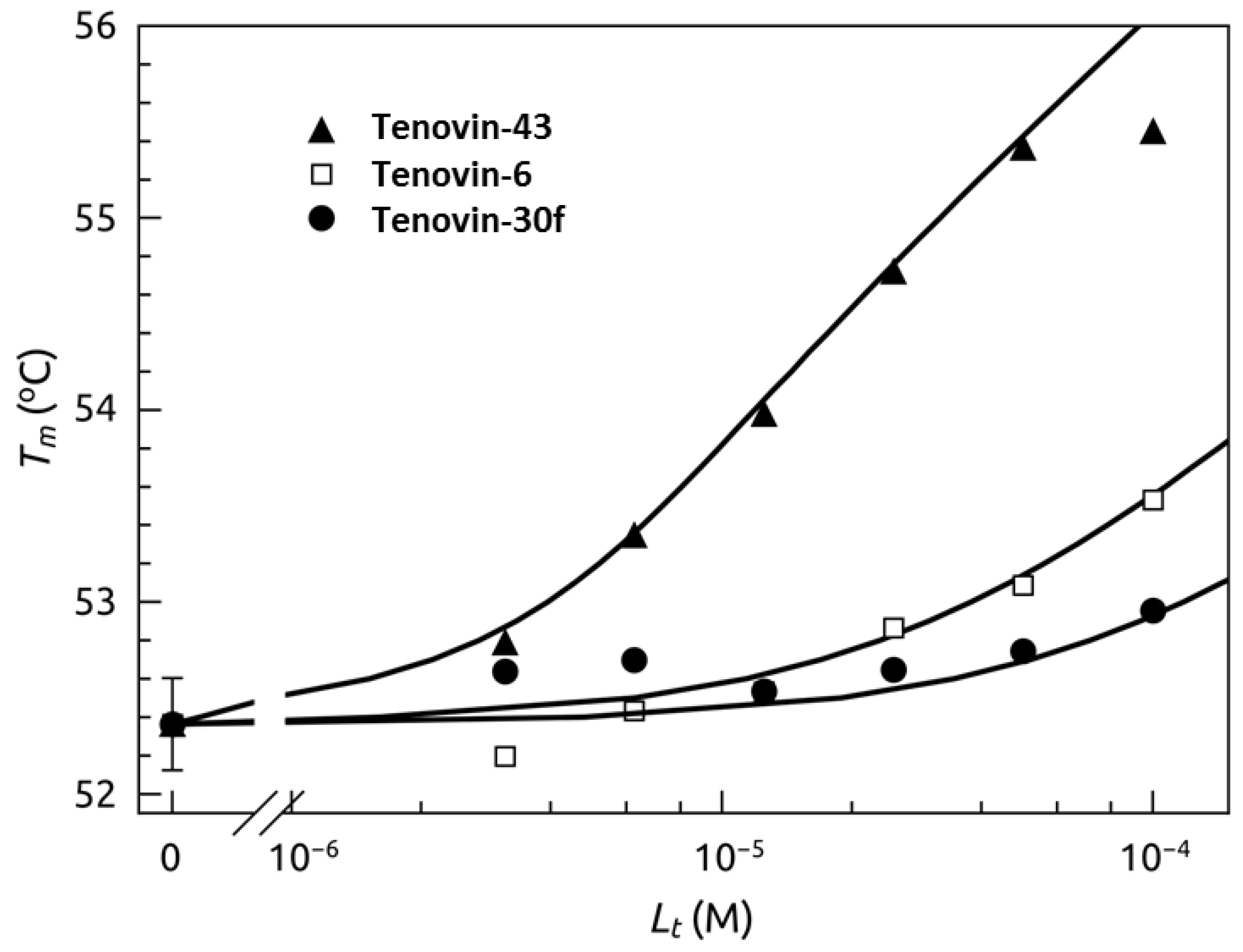
2.4. Thermal Shift Analysis of SIRT2 in the Presence of the Tenovins and AGK2

The binding of tenovins (tenovin-6, tenovin-43, and a previously reported inactive analogue tenovin-30f) to SIRT2 was also determined by a fluorescent thermal shift assay (Figure 6 and Figure S7) [9]. The dissociation constants at 37 °C, for the three ligands were: 0.67 µM for tenovin-43, 15 µM for tenovin-6, and 50 µM for the previously reported inactive tenovin-30f [9]. The calculated Kd values as determined by thermal shift correlate well with the previously obtained data using both the commercially available assay kit and the NMR method, where the most potent analogue tenovin-43 was shown to have the lowest dissociation constant. AGK2 was also assessed by thermal shift and was determined to have a Kd of >200 µM (Figures S8 and S9).
Figure 6. Ligand dosing curves showing the Tm shift dependence of SIRT2 on ligand concentration. Datapoints are experimental data obtained from curves as in Figure S6 while the lines are simulated according to the model as previously explained [26,27,28,29,30,31,32]. The dissociation constants at 37 °C, for the three ligands were: 0.67 µM for tenovin-43, 15 µM for tenovin-6 and 50 µM for tenovin-30f [9].
Molecules 17 12206 g006

3. Experimental

3.1. SIRT2 Expression and Purification

SIRT2 cDNA (residues 50-356) was expressed from Ncol and Xhol restriction sites of the pET32a vector (Novagen, Merck Millipore, Billerica, MA, USA) with an N-terminal His tag. Protein was overexpressed in BL21(DE3) pLysS bacterial cells (Sigma, Dorset, UK). Competent bacteria were transformed with the SIRT2 plasmid and starter cultures (10 mL) grown overnight at 37 °C in LB medium with ampicillin (100 mg/L) and chloramphenicol (100 mg/L). The overnight starter culture was added to 1 L of LB media containing ampicillin and chloramphenicol which was grown at 37 °C with 200 rpm shaking, until the OD600 reached 0.7–0.8. Cultures were cooled on ice before the addition of IPTG (0.1 mM) and Zn(OAc)2 (40 mM) and the culture grown at 18 °C overnight. The cells were harvested by centrifugation at 7500 rpm for 35 min before storing the resulting pellets at −20 °C. SIRT2 containing pellets were resuspended in lysis buffer (20 mM Tris pH 8, 200 mM NaCl, 10 mM imidazole, 2 mM β-mercaptoethanol, Roche EDTA-free mini protease inhibitor cocktail) and incubated on ice for 30 min. The suspension was sonicated for 10 × 1 min. The soluble fraction was collected after centrifugation at 15,000 rpm for 30 min. The resulting supernatant was filtered (0.45 µm), and loaded onto a pre-equilibrated 5mL HisTrap HP column (GE Healthcare, UK). The tagged protein eluted at 100 mM imidazole concentration. The fractions containing the enzyme were combined and dialysed in buffer (50 mM Tris pH 7.5, 100 mM NaCl, 5 mM imidazole, 2 mM β-mercaptoethanol) for 2 h. The protein was placed in fresh dialysis buffer and biotinylated thrombin added before leaving to cleave overnight at 4 °C. The enzyme was transferred into a tube where streptavidin agarose was added and the mixture gently mixed on a rocking table for 2 h at 4 °C. The suspension was centrifuged at 3,000 rpm for 10 min and the supernatant containing the purified enzyme decanted. The cleaved SIRT2 was further purified by Ni affinity chromatography as before, with cleaved protein eluting at 5 mM imidazole, followed by gel filtration chromatography on S-200 sephacryl column. The protein was concentrated by centrifugation using a spin concentrator. SIRT2 was concentrated to >12 mg/mL to ensure minimal amount of water in the NMR experiments.

3.2. SIRT1 and SIRT2 Inhibition Assay

All compounds were tested for inhibition of SIRT1 and SIRT2 using human recombinant enzymes; SirT1 available in the Fluor de Lys® fluorescence-based assay kit (Enzo Life Sciences, AK555, Exeter, UK) and SirT2 expressed and purified in house. All other required reagents were provided in the kit which was stored at −78 °C. Before use, small aliquots of each enzyme (2–3 μL) were prepared, snap frozen in liquid nitrogen and stored at −78 °C. Fresh dilutions of compounds were prepared in DMSO and further diluted in assay buffer. NAD+ (12.5 μL at 4 mM) and Fluor de Lys® SIRT1 or SIRT2 (12.5 μL at 100 μM) in assay buffer were added to a white 96 well plate, followed by compound (10 μL) and lastly enzyme (15 μL, 0.07 U/μL for kit SIRT1 and 0.3 U/μL for in-house SIRT2). After incubation for 1 h at 37 °C a developer solution (50 μL) was added to each reaction. The developer solution contained 38 μL buffer, 10 μL developer and 2 μL nicotinamide per reaction. The plate was then incubated for 45 min at rt and then read using a Spectra Max Gemini fluorimeter with an excitation wavelength of 355 nm and an emission wavelength of 460 nm. SigmaPlot software was used to generate fit curves for raw plots and the equation for each fit curve was used to calculate IC50 data.

3.3. 1H-NMR Experiments

To a D2O buffer containing 50 mM Tris (pH 8), 150 mM NaCl, 2 mM KCl and 1 mM MgCl2 was added NAD+ (1 mM), peptide (200 μM) and inhibitor (varying concentration). The inhibitor was dissolved in d6-DMSO before addition to the buffer ensuring that the final NMR sample contained only 0.5% DMSO for each experiment. The initial data was collected before addition of in house SIRT2 (10 μM) directly to the NMR tube. After the incubation was complete, the NMR tube was shaken and replaced in the spectrometer for data collection. 1D 1H-NMR with double solvent suppression (for H2O and TRIS) were recorded on a 500 MHz Bruker spectrometer at 37 °C.

3.4. Thermal Shift Experiments

The thermal shift assay was performed using Corbett Rotor-Gene 6000 (QIAGEN Rotor-Gene Q, Qiagen, Sydney, Australia) spectrofluorimeter. The prepared protein concentration was usually 5 µM and the ligand concentrations 50 µM. Reaction volume was usually 10 µL. Unfolding of the protein was monitored by measuring the fluorescence of the 1,8-anilinonaphthalene sulfonate (ANS), at 50–100 µM. The samples were heated at a rate of 1 °C/min. The samples were excited with 365 nm UV light and ANS fluorescence emission was registered at 460 nm light. Data analysis was performed as previously described [29].

3.5. General

All chemicals and solvents were purchased from Aldrich (Dorset, UK) or Alfa-Aesar (Heysham, UK) and used without further purification. All reactions were carried out under a positive pressure of nitrogen or argon in flame or oven-dried glassware. Ethanol was dried over Mg/I2; pyridine was dried over KOH pellets; all the other solvents were dried on a MBRAWN SPS-800 apparatus.
Thin layer chromatography (TLC) analysis was performed on silica pre-coated SIL G-25 UV254 sheets (layer: 0.25 mm silica gel with fluorescent indicator UV254, Alugram, Aldrich, Dorset, UK). Compounds were visualized by UV light (UV lamp, model UVGL-58, Mineralight LAMP, Multiband UV-254/365 nm) and stained with potassium permanganate. Flash column chromatography was carried out on silica gel (40–63 μm, Fluorochem, Hadfield, UK) or, where indicated, on basic alumina (Brockmann I, Sigma-Aldrich). Melting points were measured with an Electrothermal 9100 capillary melting point apparatus and are uncorrected.
Fourier Transform infra-red spectra (FT-IR) were acquired on a Perkin Elmer Paragon 1000 FT spectrometer. Absorption maxima are reported in wavenumbers (cm−1).
Unless otherwise stated, 1H-NMR spectra were measured at room temperature (298 K) on a Bruker DPX 400 (1H = 400 MHz) and Bruker Avance 300 (1H = 300.1 MHz) instruments. Deuterated solvents were used and 1H-NMR chemical shifts were internally referenced to CHCl3 (7.26 ppm) in chloroform-d1 solution. Chemical shifts are expressed as δ in unit of ppm.
13C-NMR spectra were recorded in the same conditions and in the same solvents using the PENDANT sequence mode on a Bruker DPX 400 (13C = 100 MHz). Data processing was carried out using TOPSPIN 2 NMR version (Bruker UK, Ltd, Coventry, UK). In 1H-NMR assignment the multiplicity used is indicated by the following abbreviations: s = singlet, d = doublet, dd = doublet of doublets, t = triplet, q = quartet, m = multiplet, brs = broad singlet. Signals of protons and carbons were assigned, as far as possible, by using the following two-dimensional NMR spectroscopy techniques: [1H-1H] COSY, [1H-13C] COSY (HSQC: Heteronuclear Single Quantum Coherence) and long range [1H-13C] COSY (HMBC: Heteronuclear Multiple Bond Connectivity).
Mass spectrometry (electrospray mode, ES; chemical ionization mode, CI) were recorded on a high performance orthogonal acceleration reflecting TOF mass spectrometer operating in positive and negative mode, coupled to a Waters 2975 HPLC.

3.6. Synthesis

3.6.1. General Procedure for the alkylation of 3

To a stirred solution of 3 (1 equiv.) in dry DMF (1 vol.) under N2 was added K2CO3 (2 equiv.) and the alkyl halide (1.1 equiv.). The resulting solution was stirred for 16 h at room temperature before being partitioned between ethyl acetate (1 vol.) and water (0.5 vol.). The organic layer was washed with water (0.5 vol.), brine (0.5 vol.), dried (MgSO4), filtered and the solvent removed in vacuo to give the desired product which was used without further purification.
Methyl 3,5-dichloro-4-ethoxybenzoate (4f). Prepared from compound 2 (1.3 g, 5.9 mmol), K2CO3 (1.6 g, 11.7 mmol) and iodoethane (522 μL, 6.5 mmol) in DMF (10 mL). The product was obtained as a brown oil (1.4 g, 5.6 mmol, 96%). νmax cm−1 (NaCl, thin layer) 2994, 1701, 1652, 1556, 1147, 854; δH (CDCl3, 400 MHz) 7.91 (2H, s, ArH), 4.09 (2H, q, J = 7.0 Hz, CH2), 3.85 (3H, s, CH3) and 1.41 (3H, t, J = 7.0 Hz, CH3); δC (CDCl3, 100 MHz) 164.7 (C), 155.5 (C), 130.2 (CH), 129.8 (C), 126.9 (C), 70.0 (CH2), 52.6 (CH3) and 15.5 (CH3); m/z (ES)+: 249.35 [(M + H)+, 100%].
Methyl 4-propoxy-3,5-dichlorobenzoate (4g). Prepared from 2 (500 mg, 2.3 mmol), K2CO3 (630 mg, 4.6 mmol) and iodopropane (243 μL, 2.5 mmol) in DMF (10 mL). The product was obtained as a yellow-brown oil (472 mg, 1.8 mmol, 78%). νmax cm−1 (NaCl, thin layer) 2954 2880, 1733, 1690, 1592, 1556, 1462, 1288, 1138, 986; δH (CDCl3, 400 MHz) 7.90 (2H, s, ArH), 3.97 (2H, t, J = 6.6 Hz, CH2), 3.84 (3H, s, CH3), 1.82 (2H, app. sextet, J = 7.0 Hz, CH2) and 1.02 (3H, t, J = 7.5 Hz, CH3); δC (CDCl3, 100 MHz) 164.2 (C), 155.0 (C), 130.6 (CH), 130.1 (C), 127.3 (C), 76.0 (CH2), 53.6 (CH3), 23.8 (CH2) and 10.8 (CH3); m/z (ES)+: 263.24 [(M + H)+, 100%].
Methyl 4-butoxy-3,5-dichlorobenzoate (4h). Prepared from 2 (500 mg, 2.3 mmol), K2CO3 (630 mg, 4.6 mmol) and iodobutane (283 μL, 2.5 mmol) in DMF (10 mL). The product was obtained as a brown oil (578 mg, 2.1 mmol, 91%). νmax cm−1 (NaCl, thin layer) 2959 (ArC-H), 1729, 1642, 1556, 1435, 1284, 1137, 987; δH (CDCl3, 400 MHz) 7.90 (2H, s, ArH), 4.01 (2H, t, J = 6.8 Hz, CH2), 3.84 (3H, s, CH3), 1.81–1.76 (2H, m, CH2), 1.50 (2H, app. sextet, J = 7.5 Hz, CH2) and 0.93 (3H, t, J = 7.5 Hz, CH3); δC (CDCl3, 100 MHz) 164.7 (C), 155.6 (C), 130.3 (CH), 129.7 (C), 126.9 (C), 73.8 (CH2), 52.6 (CH3), 32.11 (CH2), 19.04 (CH2) and 13.8 (CH3); m/z (ES)+: 277.06 [(M + H)+, 100%].

3.6.2. General Procedure for Ester Hydrolysis

The ester (1 equiv.) and sodium hydroxide (1.2 equiv.) were heated at reflux in a solution of methanol (1 vol.) and water (1 vol.) until the methyl ester was consumed by TLC (4–6 h). The methanol was removed in vacuo and the aqueous fraction acidified with 2 M HCl. The resulting precipitate was extracted with ethyl acetate (3 × 1 vol.) and the organic layers combined and washed with brine (0.5 vol.), dried (MgSO4), filtered and the solvent removed to yield the desired acid.
3,5-Dichloro-4-ethoxybenzoic acid (5f). Prepared from methyl 4-ethoxy-3,5-dichlorobenzoate (500 mg, 2.0 mmol) in MeOH/water (10 mL) and NaOH (96 mg, 2.4 mmol). The desired product was obtained as an off-white solid (1.8 g, 7.7 mmol, 75%). Mp 179–180 °C; νmax cm−1 (KBr) 3225, 2104, 1635, 1206; δH (CDCl3, 400 MHz) 8.12 (2H, s, ArH), 4.28 (2H, q, J = 6.9 Hz, CH2), 1.57 (3H, t, J = 6.9 Hz, CH3); δC (CDCl3, 100 MHz) 169.6 (C), 156.3 (C), 130.8 (CH), 130.0 (C), 125.9 (C), 70.2 (CH2), 15.5 (CH3); m/z (ES) 232.97 [(M−H), 100%]; HRMS (ES) [Found: (M-H), 232.9767, C9H7O3Cl2 requires 232.9772].
4-Propoxy-3,5-dichlorobenzoic acid (5g). Prepared from methyl 4-propoxy-3,5-dichlorobenzoate (400 mg, 1.5 mmol) in MeOH/water (10 mL) and NaOH (72 mg, 1.8 mmol). The product was obtained as a white solid (347 mg, 1.4 mmol, 93%). Mp 125–126 °C; νmax cm−1 (KBr) 2937, 1699, 1558, 1493, 1385, 1077, 906, 768; δH (CDCl3, 400 MHz) 7.97 (2H, s, ArH), 4.00 (2H, t, J = 6.6 Hz, CH2), 1.83 (2H, app. sextet, J = 7.1 Hz, CH2), 1.03 (3H, t, J = 7.6 Hz, CH3); δC (CDCl3, 100 MHz) 169.4, 156.4, 129.9, 125.9, 75.7, 23.4, 10.4; m/z (ES) 247.23 [(M−H), 100%]; HRMS (ES) [Found: (M−H), 246.9924, C10H9O3Cl2 requires 246.9929]
4-Butoxy-3,5-dichlorobenzoic acid (5h). Prepared from methyl 4-butoxy-3,5-dichlorobenzoate (500 mg, 1.8 mmol) in MeOH/water (10 mL) and NaOH (86 mg, 2.2 mmol). The product was obtained as a yellow solid (472 mg, 1.8 mmol, 99%). Mp 98–99 °C; νmax cm−1 (KBr) 2955, 1682, 1214, 1557, 1388, 1056, 810; δH (CDCl3, 400 MHz) 7.91 (2H, s, ArH), 4.04 (2H, t, J = 6.6 Hz, CH2), 1.8–1.7 (2H, m, CH2), 1.49 (2H, app. sextet, J = 7.0 Hz, CH2), 0.94 (3H, t, J = 7.4 Hz, CH3); δC (CDCl3, 100 MHz) 169.3 (C), 156.4 (C), 130.8 (CH), 130.2 (C), 125.9 (C), 73.8 (CH2), 32.1 (CH2), 19.03 (CH2), 13.8 (CH3); m/z (ES) 261.02 [(M–H), 100%]; HRMS (ES) [Found: (M–H), 261.0078, C11H11O3Cl2 requires 261.0085].

3.6.3. General Procedure for Synthesis of Acid Chlorides 1a1

To a stirred solution of the benzoic acid (synthesised or commercially available) (1 equiv.) in DCM (1 vol.), under N2, was added a solution of oxalyl chloride (2 equiv.) in DCM (1 vol.). A drop of dry DMF was added and the resulting solution stirred at room temperature for 90 min. The solvent was removed in vacuo and the resulting acid chloride used immediately without purification or characterisation.

3.6.4. General Procedure for Sodium Thiocyanate Coupling Reaction

To a solution of the acid chloride (1 equiv.) in dry acetone (1 vol.) under N2, was added sodium thiocyanate (1 equiv.). The resulting suspension stirred at room temperature for 30 min before being cooled to 0 °C. A solution of the amine (1 equiv.) in dry acetone (1 vol.) was added and the resulting suspension allowed to warm to room temperature and stirred for 16 h. The reaction was filtered through Celite and the filtrate concentrated to give the crude product. The product was purified by column chromatography (1–20% methanol-DCM). The purified product was dissolved in acetone and 2 M HCl in diethyl ether (1 equiv.) added slowly. The resulting precipitate was filtered and recrystallised from ethanol to afford the pure HCl salts.
3,5-Dibromo-N-((4-(5-(dimethylamino)pentanamido)phenyl)carbamothioyl)-4-methoxybenzamide hydrochloride (Tenovin-36). Prepared from 3,5-dibromo-4-methoxybenzoyl chloride (85 mg, 0.26 mmol) and sodium thiocyanate (21 mg, 0.26 mmol) in dry acetone (3 mL) followed by addition of N-(4-aminophenyl)-5-(dimethylamino)pentanamide (61 mg, 0.26 mmol) in acetone (3 mL). The crude material was purified by column chromatography (1–20% MeOH-DCM) and this material subsequently converted to the HCl salt to afford the product as a yellow solid (40 mg, 0.06 mmol, 23%). Mp 159–160 °C; νmax cm−1 (KBr) 2924, 2857, 1669, 1606, 1541, 1495, 1405, 1262, 1094, 824; δH (DMSO-d6, 400 MHz) 12.41 (1H, s, NH), 11.85 (1H, s, NH), 10.21 (1H, s, NH), 9.75 (1H, br. s, NH+), 8.34 (2H, s, ArH), 7.69 (4H, AA’BB’, J = 8.8, 29.0 Hz, ArH), 3.96 (3H, s, CH3), 3.14 (2H, m, CH2), 2.83 (6H, d, J = 4.8 Hz, (CH3)2), 2.48 (2H, m, CH2), 1.74 (4H, m, (CH2)2); δC (DMSO-d6, 400 MHz) 178.5 (C), 170.8 (C), 164.9 (C), 156.9 (C), 137.5 (C), 133.2 (CH), 132.7 (C), 130.7 (C), 124.7 (CH), 119.0 (CH), 117.4 (C), 60.6 (CH3), 56.1 (CH2), 41.9 (CH3), 35.5 (CH2), 23.2 (CH2) and 22.0 (CH2); m/z (ES)+ 586.98 [((M−HCl)+H)+, 100%].
N-((4-(5-(Dimethylamino)pentanamido)phenyl)carbamothioyl)-3,4,5-trimethoxybenzamide hydro-chloride (Tenovin-37). Prepared from 3,4,5-trimethoxybenzoyl chloride (53 mg, 0.23 mmol) and sodium thiocyanate (19 mg, 0.23 mmol) in dry acetone (3 mL) followed by addition of N-(4-aminophenyl)-5-(dimethylamino)pentanamide (54 mg, 0.23 mmol) in acetone (3 mL). The crude material was purified by column chromatography (1–20% MeOH-DCM) and this material subsequently converted to the HCl salt to afford the product as a yellow solid (36.3 mg, 0.06 mmol, 30%). Mp 115–117 °C; νmax cm−1 (KBr) 2925, 2858, 1670, 1642, 1495, 1405, 1340, 1261, 1125, 1050, 824; δH (DMSO-d6, 300 MHz) 12.65 (1H, s, NH), 11.56 (1H, s, NH), 10.15 (1H, s, NH), 9.82 (1H, br. s, NH+), 7.62 (4H, AA’BB’, J = 9.0, 24.4 Hz, ArH), 7.38 (2H, s, ArH), 3.88 (6H, s, (CH3)2), 3.75 (3H, s, CH3), 3.06 (2H, m, CH2), 2.75 (6H, d, J = 4.9 Hz, (CH3)2), 2.39 (2H, t, J = 6.5 Hz, CH2), 1.65 (4H, m, (CH2)2); δC (DMSO-d6, 100 MHz) 178.9 (C), 170.7 (C), 167.4 (C), 152.5 (C), 141.5 (C), 137.4 (C), 132.8 (C), 126.7 (C), 124.7 (CH), 119.0 (CH), 106.5 (CH), 60.1 (CH3), 56.6 (CH2), 56.1 (CH3), 41.9 (CH3), 35.5 (CH2), 23.2 (CH2), 23.2 (CH2), 21.9 (CH2); m/z (ES)+ 489.18, [((M−HCl)+H)+, 100%]; HRMS (ES)+ [Found: ((M−HCl)+H)+, 489.2172, C24H33ClN4O5S requires 489.2166].
N-(4-(5-(dimethylamino)pentanamido)phenylcarbamothioyl)-4-methoxy-3,5-dimethylbenzamide hydrochloride (Tenovin-38). Prepared from 4-methoxy-3,5-dimethylbenzoyl chloride (110 mg, 0.6 mmol) and sodium thiocyanate (45 mg, 0.6 mmol) in dry acetone (3 mL) followed by addition of N-(4-aminophenyl)-5-(dimethylamino)pentanamide (130 mg, 0.6 mmol) in acetone (3 mL). The crude material was purified by column chromatography (1–20% MeOH-DCM) and this material subsequently converted to the HCl salt. The product was obtained as a brown solid (74 mg, 0.15 mmol, 25%). Mp 190–192 °C; νmax cm−1 (KBr) 2925, 2854, 1667, 1604, 1515, 1337, 1307, 1166, 1007, 740; δH (d6-DMSO, 400 MHz) 12.64 (1H, s, NH), 11.36 (1H, s, NH), 10.20 (1H, s, NH), 9.91 (1H, br. s, NH+), 7.79 (2H, s, ArH), 7.69 (2H, d, J = 8.4 Hz, ArH), 7.63 (2H, d, J = 8.4 Hz, ArH), 3.77 (3H, s, CH3), 3.15–3.08 (2H, m, CH2), 2.79 (6H, s, (CH3)2), 2.47–2.41 (2H, m, CH2), 2.33 (6H, s, (CH3)2), 1.76–1.69 (4H, m, (CH2)2); δC (d6-DMSO, 100 MHz) 178.9 (C), 170.7 (C), 167.7 (C), 160.7 (C), 137.3 (C), 132.8 (C), 130.6 (C), 129.5 (CH), 126.9 (C), 124.7 (CH), 118.9 (CH), 59.4 (CH3), 56.2 (CH2), 42.0 (CH3), 35.5 (CH2), 23.3 (CH2), 21.9 (CH2), 15.8 (CH3); m/z (ES)+ 457.03 [((M−HCl)+H)+, 100%]; HRMS (ES)+ [Found: ((M−HCl)+H)+, 457.2265, C24H33N4O3S requires 457.2273].
N-((4-(5-(dimethylamino)pentanamido)phenyl)carbamothioyl)-3,5-difluoro-4-methoxybenzamide hydrochloride (Tenovin-39). Prepared from 3,5-difluoro-4-methoxybenzoyl chloride (132 mg, 0.64 mmol) and sodium thiocyanate (52 mg, 0.64 mmol) in dry acetone (3 mL) followed by addition of N-(4-aminophenyl)-5-(dimethylamino)pentanamide (151 mg, 0.64 mmol) in acetone (3 mL). The crude material was purified by column chromatography (1–20% MeOH-DCM) and this material subsequently converted to the HCl salt to afford the product as an off-white solid (134 mg, 0.27 mmol, 42%). Mp 153–154 °C; νmax cm−1 (KBr) 2924, 2859, 1659, 1608, 1544, 1495, 1451, 1404, 1262, 1180, 1049, 824; δH (d6-DMSO, 400 MHz) 12.33 (1H, s, NH), 11.54(1H, s, NH), 10.19 (2H, br. s., (NH)2), 7.81 (2H, d, J = 8.6 Hz, ArH), 7.62 (2H, d, J = 6.8 Hz, ArH), 7.53 (2H, d, J = 6.8 Hz, ArH), 4.08 (3H, s, CH3), 3.00 (2H, app. br. s., CH2), 2.68 (6H, s, (CH3)2), 2.35 (2H, app. br. s., CH2) and 1.62 (4H, app. br. s., (CH2)2); δC (d4-CD3OD, 100 MHz) 180.5 (C), 173.7 (C), 167.0 (C), 157.2 (C), 155.2 (C), 138.3 (C), 135.2 (C), 127.8 (C), 125.9 (CH), 121.1 (CH), 114.1 (CH), 58.7 (CH2), 53.6 (CH3), 43.5 (CH3), 36.7 (CH2), 25.2 (CH2), 23.1 (CH2); m/z (ES)+ 465.03 [((M−HCl)+H)+, 100%]; HRMS (ES)+ [Found: ((M−HCl)+H)+, 465.1760, C22H27N4O3F2S requires 465.1772].
N-((4-(5-(dimethylamino)pentanamido)phenyl)carbamothioyl)-3,5-difluoro-4-ethoxybenzamide hydrochloride (Tenovin-40). Prepared from 3,5-difluoro-4-ethoxybenzoyl chloride (150 mg, 0.68 mmol) and sodium thiocyanate (55 mg, 0.68 mmol) in dry acetone (3 mL) followed by addition of N-(4-aminophenyl)-5-(dimethylamino)pentanamide (160 mg, 0.68 mmol) in acetone (3 mL). The crude material was purified by column chromatography (1–20% MeOH-DCM) and this material subsequently converted to the HCl salt to afford the product as an off-white solid (124 mg, 0.24 mmol, 35%). Mp 206–207 °C; νmax cm−1 (KBr) 2926, 2860, 1674, 1606, 1512, 1495, 1436, 1405, 1263, 1135, 1049, 874; δH (d6-DMSO, 400 MHz) 12.34 (1H, s, NH), 11.56 (1H, br. s, NH), 10.11 (2H, s, NH), 9.70 (2H, br. s., NH+), 7.83 (2H, d, J = 9.2 Hz, ArH), 7.64 (2H, d, J = 7.9 Hz, ArH), 7.56 (2H, d, J = 7.9 Hz, ArH), 4.31 (2H, app quartet, J = 4.3 Hz, CH2), 3.08-2.99 (2H, m, CH2), 2.72 (6H, app. s, (CH3)2), 2.38 (2H, app. t., J = 6.3 Hz, CH2), 1.71–1.56 (4H, m, (CH2)2) and 1.32 (3H, t, J = 7.0 Hz, CH3); δC (d4-CD3OD, 100 MHz) 180.4 (C), 173.5 (C), 167.0 (C), 157.6 (C), 155.7 (C), 138.3 (C), 135.2 (C), 128.0 (C), 125.9 (CH), 121.1 (CH), 113.9 (CH), 58.7 (CH2), 43.5 (CH3), 36.7 (CH2), 30.8 (CH2), 25.2 (CH2), 23.1 (CH2), 15.8 (CH3); m/z (ES)+ 478.90 [((M−HCl)+H)+, 100%]; HRMS (ES)+ [Found: ((M−HCl)+H)+, 479.1940, C23H29N4O3F2S requires 479.1928].
3,5-Dichloro-N-(4-(5-(dimethylamino)pentanamido)phenylcarbamothioyl)-4-ethoxybenzamide hydrochloride (Tenovin-41). Prepared by the reaction of 3,5-dichloro-4-ethoxybenzoyl chloride (216 mg, 0.86 mmol) and sodium thiocyanate (69 mg, 0.86 mmol) in dry acetone (6 mL) followed by addition of a solution of N-(4-aminophenyl)-5-(dimethylamino)pentanamide (202 mg, 0.86 mmol) in dry acetone (6 mL) using general method F. The product was further purified by conversion to the HCl salt by the addition of 2M HCl in diethyl ether. The desired product was obtained as a brown sticky oil (42 mg, 0.08 mmol, 9.5%). νmax cm−1 (NaCl, thin layer) 2965, 2359.3, 1651, 1048, 1025, 998, 765; δH (CDCl3, 400 MHz) 12.29 (1H, s, NH), 11.68 (1H, s, NH), 10.21 (1H, s, NH), 8.08 (2H, s, ArH), 7.79–7.46 (4H, m, ArH), 4.19–4.06 (2H, m, CH2), 3.06–2.93 (2H, m, CH2), 2.69 (6H, s, (CH3)2), 2.36–2.24 (2H, m, CH2), 1.71–1.65 (2H, m, CH2), 1.41–1.28 (2H, m, CH2), 1.23–1.17 (3H, m, CH3); δC (d4-CD3OD, 100 MHz) 180.4 (C), 173.5 (C), 167.0 (C), 156.7 (C), 138.2 (C), 135.2 (C), 131.1 (C), 130.9 (C), 130.4 (CH), 125.9 (CH), 121.2 (CH), 71.2 (CH2), 58.8 (CH2), 43.5 (CH3), 36.6 (CH2), 25.3 (CH2), 23.1 (CH2), 15.8 (CH3); m/z (ES)+ 510.94 [((M−HCl)+H)+, 100%]; HRMS (ES)+ [Found: ((M−HCl)+H)+, 511.1337, C23H29N4O3SCl2 requires 511.1337].
3,5-Dichloro-N-((4-(5-(dimethylamino)pentanamido)phenyl)carbamothioyl)-4-propoxybenzamide hydrochloride (Tenovin-42). Prepared from 3,5-dichloro-4-propoxybenzoyl chloride (160 mg, 0.60 mmol) and sodium thiocyanate (49 mg, 0.60 mmol) in dry acetone (3 mL) followed by addition of N-(4-aminophenyl)-5-(dimethylamino)pentanamide (142 mg, 0.60 mmol) in acetone (3 mL). The crude material was purified by column chromatography (1–20% MeOH-DCM) and this material subsequently converted to the HCl salt to afford the product as an off-white solid (73 mg, 0.13 mmol, 21%). Mp 167–168 °C; νmax cm−1 (KBr) 2927, 2861, 1674, 1636, 1606, 1539, 1473, 1452, 1265, 1148, 1051, 873; δH (d6-DMSO, 400 MHz) 12.31 (1H, s, NH), 11.70 (1H, s, NH), 10.23 (1H, s, NH), 10.13 (1H, s, NH+), 8.09 (2H, s, ArH), 7.65 (2H, d, J = 8.9 Hz, ArH), 7.56 (2H, d, J = 8.9 Hz, ArH), 4.04 (2H, t, J = 6.4 Hz, CH2), 3.02–2.96 (2H, m, CH2), 2.72 (6H, d, J = 5.2 Hz, (CH3)2), 2.38–2.32 (2H, m, CH2), 1.86–1.75 (2H, m, CH2), 1.70–1.60 (4H, m, (CH2)2), 1.03 (3H, t, J = 7.6 Hz, CH3); δC (d6-DMSO, 100 MHz) 170.5 (C), 165.5 (C), 154.4 (C), 150.5 (C), 135.4 (C), 132.7 (C), 129.5 (C), 129.2 (CH), 128.6 (C), 120.2 (CH), 119.6 (CH), 75.4 (CH2), 56.2 (CH2), 42.0 (CH3), 35.4 (CH2), 23.3 (CH2), 22.9 (CH2), 21.9 (CH2), 10.6 (CH3); m/z (ES)+ 525.09 [((M−HCl)+H)+, 100%]; HRMS (ES)+ [Found: ((M−HCl)+H)+, 525.1509, C24H31N4O3SCl2 requires 525.1494].
4-Butoxy-3,5-dichloro-N-((4-(5-(dimethylamino)pentanamido)phenyl)carbamothioyl)benzamide hydrochloride (Tenovin-43). Prepared from 3,5-dichloro-4-butoxybenzoyl chloride (28 mg, 0.10 mmol) and sodium thiocyanate (8 mg, 0.10 mmol) in dry acetone (2 mL) followed by addition of N-(4-aminophenyl)-5-(dimethylamino)pentanamide (24 mg, 0.10 mmol) in acetone (2 mL). The crude material was purified by column chromatography (1–20% MeOH-DCM) and this material subsequently converted to the HCl salt to afford the product as a cream solid (31 mg, 0.05 mmol, 51%). Mp 174–176 °C; νmax cm−1 (KBr) 2959, 2927, 2858, 1674, 1606, 1545, 1495, 1405, 1264, 1151, 1050, 876; δH (d6-DMSO, 400 MHz) 12.25 (1H, s, NH), 11.64 (1H, s, NH), 10.08 (1H, s, NH), 9.66 (1H, s, NH+), 8.03 (2H, s, ArH), 7.59 (2H, d, J = 8.8 Hz, ArH), 7.50 (2H, d, J = 8.8 Hz, ArH), 4.01 (2H, t, J = 6.3 Hz, CH2), 3.04–2.93 (2H, m, CH2), 2.69 (6H, d, J = 4.5 Hz, (CH3)2), 2.32 (2H, app. t, J = 6.7 Hz, CH2), 1.71 (2H, app. sextet, J = 8.0 Hz, CH2), 1.66–1.50 (4H, m, (CH2)2), 1.45 (2H, app. sextet, J = 7.3 Hz, CH2), 0.89 (3H, t, J = 7.4 Hz, CH3); δC (d6-DMSO, 100 MHz) 179.5 (C) 170.5 (C), 165.6 (C), 154.4 (C), 135.3 (C), 132.5 (C), 129.5 (C), 129.2 (CH), 128.6 (C), 120.3 (CH), 119.6 (CH), 73.6 (CH2), 56.2 (CH2), 42.1 (CH3), 35.4 (CH2), 31.5 (CH2), 23.3 (CH2), 21.8 (CH2), 18.5 (CH2), 13.6 (CH3); m/z (ES)+ 539.09 [((M−HCl)+H)+, 100%]; HRMS (ES)+ [Found: ((M−HCl)+H)+, 539.1655, C25H33N4O3SCl2 requires 539.1655].
4-Butoxy-N-((4-(5-(dimethylamino)pentanamido)phenyl)carbamothioyl)benzamide hydrochloride (Tenovin-44). Prepared from 4-butoxybenzoyl chloride (45 mg, 0.21 mmol) and sodium thiocyanate (17 mg, 0.21 mmol) in dry acetone (3 mL) followed by addition of N-(4-aminophenyl)-5-(dimethylamino)pentanamide (49 mg, 0.21 mmol) in acetone (3 mL). The crude material was purified by column chromatography (1–20% MeOH-DCM) and this material subsequently converted to the HCl salt to afford the product as a brown solid (72 mg, 0.14 mmol, 68%). Mp 160–161 °C; νmax cm−1(KBr) 2956, 2927, 2861, 1670, 1606, 1546, 1495, 1408, 1251, 1190, 1050, 875; δH (d6-DMSO, 400 MHz) 12.63 (1H, s, NH), 11.33 (1H, s, NH), 10.43 (1H, s, NH+), 10.27 (1H, s, NH), 7.98 (2H, d, J = 8.8 Hz, ArH), 7.66 (2H, d, J = 8.8 Hz, ArH), 7.57 (2H, d, J = 8.8 Hz, ArH), 7.04 (2H, d, J = 8.8 Hz, ArH), 4.06 (2H, t, J = 6.5 Hz, CH2), 3.09-3.00 (2H, m, CH2), 2.70 (6H, d, J = 5.0 Hz, (CH3)2), 2.38 (2H, app. t, J = 6.7 Hz, CH2), 1.75–1.57 (6H, m, (CH2)3), 1.43 (2H, app. sextet, J = 7.6 Hz, CH2), 0.93 (3H, t, J = 7.5 Hz, CH3); δC (d4-CD3OD, 100 MHz) 180.8 (C), 173.6 (C), 169.1 (C), 165.0 (C), 138.1 (C), 135.3 (C), 131.5 (CH), 125.8 (CH), 125.2 (C), 121.4 (CH), 115.6 (CH), 69.2 (CH2), 58.7 (CH2), 43.5 (CH3), 36.7 (CH2), 32.3 (CH2), 25.2 (CH2), 23.2 (CH2), 20.2 (CH2), 14.2 (CH3); m/z (ES)+ 471.04 [((M−HCl)+H)+, 100%]; HRMS (ES)+ [Found: ((M−HCl)+H)+, 471.2429, C25H35N4O3S requires 471.2430].
1H and 13C-NMR spectra of all tenovin analogues are included in the electronic Supplementary Information.

4. Conclusions

There remains a need for novel approaches to assess the function of the sirtuin family of deacetylases. This is driven by the continuing interest in this important class of proteins. Here we show that 1H-NMR methods can be used to follow the deacetylation of a histone H4-based N-acetylated substrate. By monitoring the signal corresponding to the methyl group of the N-acetyl functional group, reaction of the substrate can be followed. Our previously reported sirtuin inhibitor, tenovin-6, was shown to inhibit this reaction. Detailed studies showed that inhibition by tenovin-6 was dose dependent and that using this system tenovin-6 had an IC50 value of 139.2 ± 9.5 µM. Further studies led to the identification of a tenovin-6 analogue that has a high nanomolar IC50 value against SIRT2 (tenovin-43). Compounds with this level of potency are rare in the sirtuin inhibitor literature to date [21]. Importantly, the relative activity of tenovins -6 and -43, as judged by our newly developed 1H NMR method and the commercially available assay, were in agreement. Tenovin-43 was also shown to inhibit SIRT2 at concentrations where no inhibition of SIRT2 by the current state of the art inhibitor AGK2 was observed. Whilst use of the NMR method is not suitable for the assessment of numerous analogues, the relative activity of inhibitors can be tested. In addition, researchers can use this new method as another means of reassuring themselves that their sirtuin modulators target the relevant enzyme activity. A thermal shift assay was also used with SIRT2 to measure the binding constants of tenovins-6, -43 and AGK2. Whilst the calculated binding constants of the tenovin analogues were in agreement with the order of potencies calculated by the commercially available assay kit and the NMR method, the binding constant for AGK2 was determined to be much larger than expected.

Supplementary Materials

Supplementary materials can be accessed at: https://www.mdpi.com/1420-3049/17/10/12206/s1.

Acknowledgments

NJW was a Royal Society University Research Fellow when this project began. The authors would like to thank SULSA and Cancer Research UK for funding (LP). We would also like to thank the Van Aalten lab (University of Dundee) for providing us with an initial construct to enable SIRT2 expression. Significant modification of this construct was carried out by ARM and LLM.

References and Notes

  1. Alcain, F.J.; Villalba, J.M. Sirtuin inhibitors. Expert Opin. Ther. Pat. 2009, 19, 283–294. [Google Scholar] [CrossRef]
  2. Cen, Y. Sirtuins inhibitors: The approach to affinity and selectivity. Biochim. Biophys. Acta 2010, 1804, 1635–1644. [Google Scholar] [CrossRef]
  3. Grubisha, O.; Smith, B.C.; Denu, J.M. Small molecule regulation of Sir2 protein deacetylases. FEBS J. 2005, 272, 4607–4616. [Google Scholar] [CrossRef]
  4. Itoh, Y.; Suzuki, T.; Miyata, N. Isoform-selective histone deacetylase inhibitors. Curr. Pharm. Des. 2008, 14, 529–544. [Google Scholar] [CrossRef]
  5. Neugebauer, R.C.; Sippl, W.; Jung, M. Inhibitors of NAD(+) dependent histone deacetylases (sirtuins). Curr. Pharm. Des. 2008, 14, 562–573. [Google Scholar] [CrossRef]
  6. Medda, F.; Joseph, T.L.; Pirrie, L.; Higgins, M.; Slawin, A.M.Z.; Lain, S.; Verma, C.; Westwood, N.J. N1-Benzyl substituted cambinol analogues as isozyme selective inhibitors of the sirtuin family of protein deacetylases. Med. Chem. Comm. 2011, 2, 611–615. [Google Scholar]
  7. Medda, F.; Russell, R.J.M.; Higgins, M.; McCarthy, A.R.; Campbell, J.; Slawin, A.M.Z.; Lane, D.P.; Lain, S.; Westwood, N.J. Novel Cambinol Analogs as Sirtuin Inhibitors: Synthesis, Biological Evaluation, and Rationalization of Activity. J. Med. Chem. 2009, 52, 2673–2682. [Google Scholar] [CrossRef]
  8. Lain, S.; Hollick, J.J.; Campbell, J.; Staples, O.D.; Higgins, M.; Aoubala, M.; McCarthy, A.; Appleyard, V.; Murray, K.E.; Baker, L.; et al. Discovery, in vivo activity, and mechanism of action of a small-molecule p53 activator. Cancer Cell 2008, 13, 454–463. [Google Scholar] [CrossRef] [Green Version]
  9. McCarthy, A.R.; Pirrie, L.; Hollick, J.J.; Ronseaux, S.; Campbell, J.; Higgins, M.; Staples, O.D.; Tran, F.; Slawin, A.M.Z.; Lain, S.; et al. Synthesis and biological characterisation of sirtuin inhibitors based on the tenovins. Bioorg. Med. Chem. 2012, 20, 1779–1793. [Google Scholar] [CrossRef]
  10. Haigis, M.C.; Sinclair, D.A. Mammalian Sirtuins: Biological Insights and Disease Relevance. Annu. Rev. Pathol. Mech. Dis. 2010, 5, 253–295. [Google Scholar] [CrossRef]
  11. Du, J.; Zhou, Y.; Su, X.; Yu, J.J.; Khan, S.; Jiang, H.; Kim, J.; Woo, J.; Kim, J.H.; Choi, B.H.; et al. Sirt5 Is a NAD-Dependent Protein Lysine Demalonylase and Desuccinylase. Science 2011, 334, 806–809. [Google Scholar] [CrossRef] [Green Version]
  12. Liu, T.; Liu, P.Y.; Marshall, G.M. The Critical Role of the Class III Histone Deacetylase SIRT1 in Cancer. Cancer Res. 2009, 69, 1702–1705. [Google Scholar]
  13. Li, L.; Wang, L.; Li, L.; Wang, Z.; Ho, Y.; McDonald, T.; Holyoake, T.L.; Chen, W.; Bhatia, R. Activation of p53 by SIRT1 Inhibition Enhances Elimination of CML Leukemia Stem Cells in Combination with Imatinib. Cancer Cell 2012, 21, 266–281. [Google Scholar] [CrossRef]
  14. Yuan, H.; Wang, Z.; Li, L.; Zhang, H.; Modi, H.; Horne, D.; Bhatia, R.; Chen, W. Activation of stress response gene SIRT1 by BCR-ABL promotes leukemogenesis. Blood 2012, 119, 1904–1914. [Google Scholar] [CrossRef]
  15. Wang, Z.; Yuan, H.; Roth, M.; Stark, J.M.; Bhatia, R.; Chen, W.Y. SIRT1 deacetylase promotes acquisition of genetic mutations for drug resistance in CML cells. Oncogene 2012, in press. [Google Scholar]
  16. Vaquero, A.; Scher, M.B.; Lee, D.H.; Sutton, A.; Cheng, H.L.; Alt, F.W.; Serrano, L.; Sternglanz, R.; Reinberg, D. SirT2 is a histone deacetylase with preference for histone H4 Lys 16 during mitosis. Genes Dev. 2006, 20, 1256–1261. [Google Scholar] [CrossRef]
  17. Harting, K.; Knoll, B. SIRT2-mediated protein deacetylation: An emerging key regulator in brain physiology and pathology. Eur. J. Cell Biol. 2010, 89, 262–269. [Google Scholar] [CrossRef]
  18. Luthi-Carter, R.; Taylor, D.M.; Pallos, J.; Lambert, E.; Amore, A.; Parker, A.; Moffitt, H.; Smith, D.L.; Runne, H.; Gokce, O.; et al. SIRT2 inhibition achieves neuroprotection by decreasing sterol biosynthesis. Proc. Natl. Acad. Sci. USA 2010, 107, 7927–7932. [Google Scholar]
  19. Outeiro, T.F.; Kontopoulos, E.; Altmann, S.M.; Kufareva, I.; Strathearn, K.E.; Amore, A.M.; Volk, C.B.; Maxwell, M.M.; Rochet, J.C.; McLean, P.J.; et al. Sirtuin 2 inhibitors rescue alpha-synuclein-mediated toxicity in models of Parkinson’s disease. Science 2007, 317, 516–519. [Google Scholar]
  20. Fluor de Lys SIRT2 fluorometric drug discovery kit, Enzo Life Sciences, BML-AK556-0001.
  21. Suzuki, T.; Khan, M.N.A.; Sawada, H.; Imai, E.; Itoh, Y.; Yamatsuta, K.; Tokuda, N.; Takeuchi, J.; Seko, T.; Nakagawa, H.; et al. Design, Synthesis, and Biological Activity of a Novel Series of Human Sirtuin-2-Selective Inhibitors. J. Med. Chem. 2012, 55, 5760–5773. [Google Scholar]
  22. Tenovin-43 was tested in cell-based assays that read out on sirtuin inhibitory activity. It was found to be inactive in a p53-dependant transcription assay. Studies in an acetylated tubulin assay were hampered by tenovin-43’s apparent toxicity at concentrations of greater than 20 µM although an increase in the levels of K40-acetylated tubulin was observed when tenovin-43 was used at a final concentration of 10 µM (see Figure S1).
  23. Auld, D.S.; Southall, N.T.; Jadhav, A.; Johnson, R.L.; Diller, D.J.; Simeonov, A.; Austin, C.P.; Inglese, J. Characterization of Chemical Libraries for Luciferase Inhibitory Activity. J. Med. Chem. 2008, 51, 2372–2386. [Google Scholar] [CrossRef]
  24. Kongkamnerd, J.; Milani, A.; Cattoli, G.; Terregino, C.; Capua, I.; Beneduce, L.; Gallotta, A.; Pengo, P.; Fassina, G.; Monthakantirat, O.; et al. The Quenching Effect of Flavonoids on 4-Methylumbelliferone, a Potential Pitfall in Fluorimetric Neuraminidase Inhibition Assays. J. Biomol. Screen. 2011, 16, 755–764. [Google Scholar] [CrossRef]
  25. Pacholec, M.; Bleasdale, J.E.; Chrunyk, B.; Cunningham, D.; Flynn, D.; Garofalo, R.S.; Griffith, D.; Griffor, M.; Loulakis, P.; Pabst, B.; et al. SRT1720, SRT2183, SRT1460, and Resveratrol Are Not Direct Activators of SIRT1. J. Biol. Chem. 2010, 285, 8340–8351. [Google Scholar]
  26. Lo, M.-C.; Aulabaugh, A.; Jin, G.; Cowling, R.; Bard, J.; Malamas, M.; Ellestad, G. Evaluation of fluorescence-based thermal shift assays for hit identification in drug discovery. Anal. Biochem. 2004, 332, 153–159. [Google Scholar]
  27. Pantoliano, M.W.; Petrella, E.C.; Kwasnoski, J.D.; Lobanov, V.S.; Myslik, J.; Graf, E.; Carver, T.; Asel, E.; Springer, B.A.; Lane, P.; et al. High-Density Miniaturized Thermal Shift Assays as a General Strategy for Drug Discovery. J. Biomol. Screen. 2001, 6, 429–440. [Google Scholar] [CrossRef]
  28. Cimmperman, P.; Baranauskien, L.; Jachimovičiūtė, S.; Jachno, J.; Torresan, J.; Michailovien, V.; Matulien, J.; Sereikait, J.; Bumelis, V.; Matulis, D. A Quantitative Model of Thermal Stabilization and Destabilization of Proteins by Ligands. Biophys. J. 2008, 95, 3222–3231. [Google Scholar]
  29. Baranauskienė, L.; Hilvo, M.; Matulienė, J.; Golovenko, D.; Manakova, E.; Dudutienė, V.; Michailovienė, V.; Torresan, J.; Jachno, J.; Parkkila, S.; et al. Inhibition and binding studies of carbonic anhydrase isozymes I, II and IX with benzimidazo[1,2-c][1,2,3]thiadiazole-7-sulphonamides. J. Enzyme Inhib. Med. Chem. 2010, 25, 863–870. [Google Scholar] [CrossRef]
  30. Matulis, D.; Kranz, J.K.; Salemme, F.R.; Todd, M.J. Thermodynamic Stability of Carbonic Anhydrase: Measurements of Binding Affinity and Stoichiometry Using ThermoFluor. Biochemistry 2005, 44, 5258–5266. [Google Scholar]
  31. Niesen, F.H.; Berglund, H.; Vedadi, M. The use of differential scanning fluorimetry to detect ligand interactions that promote protein stability. Nat. Protocols 2007, 2, 2212–2221. [Google Scholar] [CrossRef]
  32. Petrikaite, V.M. Thermodynamics of Natural and Synthetic Inhibitor Binding to Human Hsp90. In Application of Thermodynamics to Biological and Materials Science; Tadashi, M., Ed.; In Tech: New York, NY, USA, 2011. [Google Scholar]
  • Sample Availability: Samples of the compounds are available from the authors.

