Article

A New Saponin from Tea Seed Pomace (Camellia oleifera Abel) and Its Protective Effect on PC12 Cells

1
Key Laboratory of Tea Biochemistry & Biotechnology, Ministry of Education and Ministry of Agriculture, Anhui Agricultural University, Hefei 230036, China
2
College of Horticulture, Qingdao Agricultural University, Qingdao 266109, China
3
School of Life Science, Anhui Agricultural University, Hefei 230036, China
*
Authors to whom correspondence should be addressed.
Molecules 2012, 17(10), 11721-11728; https://doi.org/10.3390/molecules171011721
Submission received: 31 August 2012 / Revised: 24 September 2012 / Accepted: 24 September 2012 / Published: 1 October 2012
(This article belongs to the Special Issue Triterpenes and Triterpenoids 2013)

Abstract

:
A new triterpenoid saponin, oleiferasaponin A1, was isolated from tea seed pomace (Camellia oleifera Abel). The structure of oleiferasaponin A1 was elucidated on the basis of chemical and physicochemical evidence and was found to be 22-O-cis-2-hexenoyl-A1-barrigenol 3-O-[β-D-galactopyranosyl(1→2)] [β-D-glucopyranosyl(1→2)-α-L-arabinopyranosyl(1→3)]-β-D-glucopyranosiduronic acid. PC12 cells injured with H2O2 were used as the model to test the protective effects of oleiferasaponin A1. The results indicated that oleiferasaponin A1 can potentially prevent the H2O2-induced cell death of PC12 cells.

Graphical Abstract

1. Introduction

The Camellia plant, Camellia (C.) oleifera Abel, has been widely cultivated as an economic or ornamental plant in many parts of China, including the Hunan, Jiangxi, Anhui, Henan, Zhejiang and Fujian provinces. The seeds of this plant are used for oil manufacture, while the byproduct, tea seed pomace, is normally discarded as waste or is used for fuel and feed after treatment in traditional Chinese industries. However, there is about 8% saponin in tea seed pomace [1]. Saponin has been commercially utilized as a foam-stabilizing and emulsifying agent [1] and is extensively used in aquaculture to eliminate unwanted fish and harmful insects in prawn ponds [2]. In addition, it is used as a medicine for the treatment of intestinal disorders [3] and burn injuries [4].
The chemical constituents of the seeds of C amellia sinensis var. sinensis have been studied extensively, and many compounds such as the theasaponins A1–A3, F1–F3 [5], A4, A5, C1, E8–E9, G1, H1 [6], A6, A7, B5 [7], E1, E2 [8] and E3–E7 [9] have been reported over the years. However, very few studies have been performed on characterizing the chemical constituents and biological activities of saponin obtained from the tea seeds of C. oleifera. Huang et al. identified a new compound, sasanquasaponin [3], which has a triterpenoid structure that is similar to the structures of some ginseng saponins [10]. Furthermore, the protective effect of this compound on endothelial cell injury has been studied. Kuo et al. detected the presence of camelliasaponin B1 in a saponin mixture that was obtained from the methanol extract of tea (C. oleifera) seed pomace and studied the antifungal activities of this mixture [11]. Tea saponin has also been reported to exert many pharmacological effects, including antihyperlipidemic [12], antiallergic [13], and cardioprotective effects [14]. This paper deals with the isolation and structure elucidation of a new saponin, oleiferasaponin A1, from tea seed pomace of C. oleifera, as well as its protective effect on PC12 cells.

2. Results and Discussion
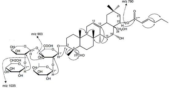
The methanol extract of tea seed pomace (Camellia oleifera Abel) was dissolved in water and was separated by a nanofiltration membrane; it was then successively subjected to purification through a macroporous resin column, a silica gel column, and repeated high pressure liquid chromatography (HPLC) to yield the new compound.
The new compound was visualized by spraying with 1% (w/v) Ce(SO4)2 in 10% (v/v) aqueous H2SO4, followed by heating at 120 °C, and it displayed purplish black spots on a thin-layer chromatography (TLC) plate, suggesting that it possessed the basic triterpenoid skeleton [15]. The IR spectrum showed absorption bands at 3386, 1717, 1655, 1077, and 1047 cm−1 due to hydroxyl; α, β-unsaturated ester; carboxy; and ether functions. The molecular formula, C59H92O26, was determined from the [M−H] ion at m/z 1215.57975 by high-resolution negative-ion electrospray ionization mass spectroscopy (ESI-MS/MS). The MS/MS fragmentation patterns (Figure 1) of the parent ion at m/z 1215.57975 confirmed the successive loss of a hexose (m/z 1035.51441 [M−H−C6H11O6]), a pentose (m/z 903.47240 [M-H-C11H19O10]), and a hexene (m/z 790.97736 [M−H−C17H28O12]) unit. The 1H-(methanol-d4) and 13C-nuclear magnetic resonance (NMR) spectra (Table 1), which were assigned following various NMR experiments, including distortionless enhancement of polarization transfer (DEPT)-90, DEPT-135, 2D homonuclear correlation spectra (H-HCOSY), heteronuclear single quantum coherence (HSQC), and heteronuclear multiple bond correlation (HMBC) spectroscopic examination, showed signals assignable to an A1-barrigenol moiety. This moiety included six methyls [δ 0.95, 0.96, 1.07, 1.08, 1.21, 1.54 (all s, H3-29, 25, 26, 30, 24, 27)], a methylene [δ 3.72 (br s, H2-28)], and two methines bearing an oxygen function [δ 3.92 (m, H-3), 5.45 (dd, J = 6.0, 3.0 Hz, H-22)], an olefin [δ 5.39 (br s, H-12)], and four glycopyranosyl moieties—β-D-glucopyranosiduronic acid [δ 4.41 (d, J = 6.0 Hz, H-1′)], β-D-galactopyranosyl [δ 5.05 (d, J = 6.0 Hz, H-1′′)], α-L-arabinopyranosyl [δ 4.54 (d, J = 6.0 Hz, H-1′′′)], and β-D-glucopyranosyl [δ 4.54 (d, J = 6.0 Hz, H-1′′′′)], together with a cis-2-hexenoyl group [δ 0.99 (t, J = 8.0 Hz, H3-6′′′′′), 1.51 (m, H2-5′′′′′), 2.66 (m, H2-4′′′′′), 6.29 (dt, J = 12.0, 4.0 Hz, H-3′′′′′), 5.85 (dt, J = 12.0, 1.6 Hz, H-2′′′′′)]. The data were very similar to those of sasanquasaponin I [13] and camelliasaponin B1 [16].In addition, the signal of C-4 was markedly shifted downfield (ΔδC = 17.2 ppm) and that of C-5 was shifted upfield (ΔδC = −6.4 ppm), and HMBC correlations from –CHO (δppm 9.48) to C-4 showed that the 23-methyl was substituted by a –CHO moiety. Furthermore, the position of the acyl group and the structure of the oligoglycoside moiety were confirmed on the basis of HMBC correlations. Long-range correlations were observed between the following proton and carbon pairs: H-22 and C-1′′′′′, H-1′ and C-3, H-1′′ and C-2′, H-1′′′ and C-3′, and H-1′′′′ and C-2′′′. On the basis of the above mentioned evidence, the chemical structure of oleiferasaponin A1 was determined to be 22-O-cis-2-hexenoyl-A1-barrigenol 3-O-[β-D-galactopyranosyl (1→2)][β-D-glucopyranosyl (1→2)-α-L-arabinopyranosyl (1→3)]-β-D-gluco-pyranosiduronic acid.
Figure 1. 2D NMR correlations and MS/MS fragmentation of oleiferasaponin A1.
Molecules 17 11721 g001
Table 1. 1H- (400MHz) and 13C-NMR (100MHz) data of oleiferasaponin A1 (in methanol-d4; δ in ppm, J in Hz).
No.δCδHNo.δCδH
139.82.55 (m)3-O-GlcA
226.11.53 (m)GlcA-1′105.34.41(d, 6.0)
386.63.92 (m)GlcA-2′78.73.8(m)
456.8 GlcA-3′84.33.71(m)
549.11.37 (m)GlcA-4′71.13.86(m)
621.70.96 (m)GlcA-5′77.43.57(m)
733.71.29 (m)GlcA-6′172.6
842.6 2′-O-Gal
948.41.11 (m)Gal-1′′103.25.05(d, 6.0)
1037.5 Gal-2′′74.03.76(m)
1125.11.99 (m)Gal-3′′75.33.76(m)
12124.85.39 (br s)Gal-4′′71.43.55(m)
13144.5 Gal-5′′76.73.68(m)
1441.7 Gal-6′′62.93.82(m)
1535.61.65(m)3′-O-Ara
16714.11(br s)Ara-1′′′102.14.54(d, 6.0)
1745.8 Ara-2′′′83.73.89(m)
1841.82.56 (m)Ara-3′′′71.53.69(m)
1942.72.26 (m)Ara-4′′′67.84(dd, 12.0, 6.0)
2032.5 Ara-5′′′64.93.26(m)
2143.02.27 (m)2′′′-O-Glc
2273.95.45(dd, 6.0, 3.0)Glc-1′′′′108.14.54(d, 6.0)
23211.29.48 (br s)Glc-2′′′′75.43.53(m)
2411.31.21(s)Glc-3′′′′76.53.32(m)
2517.70.96 (s)Glc-4′′′′71.63.64(m)
2616.91.07 (s)Glc-5′′′′78.43.69(m)
2728.21.54 (s)Glc-6′′′′63.13.77(m)
2863.13.72 (br s)22-O-(cis-2-Hexenoyl)
2934.10.95 (s)1′′′′′168.8
3025.71.08 (s)2′′′′′121.95.85(dt, 12.0, 1.6)
3′′′′′151.46.29(dt, 12.0, 4.0)
4′′′′′32.42.66(m)
5′′′′′23.81.51(m)
6′′′′′14.40.99(t, 8.0)
Oleiferasaponin A1 was tested for its protective effect on PC12 cells injured by H2O2. The cell viabilities of PC12 cells injured upon treatment with H2O2 at 5, 25, and 125 μM are shown below (Figure 2). These results show that oleiferasaponin A1 has potential cytoprotective activity against H2O2-induced damage.
Figure 2. Cell protective effects of oleiferasaponin A1 on H2O2-induced cytotoxicity in PC12 cells (n = 8). A, 5 mM H2O2; B, 5 mM H2O2 + 5 μM oleiferasaponin A1; C, 5 mM H2O2 + 25 μM oleiferasaponin A1; D, 5 mM H2O2 + 125 μM oleiferasaponin A1; The values are expressed as mean ± SD. ** p < 0.01 with respect to the H2O2 group.
Molecules 17 11721 g002

3. Experimental

3.1. General

The following spectrometric instruments were used to obtain physical data: IR spectra were obtained on a Nicolet FTIR-8700 spectrometer (Thermo Scientific Instrument Co., Boston, USA); mass spectrometry was performed using a Thermo Scientific LTQ Orbitrap XL instrument (Thermo Electron, Bremen, Germany) equipped with an ESI source operated in the negative-ion modes; 1H-NMR and 13C-NMR spectra were obtained on a AVANCE AV 400 (400/100 MHz) spectrometer (Bruker, Fallanden, Switzerland). with tetramethylsilane as an internal standard. The following materials and equipment were used for membrane separation and chromatography: 3000-Da nanofiltration membrane, (SJM, Hefei, China), AB-8 macroporous resin column (Bonc, Cangzhou, China), ordinary-phase silica gel column (200–300 mesh; Anhui Liangchen Silicon Material Co. Ltd., Huoshan, China) and a Varian Prostar HPLC instrument (Model 325) (Varian, Mulgrave, Australia).

3.2. Plant Material

Tea seed pomace (Camellia oleifera) was collected in October 2010 from a factory in Shucheng, Anhui, China.

3.3. Extraction and Isolation

The tea seed pomace (20 kg) was cut and extracted three times with methanol under reflux for 3 h. Evaporation of the solvent under reduced pressure provided a brown syrup (2.2 kg). The methanol extract (1.5 kg) was dissolved in water and purified using a nanofiltration membrane. The concentrated solution (1.1 kg) was subjected to AB-8 macroporous resin column chromatography with stepwise gradients of water and ethanol (100:0, 70:30, 30:70, and 0:100, v/v) to afford four subfractions. The third subfraction (590 g) was further subjected to ordinary-phase silica gel column chromatography [CHCl2:CH3OH:H2O (80:60:5, v/v)] to yield six fractions. A part of fraction 5 (900 mg) was purified by HPLC [MeOH:H2O (30:70)] to furnish a saponin mixture (86 mg). The mixture was further purified by HPLC [acetonitrile in 0.2% AcOH:H2O (41:59)] to give oleiferasaponin A1 (23 mg).

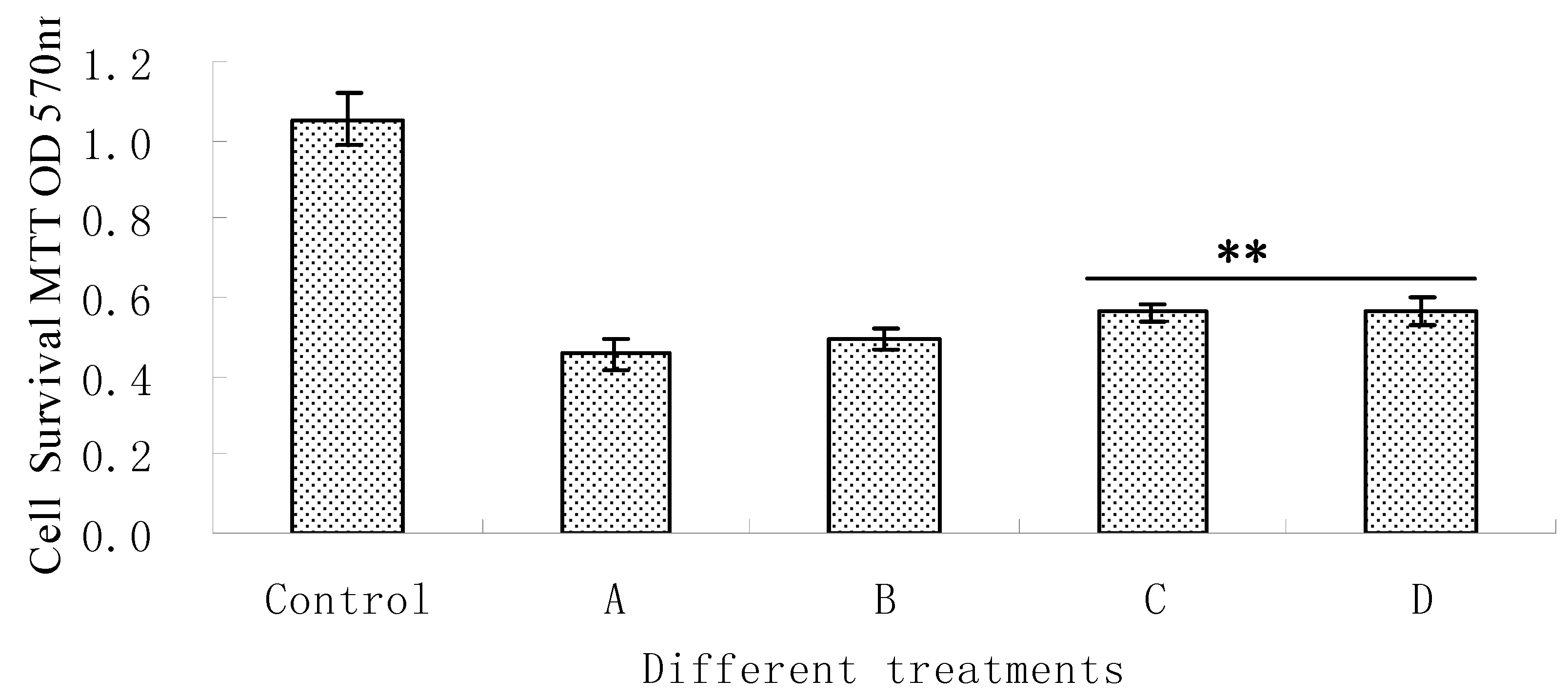
3.4. Measurement of Cell Viability

PC12 cells were cultured as previously described [17]. Cells were added into the wells of a 96-well culture plate at a density of 105 cells/mL for incubation before the cell viability experiments. The cells were permitted to adhere to plates for 16 h after seeding. Then, different volumes of the fresh compound stock were added to the plates to achieve final concentrations of 5, 25, and 125 μM. After 2 h incubation with the compound, 5 mM H2O2 was added into the plates. The cells were subjected to stress for 48 h before the experimental analyses.
The antioxidant properties of oleiferasaponin A1 on cell viability were assessed by the 3-(4,5-dimethylthiazol-2-yl)-2,5-diphenyltetrazolium bromide (MTT) assay as previously described [18]. In brief, the MTT assay was performed by the addition of MTT solution (5 μg/mL) to each well, and 2 h later, the culture was dissolved in DMSO. The absorbance of MTT was measured using a microplate absorbance reader at 570 nm. The data are presented as the percentage versus the blank control, which represents 100% cell viability.

3.5. Statistical Analysis

The mean value and standard deviation in this experiment were calculated by Excel 2007 (Microcal Software Inc., Northampton, MA, USA). Data were subject to statistics analysis by using the software package SPSS Statistics 17.0 for Windows (release 17.0.1; SPSS Inc., Chicago, IL, USA, 2008). ANOVA was carried out to determine significant difference (** p < 0.01).

4. Conclusions

The new compound, 22-O-cis-2-hexenoyl-A1-barrigenol 3-O-[β-D-galactopyranosyl(1→2)][β-D-glucopyranosyl(1→2)-α-L-arabinopyranosyl (1→3)]-β-D-glucopyranosiduronic acid, was isolated from tea seed pomace (Camellia oleifera Abel) and identified as a triterpenoid saponin on the basis of spectral analysis and chemical evidence. Oleiferasaponin A1 is a compound that can potentially prevent the H2O2-induced death of PC12 cells.

Acknowledgments

This research was supported by a project from the National Science & Technology Pillar Program during the “eleventh 5-year” plan period of China (grant NO. 2009BADB1B10). We are grateful to Zhi-Zhong Ma from Basic Medical School, Peking University Health Science Center for his generous support.

Conflicts of Interest

The authors declare no conflict of interest.
  • Samples Availability: Not available.

References

  1. Chen, Y.F.; Yang, C.H.; Chang, M.S.; Ciou, Y.P.; Huang, Y.C. Foam properties and detergent abilities of the saponins from Camellia oleifera. Int. J. Mol. Sci. 2010, 11, 4417–4425. [Google Scholar] [CrossRef]
  2. Chaicharoenpong, C.; Petsom, A. Quantitative thin layer chromatographic analysis of the saponins in tea seed meal. Phytochem. Anal. 2009, 20, 253–255. [Google Scholar] [CrossRef]
  3. Huang, Q.; He, M.; Chen, H.; Shao, L.; Liu, D.; Luo, Y.; Dai, Y. Protective effects of sasanquasaponin on injury of endothelial cells induced by anoxia and reoxygenation in vitro. Basic Clin. Pharmacol. 2007, 101, 301–308. [Google Scholar] [CrossRef]
  4. Huang, Q.; Shao, L.; He, M.; Chen, H.; Liu, D.; Luo, Y.; Dai, Y. Inhibitory effects of sasanquasaponin on over-expression of ICAM-1 and on enhancement of capillary permeability induced by burns in rats. Burns 2005, 31, 637–642. [Google Scholar] [CrossRef]
  5. Morikawa, T.; Li, N.; Nagatomo, A.; Matsuda, H.; Li, X.; Yoshikawa, M. Triterpene saponins with gastroprotective effects from tea seed (the seeds of Camellia sinensis). J. Nat. Prod. 2006, 69, 185–190. [Google Scholar] [CrossRef]
  6. Yoshikawa, M.; Morikawa, T.; Nakamura, S.; Li, N.; Li, X.; Matsuda, H. Bioactive saponins and glycosides. XXV. Acylated oleanane-type triterpene saponins from the seeds of tea plant (Camellia sinensis). Chem. Pharm. Bull. 2007, 55, 57–63. [Google Scholar]
  7. Morikawa, T.; Matsuda, H.; Li, N.; Li, X.; Yoshikawa, M. Bioactive Saponins and glycosides- Part 29- Acylated oleanane-type triterpene saponins: Theasaponins A(6), A(7), and B-5, from the seeds of Camellia sinensis. Helv. Chim. Acta 2007, 90, 2342–2348. [Google Scholar] [CrossRef]
  8. Kitagawa, I.; Hori, K.; Motozawa, T.; Murakami, T.; Yoshikawa, M. Structures of new acylated oleanene-type triterpene oligoglycosides, theasaponins E1 and E2, from the seeds of tea plant, Camellia sinensis (L.) O. Kuntze. Chem. Pharm. Bull. 1998, 46, 1901–1906. [Google Scholar]
  9. Yoshikawa, M.; Morikawa, T.; Li, N.; Nagatomo, A.; Li, X.; Matsuda, H. Bioactive saponins and glycosides. XXIII. Triterpene saponins with gastroprotective effect from the seeds of Camellia sinensis - Theasaponins E-3, E-4, E-5, E-6, and E-7. Chem. Pharm. Bull. 2005, 53, 1559–1564. [Google Scholar]
  10. Attele, A.S.; Wu, J.A.; Yuan, C.S. Ginseng pharmacology: Multiple constituents and multiple actions. Biochem. Pharmacol. 1999, 58, 1685–1693. [Google Scholar] [CrossRef]
  11. Kuo, P.C.; Lin, T.C.; Yang, C.W.; Lin, C.L.; Chen, G.F.; Huang, J.W. Bioactive saponin from tea seed pomace with inhibitory effects against Rhizoctonia solani. J. Agric. Food. Chem. 2010, 58, 8618–8622. [Google Scholar] [CrossRef]
  12. Yoshikawa, M.; Morikawa, T.; Yamamoto, K.; Kato, Y.; Nagatomo, A.; Matsuda, H. Floratheasaponins A-C, acylated oleanane-type triterpene oligoglycosides with anti-hyperlipidemic activities from flowers of the tea plant (Camellia sinensis). J. Nat. Prod. 2005, 68, 1360–1365. [Google Scholar] [CrossRef]
  13. Matsuda, H.; Nakamura, S.; Fujimoto, K.; Moriuchi, R.; Kimura, Y.; Ikoma, N.; Hata, Y.; Muraoka, O.; Yoshikawa, M. Medicinal flowers. XXXI. Acylated oleanane-type triterpene saponins, sasanquasaponins I-V, with antiallergic activity from the flower buds of Camellia sasanqua. Chem. Pharm. Bull. 2010, 58, 1617–1621. [Google Scholar]
  14. Liao, Z.; Yin, D.; Wang, W.; Zeng, G.; Liu, D.; Chen, H.; Huang, Q.; He, M. Cardioprotective effect of sasanquasaponin preconditioning via bradykinin-NO pathway in isolated rat heart. Phytother. Res. 2009, 23, 1146–1153. [Google Scholar] [CrossRef]
  15. Bhandari, S.; Babu, U.; Garg, H. A triterpene glycoside from Scrophularia koelzii. Phytochemistry 1997, 45, 1717–1719. [Google Scholar] [CrossRef]
  16. Yoshikawa, M.; Murakami, T.; Yoshizumi, S.; Murakami, N.; Yamahara, J.; Matsuda, H. Bioactive saponins and glycosides. V. Acylated polyhydroxyolean-12-ene triterpene oligoglycosides, camelliasaponins A1, A2, B1, B2, C1, and C2, from the seeds of Camellia japonica L.: structures and inhibitory activity on alcohol absorption. Chem. Pharm. Bull. 1996, 44, 1899–1907. [Google Scholar]
  17. Zhang, L.; Li, N.; Ma, Z.Z.; Tu, P.F. Comparison of the chemical constituents of aged Pu-erh Tea, ripened Pu-erh Tea, and other teas using HPLC-DAD-ESI-MS(n). J. Agric. Food. Chem. 2011, 59, 8754–8760. [Google Scholar] [CrossRef]
  18. Yang, C.S.; Tzou, B.C.; Liu, Y.P.; Tsai, M.J.; Shyue, S.K.; Tzeng, S.F. Inhibition of cadmium-induced oxidative injury in rat primary astrocytes by the addition of antioxidants and the reduction of intracellular calcium. J. Cell. Biochem. 2008, 103, 825–834. [Google Scholar] [CrossRef]

