Review

Like a Bolt from the Blue: Phthalocyanines in Biomedical Optics

by
Nawal Sekkat
1,
Hubert van den Bergh
2,
Tebello Nyokong
3 and
Norbert Lange
1,*
1
School of Pharmaceutical Sciences, University of Lausanne/Geneva, Geneva, 30, quai Ernest Ansermet, Geneva CH-1211, Switzerland
2
Laboratory of Photomedicine, Swiss Federal Institute of Technology (EPFL), Lausanne CH-1015, Switzerland
3
Department of Chemistry, Rhodes University, Grahamstown 6140, South Africa
*
Author to whom correspondence should be addressed.
Molecules 2012, 17(1), 98-144; https://doi.org/10.3390/molecules17010098
Submission received: 4 November 2011 / Revised: 5 December 2011 / Accepted: 14 December 2011 / Published: 23 December 2011

Abstract

:
The purpose of this review is to compile preclinical and clinical results on phthalocyanines (Pcs) as photosensitizers (PS) for Photodynamic Therapy (PDT) and contrast agents for fluorescence imaging. Indeed, Pcs are excellent candidates in these fields due to their strong absorbance in the NIR region and high chemical and photo-stability. In particular, this is mostly relevant for their in vivo activation in deeper tissular regions. However, most Pcs present two major limitations, i.e., a strong tendency to aggregate and a low water-solubility. In order to overcome these issues, both chemical tuning and pharmaceutical formulation combined with tumor targeting strategies were applied. These aspects will be developed in this review for the most extensively studied Pcs during the last 25 years, i.e., aluminium-, zinc- and silicon-based Pcs.

Graphical Abstract

1. Introduction

The first syntheses of metal-free and copper phthalocyanines were reported in 1907 by Braun and Tcherniac at the South Metropolitan Gas Company (United Kingdom) and in 1927 by Diesbach and von der Weid at the University of Fribourg (Switzerland). A few years later, Professor Linstead in collaboration with Imperial Chemical Industries (ICI) was the first to characterize the chemical structure of the phthalocyanine molecule, using for the first time the term “phthalocyanine” [1,2,3].
Nowadays, phthalocyanines are widely used in the dying industry. Nearly a quarter of all pigments of organic origin are related to this class of compounds. Furthermore, they are used for the fabrication of high-speed and high resolution optical media [4], as light harvesters in photovoltaic applications [5], and as experimental catalysts in redox reactions [6]. These dyes absorb strongly in the red and near infrared (NIR) part of the visible spectrum providing them with their characteristic blue or greenish color. Pcs that absorb in the NIR are especially interesting for photomedical applications such as fluorescence imaging, Photochemical Internalisation (PCI), and Photodynamic Therapy (PDT) [7,8,9,10,11,12].
Just recently Photochemical Internalisation (PCI), a novel drug delivery process, has shed light on the importance of phthalocyanines and their applications in oncology [13,14,15]. The PCI technology is based on the concomitant administration of a therapeutic agent and a photosensitizer. When internalized by endocytosis and consequently colocalized in the endosomes and/or lysosomes, light activation of the PS will subsequently lead to vesicle disruption and release of the therapeutic agent. Indeed, PCI technology enables the release of endocytosed drugs prior to lysosomal degradation, thus, increasing their therapeutic efficacy within the target cells. Among the photosensitizers used in PCI, the amphiphilic disulfonated aluminium phthalocyanine AlPcS2adj displays all the required features and characteristics such as specific insertion of the hydrophobic part of the PS into the endocytotic membrane. Due to its amphiphilic character, AlPcS2adj “intrudes into the plasma cell membrane, but is unable to penetrate through the plasma membrane and will enter the cells via adsorptive endocytosis” [16]. The concomitant administration of a drug such as gelonin or bleomycin [14,17,18] will, after internalization lead to vesicle disruption and intracellular release of the drug upon light activation [14,19,20].
As mentioned previously, phthalocyanines strongly absorb in the NIR, and have been proposed for PDT of cancer as early as 1985 [21,22]. Under some circumstances PDT treatment presents several advantages over conventional cancer therapies such as chemo, radiation and surgical treatments. It enables selective destruction of malignant tissues due to specific interaction of three individually non-toxic components i.e., a photosensitizer (PS), light and oxygen. PS selective accumulation in tumor combined with its controlled light activation enables selective destruction of tumors, sparing neighboring healthy tissue.
Although today the selectivity of PS is not fully understood, it is thought to be a multifactorial process including physico-chemical properties and binding to plasma proteins as well as the particular characteristics of tumors such as leaky vasculature, low lymphatic drainage, expression of specific enzymes and receptors and pH variation. Depending on its cellular and intracellular localization/relocalization, PS exhibits direct and indirect cell killing, vascular occlusion, release of cytokines and the response of the immune system [23,24].
Photofrin® (Table 1) was the first photosensitizer approved for clinical use in 1993 for the treatment of bladder cancer. Since then it gained marketing for the prophylactic treatment of several cancers such as the treatment of early-stage oesophagal, gastric, cervical and lung cancers [24,25]. However, this first generation photosensitizer has several limitations with respect to its clinical use since it [23,25,26,27,28]:
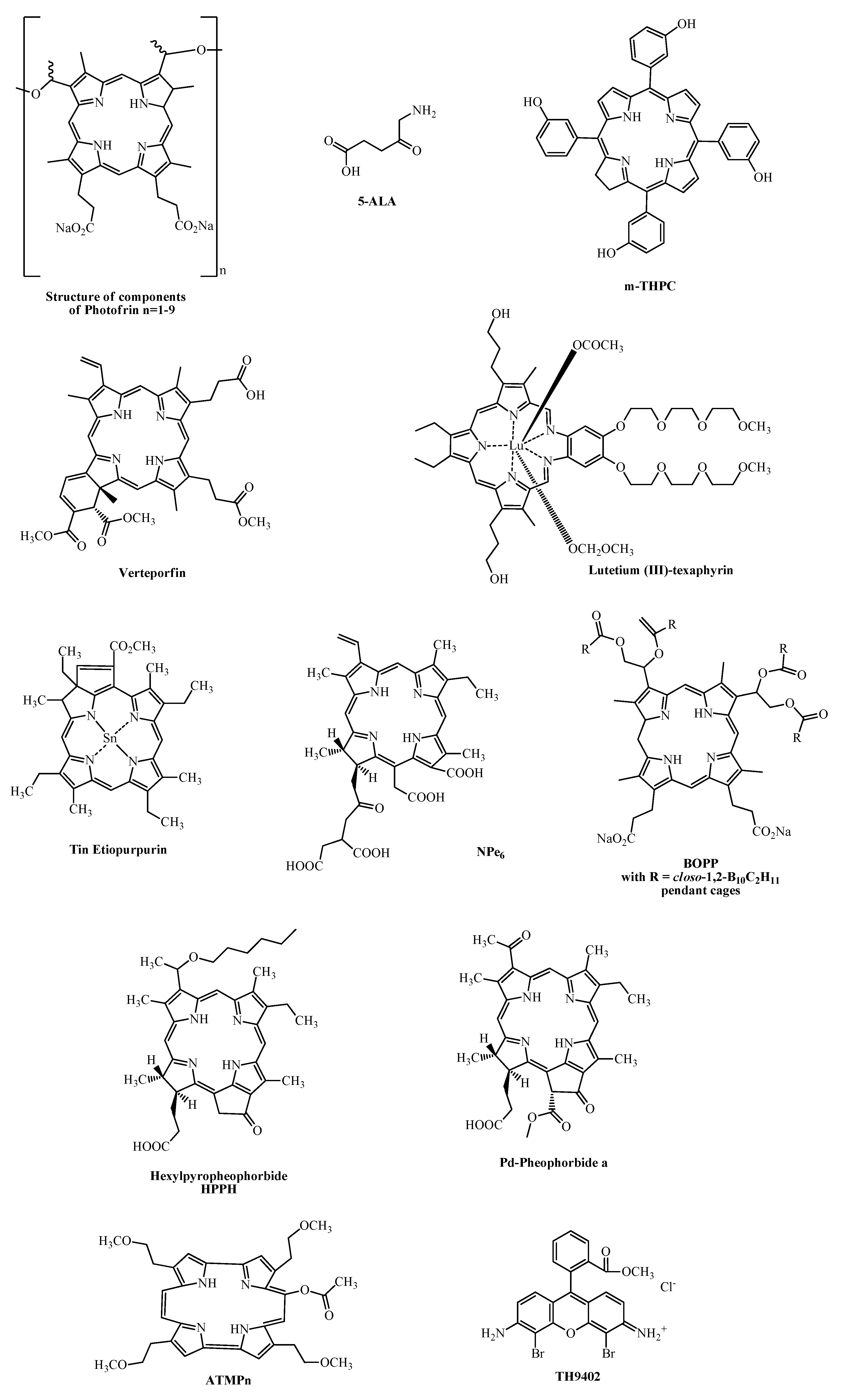
  • • Is composed of a undefined mixture of hematoporphyrin derivatives (HpD);
  • • Induces a long-lasting skin photosensitization (2 to 3 months post injection);
  • • Has a low extinction coefficient at wavelengths for optimal tissue penetration;
  • • Displays a limited selectivity for the target tissue.
Therefore, considerable efforts have been undertaken to prepare 2nd generation PS with more suitable features [29,30,31] such as:
  • • Single and chemically pure compound;
  • • Stability and good solubility in pharmaceutically acceptable formulations and in biological media;
  • • Low tendency to aggregate;
  • • High singlet oxygen quantum yield;
  • • Photostability;
  • • Fluorescence;
  • • Low phototoxicity towards healthy tissue;
  • • No dark toxicity;
  • • Fast clearance from the healthy parts of the body and specific retention in diseased tissues;
  • • Strong absorbance in NIR region and minimal absorbance between 400 and 600 nm.
In order to avoid skin photosensitization, the PS has to present the lowest absorbance in the spectral range where daylight intensity is the highest, i.e., between 400 and 600 nm. Moreover, strong absorbance in the NIR region between 600 and 900 nm favors the optimal penetration of light into tissues, thus resulting in more efficient PDT effects when treating deeper lying lesions. However, absorption by water molecules increases for wavelengths above 800 nm and at higher wavelength energy transfer to molecular oxygen is suboptimal. Hence, the window for optimal light penetration ranges from 600 to 800 nm. These considerations are based on the assumption that healthy and diseased tissues have the same absorbance of light, which in reality is not the case. Finally, 94 kJ/mol appears to be the minimal energy required by a photon to induce singlet oxygen 1O2 formation. This energy corresponds to a wavelength of approximately 1,270 nm [32,33,34,35,36,37,38,39].
In Table 1 some 1st and 2nd generation PS investigated in clinical trials and their potential therapeutic application in oncology are listed (see also Figure 1 for their chemical structures).
Table 1. Examples for PS used in clinical and preclinical trials in oncology [24,25,40,41,42,43,44,45,46,47,48,49,50,51,52].
Photosensitizers Trade NameAbsorption WavelengthPotential Indications
HpD, Porfimer sodiumPhotofrin, Photogem, Photosan, Hemporfin630 nm Cervical, brain, oesophageal, breast, head and neck, lung, bladder, superficial gastric cancers, Bowen's disease, cutaneous Kaposi's sarcoma
m-THPC, TemoporfinFoscan652 nm Oesophageal, prostate and pancreatic cancer, advanced head and neck tumors
VerteporfinVisudyne689 nmBasal and squamous cell carcinomas
HPPH, 2-(1-hexyloxyethyl)-2-devinyl pyropheophorbide-alphaPhotochlor665 nmBasal cell carcinoma, Oesophageal cancers, Head and Neck tumors
Palladium-bacteria-pheophorbideTookad763 nmProstate cancer
5-ALA,5-aminolevulinic acidLevulan630 nm Skin tumors, head and neck, gynaecological tumors and basal cell carcinomas
375-400 nm Brain, head and neck and bladder cancer photodetection
5-ALA-methylesterMetvix635 nmBasal cell carcinoma
5-ALA benzylesterBenzvix635 nmGastrointestinal tumors
5-ALA hexylesterHexvix375-400 nm Photodectection of bladder cancer
Lutetium (III)-texaphyrin or Motexafin-lutetiumLutex, Lutrin, Antrin, Optrin732 nmProstate, cervical, breast, brain cancer, melanoma
SnET2, Tin (IV) ethyl etiopurpurinPurlytin, Photrex659 nmKaposi's sarcoma, cutaneous metastatic adenocarcinomas, prostate, brain, lung cancers, basal cell carcinomas
NPe6, mono-L-aspartyl chlorin e6, talaporfin sodiumTalaporfin, Laserphyrin664 nmSolid tumors, lung cancer, cutaneous malignancies
BOPP, boronated protoporphyrinBOPP630 nm Malignant gliomas
Zinc phthalocyanineCGP55847670 nmSquamous cell carcinoma of upper aerodigestive tract
Silicon phthalocyaninePc 4 675 nmCutaneous and subcutaneous lesions from diverse solid tumor origins
Mixture of sulfonated aluminium phthalocyanine derivativesPhotosens 675 nmSkin, breast, lung, oropharingeal, breast, larynx, head and neck cancers, Sarcoma M1, epibulbal and choroidal tumors, eyes and eyelids tumors, cervical cancer
ATMPn, Acetoxy-tetrakis(β-methoxyethyl)-porphycene NA600-750 nmSkin cancer
TH9402, dibromorhodamine methyl ester NA515 nmBreast, myeloma, non-melanoma skin cancer
Figure 1. Chemical structures of clinically relevant “non-phthalocyanine” photosensitizers.
Molecules 17 00098 g001
Another appealing feature of Pcs is the ease of the introduction of peripherial and axial substituents that can be used to fine tune absorption and emissions characteristics. Phthalocyanine dyes with absorptions bands as high as 1,000 nm with suitable fluorescence quantum yields can be prepared [11]. Therefore, Pcs are potentially interesting as fluorescence reporters for in vitro and in vivo imaging that can replace commonly used fluorophors such as fluorescein and indocyanine green. Two of such compounds, “La Jolla Blue®” and IRD700DX® are commercially available as labeling agents for proteins, peptides [53,54,55], antibodies [56,57,58,59,60] and oligonucleotides [61,62,63,64,65].
This review intends to compile preclinical and clinical data accrued with Pcs as PS for PDT. However, it has to be noted that the presented concepts can be easily translated into the use of similar compounds for the fluorescence diagnosis of disease. Thereby, we will mainly focus on the most extensively studied Pcs, i.e., Pcs containing Al, Zn, and Si as central metal ion. At first we will briefly describe how chemical modulation of Pcs, alters their Structure-Activity Relationship (SAR). Then, since most Pcs are barely soluble in pharmaceutically acceptable formulations special emphasis will be placed on the impact of pharmaceutical formulation on their therapeutic efficiency. And finally, different tumor targeting strategies that have been exploited with Pcs will be discussed.

2. Phthalocyanines

Pcs belong to the group of 2nd generation PS which exhibit a high extinction coefficient around 670 and 750 nm and even up to 1,000 nm. Variation of the axial and peripheral substituents modulates the tendency for aggregation, pharmacokinetics, biodistribution, solubility, as well as fine-tuning of NIR absorbance [66]. Extinction coefficients higher than 105 M−1cm−1 have been reported [11,66]. These compounds are porphyrin-like PS, displaying tetrapyrrolic, aromatic macrocycles with each cycle linked to the other by nitrogen atoms. Each pyrrolic ring is extended by a benzene ring resulting in the red-shift of their final absorption band [31,67,68]. Figure 2 shows the general chemical structure of Pcs and nomenclature as well as typical absorption and emission spectra.
Figure 2. (a) General chemical structure of metallated phthalocyanines; (b) Typical absorption and emission spectra of metallated Pc (e.g., zinc based phthalocyanine in DMSO).
Molecules 17 00098 g002
Lowering of the symmetry of the phthalocyanine molecules results in splitting (or broadening) of the Q bands. This splitting is due to the lifting of degeneracy of the lowest unoccupied molecular orbital (LUMO) to a varying extent. It is also well established that expansion of π conjugation in phthalocyanines shifts the Q band to the red. Extension of the conjugation system is accompanied by change in colour from blue/green to colors including brown, red or purple. The location of the Q band in Pc complexes can be adjusted by attaching suitable substituents onto the peripheral and non-peripheral positions of the ring and by the change in the nature, size and number of substituents. Addition of electron donating groups such as –NH2, OR and SR at the non-peripheral (1,4,8,11,15,18,22,25) or peripheral (2,3,9,10,16,17,23,24) positions of the Pc ring results in red shift to the NIR region. Substitution at the non-peripheral position shows more red-shift than at the peripheral position [69].
Besides their strong absorption in the NIR, Pcs exhibit low absorption at wavelengths between 400 and 600 nm leading potentially to a lower skin photosensitization when exposed to sunlight [31]. Moreover, the presence of a diamagnetic central metal such as Zn2+ and Al3+ in the Pc nucleus seems to improve the triplet state life time (τt), as well as its yield (Фt) and singlet oxygen yields (ФΔ) compared to paramagnetic metals (e.g., ФΔ ≥ 0.7, Фt > 0.4 and τt = 750 μs for AlPcS4 in solution with human serum albumin) [30,48,67,70]. However, the metallation is not required for its photodynamic activity. Indeed, Feofanov et al. [71] and Karmakova et al. [72] reported efficient antitumor activity of a mixture of metal-free sulfonated phthalocyanines with an average number of sulfonated groups of 2.4 in vitro on human epidermoid carcinoma cells HEp2 and in vivo on mice bearing murine P-388 lymphoma cells, respectively.
On the other hand, Pcs with good fluorescence quantum yields up to 0.6 have been reported. This, together with their high photostability compared to commonly used labeling agents should have placed this group of compounds into the upper part of the list of NIR fluorescence reporters. As an example IR700DX® is about 20 times more stable than Cy5.5 at similar irradiation intensities and wavelengths. However, despite these photophysical and spectral characteristics the use of Pcs as labeling agents for fluorescence imaging or in PDT is still limited.

2.1. Aluminium Based Phthalocyanines

2.1.1. Aluminium Sulfonated Phthalocyanines—SAR

Sulfonated phthalocyanines bearing a central aluminium ion have been extensively studied in vitro as well as in vivo. They display potent photodynamic activity on cell lines including G361 human melanoma cells [73], pancreatic carcinoma cells (H2T) [74], and human fibrosacroma cells (HT-1080) [75], as well as in vivo on rat (CBH rats) bearing fibrosarcoma (HSN/TC/7) [76]. These early studies indicated a strong dependence of the photodynamic efficiency on the degree of sulfonation, later systematically assessed by Chan et al. [77,78,79].
At this point one has to keep in mind that earlier reports on aluminium sulfonated phthalocyanines can be sometimes misleading due to the designation as AlPcS4 or as AlPcS of the commercially available product from Ciba-Geigy, which nevertheless consisted of a mixture of mono, di-, tri- and tetrasulfonated chloroaluminium phthalocyanines with an average degree of three sulfonated substituents. Thus, in this review, we will refer to this mixture as AlPcS, whereas AlPcS4 will be used to qualify the pure tetrasulfonated Pc, keeping in mind that even these more defined compounds will be mixtures of regio- and stereoisomers. Furthermore, some early works did not disclose the identity of the axial ligands which could be chloride (Cl) as well as hydroxy (OH) groups. Since most reported studies focus on the effect of sulfonation with respect to PDT, no further distinction according to their axial ligand will be made here. Finally, the IC50 values reported in this review cannot be compared from a study to another since this value depends among other things on the applied light doses.
Interestingly, AlPcS4 has shown promising in vivo results in the treatment of malignant gliomas [80,81]. Wistar rats inoculated intracranially with C6 glioma cells, responded within 5 days to intravenous injection of AlPcS4 in saline solution at a dose of 5 mg/kg and irradiation 6 h post administration at a dose of 100 J/cm2. However, upon illumination at a light dose of 200 J/cm2, AlPcS4-mediated PDT neurological symptoms (e.g., brain damage and oedema) appeared, while light alone had no effect. Higher light doses lead to death in mice bearing intracerebrally implanted VMDk murine glioma cells as reported by Sandeman et al., presumably concomitant with hyperthermic effects in the irradiated areas [82].

2.1.2. Influence of the Degree of Sulfonation

In 1990, Chan et al. reported that in vitro monosulfonated chloroaluminium phthalocyanine (AlPcS1) was taken up faster, retained to a higher degree and showed higher phototoxicity in murine colorectal carcinoma (Colo-26) cells than higher sulfonated analogues (i.e., di, tri and tetrasulfonated chloroaluminum phthalocyanine; referred to as AlPcS2, AlPcS3 and AlPcS4 respectively) [77]. Furthermore, comparison of light doses necessary for efficient PDT on WiDr cells cultured in monolayers and as spheroids revealed that spheroids were less sensitive to PDT than cells grown in monolayers [78].
In contrast to in vitro experiments, an inverse tendency with respect to tumor accumulation and phototoxicity was observed when PDT was perfomed with this series of sulfonated Pcs in vivo. In BALB/c mice inocculated with Colo-26 tumors AlPcS1 had essentially no effect on tumor regression in contrast to the higher sulfonated compounds following the order AlPcS4 > AlPcS3 > AlPcS2 [77]. This discrepancy can be assigned to differences in pharmacokinetics and biodistribution of these compounds due to their differences in hydrophilicity [77,83]. As suggested by Chan et al., the fast clearance of highly sulfonated Pcs after intravenous administration can be circumvented by intraperitoneal application for these compounds [77].
Later, Chan et al. showed that AlPcS2 was more potent than AlPcS3 and AlPcS4, despite its lower accumulation in tumor xenografts. This suggests different PDT mechanisms of these compounds with respect to cell killing, vasculature occlusion, and intracellular localization [78]. Interestingly, the highest in vitro efficiency of AlPcS2 over other more hydrophilic aluminium sulfonated Pc was already demonstrated in 1988. The same tendency was later confirmed by Peng and Moan [83]. When injected intraperitoneally AlPcS2 was the most efficient PS followed by AlPcS4, Photofrin, and AlPcS1. The best efficiency was observed with a drug dose of 10 mg/kg and light exposure 2 h post-administration.
Moreover, the relative position of sulfonate group seems also to influence Pcs photoactivity. Indeed, AlPcS2 bearing adjacent sulfonated side-group (AlPcS2adj) rather than opposite side-substitution exhibited the best cell penetration and were the most phototoxic compounds amongst the sulfonated aluminium phthalocyanine serie, presumably due to their amphiphilic properties [84]. Therefore, the phototoxicity of mixed AlSPc in vivo should be related mostly to photoactivity of AlPcS2adj isomers [78,83]. In a recently reported study, Mathews et al. compared the phototoxic effect of AlPcS2adj to 5-aminolevulinic acid (5-ALA) on healthy brains in mice [85]. Based on PDT-induced and higher mortality rates, AlPcS2adj was considered to be a more potent photosensitizer than 5-ALA. Moreover, in a comparative study, Gupta et al. [86] reported that liposomal formulation of AlPcS2 resulted in a higher phototoxic effect as compared to its free form on human glioma cells (BMG-1) despite its lower uptake by the cells.
Despite the higher efficiency of AlPcS2 compared to other sulfonated aluminium phthalocyanines, extensive efforts and studies have been realized with the objective of further improving the pharmacokinetic properties and selectivity of the commercially available AlPcS4.
Allen et al. [26], tested sulfonamides with an alkyl chain of 4, 8, 12 and 16 carbons to one of the sulfonated groups in AlS4Pc (Figure 3) and compared induced photodynamic effects to AlPcS4 in mice bearing EMT-6 tumors. They concluded that at doses of 0.2 μmol/kg, all tested AlPcS4-derivatives induced tumor regression and were more effective than AlPcS2adj or AlPcS4. Moreover, with increasing alkyl chain length increased photodynamic efficacy was observed. In these experiments, AlPcS4 had no effect at doses up to 5 μmol/kg, and 1 μmol/kg of AlPcS2adj was needed to induce 87% of tumor cure.
Figure 3. A homologous series of AlPc alkylsulfonamides.
Molecules 17 00098 g003

2.1.3. Photosens®

Photosens®, a mixture of aluminium chloride phthalocyanines with 1 to 4 sulfonated side-groups with an average sulfonated degree of 3, was developed at the General Physics Institute of the Russian Academy of Sciences. It has been evaluated in vivo for the treatment of sarcoma M1 [87], epibulbal and choroid tumors [88], eye and eyelid related tumors [89], bladder and cervical cancer [90,91]. In a clinical trial that included 47 patients composed of 35 women presenting pre-cancerous lesions of cervix and 12 women with non-invasive cervical lesions, a Photosens® dose of 0.3 mg/kg was applied. Twenty four hours post administration, lesions were irradiated with a light dose between 150 and 200 J/cm2 at 675 nm. No PDT-related pain was reported. Out of these two subgroups, 94.2% and 83.4% of the patients, respectively, responded fully to the treatment with complete tumor regression, with significant regression for 2.9 and 8.3% of the women. Moreover, the same percentage of women showed stabilized disease status. The remaining percentages correspond to the patients who responded only partially to the treatment. Today, Photosens® is commercialized in Russia by NIOPIK.

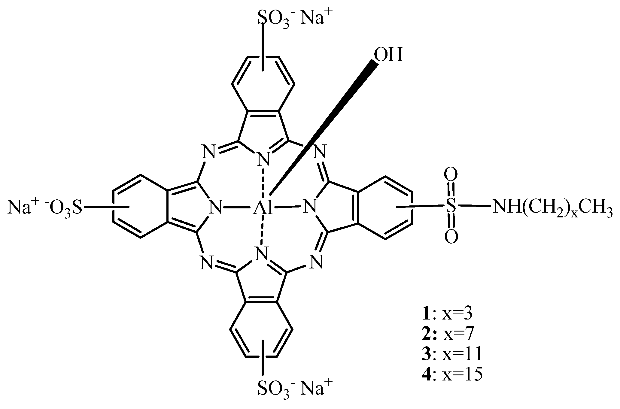
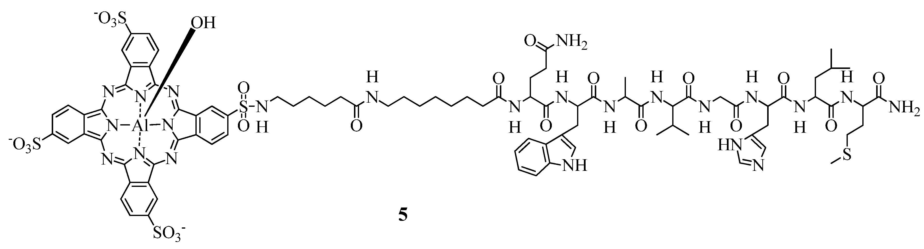
2.1.4. AlPcS-Conjugation to Tumor Targeting Moieties

Except for AlPcS1, aluminium sulfonated Pcs are readily soluble in aqueous media. Thus, most formulation efforts were aiming at increasing Pc accumulation and uptake by the tumors through the increase of the circulation half life or cellular/subcellular targeting.
The tetraglycine derivative Al(SO2Ngly)4 and a mono-substitued AlPcS4 containing one 6-aminohexanoic acid spacer (AlPcS4A1) have been exploited for protein conjugation. The conjugation of AlS4Pc to monoclonal antibodies (Mab) lead to an enhanced phototoxicity in different cell lines such as human colon carcinoma LoVo [92], modified human ovarian carcinoma cells SKOV3-CEA-1B9 (SKOV cells transfected with carcinoembryonic antigen (CEA) cDNA and expressing two target antigens CEA and ErbB2) [60], and various squamous carcinoma cells including UM-SCC-11B, UM-SCC-22A, UM-SCC-22B, A431, SCV-7 and OE [56,93].
Moreover, in vivo biodistribution studies on tumor bearing mice revealed that these conjugates presented an increased selectivity toward the malignant tissues compared to free AlPcS4. Indeed, when 12 identical Pcs were conjugated to a 35A7 Mab directed against CEA (35A7 Mab-(AlPcS4A1)12), a tumor-to-muscle ratio and tumor-to-skin ratio of 33 and 8, respectively, was reported in nude mice bearing a human colon carcinoma xenograft [92]. In a subsequent study, the photodyanmic efficiency was further improved using an internalizing antibody (FSP77) directed against ErbB2. In vitro, SKOV3-CEA-1B9 showed a fast uptake of FSP77Mab-(AlPcS4A1)6 and a higher phototoxic efficacy (i.e., 96% vs. 68% of growth inhibition) as compared to the corresponding 35A7 Mab (AlPcS4A1)8 [60]. These studies are in agreement with the results reported by Vrouenraets et al. [56,94] Pcs conjugated to internalizing Mabs, i.e., SCC-Mab 425 and Mab U36, were photodynamically more effective than the corresponding non-internalizing Mab E48. Furthermore, the Mab 425-AlPcS4 was 7,500 times more effective in vitro than free AlPcS4. It has to be noted also that the loading of PS per Mab can be tuned through different conjugation methods. Indeed, by introducing a five carbon spacer chain between the Mab and AlPcS4, Carcenac et al. [92] were able to link up to 16 AlPcS4A1 entities to the proteins whereas Vrouenraets et al. could only conjugate four Al(SO2Ngly)4 without affecting its solubility.
Later, Vrouenraets et al. tested three different Mabs [BIWA 4 (bivatuzumab), E48 and 425] on five different SCC cell lines (22A, 22B A431, SCV-7 and OE) [93]. PS conjugation to the aforementioned Mabs resulted in a higher phototoxicity compared to free AlPcS4. BIWA 4-AlPcS4 showed highest phototoxicity in all the five cell lines, exhibiting an IC50 as low as 0.06 nM as compared to IC50 ≥ 700nM for the free PS. Interestingly, the effect of the other Mabs varied depending on the cell line but not on the internalizing efficiency of the particular Mab. Mab E48-AlPcS4 conjugate was more active on 22A than on 431 cells, while Mab 425-AlPcS4 conjugate had the opposite effect. After determination of Pc’s cell binding and internalization, the authors suggested that the phototoxicity of these conjugates was more dependend on the overall cell binding than on their cellular internalization.
Overexpression of the low density lipoproteins (LDL) receptor has been reported for several tumors allowing the internalization of LDL proteins into the cell [95]. Therefore, Urizzi et al. [96] conjugated AlS4Pc to LDL either via covalent linking of a bisulfonated substituent of AlPcS4 through a 5-carbon spacer chain [(AlPcS4A2): aluminium di-(6-carboxypentylaminosulfonyl)-tetrasulfophthalocyanine) or non-covalent linking to the phospholipidic region of LDL (AlPcS4(C12)]. Loading ratios of 72:1 and 61:1 for AlPcS4A2-LDL and AlPcS4(C12)-LDL, respectively, have been reported to be the most stable formulations.
These compounds were tested in vitro on EMT-6 and A549 cells. After 2 h of incubation with the conjugates or with free AlPcS4, AlPcS4(C12)-LDL displayed a 10-fold higher phototoxicity effect than AlPcS4A2-LDL. This has been ascribed to the increased aggregation tendency, higher scavenging of ROS products, and alteration of the conjugate interaction with cellular receptors when Pc was covalently coupled to the protein.
Interestingly, free AlPcS4 and AlPcS4A2 were devoid of any photocytotoxic effects under the same conditions. A possible reason for this lack of photoxic activity could be the negative net charge of these Pcs, leading to a different cellular uptake and/or intracellular localization.
Further in vivo studies on mice bearing EMT-6 allografts were performed comparing free AlPcS4 to AlPcS4(C12) and AlPcS4(C12)-LDL conjugate. No difference in tumor response was reported for both conjugates. As compared to free AlPcS4, 25 times lower drug doses could be used to induce similar responses. Moreover, the post-treatment oedema regressed within 3–4 days post-PDT. The similar phototoxic effect of free and AlPcS4(C12)-LDL conjugate can presumably be attributed to binding of the free Pc to plasma proteins (e.g., LDL) and redistribution post injection.
Overexpression of gastrin-releasing peptide-receptors (GRPR) have been associated with many cancerous conditions such as ovarian, breast, prostate and lung cancer [97]. Since bombesin is the amphibian homologue of the human gastrin releasing peptide [98] a recent study reported an attempt to target human prostate tumor cells PC-3 by conjugating AlPcS4 to bombesin.
However, due to the conjugation of bombesin to the AlPcS4 (Figure 4), the binding affinity to GRPR was about 40 times lower for the conjugate as compared to bombesin, certainly resulting in a loss of specificity and a moderate phototoxicity [99]. Nevertheless, the photoactivity of the conjugate was 2.5 and 5 fold higher than AlPcS4A1 and the free tetrasulfonated aluminium phthalocyanine, respectively, in vitro.
Figure 4. Chemical structure of the bombesin-AlPcS4 conjugate 5 for the targeting of GRPR.
Molecules 17 00098 g004

2.1.5. AlPcS4-Formulation using Targeted Delivery Systems
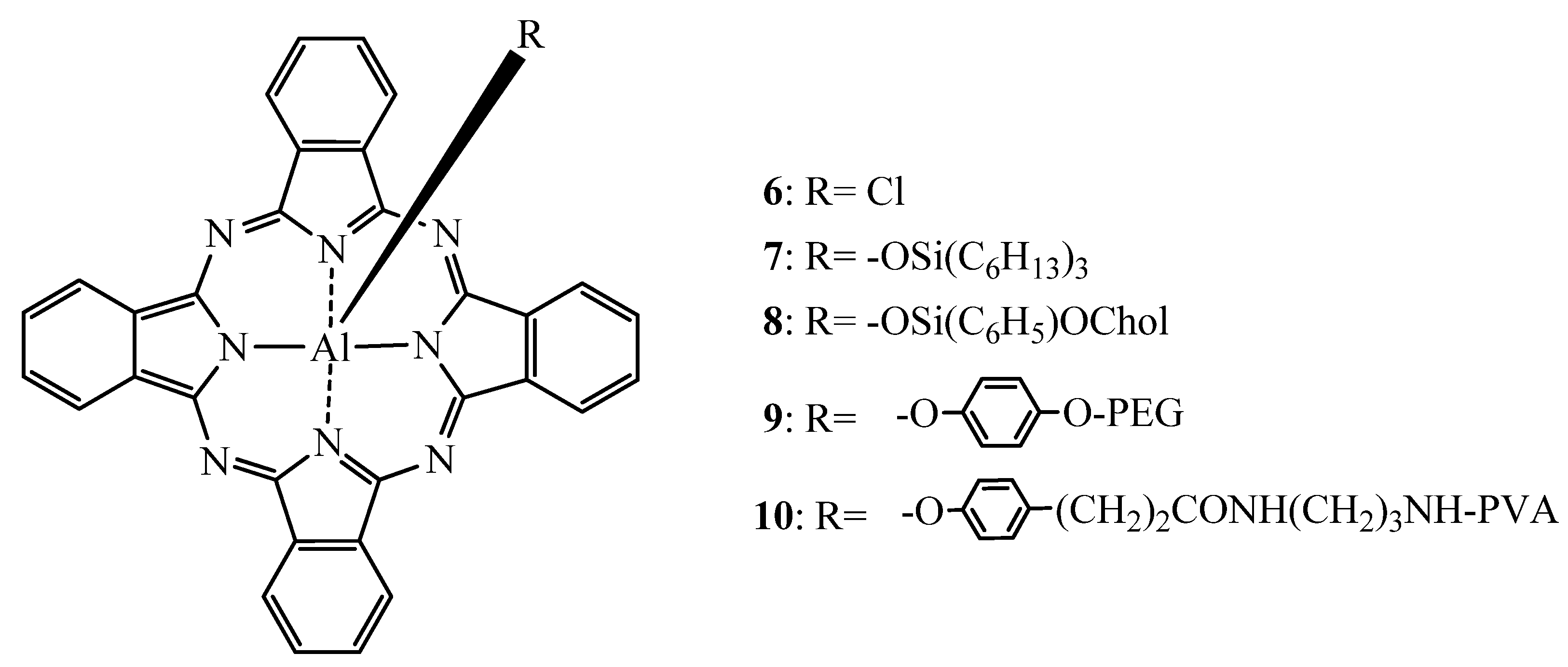
Simple liposomes are usually unstable upon systemic administration and rapidly cleared from the body. However, the incorporation of PEG-derivatized phospholipids in the bilayer of liposomal membranes generally prolongs their circulation time. This property of PEGylated liposomes coupled to additional tumor targeting moiety such as transferrin was exploited by Gijsens et al. in vitro [100] and in vivo [101]. In HeLa cells, expressing approximatively 2 × 10⁵ transferrin receptors per cell [102], transferrin conjugated PEG-Liposomes containing AlPcS4 (Tf-Lip-AlPcS4) were 10 times more efficient than free AlPcS4 (IC50 of 0.63 μM vs. 6.3 μM, respectively) while non-Tf conjugated AlPcS4 liposomes (Lip-AlPcS4) were less potent than free PS. Indicating that non-targeted liposomes are only poorly taken up and additionally, show only limited release of their content into the cells [100].
Initially, the same tendencies were confirmed when these formulations were tested on AY-27 cells [101]. However, in a rat bladder model using the same cell line no or only moderate accumulation of all liposomal formulations was observed. However, pretreatment with a chondroitinase breaking the epithelial glycocalyx resulted in a selective retention of Tf-Lip-AlPcS4. On the contrary, free AlPcS4 was detectable with or without the enzymatic pretreatment of the bladder but with a lower selectivity. The reported tumor-to-normal tissue ratio and tumor-to-submucosa/muscle ratio for Tf-Lip-AlPcS4 were 18:1 and 78:1, respectively.
Qualls et al. [103] encapsulated AlPcS4 into pH sensitive liposomes using 1,2-di-O-(Z-1′-hexadecenyl)-sn-glycero-3-phosphocholine (DPPlsC) an acid labile lipid. Moreover, folate-polyethyleneglycol 3350-distearoylphosphatidylethalonamine (Folate-PEG3350-DSPE) was incorporated for targeting of folate-receptors in order to promote the receptor-mediated Pc internalization. The liposomes were subsequently tested in vitro on folate receptor expressing human nasopharyngeal cancer cells. It was found that DPPIsC-folate liposomes were taken up faster and were photodynamically more active than the free AlPcS4 confirming earlier studies with similar liposomes on colorectal carcinoma cells C170 cells by Morgan et al. [104]. Moreover, it was shown in vitro that the targeting moiety rather than the pH sensitivity of the delivery system dominated the photocytotoxicity of the PS [103].

2.1.6. AlClPc—SAR

Modulation of the axial substituents is another way to modulate the pharmacological, pharmaceutical as well as optical properties of Pcs. As confimed by Chan et al., Ben Hur initially reported that a simple Pc carrying a chlorine as counterion (AlClPc, 6, Figure 5) was photodynamically more active than its sulfonated counterparts [79,105]. In order to further improve these compounds systematic studies with different alterations of the axial substituents have been undertaken. Decreau et al. [106] reported increasing phototoxicity with increasing the hydrophobicity of AlClPc derivatives. Indeed, when incorporated into liposomal (Egg Yolk Lecithin based liposomes) or emulsion (Cremophor EL based emulsion) formulations, the hydrophobic aluminium Pc (AlPc) derivatives such as 7 and its cholesterol derivative 8 (Figure 5) were more phototoxic than 6. After 1 h incubation of achromic M6 melanocytes with liposomal formulations of 6 or its derivatives the drug dose required to induce 50% of the cell death was 10−8, 3 × 10⁻⁸ and 7 × 10⁻⁸ M for 8, 7, and 6, respectively. This was further improved by a factor of approximatively 10 by using Cremophor EL based emulsions.
Figure 5. Chemical structure of AlPc 6–10 [106,107].
Molecules 17 00098 g005
Brasseur et al. [107] conjugated polyethylene glycol [PEG; molecular weight (M.W.) of 2,000] or polyvinylalcohol (PVA; 13,000–23,000) axially to AlPc (Figure 5). The resulting steric hindrance combined with the increased water-solubility provided by the axial polymeric ligands, was expected to enhance the efficacy of such AlPc derivatives. In vitro, after 1 h of incubation, the hydrophobic 6 was 3 times more potent in EMT-6 cells to induce 90% of cell death than its axially derivatized counterparts. Interestingly, in in vivo experiments, 10 showed the longest half-life (6.8 h vs. 23 min for AlPc-PEG and 2.6 h for 6 in Cremophor EL), the lowest retention in the liver, spleen and lung and most importantly the highest tumor accumulation. Tumor-to-skin and a tumor-to-muscle ratios of 12 and 27 respectively after 24 h post injection in EMT-6 tumor bearing mice have been reported whereas, 9 exhibited the lowest half-life and accumulation in tumor. Thus, the axial substitution of AlPc with PVA could exert the same efficiency as 6 formulated in Cremophor thereby avoiding potential risk factors such as anaphylactic reactions [108].

2.1.7. AlClPc-Formulation

Besides the use of Cremophor EL based emulsions, some efforts have been reported on the formulation of AlClPc (6) in suitable delivery vehicles aiming at improving the photodynamic outcome of AlClPc-based PDT such as polymeric pH-sensitive micelles.
Micelles are usually aimed at entrapping hydrophobic drugs in their lipophilic core mostly to enhance their bioavailability and to make them suitable for systemic administration. Furthermore, owing the Enhanced Permeability and Retention effect (EPR effect), micelles from 5 to 50–100 nm allows the preferential accumulation of drug into tumors [109,110]. Indeed, abnormalities at tumor sites i.e., defective and leaky neovasculature, low lymphatic drainage and limited venous return, enable drug accumulation and retention in tumors [111,112]. Moreover, pH-sensitive micelles are expected to enable specific tumor delivery of the therapeutic agent based on the physiological differences between healthy and cancerous tissues [113].
It was demonstrated in vitro that pH-sensitive Polymeric Micelles (PM) of N-isopropylacrylamide (NIPAM) copolymers loaded with 6 were photodynamically more active than 6 in a Cremophor formulation on EMT-6 cells [114]. Furthermore, the terminally alkylated NIPAM copolymers loaded with 6 were less efficient in vitro than its randomly alkylated counterpart.
In vivo, 6 encapsulated in PM exhibited a rapid clearance and a low accumulation compared to the Cremophor formulation. However, despite these unfavorable pharmacokinetic characteristics, 6 in PM showed a “similar phototoxic activity to that of the Cremophor formulation” [115].
Trials to decrease PM clearance and thus improve their phototoxicity by incorporating hydrophilic N-vinyl-2-pyrrolidone (VP) units into NIPAM copolymers failed; mainly due to increased accumulation in the lungs and unchanged uptake by tumor cells [116].
The intrinsic insolubility of AlClPc in aqueous media represents certainly a drawback if systemic administration of the drug is desired. However, in case of topical application, for the treatment of non-melanoma skin cancer the aggregation of the Pc can be circumvented by the use of pharmaceutically acceptable solvents that both enable the dissolution of the drug and additionally enhance the permeability of the skin such as DMSO [117]. In a study conducted by Kyriazi et al. [118], AlClPc was diluted in a mixture of DMSO, Tween 80 and water and tested on albino mice (SKH-HR1) and SKH-HR2 (with melanin) bearing UV-induced skin carcinomas. Irradiation was performed both on the tumor and also on the normal skin surrounding the tumor. Optimal tumor response in terms of “highest percentage of mice in complete remission” was obtained, with a light dose of 150 J/cm2 and a fluence rate of 75 W/cm2. Furthermore, the formulation enabled a 40 times more selective uptake of the drug by the tumors compared to normal skin after 1 h post-administration. Interestingly, better therapeutic outcome was achieved when lower fluence rates were applied. This observation is in agreement with other studies conducted on mTHPC and 5-ALA photosensitisers and was related to the lower consumption of oxygen under these conditions [119,120].

2.2. Zinc Based Phthalocyanines
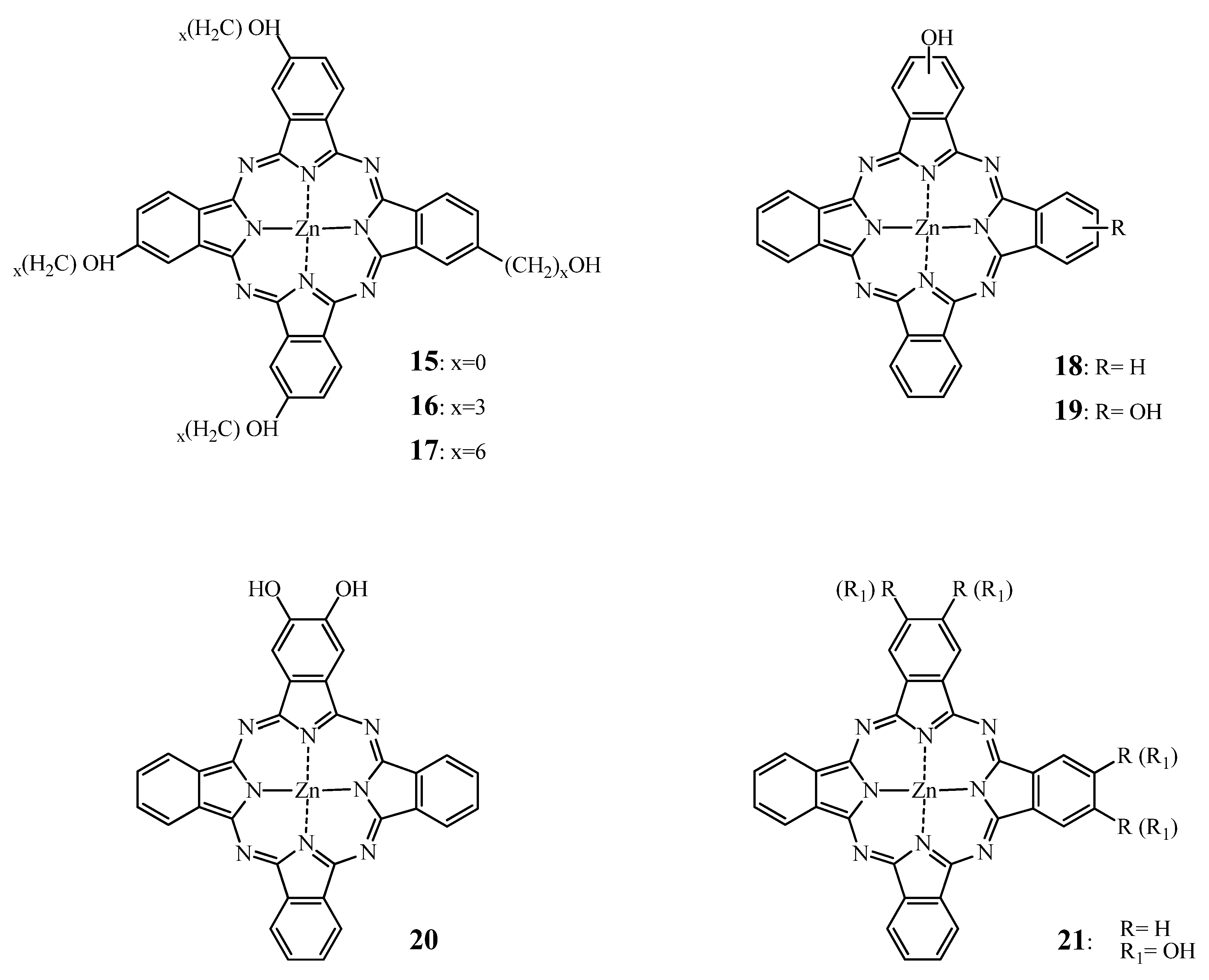
2.2.1. ZnPc—SAR

As mentionned in Section 2.1.1, providing permanent negative charges through sulfonation to Pcs increases their solubility in aqueous media. Wöhrle et al. reported [121] that amphiphilically sulfonated ZnPcs such as the mono- and disulfonated phthalocyanines are more potent and effective than their tri- and tetrasulfonated counterparts in vivo. Comparing Zn and Al substituted analogs, they found the following pattern of photodynamic efficacy: AlPcS2 ≈ ZnPcS1 > AlPcS1 > AlPcS3 ≈ ZnPcS2 > AlPcS4 > ZnPcS4. In 1997, Kudrevich et al. [122] conducted in vitro and in vivo experiments on Balb/c mice bearing EMT-6 tumors using a series of trisulfonated, amphiphilic water-soluble ZnPc derivatives (Figure 6).
Figure 6. Structures of compounds 11, 12 and 13 [122].
Molecules 17 00098 g006
In vitro compounds 11 and 12 were two and more than five times more potent than the tri and tetrasulfonated zinc phthalocyanines, respectively, whereas compound 13, exhibited the lowest phototoxicity probably due to its poor cellular uptake caused by the “bulky diphenylpyrazino substituent”. In vivo, no tumor regression could be noticed with 13 and ZnPcS3. However, in almost 90% of the mice treated with 5 μmol/kg of compounds 11, 12, and ZnPcS4 no palpable tumors have been detected three weeks post-PDT. It was suggested that the amphiphilic character of 11 and 12 decreases the aggregation tendency, thus increasing photoactivity and tumor uptake. Moreover, due to the higher conjugation extent, a bathochromic shift of the Q-band (i.e., 706 nm) was induced.
Based on the experiments evaluating the impact of amphiphilicity on photodynamic activity with aluminium sulfonated phthalocyanines, there have been numerous attempts to increase the amphilic character of zinc sulfonated phthalocyanines. In one of their studies, Cauchon et al. [123] compared the photodynamic efficiency of different trisulfonated phthalocyanines bearing one alkyl chain containing between n = 2 and 16 carbon atoms (ZnPcS3(Cn)).
In vitro studies revealed that ZnPcS3(C9) exhibited the highest cellular uptake and photocytotoxic effect on EMT-6 cells, while the other hydrocarbon substituted phthalocyanines were less efficient. The structure-activity relationship to induce 90% cell death of these compounds followed a typical hyperbolic curve also observed for other homologous series of photosensitizers [124]. However, the most potent compound in this study was the disulfonated zinc phthalocyanine with two sulfonic groups positioned adjacently despite a lower cellular uptake which can be explained by different intracellular localization upon internalization.
In a series of different ZnPcs, disulfo-di-phthalimidomethyl zinc phthalocyanine (ZnPcS2P2 = 14) (Figure 7) has been shown the highest photodynamic activity in Human myelogenous leukemia HL60 cells, K562, FGC85, SGC7901 and LCC carcinoma cell lines [125,126,127]. Moreover, when administered to mice bearing S180 and U14 solid tumors [126] in a Cremophor-based emulsion, the tumor weight in PDT treated animals decreased compared to the controls. The highest inhibitory rate was achieved with a drug dose of 2 mg/kg in both models. Moreover, no acute toxicity was reported after intravenous or intraperitoneal administration of 14 with drug dose up to 100 mg/kg [126]. The repeated-dose toxicity of this compound was assessed a few years later in healthy Wistar rats [128] using the same delivery system. Interestingly, an acute toxicity of 14 was found at a dose of approximatively 52 mg/kg based on their investigations on mice [126] and on an “anticipated human clinical application” (data not disclosed).
Figure 7. Chemical structure of phthalocyanine 14i.e., ZnPcS2P2.
Molecules 17 00098 g007
It was reported that a 10 times repeated administration of 4 mg/kg, which corresponds to 100 times that anticipated clinical “therapeutic” dose by the authors, induced notable hepatic and spleen damage.
Besides sulfonation, another means to enhance the solubility of Pcs in aqueous media and increase their amphiphilicity is hydroxylation. Boyle et al. [129] and Hu et al. [130], reported the synthesis and evaluation of several hydroxysubstituted zinc phthalocyanines.
When incorporated in Cremophor EL emulsions, all compounds showed strong photodynamic action both in vitro and in vivo except for the directly substituted tetrahydroxyphthalocyanine 15 (Figure 8). Indeed, Boyle et al. [129] showed that 15 was phototoxic in vitro on Chinese hamster lung fibroblasts V79; but failed in vivo on Balb/c mice bearing EMT-6 tumors, to induce any perceptible tumor response. Meanwhile, the tetra(3-hydroxypropyl) zinc phthalocyanine 16 (Figure 8) and tetra(6-hydroxyhexyl)zinc phthalocyanine 17 (Figure 8) induced tumor cure as measured by tumor necrosis within 48 h and no tumor regrowth up to 30 days at 0.5 μmol/kg and 1 μmol/kg (1.0 mg/kg), respectively, when applied intravenously. Compared to a mixture of mono, di, tri and tetrasulfonated ZnPcs, 16 and 17 were ten and five times more potent than ZnPcS. Furthermore, it seemed that the hydroxyderivatives function via vascular shutdown rather than direct cell killing as observed for the sulfonated zinc phthalocyanines.
Figure 8. Chemical structure of hydroxylated phthalocyanines 15–21.
Molecules 17 00098 g008
Moreover, Winkelman et al. [131] postulated that a critical distance of 1.2 nm is required between oxygen atoms of sulfonate, carboxyl or hydroxyl substituents and the core of phthalocyanines or porphyrins in order to present biological activity. This hypothesis could potentially explain the low photoactivity exhibited by 15 as well as the phototoxicity of the alkylhydroxy ZnPc derivatives.
A few years later, Hu et al. [130] tested mono-, di- and tri-substituted hydroxy ZnPcs derivatives 1821 (Figure 8) formulated in Cremophor emulsions in EMT-6 tumor cells in vitro and in vivo. It was observed that the phototoxicity decreased with the introduction of hydroxyl groups (i.e., 15 vs.20) and that adjacent positioning of the hydroxyl functions such as in 21 increased Pcs phototoxic effect.
A third possible chemical modulation of ZnPc consists in fluorination of the PS. Several articles report the synthesis and evaluation of this class of ZnPcs [132,133] (Figure 9). The main reasons being that fluorination increases the water solubility and triplet state quantum yield of the phthalocyanine while maintaining similar behavior in biological medias as hydrogen atoms [134].
The most extensive studies on fluorinated ZnPc in PDT have been performed by the van Lier group (using hexadecafluorinated zinc phthalocyanine (ZnPcF16)(25, Figure 10) [135,136,137,138].
Figure 9. Structure of the fluorinated ZnPc 22, 23 and 24.
Molecules 17 00098 g009
Figure 10. Structure of the zinc hexadecafluorinated Pc 25 and its monosulfonated analogue 26.
Molecules 17 00098 g010
In their most recent publication on this subject, Allémann et al. [137] compared Cremophor based emulsion of compounds 25 and 26. It is noteworthy to mention that Pc 26, bears one sulfonated group to provide some amphiphilicity, thus, probably improving cell penetration. To BALB/c mice bearing EMT-6 tumor allografts, 1 μmol/kg of 25 or 26 were applied and their bioavailability as well as their PDT effect has been assessed. After one week, the area under the plasma concentration as a function of time of 25 and 26 were 237 and 183 nmol∙h/g with half-lives of 9.25 and 12 h, respectively. The highest PS tumor accumulation was recorded 24 h post-injection for both Pcs. However, 26 exhibited better tumor selectivity with reported tumor-to-muscle and tumor-to-skin ratios of approximatively 13 and 4 for 26 and, 7 and 2.5 for 25, respectively. Strikingly, a 66% tumor response was recorded for 26 at 1 μmol/kg for a light dose of 100 J/cm2 but was associated with 33% mortality. Hence, it was considered that the best PDT results (i.e., tumor response and animal viability) were obtained using 0.1 μmol/kg of 26 irradiated at 400 J/cm2. As compared to previous results reported by Boyle et al. [138], 26 is twenty times more phototoxic than 25 while displaying equivalent tumor uptake. The impressive 26 phototoxicity observed was attributed to “extensive cellular damage”. In tumor free rats, 26 formulated in Cremophor was devoid of any lethality after irradiation. Alterations of tumor surrounding tissues and extensive edema were reported, but were associated with rapid recovery [137]. A proposed way to avoid these collateral damages is to tune its biodistribution and elimination using different formulations forms, such as PEG-coated nanoparticles (NP). In Section 2.2.3 of this review, formulation of 25 in NP is further discussed.
Allémann et al. [137] also investigated the influence of incubation media on PDT efficiency of ZnPc in vitro by comparing 1-methyl-2-pyrrolidinone to pyridine. Dilution of Cremophor emulsion of ZnPc at 10 μM in 1-methyl-2-pyrrolidinone solution led to complete loss of in vitro PDT efficacy presumably due precipitation and aggregation as noticed by an altered absorption spectrum. This observation confirmed earlier in vivo studies by Boyle et al. [138] that obtained no significant tumor response in BALB/c mice bearing EMT-6 tumors when ZnPc was diluted in 1-methyl-2-pyrrolidinone. In the meantime, complete tumor ablation was achieved when pyridine was used to dilute ZnPc Cremophor-based emulsion at a drug dose of 2 μmol/kg. The authors assumed a possible “coordination of pyridine to the axial ligands of the central metal ion” resulting in an increase photodynamic activity.
Choi et al. and Liu et al. [139,140] reported recent in vitro investigations on the influence of glycosylated ZnPc based on the strategy to concomitantly increase the water-solubility and the selectivity of these compounds through targeting of glucose transporter [141,142]. In accordance with zinc sulfonated phthalocyanines, tetra-glycosylated ZnPc derivatives were substantially less photoactive than the mono-glycosylated and di-glucosylated ZnPcs derivatives in vitro [139,140]. Moreover, the α and β positioning of the glycose substituents seemed to influence the tendency to aggregate and consequently affects their phototoxicity. Indeed, α substitution appeared to prevent Pc aggregation as compared to the β positioned analogues. Promising phototoxicities in the nanomolar range were achieved in HT29 and HepG2 cells glucosylated di- α substituted ZnPc.
Ometto et al. [143] as well as Fabris et al. [144] have tested octapentyl (27) and octadecyl (28) substituted ZnPc (Figure 11). Administered intramuscularly to MS-2 fibrosarcoma bearing Balb/c mice in a Cremophor emulsion both compounds were highly selective for the tumor tissue. Limited skin photosensitization was confirmed with healthy Balb/c mice under the same experimental conditions. The strong binding of both compounds to LDL is presumably responsible for this selectivity [145]. Both 27 and 28 induced a shrinkage of the tumor volume after PDT [143,144]. However, both derivatives accumulated to a higher extend in the liver and spleen than in the tumor even one week post-PDT, presumably because the bile-gut is the primary elimination pathway characteristic for lipidic drug delivery systems [143,146].
Figure 11. Chemical structure of octapentyl and octadecyl ZnPcs 27 and 28.
Molecules 17 00098 g011
Liu et al. have achieved synthesis and in vitro evaluation of zinc octa[(biscarboxylate)-phenoxy]phthalocyanine and its sodium salts [147] (i.e, compounds 29 and 30, see Figure 12).
Figure 12. Chemical structure of phthalocyanines 29 and 30.
Molecules 17 00098 g012
This structural extension with phenylcarboxylate groups resulted in a diminished stacking and aggregation tendency, due to possible non-planar orientation of this group as well as extended “inter-ring distance”. However, at physiological pH, only 29 was water-soluble. The authors presented this aspect as the major drawback of 29 to HEp2 cell penetration, whereas the more hydrophobic 30 was taken up to a higher extent. However, cell penetration could have been also hampered by the presence of negative charges on 29 at physiological pH. Despite differences in cellular uptake both Pcs displayed similar but moderate phototoxicities. However, this low potential for photosensitization of biological materials can be efficiently used for fluorescence diagnostics. Very recently ZnPc similar to 29 zinc tetra[(monocarboxylate)phenoxy]phthalocyanine has been exploited for the use in PCR analysis [64]. This compound had similar but narrower absorption and emission bands in the NIR as compared to a conventional cyanine dye. Most importantly, the Pcs dyes were thermally and chemically stable and showed essentially no photobleaching.
The outstanding tendency of Pcs to form photoinactive aggregates can also be exploited for the design of “molecular beacons”. These compounds are powerful tools for real time detection of RNA/DNA in vitro and in vivo. In molecular beacons, a quencher/donor pair is positioned on the distal ends of a short, hairpin forming oligonucleotide. In absence of a complementary sequence the close proximity of the fluorophore and its corresponding quencher makes the molecular beacon optically silent. However, hybridization with the complementary sequence then restores the fluorescence of the reporter. Despite their high specificity the sensitivity of conventional molecular beacons is often compromised by a poor sensitivity provided by insufficient quenching. Futhermore, longer observation periods are often impeded by most fluorescent dyes’ strong photobleaching. Therefore, in an earlier study the same group reported the use of a pair of Pc similar in structure to ZnPc in molecular beacons [148,149]. After optimization of the reaction conditions good yields for molecular beacons were achieved. However, since Pcs have long interaction ranges the authors had to use longer oligonucleotide sequences as complementary sequence to observe a fluorescence increase. A signal to background ration as high as 59 was reported. Furthermore, the perfect matched complementary sequence was five times more fluorescent than a single base mismatch. Therefore, water soluble Pcs can be efficiently employed as fluorescence reporters in vitro but their whole potential in this area remains to be demonstrated.
Lo and co-workers reported the photoactivity of monosubstituted ZnPc with a 1,3-bis(dimethyl-amino)-2-propoxy group at the α or β position (Figure 13), “and the corresponding di-N-methylated derivatives” on human colorectal carcinoma cells HT29 [150]. The α and β positioning is an analogy to the α and β positions for sugar moieties. Indeed in sugars, α and β correspond to lower and upper position respectively of the hydroxylic group on the anomeric carbon (C1) of the cyclic sugar moiety, the cylcle defining the referential plane [151].
Figure 13. Structure of compounds 31–34.
Molecules 17 00098 g013
The absorption and fluorescence spectra showed a lower aggregation tendency for the β-substituted compounds as compared to their α-substituted analogues. Consequently, the IC50 of 32 and 34 were 0.15 and 0.08 μM, respectively, while 31 and 33 induced 50% of cell inactivation at doses of 0.48 and 0.64 μM, respectively.
Like AlPcs, PEGylated ZnPcs have also been designed aiming at increasing their water solubility [152]. Liu et al. have prepared a series of asymmetrically substituted ZnPcs using methylated polyethylene glycol. However, these compounds were essentially not water soluble. Therefore, just recently the synthesis of tetra and octa substituted PEGylated ZnPcs has been reported [153]. In these compounds the terminal methyl group of the tetraethylene glycol side chains has been omitted in order to provide a higher hydrophilicity through hydroxyl end groups. Despite these modifications, aggregation in water was still observed that could be circumvented by the addition of Triton-X100. The less aggregated compound that was non-peripherally substituted with polyethylene glycol showed a strong bathochromic shift of more than 20 nm. But in vitro evaluation showed an IC50 value that was in the order of two orders of magnitude higher than reported for the methylated counterparts. Recently, Ng and co-workers reported the synthesis and efficient photocytotoxic effect of several pegylated ZnPcs where IC50 values ranged from 0.25 to 3.72 μM on HT29 and HepG2 cells [154].
The net charge of pharmacological compounds is of importance with respect to the pharmacokinetic properties of a drug as well as in terms of uptake and intracellular localization. Several studies report the impact of the charge on Pc-mediated PDT. Banfi et al. [155] described the synthesis and in vitro assessment of some ZnPc derivatives on human colon adenocarcinoma cells HCT116. The most potent ZnPc derivatives among the synthesized series are shown in Figure 14.
Figure 14. Structure of compounds 35, 36 and 37.
Molecules 17 00098 g014
Compound 35 was approximately five times more potent than compound 36 and approximately eight times more efficient than compound 37.
Brown and coworkers [156,157] reported that the cationic pyridinium ZnPc 42 (see Figure 15) was more phototoxic to RIF-1 cells than the anionic tetraglycine analogue 39 or the neutral hydrophobic tetradioctylamine 43 [156]. In vivo all the tested compounds induced partial tumor regression, but only compound 42 led to complete remission, most presumably through vascular occlusion in the irradiated areas. This observation is in agreement with a study of Sibrian-Vazques et al. that showed that the cellular uptake of 44 was higher and faster than 46 (see Figure 16) [158].
Figure 15. Chemical structure of phthalocyanines 38–43.
Molecules 17 00098 g015
Figure 16. Chemical structure of compounds 44–48.
Molecules 17 00098 g016

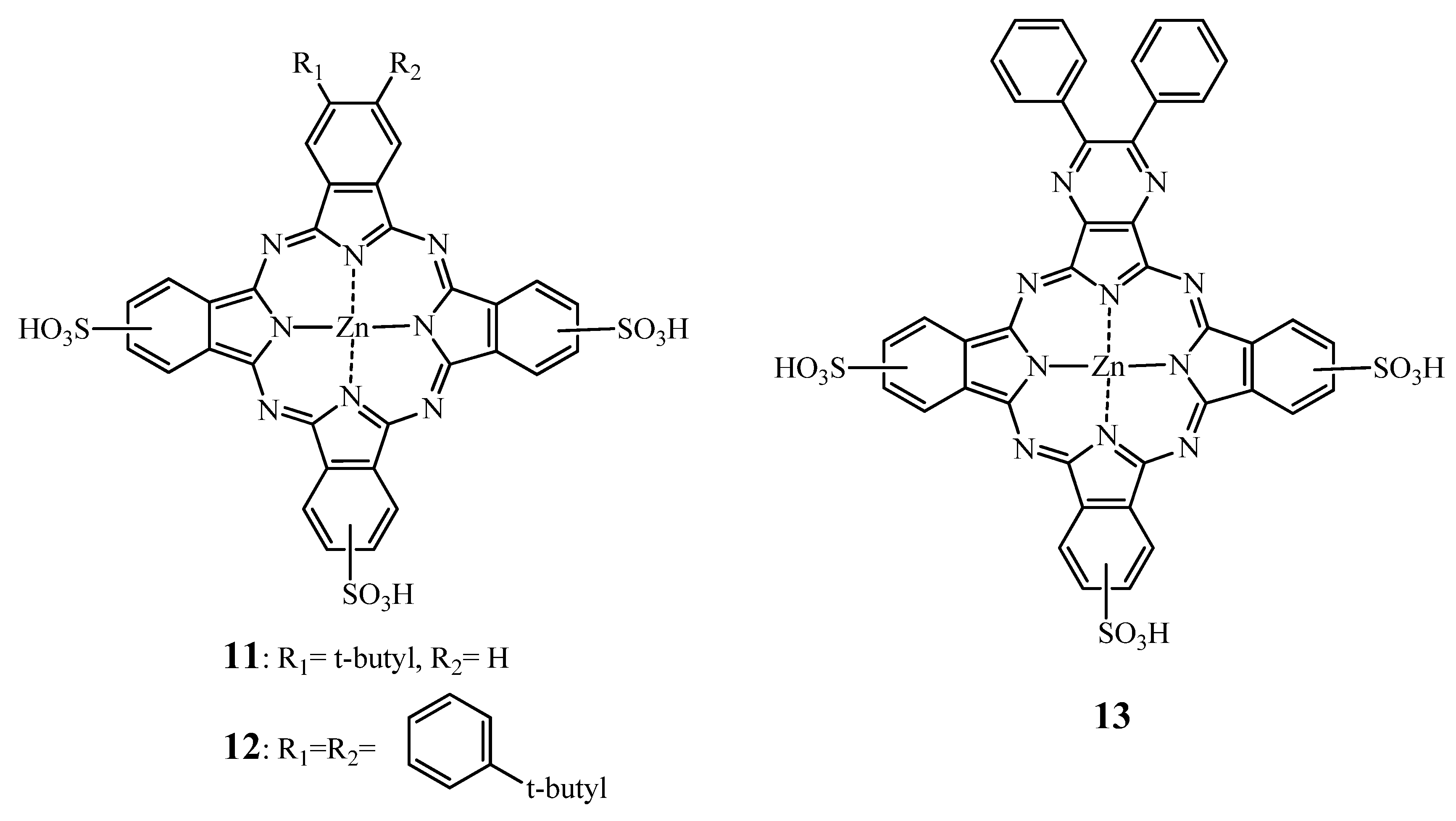
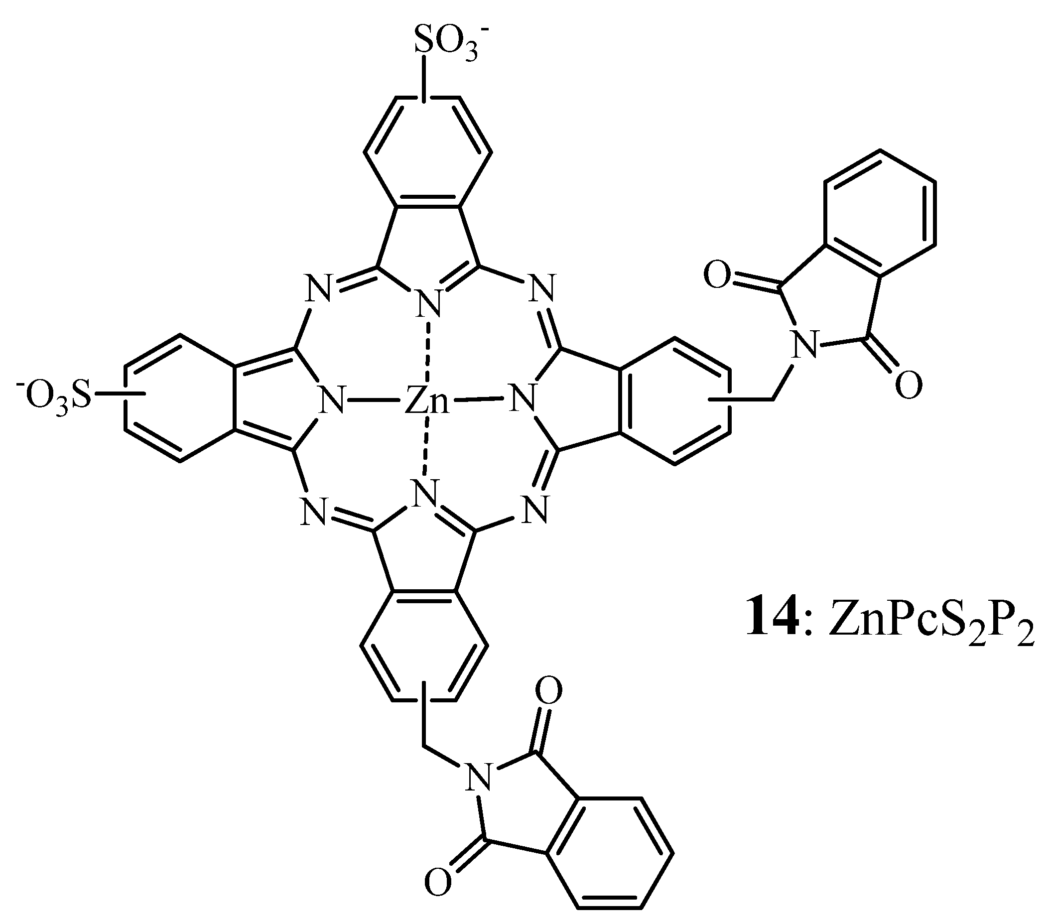
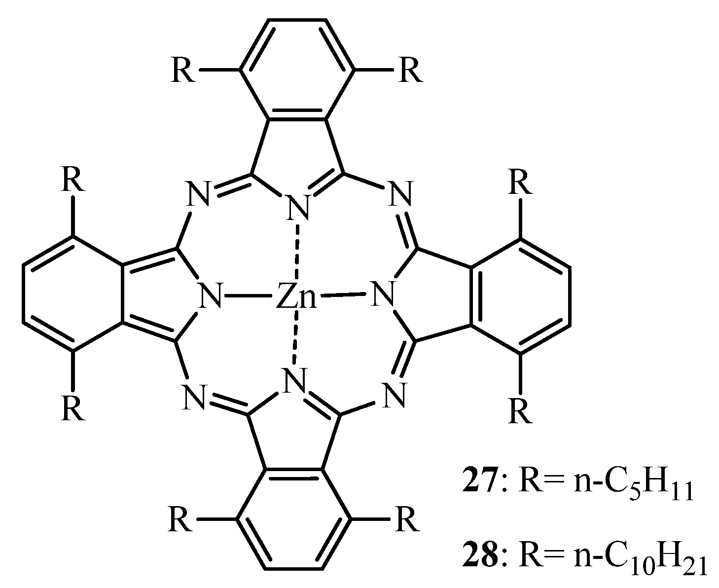
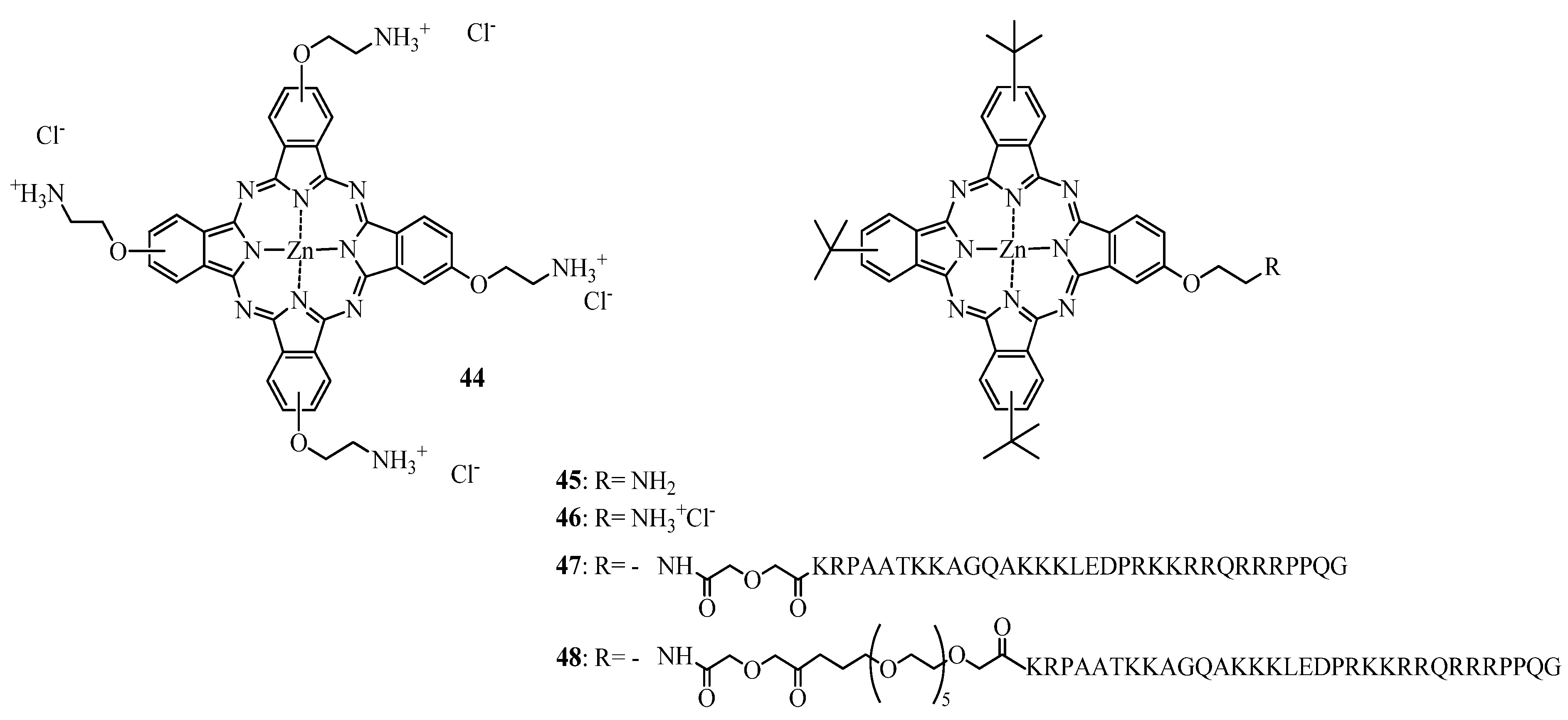
2.2.2. Tumor Targeting with Zinc Based Pc-Conjugates

In the same report the preparation and in vitro evaluation of zinc based phthalocyanines conjugates with cell-penetrating peptides (CPP) (see Figure 16) was described.
Conjugates 47 and 48 were taken up by HEp2 cells to a higher extent than the unconjugated ZnPc. The introduction of short polyethylene glycol linker between the ZnPc and CPP has been shown to further increase the uptake. At a light dose of approximately 1 J/cm2, 47 and 48 were the most active compounds, with IC50 values of 1.44 μM, 1.87 μM, respectively. In contrast, 44 did not alter the cell viability at doses up to 10 μM.

2.2.3. Formulation of ZnPcs

Phototoxicity and fluorescence of Pcs is mostly hampered by their low water-solubility and tendency to aggregate. Their incorporation into suitable and biodegrable formulations is commonly accepted as a necessity in the development of new therapeutics. Indeed, development of drug delivery systems such as liposomes, micelles and nanoparticles could improve the unfavorable biodistribution of free Pcs (i.e., improvement of PS pharmacokinetic properties, better targeting of diseased tissues due to size of the particles, association to serum proteins and specific activation of the PS through localized delivery of the PS) as well as avoidance of aggregation and loss of phototoxic activity/fluorescence which should result in a better therapeutic outcome (see following paragraphs).
Zinc based phthalocyanines have been mostly evaluated as liposomal formulations. Liposomes are phospholipids vesicles that present the advantage to entrap either hydrophilic drugs in the core of the phospholipid vesicle or hydrophobic drugs within the lipid membrane. Incorporation of drugs into liposomes results in passive targeting of tumors [159,160]. In studies reported in this review, only small unilamellar vesicles (i.e., one phospholipid bilayer) were used.
Liposomal formulation of ZnPc showed photocytotoxic effect in vitro on a large variety of tumor cell lines [161,162,163]. The drug CGP55847 commercialized by CIBA has been the first Pc reaching clinical trials although no clinical results have been reported. Ben-Hur and Chan reported that the “early phase I/II clinical studies […] were discontinued but not for medical reasons” [164].
In vivo, ZnPc being incorporated in dipalmitoyl-phosphatidylcholine (DPPC) liposomes showed complete response at drug doses as low as 0.14 mg/kg in mice inoculated with MS-2 fibrosarcoma. At a slightly lower dose of 0.12 mg/kg (i.v.), ZnPc was found to be “associated exclusively with lipoprotein fraction”, i.e., whether it was complexed with low density lipoproteins (LDL) or incorporated into liposomal structures, and 70% of the drug was cleared from the body within 12 h independently from its carrier system, while the remaining ZnPc was slowly eliminated. However, ZnPc associated with LDL presented a higher selectivity toward tumor than ZnPc liposomes with a tumor-to-liver ratio of 2.27 and 1.04 and a tumor-to-muscle ratio of 4.20 and 3.84 after 24 h post-injection, respectively. Nonetheless, the selectivity was somewhat limited by the redistribution of the photosensitizer among other lipoproteins, especially high density lipoproteins. As liposomal formulations of benzoporphyrin derivative, liposomal CGP55847 seems to be transferred to serum lipoproteins and more predominantly to low density lipoproteins [165,166,167,168,169] explaining the propensity of the PS to selectively accumulate in tumors [163,170]. Rodal et al. suggested that the observed internalization of ZnPc was not following the endocytotic pathway of LDL but rather occurred via diffusion through the cellular membrane after binding to the LDL receptor [161].
In vivo studies on different liposomal formulations consisting of a mixture 1-palmitoyl-2-oleoyl-sn-glycero-3-phosphocholine (POPC) and 1,2-dioleoyl-sn-glycero-3-phospho-L-serine (OOPS) revealed that intratumoral distribution pattern of ZnPc in C57BL/6 mice bearing Ehrlich carcinomas or B16 melanomas was a time dependent process [171]. In agreement with van Leengoed et al. [172], 3 h after intravenous injection of 0.5 mg/kg of ZnPc, the photosensitizer was present in and around the tumor vasculature but not 24 h post-injection. Indeed, using a dorsal skinfold chamber model, van Leengoed et al. [172], could detect vascular occlusion within five minutes after photoactivation which resolved 30 min after irradiation. Moreover, maximal vasculature damage and tumor-to-muscle ratio from 8:1 to 14:1 have been reported for this liposomal delivery system [173].
Nanoparticles are another type of drug delivery system that enables the incorporation of higher drug doses of hydrophobic drugs. Passive targeting of tumors is also achieved due to their size (i.e., below 1,000 nm, generally 200 nm) through EPR effect. A strategy to increase the circulation time of the nanoparticles as well as other drug delivery systems is the incorporation of PEG moieties on their surface. It is commonly accepted that PEGylation creates hydrophilic barrier that reduces the immunogenicity of the drug delivery system and lowers its clearance from the body [174,175,176].
Polylactic acid (PLA) based nanoparticular formulations of ZnPc were investigated by Allémann et al. [135,136]. The coating of nanoparticles with PEG moieties resulted in the decrease of the reticuloendothelial system and an increase in the PS tumor retention [135]. Incorporation of 25 (Figure 10) into PEG coated nanoparticles or using a Cremophor EL based emulsion increased the bioavailability as compared to uncoated-NP by a factor of four in BALB/c mice bearing EMT-6 tumors. The tumor-to-skin and tumor-to-muscle ratios for the PEG-NP and Cremophor based formulation were 2 to 21, respectively, with a maximum concentration in tumor 48 h post-injection [135]. However, the nanoparticular drug delivery system was nearly five times more efficient for PDT-mediated treatment of mice bearing EMT-6 tumors [136]. This is in agreement with a study reported by Fadel et al. [177] describing that tumor bearing animals treated with ZnPcs laden poly(lactic-coglycolic acid) nanoparticles showed the best PDT outcome, highest tumor growth delay and longest survival times as compared to mice treated with the free PS.
An interesting approach was recently described by Kataoka and co-workers [178,179,180,181] where dendrimer based zinc phthalocyanines showed promising results in PDT and PCI applications. Indeed, they could manage to prepare anionic dendrimer zinc phthalocyanine (DPc) incorporated into positively charged poly(ethylene glycol)-poly(L-lysine) (PEG-PLL) block copolymeric micelles referred to as polyion complex micelles (PIC) [180]. The substitution of large dendritic parts avoids the aggregation and increases their water solubility [181]. These micelles were tested both in vitro on human lung adenocarcinoma cells A549 and in vivo on (Balb/c nu/nu) mice bearing A549 tumors. It was shown that 7.6 times more of encapsulated DPc in micelles (DPc/m) was taken up and that these are 78 times more phototoxic in vitro than free DPc. In addition, subsequent in vivo studies confirmed the benefits of micelle encapsulation of DPc by delayed tumor growth and limited skin photosensitization.
Another interesting approach for drug delivery of hydrophobic PS is their conjugation to cyclodextrins. This strategy enables the water-solubilisation of lipophilic PS and thus its systemic administration at high concentrations. In one report, Baugh and al. proceeded to the conjugation of zinc based phthalocyanines to cyclodextrins. The double carbon bounding linkage between the dimers was cleaved, upon light activation and in presence of oxygen; resulting in the release of the Pc. Hence, this approach could be a promising systemic carrier of hydrophobic PS for photodynamic therapy [182,183].
Formulation of ZnPcs in gels or (micro)emulsions could be of interest in topical application. Indeed, several studies reported improved photophysical and aggregation properties of ZnPcs [184,185] as well as successful delivery to the skin of sulfonated ZnPc either via simple skin penetration [184] or through iontophoresis [186].

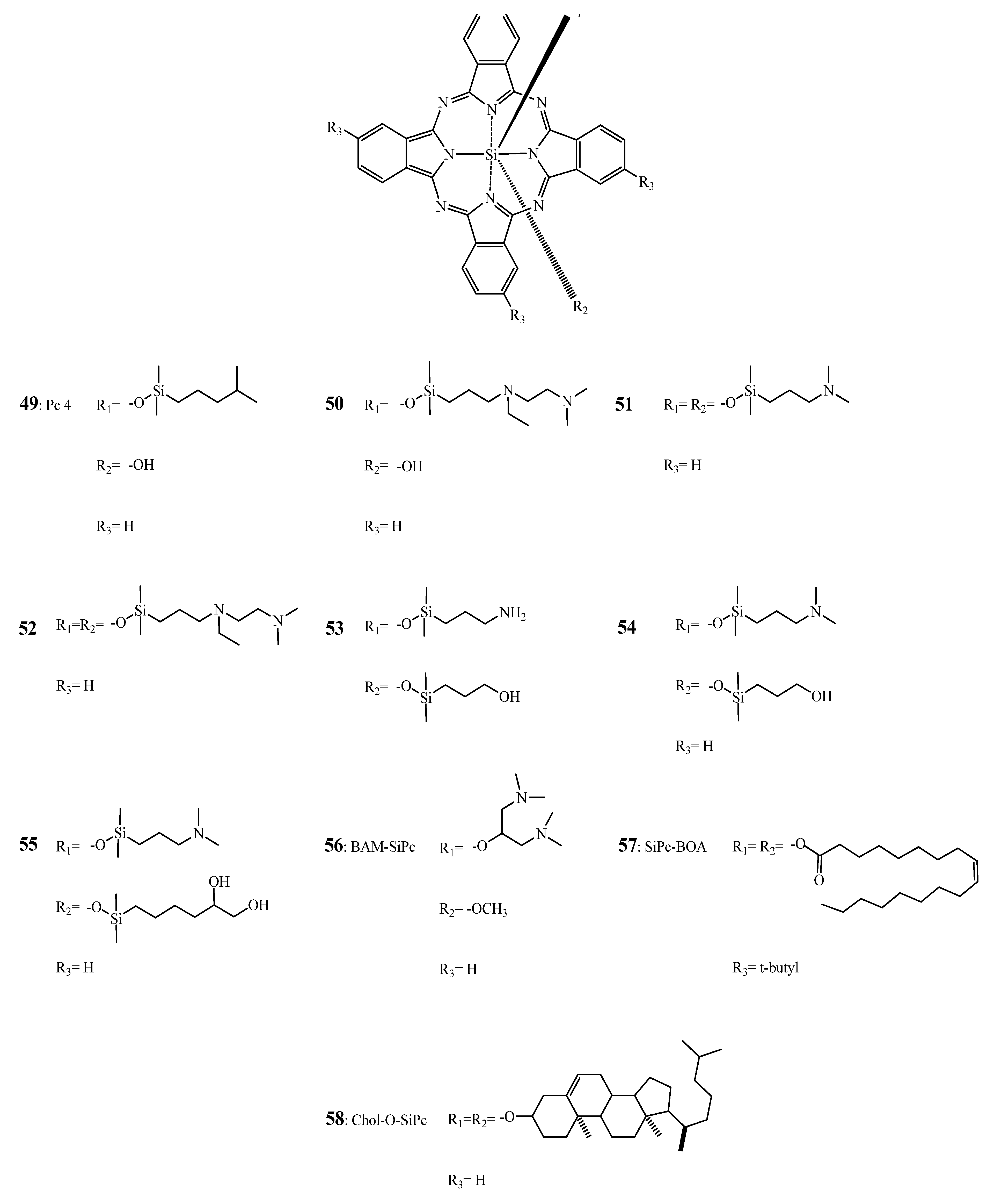
2.3. Silicon Based Phthalocyanines: Pc 4 and its Analogues

2.3.1. SiPc-SAR

Several silicon based phthalocyanines (SiPc) mostly aiming at revealing the influence of axial ligands have been proposed and tested both in vitro and in vivo. Probably, the most famous representative of these compounds with some commercial value is “La Jolla Blue”. Water-solubility of this dye absorbing at 680 nm as well as prevention of aggregation is provided by two axial polyethylene oxide moieties. Then, two peripherial carboxy groups at the macrocycle can be used for the coupling to biological molecules such as antibodies. This has been used for the design of an antibody for an FDA cleared in vitro immunofluorescence assay.
It has been shown by He et al. [187], that silicon based phthalocyanines with short aminosiloxy ligands [49 (Pc 4) and 51] are more phototoxic effect in Chinese hamster lung fibroblasts V79 and Murine leukemic lymphoblasts L5178Y-R as compared to compounds such as 50 and 52 with longer axial ligands (see Figure 17).
Figure 17. Chemical structure of Pc 4 (49) and derivatives 50–58 [187,188,189,190,191,192,193,194].
Molecules 17 00098 g017
In another report [194], the PDT effects of these compounds in vivo on C3H/HeN mice bearing RIF-1 tumors were examined. The phthalocyanines were solubilized in a Cremophor EL emulsion and light was given 24 h post Pc injection. Except 52, all axially substituted Si-Pc resulted in a complete remission. The authors suggested that 51-mediated PDT may induce cellular destruction via a slower mechanism than Pc 4-mediated PDT.
In 2009, Rodriguez et al. [188], compared Pc 4 to compounds 51, 53, 54 and 55 on the human breast cancer cell line MCF-7c3. These analogues showed, among other features, higher cellular uptake and phototoxicity than Pc 4. Biaxial substitution reduced aggregation which, in turn, can explain their higher photodynamic efficiency. Furthermore, the presence of a hydroxyl group at one side seems to enhance the phototoxicity of the SiPcs. However, as shown by confocal microscopy, except for 51, Pc 4 analogues seem to act through a different destructive pathway due to their principle association with lysosomes rather than with mitochondria/endoplasmic reticulum. Interestingly, when clonogenic assays were performed 51 and Pc 4 had a similar behavior. In contrast, 51 exhibited a 4-fold lower IC50 in comparison to Pc 4 (49) as demonstrated by means of a MTT test. Using bi-axially substituted polyamine SiPcs with fluorescence quantum yields in aqueous media of 0.12–0.21, IC50 ranging from 450 to 1 nM have been reported in HT29 cells by Ng and co-workers [195].
Moreover, the same group of research developed several SiPcs substituted axially with β-cyclodextrins [196,197,198]. They could reach a photocytotoxic effect on human colon adenocarcinoma and hepatocarcinoma cells (HT29 and HepG2 respectively) with reported IC50 ranging from 21 nM to 1.32 μM. In addition, early in vivo studies conducted by Lau et al. [197] show promising results as drug doses as low as 1 μmol/kg for a light dose of 30 J/cm2 are efficient enough to suppress tumor growth in mice bearin HT29 xenografts.
Another promising approach used by the same group is the glucoconjugation of SiPcs [190,199]. In their report, Chan et al. [199] reported IC50 values as low as 6 nM on the same cell lines and could “retard tumor growth” in the same animal model.
Pc 4 has been developed at Case Western Reserve University at the beginning of the 90s. Since then Pc 4-mediated PDT has been reported effective in vitro against various tumor cell lines of different origin [187,200,201,202,203,204,205]. In experimental animal models for ovarian and colon cancer, no photodynamic effect was observed with doses of 0.4 mg/kg [202], whereas with doses of 0.6 or 1 mg/kg complete remission or at least significant tumor volume reduction occurred between 3 to 7 days post-PDT. Furthermore, Pc 4-PDT was associated with a delay of tumor regrowth from 9 [203] up to 90 days [202].
A recent study conducted on immunideficient mice bearing papillomas [206] induced by the administration of cottontail rabbit papillomavirus led to the conclusion that Pc 4 at a dose of 1 mg/kg and at a light dose of 150 J/cm2 given 48 h administration resulted in complete remission in 87% of the cases with no observable re-growth during 79 days. In addition, it was suggested that the rapid destruction observed in Pc 4-mediated PDT could be explained by the mitochondrial/endoplasmic reticulum [188] localization of Pc 4 mainly leading to apoptosis and necrosis [207,208,209]. However in some cases vascular occlusion was also reported [188].
Moreover, Anderson et al. [210] demonstrated that Pc 4 exhibited less skin photosensititization as compared to Photofrin®. Topically applied, Oleinick and co-workers established that Pc 4 is “effectively delivered into the human skin” [211] and was consequently investigated clinically. In these trials, Pc 4-mediated PDT was reported to be well tolerated in patients and could have promising application in mycosis fungoides treatment [212].
Recently, Fong and coworkers reported the synthesis and characterization of several silicon-based phthalocyanines [189,213,214,215,216,217]. Out of this series, BAM-SiPc 56 (see Figure 17) having an IC50 as low as 0.015 μM on HepG2, Hep3B, HT29 and J744 was the most potent. However, this compound was phototoxic on normal liver cells (i.e., WRL-68 with IC50 of 0.035 microM under the same experimental conditions) [189,192,218]. In nude mice bearing hepatocarcinoma HepG2 and colorectal adenocarcinoma HT29 tumors the same compound showed remission/regression and tumor growth delay 15 days after PDT at a drug dose of 1 μmol/kg [191].
Barge et al. [193] could also covalently link axial cholesterol moiety (Chol-O-SiPc) (Figure 17) and reported a seven fold higher potency of this derivative as compared to AlClPc (with IC50 of approximatively 8 nM) in vitro, on the pigmented melanoma cell lines M3Dau and SK-MEL-2.

2.3.2. SiPc Formulations

Pc 4 is the most extensively studied silicon-based phthalocyanine. Due to its insolubility in physiological media, Pc 4 tends to aggregate with subsequent loss of photodynamic activity and altered biodistribution. In order to avoid these issues, Pc 4 was incorporated in oil-based formulations, PEG-PCL (polyethylene glycol-block-poly-ε-caprolactone), micelles, and nanoparticles.
In a recent study Master et al. [219] were able to encapsulate with 70% efficiency Pc 4 in PEG-PCL micelles. The authors observed that in human breast cancer cells (MCF-7c3), Pc 4 delivered in PEG-PCL micelles seemed to colocalize with lysosomes and mitochondria, while Pc 4 in solution accumulated mainly in mitochondria and endoplasmic reticulum as well as in Golgi apparatus. However, no different photocytotoxic effect was observed for the two formulations.
Pc 4 was also encapsulated into silica and gold nanoparticles known for their biocompatibility and their stability [220]. Zhao et al. [221] showed that encapsulation into silica nanoparticles of 25–30 nm diameter lead to enhanced photodynamic activity in vitro as compared to the free Pc 4. Moreover, Pc 4 silica nanoparticles were found to be less sensitive to photobleaching. The reported IC50 values for the nanoparticluar delivery system were around 5 nM for A375 cells and 10 nM for B16F10.
An in vivo biodistribution study with Pc 4 adsorbed on PEG coated gold nanoparticles revealed that Pc 4 gold nanoparticles accumulated in the tumor faster than Pc 4 formulated in Cremophor EL. While the peak concentration in the tumors was only reached after 2 days using Cremophor the gold nanoparticles containing Pc 4 reached this peak within 2 h. It was also observed that the singlet oxygen yield of the two formulations was identical suggesting that the nanoparticles protect Pc 4 until its release in the target tissue [222].
Li et al. reported the synthesis and PDT effect of a neutral and lipophilic tetra-t-butyl silica phthalocyanine bisoleate referred to as SiPc-BOA (Figure 17) [223]. It was noticed that axial substitution of the oleate moieties decreased the propensity of the photosensistizer to aggregate and enabled its binding to the lipidic layer (i.e., LDL delivery system). Using this strategy, Li et al. increased the loading of the LDL nanovesicules with a molar ratio of 400 SiPc-BOA per one LDL [223]. These SiPcBOA laden nanoparticles were 10 times more effective than the free compound in HepG2 cells suggesting a LDL receptor-mediated photodynamic effect.
A multifunctional approach was proposed by Zheng et al. [224] through additional coupling of folic acid to lysine residues of the apolipoprotein B of the LDL part in SiPc-BOA-LDL conjugates. The validity of their hypothesis was tested in a system consisting of cells expressing the folate receptor (κB cells), those expressing the LDL receptor (HepG2 cells), and cells that lack the folate receptor (HT1080, CHO cells). An interesting review of the aforementioned results could be found in the following book [225].

3. Applications of Pcs in Fluorescence Imaging

Most investigations on phthalocyanine dyes in biomedicine focus on their application in PDT for cancer. However, just recently research was also extended to parasite and bacterial disease treatment such as leishmaniosis [226,227,228,229,230]. Furthermore, in this context, an at least as important research area, i.e., fluorescence imaging, is sometimes ignored. Fluorescence imaging allows the non-invasive detection of superficial disease in a preclinical or clinical setting. It is sensitive, can be perfomed in real time, and its resolution can be tuned to a molecular level. As few as a thousand cells can be detected by this methodology. Today, fluorescence imaging is successfully implemented in preclinical in vitro and in vivo research to specifically monitor the therapeutic outcome of new drugs in animals or to reveal disease mechanisms. Furthermore, it is used clinically for the detection of several diseases, including age-related macular degeneration [231] and cancer [232]. It has been shown to improve the detection rate of barely visible lesions, surgery and recurrence rate in malignant glioblastoma [233,234] and bladder cancer [235]. Despite the recent hype of fluorescent proteins introduced into molecular biology, exogenous fluorescent dyes [236,237] or exogenously-induced fluorescent dyes [238] still play an important role in this research area.
For optimal monitoring of diseases in vivo it will be advantageous that fluorescent dyes absorb and emit in the NIR region of the visble light spectrum in order to optimally penetrate into the tissue and induce only minimal autofluorescence upon excitation with light. Therefore, scientists have developed panoply of different NIR fluorescent dyes for the labeling of proteins, small bioactive peptides, or oligonucleotides [239]. Although there are many classes of NIR dyes including oxazines and rhodamines commercially available, the most commonly used belong to the class of cyanine dyes. One of these, indocyanine green, is approved for the detection of occult choroidal neovasculariuation secondary to age-related macular degeneration. Today these dyes cover an absorption spectrum ranging between 650 and 800 nm. As described above, phthalocyanine dyes can be fine-tuned to the desired absorption/emission wavelengths and can be made water-soluble [240]. Furthermore, they are chemically stable in most solvents and are not as prone to degradation in highly acidic/basic media. Some of these compounds can have a fluorescent quantum yield as high as 70% and are extremely photostable. And finally, compared to conventional cyanine dyes they have a long fluorescence lifetime and an extremely high extinction coefficient. Despite, these advantages there are only two compounds, La Jolla Blue® and IRDye 700DX®, belonging to this interesting class commercially available. Although these compounds have relatively high fluorescence quatum yields, they can still be used as efficient photosensitizers, as demonstrated by, Mitsunaga et al. [58] by conjugation of IRDye 700DX® to monoclonal antibodies targeting epidermal growth factor receptors. Using these conjugates efficient killing of tumor cells was demonstrated in vitro and in vivo when NIR was used as light source.
Their tendency to aggregate seems to be ideally suited for a relatively new class of fluorescent reporters referred to as “smart probes” [241]. This approach has been pioneered by Ralph Weissleder’s research group and is based on the selfquenching/dequenching paradigm [242]. In these compounds, typically several fluorescent dyes are coupled either directly or via chemically or enzymatically labile linker to a polymeric carrier. In their native state these reporters are optically silent due selfquenching of the fluorescent dyes. However, as soon as a chemical or enzymatic trigger results in the release of the fluorescent dye from its polymeric carrier this selfquenching is abrogated. Today, this methodology has been extensively used for the detection of proteolytic activity in vivo. One can deduce from the discussions on the photodynamic activity of aggregated Pc above, that these compounds can also be used in “smart probes” with a better selfquenching allowing the use of low molecular weight carriers with improved pharmacokinetics. Another phenomenon of aggregated Pc, will further favor their use in “smart probes”. As the extinction coefficient of the Q-band undergoes significant reduction in the aggregated form, these fluorescence reporters not only show reduced fluorescence intensities in the non-activated form but also are less sensitive to excitation. Furthermore, due to their lipophilicity, once cleaved, Pc dyes will stay longer at the activation site due to reduced clearance. Currently, we are evaluating this approach for simultaneously treating and monitoring diseases with Pc-based polymeric photosensitizer prodrugs [243,244,245].

4. Conclusions

Throughout this review, it has been shown that phthalocyanines are promising photosensitizers for photodynamic therapy applications. Some of them are even currently used in PDT (e.g., Photosens®) or tested in clinical trials (e.g., Pc 4). At a chemical level, it is concluded that Pcs’ mainly amphiphilic character leads to a higher efficiency in vivo. In addition, hampering their stacking via axial ligation and including positive charge(s) seems to influence their photocytotoxicity, by increasing their cellular uptake and internalization. It is also of major importance to consider their subsequent cellular (re)localization in order to understand and evaluate their photodynamic activity. Hence, by only considering their chemistry, phthalocyanines exhibit this flexibility, enabling further screening and investigations, which rises hope for cancer treatment. Perspectives such as chemical coupling of two photosensitizers as well as screening of new phthalocyanines are currently examined and could be interesting prospects for PDT cancer treatment [246,247,248,249].
From a pharmaceutical point of view, suitable and optimal formulations of Pcs can increase dramatically the therapeutic efficacy. Suitable drug delivery systems can act as a solubilizing matrix for the PS as well as a shield and protection from degradation.

Acknowledgements

NL research is supported by the Swiss Science Foundation grants #205320-122144, #326030-117436, #CR32I3-129987, #205321-126834, #310030-119938, and #K-32K1-116460. TN and NL are grateful for the financial support of grant #IZLSZ2-123011 and Council for Scientific and Industrial Research in South Africa promoting the collaboration between South African and Swiss scientists.

References and Notes

  1. McKeown, N.B. The synthesis of symmetrical phthalocyanines. In The Porphyrin Handbook; Kadish, K.M., Smith, K.M., Guilard, R., Eds.; Academic Press: San Diego, CA, USA, 2003; pp. 61–124. [Google Scholar]
  2. Thomas, A.L. Phthalocyanine Research and Applications; CRC Press: Baton Rouge, FL, USA, 1990; p. 2. [Google Scholar]
  3. McKeown, N.B. An introduction to the phthalocyanines. In Phthalocyanine Materials: Synthesis, Structure and Function; McKeown, N.B., Ed.; Cambridge University Press: Cambridge, UK, 1998; pp. 1–10. [Google Scholar]
  4. Geng, Y.Y.; Gu, D.H.; Wu, Y.Q.; Gan, F.X. High speed recording property of phthalocyanine thin film for compact disc recordable. Proc. SPIE Int. Soc. Opt. Eng. 2003, 63, 63–66. [Google Scholar]
  5. Petritsch, K.; Friend, R.H.; Lux, A.; Rozenberg, G.; Moratti, S.C.; Holmes, A.B. Liquid crystalline phthalocyanines in organic solar cells. Synth. Met. 1999, 102, 1776–1777. [Google Scholar]
  6. Nyokong, T.; Vilakazi, S. Phthalocyanines and related complexes as electrocatalysts for the detection of nitric oxide. Talanta 2003, 61, 27–35. [Google Scholar] [CrossRef]
  7. Ali, H.; van Lier, J.E. Porphyrins and phthalocyanines as photosensitizers and radiosensitizers. In Handbook of Porphyrin Science with Applications to Chemistry, Physics, Materials Science, Engineering, Biology and Medecine; Kadish, K.M., Smith, K.M., Guilard, R., Eds.; World Scientific: Singapore, 2010; pp. 1–119. [Google Scholar]
  8. Alonso, C.; Boyle, R.W. Bioconjugates of porphyrins and related molecules for photodynamic Therapy. In Handbook of Porphyrin Science with Applications to Chemistry, Physics, Materials Science, Engineering, Biology and Medecine; Kadish, K.M., Smith, K.M., Guilard, R., Eds.; World Scientific: Singapore, 2010; pp. 121–190. [Google Scholar]
  9. Ethirajan, M.; Patel, N.J.; Pandey, R.K. Porphyrin-based multifunctional agents for tumor-imaging and photodynamic therapy (PDT). In Handbook of Porphyrin Science with Applications to Chemistry, Physics, Materials Science, Engineering, Biology and Medecine; Kadish, K.M., Smith, K.M., Guilard, R., Eds.; World Scientific: Singapore, 2010; pp. 249–323. [Google Scholar]
  10. Jux, N.; Röder, B. Targeting strategies for tetrapyrrole-based photodynamic therapy. In Handbook of Porphyrin Science with Applications to Chemistry, Physics, Materials Science, Engineering, Biology and Medecine; Kadish, K.M., Smith, K.M., Guilard, R., Eds.; World Scientific: Singapore, 2010; pp. 325–401. [Google Scholar]
  11. Nyokong, T.; Antunes, E. Photochemical and photophysical properties of metallophthalocyanines. In Handbook of Porphyrin Science with Applications to Chemistry, Physics, Materials Science, Engineering, Biology and Medecine; Kadish, K.M., Smith, K.M., Guilard, R., Eds.; World Scientific: Singapore, 2010; pp. 247–349. [Google Scholar]
  12. Fukuda, T.; Kobayashi, N. UV-visible absorption spectroscopic properties of phthalocyanines and related macrocycles. In Handbook of Porphyrin Science with Applications to Chemistry, Physics, Materials Science, Engineering, Biology and Medecine; Kadish, K.M., Smith, K.M., Guilard, R., Eds.; World Scientific: Singapore, 2010; pp. 1–645. [Google Scholar]
  13. Dietze, A.; Peng, Q.; Selbo, P.K.; Kaalhus, O.; Muller, C.; Bown, S.; Berg, K. Enhanced photodynamic destruction of a transplantable fibrosarcoma using photochemical internalisation of gelonin. Br. J. Cancer 2005, 92, 2004–2009. [Google Scholar]
  14. Selbo, P.K.; Sivam, G.; Fodstad, O.; Sandvig, K.; Berg, K. In vivo documentation of photochemical internalization, a novel approach to site specific cancer therapy. Int. J. Cancer 2001, 92, 761–766. [Google Scholar] [CrossRef]
  15. Adigbli, D.K.; Wilson, D.G.; Farooqui, N.; Sousi, E.; Risley, P.; Taylor, I.; Macrobert, A.J.; Loizidou, M. Photochemical internalisation of chemotherapy potentiates killing of multidrug-resistant breast and bladder cancer cells. Br. J. Cancer 2007, 97, 502–512. [Google Scholar]
  16. Norum, O.J.; Selbo, P.K.; Weyergang, A.; Giercksky, K.E.; Berg, K. Photochemical internalization (PCI) in cancer therapy: From bench towards bedside medicine. J. Photochem. Photobiol. B 2009, 96, 83–92. [Google Scholar]
  17. Norum, O.J.; Bruland, O.S.; Gorunova, L.; Berg, K. Photochemical internalization of bleomycin before external-beam radiotherapy improves locoregional control in a human sarcoma model. Int. J. Radiat. Oncol. Biol. Phys. 2009, 75, 878–885. [Google Scholar]
  18. Norum, O.J.; Giercksky, K.E.; Berg, K. Photochemical internalization as an adjunct to marginal surgery in a human sarcoma model. Photochem. Photobiol. Sci. 2009, 8, 758–762. [Google Scholar]
  19. Prasmickaite, L.; Hogset, A.; Selbo, P.K.; Engesaeter, B.O.; Hellum, M.; Berg, K. Photochemical disruption of endocytic vesicles before delivery of drugs: A new strategy for cancer therapy. Br. J. Cancer 2002, 86, 652–657. [Google Scholar]
  20. Hogset, A.; Prasmickaite, L.; Selbo, P.K.; Hellum, M.; Engesaeter, B.O.; Bonsted, A.; Berg, K. Photochemical internalisation in drug and gene delivery. Adv. Drug Deliv. Rev. 2004, 56, 95–115. [Google Scholar]
  21. Ben-Hur, E.; Rosenthal, I. The phthalocyanines: A new class of mammalian cells photosensitizers with a potential for cancer phototherapy. Int. J. Radiat. Biol. Relat. Stud. Phys. Chem. Med. 1985, 47, 145–147. [Google Scholar] [CrossRef]
  22. Brasseur, N.; Ali, H.; Autenrieth, D.; Langlois, R.; van Lier, J.E. Biological activities of phthalocyanines-III. Photoinactivation of V-79 Chinese hamster cells by tetrasulfophthalocyanines. Photochem. Photobiol. 1985, 42, 515–521. [Google Scholar] [CrossRef]
  23. Ackroyd, R.; Kelty, C.; Brown, N.; Reed, M. The history of photodetection and photodynamic therapy. Photochem. Photobiol. 2001, 74, 656–669. [Google Scholar]
  24. Dolmans, D.E.; Fukumura, D.; Jain, R.K. Photodynamic therapy for cancer. Nat. Rev. Cancer 2003, 3, 380–387. [Google Scholar]
  25. Sharman, W.M.; Allen, C.M.; van Lier, J.E. Photodynamic therapeutics: Basic principles and clinical applications. Drug Discov. Today 1999, 4, 507–517. [Google Scholar]
  26. Allen, C.M.; Langlois, R.; Sharman, W.M.; La, M.C.; van Lier, J.E. Photodynamic properties of amphiphilic derivatives of aluminum tetrasulfophthalocyanine. Photochem. Photobiol. 2002, 76, 208–216. [Google Scholar]
  27. Brown, S.B.; Brown, J.E.; Vernon, D.I. Photosensitising drugs—Their potential in oncology. Expert Opin. Invest. Drugs 1999, 8, 1967–1979. [Google Scholar]
  28. Allison, R.R. Photosenitizers in clinical PDT. Photodiag. Photod. Ther. 2004, 1, 27–42. [Google Scholar]
  29. Lange, N. Controlled drug delivery in photodynamic therapy and fluorescence-based diagnosis of cancer. In Handbook of Biomedical Fluorescence; Mary-Ann, M., Brian, W.P., Eds.; CRC Press: Boca Raton, FL, USA, 2003; pp. 563–635. [Google Scholar]
  30. Josefsen, L.B.; Boyle, R.W. Photodynamic therapy and the development of metal-based photosensitisers. Met. Based Drugs 2008, 2008, 1–24. [Google Scholar]
  31. Ethirajan, M.; Saenz, C.; Gupta, A.; Dobhal, M.P.; Pandey, R.K. Photosensitizers for photodynamic therapy and imaging. In Advances in Photodynamic Therapy: Basic, Translational, and Clinical; Michael, R.H., Pawel, M., Eds.; Artech House: Boston, MA, USA, 2008; pp. 13–39. [Google Scholar]
  32. Stolik, S.; Delgado, J.A.; Perez, A.; Anasagasti, L. Measurement of the penetration depths of red and near infrared light in human "ex vivo" tissues. J. Photochem. Photobiol. B 2000, 57, 90–93. [Google Scholar] [CrossRef]
  33. Sevick-Muraca, E.M.; Godavarty, A.; Houston, J.P.; Thompson, A.B.; Roy, R. Near-infrared imaging with fluorescent contrast agents. In Handbook of Biomedical Fluorescence; Mycek, M.-A., Pogue, B.W., Eds.; CRC Press: Boca Raton, FL, USA, 2003; pp. 445–527. [Google Scholar]
  34. MacDonald, I.J.; Dougherty, T.J. Basic principles of photodynamic therapy. J. Porphyr. Phthalocya. 2001, 5, 105–129. [Google Scholar]
  35. Lang, K.; Mosinger, J.; Wagnerova, D.M. Photophysical properties of porphyrinoid sensitizers non-covalently bound to host molecules, models for photodynamic therapy. Coord. Chem. Rev. 2004, 248, 321–350. [Google Scholar]
  36. Jori, G. Far-red-absorbing photosensitizers—Their use in the photodynamic therapy of tumors. J. Photochem. Photobiol. A 1992, 62, 371–378. [Google Scholar]
  37. Haddad, R.; Blumenfeld, A.; Siegal, A.; Kaplan, O.; Cohen, M.; Skornick, Y.; Kashtan, H. In vitro and in vivo effects of photodynamic therapy on murine malignant melanoma. Ann. Surg. Oncol. 1998, 5, 241–247. [Google Scholar] [CrossRef]
  38. Boyle, R.W.; Dolphin, D. Structure and biodistribution relationships of photodynamic sensitizers. Photochem. Photobiol. 1996, 64, 469–485. [Google Scholar]
  39. Bonnett, R. Photosensitizers of the porphyrin and phthalocyanine series for photodynamic therapy. Chem. Soc. Rev. 1995, 24, 19–33. [Google Scholar]
  40. Brasseur, N.; Menard, I.; Forget, A.; El Jastimi, R.; Hamel, R.; Molfino, N.A.; van Lier, J.E. Eradication of multiple myeloma and breast cancer cells by TH9402-mediated photodynamic therapy: Implication for clinical ex vivo purging of autologous stem cell transplants. Photochem. Photobiol. 2000, 72, 780–787. [Google Scholar] [CrossRef]
  41. Brown, S.B.; Brown, E.A.; Walker, I. The present and future role of photodynamic therapy in cancer treatment. Lancet Oncol. 2004, 5, 497–508. [Google Scholar]
  42. Ferreira, J.; Menezes, P.F.C.; Kurachi, C.; Sibata, C.H.; Allison, R.R.; Bagnato, V.S. Comparative study of photodegradation of three hematoporphyrin derivative: Photofrin (R), Photogem (R) and Photosan. Laser Phys. Lett. 2007, 4, 743–748. [Google Scholar]
  43. Fickweiler, S.; Abels, C.; Karrer, S.; Baumler, W.; Landthaler, M.; Hofstadter, F.; Szeimies, R.M. Photosensitization of human skin cell lines by ATMPn (9-acetoxy-2,7,12,17-tetrakis-(beta-methoxyethyl)-porphycene) in vitro: Mechanism of action. J. Photochem. Photobiol. B 1999, 48, 27–35. [Google Scholar] [CrossRef]
  44. Jori, G. Tumour photosensitizers: Approaches to enhance the selectivity and efficiency of photodynamic therapy. J. Photochem. Photobiol. B 1996, 36, 87–93. [Google Scholar]
  45. Karrer, S.; Abels, C.; Szeimies, R.M.; Baumler, W.; Dellian, M.; Hohenleutner, U.; Goetz, A.E.; Landthaler, M. Topical application of a first porphycene dye for photodynamic therapy—Penetration studies in human perilesional skin and basal cell carcinoma. Arch. Dermatol. Res. 1997, 289, 132–137. [Google Scholar]
  46. Mathai, S.; Bird, D.K.; Stylli, S.S.; Smith, T.A.; Ghiggino, K.P. Two-photon absorption cross-sections and time-resolved fluorescence imaging using porphyrin photosensitisers. Photochem. Photobiol. Sci. 2007, 6, 1019–1026. [Google Scholar]
  47. Mody, T.D. Pharmaceutical development and medical applications of porphyrin-type macrocycles. J. Porphyr. Phthalocya. 2000, 4, 362–367. [Google Scholar]
  48. Nyman, E.S.; Hynninen, P.H. Research advances in the use of tetrapyrrolic photosensitizers for photodynamic therapy. J. Photochem. Photobiol. B 2004, 73, 1–28. [Google Scholar]
  49. Ochsner, M. Light scattering of human skin: A comparison between zinc(II)-phthalocyanine and Photofrin II(R). J. Photochem. Photobiol. B 1996, 32, 3–9. [Google Scholar]
  50. Palumbo, G. Photodynamic therapy and cancer: A brief sightseeing tour. Expert Opin. Drug Del 2007, 4, 131–148. [Google Scholar] [CrossRef]
  51. Rosenthal, M.A.; Kavar, B.; Hill, J.S.; Morgan, D.J.; Nation, R.L.; Stylli, S.S.; Basser, R.L.; Uren, S.; Geldard, H.; Green, M.D.; et al. Phase I and pharmacokinetic study of photodynamic therapy for high-grade gliomas using a novel boronated porphyrin. J. Clin. Oncol. 2001, 19, 519–524. [Google Scholar]
  52. Usuda, J.; Kato, H.; Okunaka, T.; Furukawa, K.; Tsutsui, H.; Yamada, K.; Suga, Y.; Honda, H.; Nagatsuka, Y.; Ohira, T.; et al. Photodynamic therapy (PDT) for lung cancers. J. Thorac. Oncol. 2006, 1, 489–493. [Google Scholar] [CrossRef]
  53. Ogunsipe, A.; Nyokong, T. Photophysicochemical consequences of bovine serum albumin binding to non-transition metal phthalocyanine sulfonates. Photochem. Photobiol. Sci. 2005, 4, 510–516. [Google Scholar] [CrossRef]
  54. Huang, J.D.; Wang, S.; Lo, P.C.; Fong, W.P.; Ko, W.H.; Ng, D.K.P. Halogenated silicon(iv) phthalocyanines with axial poly(ethylene glycol) chains. Synthesis, spectroscopic properties, complexation with bovine serum albumin and in vitro photodynamic activities. New J. Chem. 2004, 28, 348–354. [Google Scholar] [CrossRef]
  55. Chen, X.L.; Li, D.H.; Zhu, Q.Z.; Yang, H.H.; Zheng, H.; Wang, Z.H.; Xu, J.G. Determination of proteins at nanogram levels by a resonance light-scattering technique with tetra-substituted sulphonated aluminum phthalocyanine. Talanta 2001, 53, 1205–1210. [Google Scholar] [CrossRef]
  56. Vrouenraets, M.B.; Visser, G.W.; Stigter, M.; Oppelaar, H.; Snow, G.B.; van Dongen, G.A. Targeting of aluminum (III) phthalocyanine tetrasulfonate by use of internalizing monoclonal antibodies: Improved efficacy in photodynamic therapy. Cancer Res. 2001, 61, 1970–1975. [Google Scholar]
  57. Peng, X.; Draney, D.R.; Volcheck, W.M.; Bashford, G.R.; Lamb, D.T.; Zhang, Y.; Johnson, C.M. Phthalocyanine dye as an extremely photostable and highly fluorescent near-infrared labeling reagent. Proc. SPIE Int. Soc. Opt. Eng. 2006, 6097, 60970. [Google Scholar]
  58. Mitsunaga, M.; Ogawa, M.; Kosaka, N.; Rosenblum, L.T.; Choyke, P.L.; Kobayashi, H. Cancer cell-selective in vivo near infrared photoimmunotherapy targeting specific membrane molecules. Nat. Med. 2011, 17, 1685–1691. [Google Scholar] [CrossRef]
  59. Duan, W.; Smith, K.; Savoie, H.; Greenman, J.; Boyle, R.W. Near IR emitting isothiocyanato-substituted fluorophores: Their synthesis and bioconjugation to monoclonal antibodies. Org. Biomol. Chem. 2005, 3, 2384–2386. [Google Scholar]
  60. Carcenac, M.; Dorvillius, M.; Garambois, V.; Glaussel, F.; Larroque, C.; Langlois, R.; Hynes, N.E.; van Lier, J.E.; Pelegrin, A. Internalisation enhances photo-induced cytotoxicity of monoclonal antibody-phthalocyanine conjugates. Br. J. Cancer 2001, 85, 1787–1793. [Google Scholar]
  61. Devlin, R.; Studholme, R.M.; Dandliker, W.B.; Fahy, E.; Blumeyer, K.; Ghosh, S.S. Homogeneous detection of nucleic acids by transient-state polarized fluorescence. Clin. Chem. 1993, 39, 1939–1943. [Google Scholar]
  62. Hammer, R.P.; Owens, C.V.; Hwang, S.H.; Sayes, C.M.; Soper, S.A. Asymmetrical, water-soluble phthalocyanine dyes for covalent labeling of oligonucleotides. Bioconjug. Chem. 2002, 13, 1244–1252. [Google Scholar] [CrossRef]
  63. Koval, V.V.; Chernonosov, A.A.; Abramova, T.V.; Ivanova, T.M.; Fedorova, O.S.; Derkacheva, V.M.; Lukyanets, E.A. Photosensitized and catalytic oxidation of DNA by metallophthalocyanine-oligonucleotide conjugates. Nucleos. Nucleot. Nucl. 2001, 20, 1259–1262. [Google Scholar]
  64. Nesterova, I.V.; Verdree, V.T.; Pakhomov, S.; Strickler, K.L.; Allen, M.W.; Hammer, R.P.; Soper, S.A. Metallo-phthalocyanine near-IR fluorophores: Oligonucleotide conjugates and their applications in PCR assays. Bioconjug. Chem. 2007, 18, 2159–2168. [Google Scholar]
  65. Walker, G.T.; Fraiser, M.S.; Schram, J.L.; Little, M.C.; Nadeau, J.G.; Malinowski, D.P. Strand displacement amplification—An isothermal, in vitro DNA amplification technique. Nucleic Acids Res. 1992, 20, 1691–1696. [Google Scholar] [CrossRef]
  66. Rio, Y.; Rodriguez-Morgade, M.S.; Torres, T. Modulating the electronic properties of porphyrinoids: A voyage from the violet to the infrared regions of the electromagnetic spectrum. Org. Biomol. Chem. 2008, 6, 1877–1894. [Google Scholar]
  67. Ali, H.; van Lier, J.E. Metal complexes as photo- and radiosensitizers. Chem. Rev. 1999, 99, 2379–2450. [Google Scholar]
  68. Claessens, C.G.; Blau, W.J.; Cook, M.; Hanack, M.; Nolte, R.J.M.; Torres, T.; Whörle, D. Phthalocyanines and phthalocyanine analogues: The quest for applicable optical properties. Monatsh. Chem./Chem. Monthly 2001, 132, 3–11. [Google Scholar]
  69. Nyokong, T.; Isago, H. The renaissance in optical spectroscopy of phthalocyanines and other tetraazaporphyrins. J. Porphyr. Phthalocya. 2004, 8, 1083–1090. [Google Scholar] [CrossRef]
  70. Foley, M.S.; Beeby, A.; Parker, A.W.; Bishop, S.M.; Phillips, D. Excited triplet state photophysics of the sulphonated aluminium phthalocyanines bound to human serum albumin. J. Photochem. Photobiol. B 1997, 38, 10–17. [Google Scholar]
  71. Feofanov, A.; Grichine, A.; Karmakova, T.; Kazachkina, N.; Pecherskih, E.; Yakubovskaya, R.; Luk'yanets, E.; Derkacheva, V.; Egret-Charlier, M.; Vigny, P. Chelation with metal is not essential for antitumor photodynamic activity of sulfonated phthalocyanines. Photochem. Photobiol. 2002, 75, 527–533. [Google Scholar]
  72. Karmakova, T.; Feofanov, A.; Nazarova, A.; Grichine, A.; Yakubovskaya, R.; Luk'yanets, E.; Maurizot, J.C.; Vigny, P. Distribution of metal-free sulfonated phthalocyanine in subcutaneously transplanted murine tumors. J. Photochem. Photobiol. B 2004, 75, 81–87. [Google Scholar]
  73. Kolarova, H.; Nevrelova, P.; Bajgar, R.; Jirova, D.; Kejlova, K.; Strnad, M. In vitro photodynamic therapy on melanoma cell lines with phthalocyanine. Toxicol. In vitro 2007, 21, 249–253. [Google Scholar] [CrossRef]
  74. Ward, A.J.; Matthews, E.K. Cytotoxic, nuclear, and growth inhibitory effects of photodynamic drugs on pancreatic carcinoma cells. Cancer Lett. 1996, 102, 39–47. [Google Scholar] [CrossRef]
  75. Glassberg, E.; Lewandowski, L.; Lask, G.; Uitto, J. Laser-induced photodynamic therapy with aluminum phthalocyanine tetrasulfonate as the photosensitizer: Differential phototoxicity in normal and malignant human cells in vitro. J. Invest. Dermatol. 1990, 94, 604–610. [Google Scholar]
  76. Tralau, C.J.; Macrobert, A.J.; Coleridge-Smith, P.D.; Barr, H.; Bown, S.G. Photodynamic therapy with phthalocyanine sensitisation: Quantitative studies in a transplantable rat fibrosarcoma. Br. J. Cancer 1987, 55, 389–395. [Google Scholar]
  77. Chan, W.S.; Marshall, J.F.; Svensen, R.; Bedwell, J.; Hart, I.R. Effect of sulfonation on the cell and tissue distribution of the photosensitizer aluminum phthalocyanine. Cancer Res. 1990, 50, 4533–4538. [Google Scholar]
  78. Chan, W.S.; West, C.M.; Moore, J.V.; Hart, I.R. Photocytotoxic efficacy of sulphonated species of aluminium phthalocyanine against cell monolayers, multicellular spheroids and in vivo tumours. Br. J. Cancer 1991, 64, 827–832. [Google Scholar] [CrossRef]
  79. Chan, W.S.; Brasseur, N.; La, M.C.; Ouellet, R.; van Lier, J.E. Efficacy and mechanism of aluminium phthalocyanine and its sulphonated derivatives mediated photodynamic therapy on murine tumours. Eur. J. Cancer 1997, 33, 1855–1859. [Google Scholar]
  80. Stylli, S.S.; Hill, J.; Sawyer, W.; Kaye, A. Aluminium phthalocyanine mediated photodynamic therapy in experimental malignant glioma. J. Clin. Neurosci. 1995, 2, 146–151. [Google Scholar]
  81. Stylli, S.S.; Hill, J.S.; Sawyer, W.H.; Kaye, A.H. Phthalocyanine photosensitizers for the treatment of brain tumours. J. Clin. Neurosci. 1995, 2, 64–72. [Google Scholar]
  82. Sandeman, D.R.; Bradford, R.; Buxton, P.; Bown, S.G.; Thomas, D.G. Selective necrosis of malignant gliomas in mice using photodynamic therapy. Br. J. Cancer 1987, 55, 647–649. [Google Scholar]
  83. Peng, Q.; Moan, J. Correlation of distribution of sulphonated aluminium phthalocyanines with their photodynamic effect in tumour and skin of mice bearing CaD2 mammary carcinoma. Br. J. Cancer 1995, 72, 565–574. [Google Scholar]
  84. Paquette, B.; Ali, H.; Langlois, R.; van Lier, J.E. Biological activities of phthalocyanines-VIII. Cellular distribution in V-79 Chinese hamster cells and phototoxicity of selectively sulfonated aluminum phthalocyanines. Photochem. Photobiol. 1988, 47, 215–220. [Google Scholar] [CrossRef]
  85. Mathews, M.S.; Chighvinadze, D.; Gach, H.M.; Uzal, F.A.; Madsen, S.J.; Hirschberg, H. Cerebral edema following photodynamic therapy using endogenous and exogenous photosensitizers in normal brain. Lasers Surg. Med. 2011, 43, 892–900. [Google Scholar] [CrossRef]
  86. Gupta, S.; Dwarakanath, B.S.; Chaudhury, N.K.; Mishra, A.K.; Muralidhar, K.; Jain, V. In vitro and in vivo targeted delivery of photosensitizers to the tumor cells for enhanced photodynamic effects. J. Cancer Res. Ther. 2011, 7, 314–324. [Google Scholar] [CrossRef]
  87. Lagoda, T.S.; Kaplan, M.A.; Krivosheev, I.; Zhavoronkov, L.P.; Bokova, M.B. The optimization of a plan for the photodynamic therapy of sarcoma M1 using photosens. Vopr. Onkol. 2000, 46, 327–331. [Google Scholar]
  88. Budzinskaia, M.V.; Likhvantseva, V.G.; Shevxchik, S.A.; Loshchenov, V.B.; Kuz'min, S.G.; Vorozhtsov, G.N. Experimental assessment of the capacities of use of photosense. Communication 2. Photodynamic therapy for epibulbar and choroid tumors. Vestn. Oftalmol. 2005, 121, 17–19. [Google Scholar]
  89. Likhvantseva, V.G.; Osipova, E.A.; Petrenko, M.A.; Merzliakova, O.I.; Kuz'min, S.G.; Vorozhtsov, G.N. Analysis of changes in the accumulation of the photosensitizer Photosens, its elimination kinetics and distribution in the tissues of the eye and eyelids in health and in some tumorous processes. Vestn. Oftalmol. 2008, 124, 38–44. [Google Scholar]
  90. Apolikhin, O.I. Adjuvant photodynamic therapy (PDT) with photosensitizer photosens for superficial bladder cancer. Experimental investigations to treat prostate cancer by PDT with photosens. Soc. Photo-Opt. Instrum. 2007, 8, 45. [Google Scholar]
  91. Trushina, O.I.; Novikova, E.G.; Sokolov, V.V.; Filonenko, E.V.; Chissov, V.I.; Vorozhtsov, G.N. Photodynamic therapy of virus-associated precancer and early stages cancer of cervix uteri. Photodiagn. Photodyn. Ther. 2008, 5, 256–259. [Google Scholar]
  92. Carcenac, M.; Larroque, C.; Langlois, R.; van Lier, J.E.; Artus, J.C.; Pelegrin, A. Preparation, phototoxicity and biodistribution studies of anti-carcinoembryonic antigen monoclonal antibody-phthalocyanine conjugates. Photochem. Photobiol. 1999, 70, 930–936. [Google Scholar]
  93. Vrouenraets, M.B.; Visser, G.W.; Stigter, M.; Oppelaar, H.; Snow, G.B.; van Dongen, G.A. Comparison of aluminium (III) phthalocyanine tetrasulfonate- and meta-tetrahydroxyphenylchlorin-monoclonal antibody conjugates for their efficacy in photodynamic therapy in vitro. Int. J. Cancer 2002, 98, 793–798. [Google Scholar] [CrossRef]
  94. Vrouenraets, M.B.; Visser, G.W.; Loup, C.; Meunier, B.; Stigter, M.; Oppelaar, H.; Stewart, F.A.; Snow, G.B.; van Dongen, G.A. Targeting of a hydrophilic photosensitizer by use of internalizing monoclonal antibodies: A new possibility for use in photodynamic therapy. Int. J. Cancer 2000, 88, 108–114. [Google Scholar]
  95. Gueddari, N.; Favre, G.; Hachem, H.; Marek, E.; Le, G.F.; Soula, G. Evidence for up-regulated low density lipoprotein receptor in human lung adenocarcinoma cell line A549. Biochimie 1993, 75, 811–819. [Google Scholar] [CrossRef]
  96. Urizzi, P.; Allen, C.M.; Langlois, R.; Ouellet, R.; La Madeleine, C.; van Lier, J.E. Low-density lipoprotein-bound aluminum sulfophthalocyanine: Targeting tumor cells for photodynamic therapy. J. Porphyr. Phthalocya. 2001, 5, 154–160. [Google Scholar] [CrossRef]
  97. Xiao, D.; Wang, J.; Hampton, L.L.; Weber, H.C. The human gastrin-releasing peptide receptor gene structure, its tissue expression and promoter. Gene 2001, 264, 95–103. [Google Scholar]
  98. Yegen, B.C. Bombesin-like peptides: Candidates as diagnostic and therapeutic tools. Curr. Pharm. Des. 2003, 9, 1013–1022. [Google Scholar]
  99. Dubuc, C.; Langlois, R.; Benard, F.; Cauchon, N.; Klarskov, K.; Tone, P.; van Lier, J.E. Targeting gastrin-releasing peptide receptors of prostate cancer cells for photodynamic therapy with a phthalocyanine-bombesin conjugate. Bioorg. Med. Chem. Lett. 2008, 18, 2424–2427. [Google Scholar]
  100. Gijsens, A.; Derycke, A.; Missiaen, L.; De, V.D.; Huwyler, J.; Eberle, A.; De, W.P. Targeting of the photocytotoxic compound AlPcS4 to Hela cells by transferrin conjugated PEG-liposomes. Int. J. Cancer 2002, 101, 78–85. [Google Scholar]
  101. Derycke, A.S.; Kamuhabwa, A.; Gijsens, A.; Roskams, T.; De, V.D.; Kasran, A.; Huwyler, J.; Missiaen, L.; de Witte, P.A. Transferrin-conjugated liposome targeting of photosensitizer AlPcS4 to rat bladder carcinoma cells. J. Natl. Cancer Inst. 2004, 96, 1620–1630. [Google Scholar]
  102. Bridges, K.R.; Smith, B.R. Discordance between transferrin receptor expression and susceptibility to lysis by natural-killer cells. J. Clin. Invest. 1985, 76, 913–918. [Google Scholar]
  103. Qualls, M.M.; Thompson, D.H. Chloroaluminum phthalocyanine tetrasulfonate delivered via acid-labile diplasmenylcholine-folate liposomes: Intracellular localization and synergistic phototoxicity. Int. J. Cancer 2001, 93, 384–392. [Google Scholar]
  104. Morgan, J.; Gray, A.G.; Huehns, E.R. Specific targeting and toxicity of sulphonated aluminium phthalocyanine photosensitised liposomes directed to cells by monoclonal antibody in vitro. Br. J. Cancer 1989, 59, 366–370. [Google Scholar] [CrossRef]
  105. Ben-Hur, E.; Rosenthal, I. Photosensitization of Chinese hamster cells by water-soluble phthalocyanines. Photochem. Photobiol. 1986, 43, 615–619. [Google Scholar]
  106. Decreau, R.; Richard, M.J.; Julliard, M. Photodynamic therapy against achromic M6 melanocytes: Phototoxicity of lipophilic axially substituted aluminum phthalocyanines and hexadecahalogenated zinc phthalocyanines. J. Porphyr. Phthalocya. 2001, 5, 390–396. [Google Scholar]
  107. Brasseur, N.; Ouellet, R.; La, M.C.; van Lier, J.E. Water-soluble aluminium phthalocyanine-polymer conjugates for PDT: Photodynamic activities and pharmacokinetics in tumour-bearing mice. Br. J. Cancer 1999, 80, 1533–1541. [Google Scholar]
  108. Dye, D.; Watkins, J. Suspected anaphylactic reaction to cremophor El. Brit. Med. J. 1980, 280, 1353. [Google Scholar]
  109. Torchilin, V.P. Cell penetrating peptide-modified pharmaceutical nanocarriers for intracellular drug and gene delivery. Biopolymers 2008, 90, 604–610. [Google Scholar]
  110. van Nostrum, C.F. Polymeric micelles to deliver photosensitizers for photodynamic therapy. Adv. Drug Delivery Rev. 2004, 56, 9–16. [Google Scholar]
  111. Iyer, A.K.; Khaled, G.; Fang, J.; Maeda, H. Exploiting the enhanced permeability and retention effect for tumor targeting. Drug Discov. Today 2006, 11, 812–818. [Google Scholar]
  112. Matsumura, Y.; Maeda, H. A new concept for macromolecular therapeutics in cancer-chemotherapy—Mechanism of tumoritropic accumulation of proteins and the antitumor agent Smancs. Cancer Res. 1986, 46, 6387–6392. [Google Scholar]
  113. Oerlemans, C.; Bult, W.; Bos, M.; Storm, G.; Nijsen, J.F.W.; Hennink, W.E. Polymeric micelles in anticancer therapy: Targeting, imaging and triggered release. Pharm. Res. 2010, 27, 2569–2589. [Google Scholar]
  114. Taillefer, J.; Jones, M.C.; Brasseur, N.; van Lier, J.E.; Leroux, J.C. Preparation and characterization of pH-responsive polymeric micelles for the delivery of photosensitizing anticancer drugs. J. Pharm. Sci. 2000, 89, 52–62. [Google Scholar]
  115. Taillefer, J.; Brasseur, N.; van Lier, J.E.; Lenaerts, V.; Le, G.D.; Leroux, J.C. In-vitro and in-vivo evaluation of pH-responsive polymeric micelles in a photodynamic cancer therapy model. J. Pharm. Pharmacol. 2001, 53, 155–166. [Google Scholar]
  116. Le, G.D.; Taillefer, J.; van Lier, J.E.; Lenaerts, V.; Leroux, J.C. Optimizing pH-responsive polymeric micelles for drug delivery in a cancer photodynamic therapy model. J. Drug Target 2002, 10, 429–437. [Google Scholar]
  117. Williams, A.C.; Barry, B.W. Penetration enhancers. Adv. Drug Delivery Rev. 2004, 56, 603–618. [Google Scholar] [CrossRef]
  118. Kyriazi, M.; Alexandratou, E.; Yova, D.; Rallis, M.; Trebst, T. Topical photodynamic therapy of murine non-melanoma skin carcinomas with aluminum phthalocyanine chloride and a diode laser: Pharmacokinetics, tumor response and cosmetic outcomes. Photodermatol. Photoimmunol. Photomed. 2008, 24, 87–94. [Google Scholar]
  119. Robinson, D.J.; de Bruijn, H.S.; van der Veen, N.; Stringer, M.R.; Brown, S.B.; Star, W.M. Fluorescence photobleaching of ALA-induced protoporphyrin IX during photodynamic therapy of normal hairless mouse skin: The effect of light dose and irradiance and the resulting biological effect. Photochem. Photobiol. 1998, 67, 140–149. [Google Scholar] [CrossRef]
  120. Kruijt, B.; van der Ploeg-van den, Heuvel; de Bruijn, H.S.; Sterenborg, H.J.; Amelink, A.; Robinson, D.J. Monitoring interstitial m-THPC-PDT in vivo using fluorescence and reflectance spectroscopy. Lasers Surg. Med. 2009, 41, 653–664. [Google Scholar] [CrossRef]
  121. Wohrle, D.; Hirth, A.; Bogdahn-Rai, T.; Schnurpfeil, G.; Shopova, M. Photodynamic therapy of cancer: second and third generations of photosensitizers. Russ. Chem. Bull. 1998, 47, 807–816. [Google Scholar]
  122. Kudrevich, S.; Brasseur, N.; La, M.C.; Gilbert, S.; van Lier, J.E. Syntheses and photodynamic activities of novel trisulfonated zinc phthalocyanine derivatives. J. Med. Chem. 1997, 40, 3897–3904. [Google Scholar]
  123. Cauchon, N.; Tian, H.; Langlois, R.; La, M.C.; Martin, S.; Ali, H.; Hunting, D.; van Lier, J.E. Structure-photodynamic activity relationships of substituted zinc trisulfophthalocyanines. Bioconjug. Chem. 2005, 16, 80–89. [Google Scholar]
  124. Potter, W.R.; Henderson, B.W.; Bellnier, D.A.; Pandey, R.K.; Vaughan, L.A.; Weishaupt, K.R.; Dougherty, T.J. Parabolic quantitative structure-activity relationships and photodynamic therapy: Application of a three-compartment model with clearance to the in vivo quantitative structure-activity relationships of a congeneric series of pyropheophorbide derivatives used as photosensitizers for photodynamic therapy. Photochem. Photobiol. 1999, 70, 781–788. [Google Scholar]
  125. Liu, E.S.; Dai, Z.F.; Huang, J.D.; Chen, N.S.; Huang, J.L.; Huang, Z.Q.; Sun, J.C. Syntheses of metal phthalocyanines and their photoinactivations on cancer cells. Acta Biochim. Bioph. Sin. 1998, 30, 31–34. [Google Scholar]
  126. Huang, J.L.; Chen, N.S.; Huang, J.D.; Liu, E.S.; Xue, J.P.; Yang, S.L.; Huang, Z.Q.; Sun, J.C. Metal phthalocyanine as photosensitizer for photodynamic therapy (PDT)—Preparation, characterization and anticancer activities of an amphiphilic phthalocyanine ZnPcS2P2. Sci. China Ser. B 2001, 44, 113–122. [Google Scholar]
  127. Huang, H.F.; Chen, Y.Z.; Wu, Y. Experimental studies of the effects of ZnPcS2P2-based-photodynamic therapy on bone marrow purging. Chin. Med. J. (Engl.) 2005, 118, 105–110. [Google Scholar]
  128. Zhang, Z.; Jin, H.; Bao, J.; Fang, F.; Wei, J.; Wang, A. Intravenous repeated-dose toxicity study of ZnPcS2P2-based-photodynamic therapy in Wistar rats. Photochem. Photobiol. Sci. 2006, 5, 1006–1017. [Google Scholar] [CrossRef]
  129. Boyle, R.W.; Leznoff, C.C.; van Lier, J.E. Biological activities of phthalocyanines-XVI. Tetrahydroxy- and tetraalkylhydroxy zinc phthalocyanines. Effect of alkyl chain length on in vitro and in vivo photodynamic activities. Br. J. Cancer 1993, 67, 1177–1181. [Google Scholar] [CrossRef]
  130. Hu, M.; Brasseur, N.; Yildiz, S.Z.; van Lier, J.E.; Leznoff, C.C. Hydroxyphthalocyanines as potential photodynamic agents for cancer therapy. J. Med. Chem. 1998, 41, 1789–1802. [Google Scholar]
  131. Winkelman, J.W.; Arad, D.; Kimel, S. Stereochemical factors in the transport and binding of photosensitizers in biological systems and in photodynamic therapy. J. Photochem. Photobiol. B 1993, 18, 181–189. [Google Scholar]
  132. Fukushima, K.; Tabata, K.; Okura, I. Photochemical properties of water-soluble fluorinated zinc phthalocyanines and their photocytotoxicity against HeLa cells. J. Porphyr. Phthalocya. 1998, 2, 219–222. [Google Scholar]
  133. Gao, L.; Qian, X.; Zhang, L.; Zhang, Y. Tetra-trifluoroethoxyl zinc phthalocyanine: potential photosensitizer for use in the photodynamic therapy of cancer. J. Photochem. Photobiol. B 2001, 65, 35–38. [Google Scholar]
  134. O’Hagan, D.; Rzepa, H.S. Some influences of fluorine in bioorganic chemistry. Chem. Commun. 1997, 645–652. [Google Scholar]
  135. Allemann, E.; Brasseur, N.; Benrezzak, O.; Rousseau, J.; Kudrevich, S.V.; Boyle, R.W.; Leroux, J.C.; Gurny, R.; van Lier, J.E. PEG-coated poly(lactic acid) nanoparticles for the delivery of hexadecafluoro zinc phthalocyanine to EMT-6 mouse mammary tumours. J. Pharm. Pharmacol. 1995, 47, 382–387. [Google Scholar]
  136. Allemann, E.; Rousseau, J.; Brasseur, N.; Kudrevich, S.V.; Lewis, K.; van Lier, J.E. Photodynamic therapy of tumours with hexadecafluoro zinc phthalocynine formulated in PEG-coated poly(lactic acid) nanoparticles. Int. J. Cancer 1996, 66, 821–824. [Google Scholar]
  137. Allemann, E.; Brasseur, N.; Kudrevich, S.V.; La, M.C.; van Lier, J.E. Photodynamic activities and biodistribution of fluorinated zinc phthalocyanine derivatives in the murine EMT-6 tumour model. Int. J. Cancer 1997, 72, 289–294. [Google Scholar]
  138. Boyle, R.W.; Rousseau, J.; Kudrevich, S.V.; Obochi, M.; van Lier, J.E. Hexadecafluorinated zinc phthalocyanine: Photodynamic properties against the EMT-6 tumour in mice and pharmacokinetics using 65Zn as a radiotracer. Br. J. Cancer 1996, 73, 49–53. [Google Scholar]
  139. Liu, J.Y.; Lo, P.C.; Fong, W.P.; Ng, D.K. Effects of the number and position of the substituents on the in vitro photodynamic activities of glucosylated zinc(II) phthalocyanines. Org. Biomol. Chem. 2009, 7, 1583–1591. [Google Scholar] [CrossRef]
  140. Choi, C.F.; Huang, J.D.; Lo, P.C.; Fong, W.P.; Ng, D.K. Glycosylated zinc(II) phthalocyanines as efficient photosensitisers for photodynamic therapy. Synthesis, photophysical properties and in vitro photodynamic activity. Org. Biomol. Chem. 2008, 6, 2173–2181. [Google Scholar] [CrossRef]
  141. Airley, R.E.; Mobasheri, A. Hypoxic regulation of glucose transport, anaerobic metabolism and angiogenesis in cancer: Novel pathways and targets for anticancer therapeutics. Chemotherapy 2007, 53, 233–256. [Google Scholar]
  142. Medina, R.A.; Owen, G.I. Glucose transporters: Expression, regulation and cancer. Biol. Res. 2002, 35, 9–26. [Google Scholar]
  143. Ometto, C.; Fabris, C.; Milanesi, C.; Jori, G.; Cook, M.J.; Russell, D.A. Tumour-localising and -photosensitizing properties of a novel zinc(II) octadecylphthalocyanine. Br. J. Cancer 1996, 74, 1891–1899. [Google Scholar]
  144. Fabris, C.; Ometto, C.; Milanesi, C.; Jori, G.; Cook, M.J.; Russell, D.A. Tumour-localizing and tumour-photosensitizing properties of zinc(II)-octapentyl-phthalocyanine. J. Photochem. Photobiol. B 1997, 39, 279–284. [Google Scholar]
  145. Jori, G.; Reddi, E. The role of lipoproteins in the delivery of tumour-targeting photosensitizers. Int. J. Biochem. 1993, 25, 1369–1375. [Google Scholar]
  146. Polo, L.; Reddi, E.; Garbo, G.M.; Morgan, A.R.; Jori, G. The distribution of the tumour photosensitizers Zn(II)-phthalocyanine and Sn(IV)-etiopurpurin among rabbit plasma proteins. Cancer Lett. 1992, 66, 217–223. [Google Scholar] [CrossRef]
  147. Liu, W.; Jensen, T.J.; Fronczek, F.R.; Hammer, R.P.; Smith, K.M.; Vicente, M.G. Synthesis and cellular studies of nonaggregated water-soluble phthalocyanines. J. Med. Chem. 2005, 48, 1033–1041. [Google Scholar]
  148. Nesterova, I.V.; Erdem, S.S.; Pakhomov, S.; Hammer, R.P.; Soper, S.A. Phthalocyanine dimerization-based molecular beacons using near-IR fluorescence. J. Am. Chem. Soc. 2009, 131, 2432–2433. [Google Scholar]
  149. Tyagi, S.; Kramer, F.R. Molecular beacons: Probes that fluoresce upon hybridization. Nat. Biotechnol. 1996, 14, 303–308. [Google Scholar]
  150. Lo, P.C.; Zhao, B.; Duan, W.; Fong, W.P.; Ko, W.H.; Ng, D.K. Synthesis and in vitro photodynamic activity of mono-substituted amphiphilic zinc(II) phthalocyanines. Bioorg. Med. Chem. Lett. 2007, 17, 1073–1077. [Google Scholar]
  151. Jeremy, M.B.; John, L.T.; Lubert, S. Biochemistry, 5th ed; W. H. Freeman and Company: New York, NY, USA, 2002. [Google Scholar]
  152. Liu, J.Y.; Jiang, X.J.; Fong, W.P.; Ng, D.K. Highly photocytotoxic 1,4-dipegylated zinc(II) phthalocyanines. Effects of the chain length on the in vitro photodynamic activities. Org. Biomol. Chem. 2008, 6, 4560–4566. [Google Scholar] [CrossRef]
  153. Tuncel, S.; Dumoulin, F.; Gailer, J.; Sooriyaarachchi, M.; Atilla, D.; Durmus, M.; Bouchu, D.; Savoie, H.; Boyle, R.W.; Ahsen, V. A set of highly water-soluble tetraethyleneglycol-substituted Zn(II) phthalocyanines: Synthesis, photochemical and photophysical properties, interaction with plasma proteins and in vitro phototoxicity. Dalton Trans. 2011, 40, 4067–4079. [Google Scholar]
  154. Bai, M.; Lo, P.C.; Ye, J.; Wu, C.; Fong, W.P.; Ng, D.K. Facile synthesis of pegylated zinc(II) phthalocyanines via transesterification and their in vitro photodynamic activities. Org. Biomol. Chem. 2011, 9, 7028–7032. [Google Scholar] [CrossRef]
  155. Banfi, S.; Caruso, E.; Buccafurni, L.; Ravizza, R.; Gariboldi, M.; Monti, E. Zinc phthalocyanines-mediated photodynamic therapy induces cell death in adenocarcinoma cells. J. Organomet. Chem. 2007, 692, 1269–1276. [Google Scholar]
  156. Ball, D.J.; Mayhew, S.; Wood, S.R.; Griffiths, J.; Vernon, D.I.; Brown, S.B. A comparative study of the cellular uptake and photodynamic efficacy of three novel zinc phthalocyanines of differing charge. Photochem. Photobiol. 1999, 69, 390–396. [Google Scholar]
  157. Cruse-Sawyer, J.E.; Griffiths, J.; Dixon, B.; Brown, S.B. The photodynamic response of two rodent tumour models to four zinc (II)-substituted phthalocyanines. Br. J. Cancer 1998, 77, 965–972. [Google Scholar]
  158. Sibrian-Vazquez, M.; Ortiz, J.; Nesterova, I.V.; Fernandez-Lazaro, F.; Sastre-Santos, A.; Soper, S.A.; Vicente, M.G. Synthesis and properties of cell-targeted Zn(II)-phthalocyanine-peptide conjugates. Bioconjug. Chem. 2007, 18, 410–420. [Google Scholar]
  159. Torchilin, V.P. Targeted pharmaceutical nanocarriers for cancer therapy and imaging. AAPS J. 2007, 9, E128–E147. [Google Scholar] [CrossRef]
  160. Liu, R.; Cannon, J.B.; Paspal, S.Y.L. Liposomes in solubilization. In Water-Insoluble Drug Formulation; Liu, R., Ed.; CRC Press: Boca Raton, FL, USA, 2008; pp. 375–409. [Google Scholar]
  161. Rodal, G.H.; Rodal, S.K.; Moan, J.; Berg, K. Liposome-bound Zn (II)-phthalocyanine. Mechanisms for cellular uptake and photosensitization. J. Photochem. Photobiol. B 1998, 45, 150–159. [Google Scholar] [CrossRef]
  162. Valduga, G.; Reddi, E.; Garbisa, S.; Jori, G. Photosensitization of cells with different metastatic potentials by liposome-delivered Zn(II)-phthalocyanine. Int. J. Cancer 1998, 75, 412–417. [Google Scholar]
  163. Milanesi, C.; Zhou, C.; Biolo, R.; Jori, G. Zn(II)-phthalocyanine as a photodynamic agent for tumours. II. Studies on the mechanism of photosensitised tumour necrosis. Br. J. Cancer 1990, 61, 846–850. [Google Scholar] [CrossRef]
  164. Ben-Hur, E.; Chan, W.S. Phthalocyanines in photobiology and their medical applications. In The Porphyrin Handbook; Kadish, K.M., Smith, K.M., Guilard, R., Eds.; Academic Press: San Diego, CA, USA, 2003; pp. 1–35. [Google Scholar]
  165. Allison, B.A.; Pritchard, P.H.; Richter, A.M.; Levy, J.G. The plasma distribution of benzoporphyrin derivative and the effects of plasma-lipoproteins on its biodistribution. Photochem. Photobiol. 1990, 52, 501–507. [Google Scholar]
  166. Allison, B.A.; Pritchard, P.H.; Levy, J.G. Evidence for low-density-lipoprotein receptor-mediated uptake of benzoporphyrin derivative. Br. J. Cancer 1994, 69, 833–839. [Google Scholar]
  167. Polo, L.; Bianco, G.; Reddi, E.; Jori, G. The effect of different liposomal formulations on the interaction of Zn(II)-phthalocyanine with isolated low and high density lipoproteins. Int. J. Biochem. Cell Biol. 1995, 27, 1249–1255. [Google Scholar]
  168. Reddi, E.; Zhou, C.; Biolo, R.; Menegaldo, E.; Jori, G. Liposome- or LDL-administered Zn (II)-phthalocyanine as a photodynamic agent for tumours. I. Pharmacokinetic properties and phototherapeutic efficiency. Br. J. Cancer 1990, 61, 407–411. [Google Scholar] [CrossRef]
  169. Schmidt-Erfurth, U.; Michels, S.; Indorf, L.; Eggers, R.; Birngruber, R. Mechanism of photodynamic occlusion using liposomal Zn(II)-phtalocyanine. Curr. Eye Res. 2005, 30, 601–612. [Google Scholar]
  170. Gal, D.; Macdonald, P.C.; Porter, J.C.; Simpson, E.R. Cholesterol-metabolism in cancer-cells in monolayer-culture. III. Low-density lipoprotein metabolism. Int. J. Cancer 1981, 28, 315–319. [Google Scholar] [CrossRef]
  171. Love, W.G.; Duk, S.; Biolo, R.; Jori, G.; Taylor, P.W. Liposome-mediated delivery of photosensitizers: Localization of zinc (II)-phthalocyanine within implanted tumors after intravenous administration. Photochem. Photobiol. 1996, 63, 656–661. [Google Scholar]
  172. van Leengoed, H.L.; Cuomo, V.; Versteeg, A.A.; van der Veen, N.; Jori, G.; Star, W.M. In vivo fluorescence and photodynamic activity of zinc phthalocyanine administered in liposomes. Br. J. Cancer 1994, 69, 840–845. [Google Scholar] [CrossRef]
  173. Schieweck, K.; Capraro, H.G.; Isele, U.; van Hoogevest, P.; Ochsner, M.; Maurer, T.; Batt, E. CGP 55847, liposome-delivered zinc(II) phthalocyanine as phototherapeutic agent for tumours. Proc. SPIE Int. Soc. Opt. Eng. 2078, 107–118. [Google Scholar]
  174. Gelperina, S.; Kisich, K.; Iseman, M.D.; Heifets, L. The potential advantages of nanoparticle drug delivery systems in chemotherapy of tuberculosis. Am. J. Respir. Crit. Care Med. 2005, 172, 1487–1490. [Google Scholar] [CrossRef]
  175. Betancourt, T.; Byrne, J.D.; Sunaryo, N.; Crowder, S.W.; Kadapakkam, M.; Patel, S.; Casciato, S.; Brannon-Peppas, L. PEGylation strategies for active targeting of PLA/PLGA nanoparticles. J. Biomed. Mater. Res. Part A 2009, 91A, 263–276. [Google Scholar]
  176. Fattal, E.; Vauthier, C. Nanoparticles as Drug Delivery Systems. In Encyclopedia of Pharmaceutical Technology; Swarbrick, J., Boylan, J.C., Eds.; Marcel Dekker, Inc.: New York, NY, USA, 2011; pp. 1864–1882. [Google Scholar]
  177. Fadel, M.; Kassab, K.; Fadeel, D.A. Zinc phthalocyanine-loaded PLGA biodegradable nanoparticles for photodynamic therapy in tumor-bearing mice. Lasers Med. Sci. 2010, 25, 283–292. [Google Scholar]
  178. Lu, H.L.; Syu, W.J.; Nishiyama, N.; Kataoka, K.; Lai, P.S. Dendrimer phthalocyanine-encapsulated polymeric micelle-mediated photochemical internalization extends the efficacy of photodynamic therapy and overcomes drug-resistance in vivo. J. Control. Release 2011, 155, 458–464. [Google Scholar] [CrossRef]
  179. Herlambang, S.; Kumagai, M.; Nomoto, T.; Horie, S.; Fukushima, S.; Oba, M.; Miyazaki, K.; Morimoto, Y.; Nishiyama, N.; Kataoka, K. Disulfide crosslinked polyion complex micelles encapsulating dendrimer phthalocyanine directed to improved efficiency of photodynamic therapy. J. Control. Release 2011, 155, 449–457. [Google Scholar]
  180. Nishiyama, N.; Nakagishi, Y.; Morimoto, Y.; Lai, P.S.; Miyazaki, K.; Urano, K.; Horie, S.; Kumagai, M.; Fukushima, S.; Cheng, Y.; et al. Enhanced photodynamic cancer treatment by supramolecular nanocarriers charged with dendrimer phthalocyanine. J. Control. Release 2009, 133, 245–251. [Google Scholar] [CrossRef]
  181. Jang, W.D.; Nakagishi, Y.; Nishiyama, N.; Kawauchi, S.; Morimoto, Y.; Kikuchi, M.; Kataoka, K. Polyion complex micelles for photodynamic therapy: Incorporation of dendritic photosensitizer excitable at long wavelength relevant to improved tissue-penetrating property. J. Control. Release 2006, 113, 73–79. [Google Scholar] [CrossRef]
  182. Baugh, S.D.; Yang, Z.; Leung, D.K.; Wilson, D.M.; Breslow, R. Cyclodextrin dimers as cleavable carriers of photodynamic sensitizers. J. Am. Chem. Soc. 2001, 123, 12488–12494. [Google Scholar]
  183. Ruebner, A.; Kirsch, D.; Andrees, S.; Decker, W.; Roeder, B.; Spengler, B.; Kaufmann, R.; Moser, J.G. Dimeric cyclodextrin carriers with high binding affinity to porphyrinoid photosensitizers. J. Inclus. Phenom. Mol. 1997, 27, 69–84. [Google Scholar]
  184. Rossetti, F.C.; Lopes, L.B.; Carollo, A.R.; Thomazini, J.A.; Tedesco, A.C.; Bentley, M.V. A delivery system to avoid self-aggregation and to improve in vitro and in vivo skin delivery of a phthalocyanine derivative used in the photodynamic therapy. J. Control. Release 2011, 155, 400–408. [Google Scholar] [CrossRef]
  185. Rodriguez, M.E.; Diz, V.E.; Awruch, J.; Dicelio, L.E. Photophysics of zinc (II) phthalocyanine polymer and gel formulation. Photochem. Photobiol. 2010, 86, 513–519. [Google Scholar] [CrossRef]
  186. Souza, J.G.; Gelfuso, G.M.; Simao, P.S.; Borges, A.C.; Lopez, R.F. Iontophoretic transport of zinc phthalocyanine tetrasulfonic acid as a tool to improve drug topical delivery. Anti-Cancer Drugs 2011, 22, 783–793. [Google Scholar] [CrossRef]
  187. He, J.; Larkin, H.E.; Li, Y.S.; Rihter, D.; Zaidi, S.I.; Rodgers, M.A.; Mukhtar, H.; Kenney, M.E.; Oleinick, N.L. The synthesis, photophysical and photobiological properties and in vitro structure-activity relationships of a set of silicon phthalocyanine PDT photosensitizers. Photochem. Photobiol. 1997, 65, 581–586. [Google Scholar] [CrossRef]
  188. Rodriguez, M.E.; Zhang, P.; Azizuddin, K.; Delos Santos, G.B.; Chiu, S.M.; Xue, L.Y.; Berlin, J.C.; Peng, X.; Wu, H.; Lam, M.; et al. Structural factors and mechanisms underlying the improved photodynamic cell killing with silicon phthalocyanine photosensitizers directed to lysosomes versus mitochondria. Photochem. Photobiol. 2009, 85, 1189–1200. [Google Scholar] [CrossRef]
  189. Lo, P.C.; Huang, J.D.; Cheng, D.Y.; Chan, E.Y.; Fong, W.P.; Ko, W.H.; Ng, D.K. New amphiphilic silicon(IV) phthalocyanines as efficient photosensitizers for photodynamic therapy: Synthesis, photophysical properties, and in vitro photodynamic activities. Chemistry 2004, 10, 4831–4838. [Google Scholar] [CrossRef]
  190. Lo, P.C.; Chan, C.M.; Liu, J.Y.; Fong, W.P.; Ng, D.K. Highly photocytotoxic glucosylated silicon(IV) phthalocyanines. Effects of peripheral chloro substitution on the photophysical and photodynamic properties. J. Med. Chem. 2007, 50, 2100–2107. [Google Scholar] [CrossRef]
  191. Leung, S.C.H.; Lo, P.C.; Ng, D.K.P.; Liu, W.K.; Fung, K.P.; Fong, W.P. Photodynamic activity of BAM-SiPc, an unsymmetrical bisamino silicon(IV) phthalocyanine, in tumour-bearing nude mice. Br. J. Pharmacol. 2008, 154, 4–12. [Google Scholar] [CrossRef]
  192. Lai, J.C.; Lo, P.C.; Ng, D.K.; Ko, W.H.; Leung, S.C.; Fung, K.P.; Fong, W.P. BAM-SiPc, a novel agent for photodynamic therapy, induces apoptosis in human hepatocarcinoma HepG2 cells by a direct mitochondrial action. Cancer Biol. Ther. 2006, 5, 413–418. [Google Scholar] [CrossRef]
  193. Barge, J.; Decreau, R.; Julliard, M.; Hubaud, J.C.; Sabatier, A.S.; Grob, J.J.; Verrando, P. Killing efficacy of a new silicon phthalocyanine in human melanoma cells treated with photodynamic therapy by early activation of mitochondrion-mediated apoptosis. Exp. Dermatol. 2004, 13, 33–44. [Google Scholar]
  194. Anderson, C.Y.; Freye, K.; Tubesing, K.A.; Li, Y.S.; Kenney, M.E.; Mukhtar, H.; Elmets, C.A. A comparative analysis of silicon phthalocyanine photosensitizers for in vivo photodynamic therapy of RIF-1 tumors in C3H mice. Photochem. Photobiol. 1998, 67, 332–336. [Google Scholar] [CrossRef]
  195. Jiang, X.J.; Yeung, S.L.; Lo, P.C.; Fong, W.P.; Ng, D.K. Phthalocyanine-polyamine conjugates as highly efficient photosensitizers for photodynamic therapy. J. Med. Chem. 2011, 54, 320–330. [Google Scholar]
  196. Leng, X.; Choi, C.F.; Lo, P.C.; Ng, D.K. Assembling a mixed phthalocyanine-porphyrin array in aqueous media through host-guest interactions. Org. Lett. 2007, 9, 231–234. [Google Scholar] [CrossRef]
  197. Lau, J.T.; Lo, P.C.; Fong, W.P.; Ng, D.K. Preparation and photodynamic activities of silicon(IV) phthalocyanines substituted with permethylated beta-cyclodextrins. Chemistry 2011, 17, 7569–7577. [Google Scholar] [CrossRef]
  198. Lau, J.T.; Lo, P.C.; Tsang, Y.M.; Fong, W.P.; Ng, D.K. Unsymmetrical beta-cyclodextrin-conjugated silicon(IV) phthalocyanines as highly potent photosensitisers for photodynamic therapy. Chem. Commun. (Camb.) 2011, 47, 9657–9659. [Google Scholar]
  199. Chan, C.M.; Lo, P.C.; Yeung, S.L.; Ng, D.K.; Fong, W.P. Photodynamic activity of a glucoconjugated silicon(IV) phthalocyanine on human colon adenocarcinoma. Cancer Biol. Ther. 2010, 10, 126–134. [Google Scholar] [CrossRef]
  200. Agarwal, M.L.; Clay, M.E.; Harvey, E.J.; Evans, H.H.; Antunez, A.R.; Oleinick, N.L. Photodynamic therapy induces rapid cell death by apoptosis in L5178Y mouse lymphoma cells. Cancer Res. 1991, 51, 5993–5996. [Google Scholar]
  201. Ahmad, N.; Feyes, D.K.; Agarwal, R.; Mukhtar, H. Photodynamic therapy results in induction of WAF1/CIP1/P21 leading to cell cycle arrest and apoptosis. Proc. Natl. Acad. Sci. USA 1998, 95, 6977–6982. [Google Scholar]
  202. Colussi, V.C.; Feyes, D.K.; Mulvihill, J.W.; Li, Y.S.; Kenney, M.E.; Elmets, C.A.; Oleinick, N.L.; Mukhtar, H. Phthalocyanine 4 (Pc4) photodynamic therapy of human OVCAR-3 tumor xenografts. Photochem. Photobiol. 1999, 69, 236–241. [Google Scholar]
  203. Whitacre, C.M.; Feyes, D.K.; Satoh, T.; Grossmann, J.; Mulvihill, J.W.; Mukhtar, H.; Oleinick, N.L. Photodynamic therapy with the phthalocyanine photosensitizer Pc 4 of SW480 human colon cancer xenografts in athymic mice. Clin. Cancer Res. 2000, 6, 2021–2027. [Google Scholar]
  204. Whitacre, C.M.; Satoh, T.H.; Xue, L.; Gordon, N.H.; Oleinick, N.L. Photodynamic therapy of human breast cancer xenografts lacking caspase-3. Cancer Lett. 2002, 179, 43–49. [Google Scholar]
  205. Fei, B.; Wang, H.; Meyers, J.D.; Feyes, D.K.; Oleinick, N.L.; Duerk, J.L. High-field magnetic resonance imaging of the response of human prostate cancer to Pc 4-based photodynamic therapy in an animal model. Lasers Surg. Med. 2007, 39, 723–730. [Google Scholar]
  206. Lee, R.G.; Vecchiotti, M.A.; Heaphy, J.; Panneerselvam, A.; Schluchter, M.D.; Oleinick, N.L.; Lavertu, P.; Alagramam, K.N.; Arnold, J.E.; Sprecher, R.C. Photodynamic therapy of cottontail rabbit papillomavirus-induced papillomas in a severe combined immunodeficient mouse xenograft system. Laryngoscope 2010, 120, 618–624. [Google Scholar]
  207. Agarwal, R.; Korman, N.J.; Mohan, R.R.; Feyes, D.K.; Jawed, S.; Zaim, M.T.; Mukhtar, H. Apoptosis is an early event during phthalocyanine photodynamic therapy-induced ablation of chemically induced squamous papillomas in mouse skin. Photochem. Photobiol. 1996, 63, 547–552. [Google Scholar]
  208. George, J.E., III; Ahmad, Y.; Varghai, D.; Li, X.; Berlin, J.; Jackowe, D.; Jungermann, M.; Wolfe, M.S.; Lilge, L.; Totonchi, A.; et al. Pc 4 photodynamic therapy of U87-derived human glioma in the nude rat. Lasers Surg. Med. 2005, 36, 383–389. [Google Scholar] [CrossRef]
  209. Oleinick, N.L.; Morris, R.L.; Belichenko, T. The role of apoptosis in response to photodynamic therapy: What, where, why, and how. Photochem. Photobiol. Sci. 2002, 1, 1–21. [Google Scholar] [CrossRef]
  210. Anderson, C.; Hrabovsky, S.; McKinley, Y.; Tubesing, K.; Tang, H.P.; Dunbar, R.; Mukhtar, H.; Elmets, C.A. Phthalocyanine photodynamic therapy: Disparate effects of pharmacologic inhibitorson cutaneous photosensitivity and on tumor regression. Photochem. Photobiol. 1997, 65, 895–901. [Google Scholar]
  211. Lam, M.; Hsia, A.H.; Liu, Y.; Guo, M.; Swick, A.R.; Berlin, J.C.; McCormick, T.S.; Kenney, M.E.; Oleinick, N.L.; Cooper, K.D.; et al. Successful cutaneous delivery of the photosensitizer silicon phthalocyanine 4 for photodynamic therapy. Clin. Exp. Dermatol. 2011, 36, 645–651. [Google Scholar] [CrossRef]
  212. Baron, E.D.; Malbasa, C.L.; Santo-Domingo, D.; Fu, P.; Miller, J.D.; Hanneman, K.K.; Hsia, A.H.; Oleinick, N.L.; Colussi, V.C.; Cooper, K.D. Silicon phthalocyanine (Pc 4) photodynamic therapy is a safe modality for cutaneous neoplasms: Results of a phase 1 clinical trial. Lasers Surg. Med. 2010, 42, 728–735. [Google Scholar]
  213. Choi, C.F.; Tsang, P.T.; Huang, J.D.; Chan, E.Y.; Ko, W.H.; Fong, W.P.; Ng, D.K. Synthesis and in vitro photodynamic activity of new hexadeca-carboxy phthalocyanines. Chem. Commun. (Camb.) 2004, 19, 2236–2237. [Google Scholar]
  214. Huang, J.D.; Fong, W.P.; Chan, E.Y.M.; Choi, M.T.M.; Chan, W.K.; Chan, M.C.; Ng, K.P. Photodynamic activities of a dicationic silicon(IV) phthalocyanine and its bovine serum albumin conjugates. Tetrahedron Lett. 2003, 44, 8029–8032. [Google Scholar]
  215. Huang, J.D.; Wang, S.Q.; Lo, P.C.; Fong, W.P.; Ko, W.H.; Ng, D.K.P. Halogenated silicon(IV) phthalocyanines with axial poly(ethylene glycol) chains. Synthesis, spectroscopic properties, complexation with bovine serum albumin and in vitro photodynamic activities. New J. Chem. 2004, 28, 348–354. [Google Scholar] [CrossRef]
  216. Lee, P.P.S.; Ngai, T.; Huang, J.D.; Wu, C.; Fong, W.P.; Ng, D.K.P. Synthesis, characterization, biodegradation, and in vitro photodynamic activities of silicon(IV) phthalocyanines conjugated axially with poly(epsilon-caprolactone). Macromolecules 2003, 36, 7527–7533. [Google Scholar] [CrossRef]
  217. Lee, P.P.S.; Lo, P.C.; Chan, E.Y.M.; Fong, W.P.; Ko, W.H.; Ng, D.K.P. Synthesis and in vitro photodynamic activity of novel galactose-containing phthalocyanines. Tetrahedron Lett. 2005, 46, 1551–1554. [Google Scholar] [CrossRef]
  218. Lo, P.C.; Leung, S.C.H.; Chan, E.Y.M.; Fong, W.P.; Ko, W.H.; Ng, D.K.P. Photodynamic effects of a novel series of silicon(IV) phthalocyanines against human colon adenocarcinoma cells. Photodiagn. Photodyn. Ther. 2007, 4, 117–123. [Google Scholar] [CrossRef]
  219. Master, A.M.; Rodriguez, M.E.; Kenney, M.E.; Oleinick, N.L.; Sen Gupta, A. Delivery of the photosensitizer Pc4 in PEG-PCL micelles for in vitro PDT studies. J. Pharm. Sci. 2010, 99, 2386–2398. [Google Scholar]
  220. Roy, I.; Ohulchanskyy, T.Y.; Pudavar, H.E.; Bergey, E.J.; Oseroff, A.R.; Morgan, J.; Dougherty, T.J.; Prasad, P.N. Ceramic-based nanoparticles entrapping water-insoluble photosensitizing anticancer drugs: A novel drug-carrier system for photodynamic therapy. J. Am. Chem. Soc. 2003, 125, 7860–7865. [Google Scholar]
  221. Zhao, B.Z.; Yin, J.J.; Bilski, P.J.; Chignell, C.F.; Roberts, J.E.; He, Y.Y. Enhanced photodynamic efficacy towards melanoma cells by encapsulation of Pc4 in silica nanoparticles. Toxicol. Appl. Pharm. 2009, 241, 163–172. [Google Scholar]
  222. Cheng, Y.; Samia, A.C.; Meyers, J.D.; Panagopoulos, I.; Fei, B.W.; Burda, C. Highly efficient drug delivery with gold nanoparticle vectors for in vivo photodynamic therapy of cancer. J. Am. Chem. Soc. 2008, 130, 10643–10647. [Google Scholar]
  223. Li, H.; Marotta, D.E.; Kim, S.; Busch, T.M.; Wileyto, E.P.; Zheng, G. High payload delivery of optical imaging and photodynamic therapy agents to tumors using phthalocyanine-reconstituted low-density lipoprotein nanoparticles. J. Biomed. Opt. 2005, 10, 41203. [Google Scholar] [CrossRef]
  224. Zheng, G.; Chen, J.; Li, H.; Glickson, J.D. Rerouting lipoprotein nanoparticles to selected alternate receptors for the targeted delivery of cancer diagnostic and therapeutic agents. Proc. Natl. Acad. Sci. USA 2005, 102, 17757–17762. [Google Scholar]
  225. Juan, C.; Ian, R.C.; Gang, Z. Novel targeting and activation strategies for photodynamic therapy. In Proceedings of Light-activated Tissue Regeneration and Therapy Conference; Ronald, W., Darrell, B.T., Eds.; Springer: New York, NY, USA, 2008; pp. 127–147. [Google Scholar]
  226. Dutta, S.; Ongarora, B.G.; Li, H.; Vicente, M.G.; Kolli, B.K.; Chang, K.P. Intracellular targeting specificity of novel phthalocyanines assessed in a host-parasite model for developing potential photodynamic medicine. PLoS One 2011, 6, e20786. [Google Scholar]
  227. Giuliani, F.; Martinelli, M.; Cocchi, A.; Arbia, D.; Fantetti, L.; Roncucci, G. In vitro resistance selection studies of RLP068/Cl, a new Zn(II) phthalocyanine suitable for antimicrobial photodynamic therapy. Antimicrob. Agents Chemother. 2010, 54, 637–642. [Google Scholar] [CrossRef]
  228. Mantareva, V.; Angelov, I.; Kussovski, V.; Dimitrov, R.; Lapok, L.; Wöhrle, D. Photodynamic efficacy of water-soluble Si(IV) and Ge(IV) phthalocyanines towards Candida albicans planktonic and biofilm cultures. Eur. J. Med. Chem. 2011, 46, 4430–4440. [Google Scholar] [CrossRef]
  229. Longo, J.P.; Leal, S.C.; Simioni, A.R.; de Fatima, M.A.-S.; Tedesco, A.C.; Azevedo, R.B. Photodynamic therapy disinfection of carious tissue mediated by aluminum-chloride-phthalocyanine entrapped in cationic liposomes: An in vitro and clinical study. Lasers Med. Sci. 2011. Epub ahead of print.. [Google Scholar]
  230. Montanari, J.; Maidana, C.; Esteva, M.I.; Salomon, C.; Morilla, M.J.; Romero, E.L. Sunlight triggered photodynamic ultradeformable liposomes against Leishmania braziliensis are also leishmanicidal in the dark. J. Control. Release 2010, 147, 368–376. [Google Scholar] [CrossRef]
  231. van den, B.H. Photodynamic therapy of age-related macular degeneration: History and principles. Semin. Ophthalmol. 2001, 16, 181–200. [Google Scholar]
  232. Lange, N.; Jichlinski, P.; Zellweger, M.; Forrer, M.; Marti, A.; Guillou, L.; Kucera, P.; Wagnieres, G.; van den, B.H. Photodetection of early human bladder cancer based on the fluorescence of 5-aminolaevulinic acid hexylester-induced protoporphyrin IX: A pilot study. Br. J. Cancer 1999, 80, 185–193. [Google Scholar] [CrossRef]
  233. Stepp, H.; Beck, T.; Pongratz, T.; Meinel, T.; Kreth, F.W.; Tonn, J.C.; Stummer, W. ALA and malignant glioma: Fluorescence-guided resection and photodynamic treatment. J. Environ. Pathol. Toxicol. Oncol. 2007, 26, 157–164. [Google Scholar] [CrossRef]
  234. Stummer, W.; Novotny, A.; Stepp, H.; Goetz, C.; Bise, K.; Reulen, H.J. Fluorescence-guided resection of glioblastoma multiforme by using 5-aminolevulinic acid-induced porphyrins: A prospective study in 52 consecutive patients. J. Neurosurg. 2000, 93, 1003–1013. [Google Scholar]
  235. Marcus, S.L.; Sobel, R.S.; Golub, A.L.; Carroll, R.L.; Lundahl, S.; Shulman, D.G. Photodynamic therapy (PDT) and photodiagnosis (PD) using endogenous photosensitization induced by 5-aminolevulinic acid (ALA): Current clinical and development status. J. Clin. Laser Med. Surg. 1996, 14, 59–66. [Google Scholar]
  236. Zellweger, M.; Radu, A.; Monnier, P.; van den, B.H.; Wagnieres, G. Fluorescence pharmacokinetics of Lutetium Texaphyrin (PCI-0123, Lu-Tex) in the skin and in healthy and tumoral hamster cheek-pouch mucosa. J. Photochem. Photobiol. B 2000, 55, 56–62. [Google Scholar]
  237. Glanzmann, T.; Forrer, M.; Blant, S.A.; Woodtli, A.; Grosjean, P.; Braichotte, D.; van den, B.H.; Monnier, P.; Wagnieres, G. Pharmacokinetics and pharmacodynamics of tetra(m-hydroxyphenyl)chlorin in the hamster cheek pouch tumor model: Comparison with clinical measurements. J. Photochem. Photobiol. B 2000, 57, 22–32. [Google Scholar] [CrossRef]
  238. Fotinos, N.; Campo, M.A.; Popowycz, F.; Gurny, R.; Lange, N. 5-Aminolevulinic acid derivatives in photomedicine: Characteristics, application and perspectives. Photochem. Photobiol. 2006, 82, 994–1015. [Google Scholar]
  239. Rao, J.; Dragulescu-Andrasi, A.; Yao, H. Fluorescence imaging in vivo: Recent advances. Curr. Opin. Biotechnol. 2007, 18, 17–25. [Google Scholar] [CrossRef]
  240. Dumoulin, F.; Durmus, M.; Ahsen, V.; Nyokong, T. Synthetic pathways to water-soluble phthalocyanines and close analogs. Coordin. Chem. Rev. 2010, 254, 2792–2847. [Google Scholar]
  241. Gabriel, D.; Zuluaga, M.F.; Lange, N. On the cutting edge: protease-sensitive prodrugs for the delivery of photoactive compounds. Photochem. Photobiol. Sci. 2011, 10, 689–703. [Google Scholar] [CrossRef]
  242. Weissleder, R.; Tung, C.H.; Mahmood, U.; Bogdanov, A., Jr. In vivo imaging of tumors with protease-activated near-infrared fluorescent probes. Nat. Biotechnol. 1999, 17, 375–378. [Google Scholar] [CrossRef]
  243. Campo, M.A.; Gabriel, D.; Kucera, P.; Gurny, R.; Lange, N. Polymeric photosensitizer prodrugs for photodynamic therapy. Photochem. Photobiol. 2007, 83, 958–965. [Google Scholar] [CrossRef]
  244. Gabriel, D.; Campo, M.A.; Gurny, R.; Lange, N. Tailoring protease-sensitive photodynamic agents to specific disease-associated enzymes. Bioconjug. Chem. 2007, 18, 1070–1077. [Google Scholar]
  245. Gabriel, D.; Busso, N.; So, A.; van den, B.H.; Gurny, R.; Lange, N. Thrombin-sensitive photodynamic agents: A novel strategy for selective synovectomy in rheumatoid arthritis. J. Control. Release 2009, 138, 225–234. [Google Scholar]
  246. Segalla, A.; Milanesi, C.; Jori, G.; Capraro, H.G.; Isele, U.; Schieweck, K. CGP 55398, a liposomal Ge(IV) phthalocyanine bearing two axially ligated cholesterol moieties: A new potential agent for photodynamic therapy of tumours. Br. J. Cancer 1994, 69, 817–825. [Google Scholar] [CrossRef]
  247. de Oliveira, K.T.; de Assis, F.F.; Ribeiro, A.O.; Neri, C.R.; Fernandes, A.U.; Baptista, M.S.; Lopes, N.P.; Serra, O.A.; Iamamoto, Y. Synthesis of phthalocyanines-ALA conjugates: Water-soluble compounds with low aggregation. J. Org. Chem. 2009, 74, 7962–7965. [Google Scholar]
  248. Seotsanyana-Mokhosi, I.; Kresfelder, T.; Abrahamse, H.; Nyokong, T. The effect of Ge, Si and Sn phthalocyanine photo sensitizers on cell proliferation and viability of human oesophageal carcinoma cells. J. Photochem. Photobiol. B 2006, 83, 55–62. [Google Scholar]
  249. Durmus, M.; Ahsen, V. Water-soluble cationic gallium(III) and indium(III) phthalocyanines for photodynamic therapy. J. Inorg. Biochem. 2010, 104, 297–309. [Google Scholar] [CrossRef]

