Article

Antimicrobial and Antioxidant Activities of New Metal Complexes Derived from 3-Aminocoumarin

by
Abdul Amir H. Kadhum
1,
Abu Bakar Mohamad
1,
Ahmed A. Al-Amiery
1,2,* and
Mohd S. Takriff
1
1
Department of Chemical and Process Engineering, Faculty of Engineering and Built Environment, Universiti Kebangsaan Malaysia, Bangi, Selangor 43600, Malaysia
2
Biotechnology Division, Applied Science Department, University of Technology, Baghdad 10066, Iraq
*
Author to whom correspondence should be addressed.
Molecules 2011, 16(8), 6969-6984; https://doi.org/10.3390/molecules16086969
Submission received: 20 June 2011 / Revised: 14 July 2011 / Accepted: 15 July 2011 / Published: 15 August 2011

Abstract

:
3-Aminocoumarin (L) has been synthesized and used as a ligand for the formation of Cr(III), Ni(II), and Cu(II) complexes. The chemical structures were characterized using different spectroscopic methods. The elemental analyses revealed that the complexes where M=Ni(II) and Cu(II) have the general formulae [ML2Cl2], while the Cr(III) complex has the formula [CrL2Cl2]Cl. The molar conductance data reveal that all the metal chelates, except the Cr(III) one, are non-electrolytes. From the magnetic and UV-Visible spectra, it is found that these complexes have octahedral structures. The stability for the prepared complexes was studied theoretically using Density Function Theory. The total energy for the complexes was calculated and it was shown that the copper complex is the most stable one. Complexes were tested against selected types of microbial organisms and showed significant activities. The free radical scavenging activity of metal complexes have been determined by measuring their interaction with the stable free radical DPPH and all the compounds have shown encouraging antioxidant activities.

1. Introduction

Drug resistance has become a growing problem in the treatment of infectious diseases caused by bacteria and fungi [1]. The serious medical problem of bacterial and fungal resistance and the rapid rate at which it develops has led to increasing levels of resistance to classical antibiotics [2,3], and the discovery and development of effective antibacterial and antifungal drugs with novel mechanisms of action have thus become urgent tasks for infectious disease research programs [4]. Coumarins present a variety of bioactivities, including anticoagulant, estrogenic, dermal photosensitizing, antimicrobial, vasodilator, molluscicidal, antihelmintic, sedative and hypnotic, analgesic and hypothermic actions [5,6,7,8,9,10,11,12,13,14,15,16]. In addition, coumarins have been shown to inhibit N-methyl-N-nitrosourea, aflatoxin B1 and 7,12-dimethylbenz(a)anthracene-induced mammary carcinogenesis in rats [17,18]. More recently, coumarin derivatives had been evaluated in the treatment of human immunodeficiency virus, due to their ability to inhibit human immunodeficiency virus [19,20]. Since the late 1980s, a number of in vitro and in vivo studies have investigated the possible use of coumarins in the treatment of cancer [21]. Coumarin derivatives exhibit not only excellent biological and medical activities [22], but also have the superior thermal stability and outstanding optical properties, including extended spectral responses, high quantum yields, and superior photostability. Optical applications of these compounds, such as laser dyes, nonlinear optical chromophore, fluorescent whiteners, fluorescent probes, polymer science, optical recording and solar energy collectors, have been widely investigated [23,24,25,26,27,28]. More importantly, coumarin dyes are used as blue, green and red dopants in organic light-emitting diodes (OLEDs) [29,30]. Based on the structure of coumarin where there exists delocalization of π-electrons (resonance effect), the potential for this molecule to be used as chemical inhibitor can be established [31].
3-Aminocoumarin derivatives have been found to also possess a wide range of biological activities, including CNS depressant, [32] antibacterial, [33] antiallergic [34] and insect-growth regulatory effects [35]. Medicinal metal complexes have become an interesting research area since the discovery of cisplatin [36]. Since then, many complexes have been synthesized and tested on a number of biological systems. Copper complexes are known to have a broad spectrum of biological action [37].
The preparation of 3-aminocoumarin and its use as a ligand for the formation of Cr(III), Ni(II), and Cu(II) complexes, is presented in this study. The chemical structures of the newly synthesized complexes were confirmed. The microbial activities of all synthesized compounds and their in vitro antioxidant activities were also investigated.

2. Results and Discussion
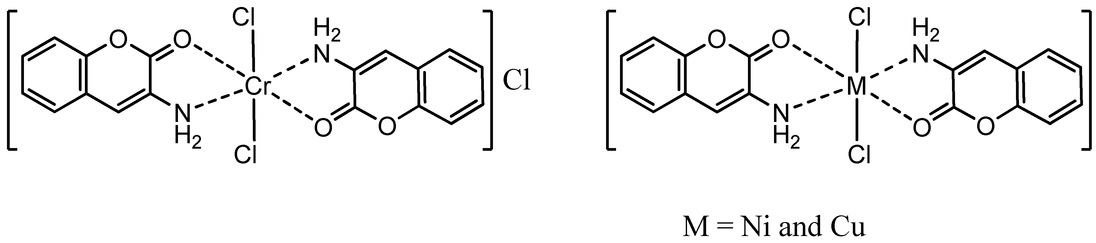
2.1. Chemistry

3-Aminocoumarin (Figure 1) is the key intermediate for the metal complexes synthesized in this work.
Figure 1. The structure of 3-aminocoumarin.
Molecules 16 06969 g001
The data for the minimized geometry and the 3d-geometrical structure of 3-aminocoumarin (Figure 2) show that the atomic charges have been affected by the presence of the ring substituent, as shown in Table 1.
Figure 2. The 3d structure of 3-aminocoumarin.
Molecules 16 06969 g002
Table 1. The atomic charges of 3-aminocoumarin.
AtomChargeAtomChargeAtomCharge
O(1)−0.0576918C(7)−0.115268H(13)0.0181674
C(2)0.546493C(8)−0.0752691H(14)0.0223331
O(3)−0.580016C(9)−0.101239H(15)0.0253347
C(4)0.144636C(10)−0.110194H(16)0.0249427
C(5)−0.359074C(11)0.197216H(17)0.0281505
C(6)0.00167107N(12)0.205349H(18)0.092316
H(19)0.0921441
The data obtained show that the highest atomic charge in the 3-aminocoumarin molecule is located at [O(3)-0.580016], while the next highest charge value are at [C(5)-0.359074] and [C(7)-0.115268]. These data show clearly that oxygen atom [O(3)] is the most reactive toward the bonding with the metal. The determined bond angle, twist angle, 3D geometrical structure (Figure 2) and stereochemistry [C(4)-C(5):(E)], indicate that this molecule is planar.
The synthesis of 3-aminocoumarin was performed by vigorously refluxing 2-hydroxybenzaldehyde with 2-acetamidoacetic acid, using piperidine as the strong base. The end of the reaction involves the removal of the acetyl group and the production of 3-aminocoumarin in 17% yield (Scheme 1).
Scheme 1. Formation of the ligand.
Molecules 16 06969 g010
The mechanism of the reaction may be explained by a carbanion mechanism (Scheme 2).
Scheme 2. Reaction mechanism of formation of the ligand.
Molecules 16 06969 g011
The complexes were synthesized by the reactions of 3-aminocoumarin with the metal ions, in which the ligand behaves as a bidentate ligand through its oxygen and nitrogen atoms. The analytical data of these complexes are presented in Table 2.
Table 2. Analytical data for the metal complexes.
No.CompoundsM:LM.P. °CYield %Elemental analysis (calculated)Elemental analysis (found)
C%H%N%M%C%H%N%M%
C1[Cr(L)2Cl2]Cl1:21894044.982.945.8310.8245.022.425.3010.65
C2[Ni(L)2Cl2]1:2221d5547.843.126.2012.9947.593.076.0912.53
C3[Cu(L)2Cl2]1:2209d6747.333.096.1313.9146.982.805.9213.99
All the complexes are fairly stable and can be stored for long periods at room temperature. The solid colored complexes are stable towards heat, air and moisture, and they are generally soluble in common organic solvents such as dimethylformamide or dimethylsulfoxide. The stability for the prepared complexes was studied theoretically using Density Function Theory (DFT). The total energy for the complexes was calculated and it was shown that the copper complex is the most stable and the chromium complex is the least stable as follows: Cu(II) > Ni(II) > Cr(III). In all our work on metal complexes (in which we have used different ligands with the ion metals Cu, Ni, and Cr), we found out that the copper complex is consistently the most stable.
Figure 3. The proposed structure for the complexes.
Molecules 16 06969 g003

2.1.1. Elemental Analysis

The compositions of the complexes are summarized in Table 2. The analytical data of the complexes are consistent with the proposed molecular structures (Figure 3), assuming molar metal to ligand ratios of 1:2. The C, H, N and M contents (both theoretically calculated values and actual values) are in accordance with the formula ML2Cl2 indicating that the 3-aminocoumarin complexes are neutral, except for the Cr(III) complex with formula [ML2Cl2]Cl (Table 2). This can be explained by the absence of any deprotonating agent during the synthesis.

2.1.2. Infrared Spectra

A study and comparison of the infrared spectra of 3-aminocoumarin and its complexes imply that 3-aminocoumarin is bidentate, with the carbonyl oxygen and amine nitrogen as the two coordination sites. In the infrared spectra of the complexes a considerable negative shift of the C=O group is observed, indicating a decrease in the stretching force-constant of the C=O bond as a consequence of coordination through the carbonyl oxygen atom. The carbonyl band for the parent 3-aminocoumarin ligand was located at 1709 cm−1, but for the Cr(III), Ni(II) and Cu(II) complexes these peaks were shifted to lower frequencies, namely 1691 cm−1, 1701 cm−1, and 1685 cm−1, respectively. The NH2 bands of 3-aminocoumarin were at 3407 and 3299 cm−1, but for the complexes the NH2 bands also shift to lower frequencies [Cr-complex ones were at 3315 and 3201 cm−1, Ni-complex at 3310 and 3181 cm−1 and finally the Cu-complex ones were at 3326 and 3217 cm−1, respectively] [38]. Additional evidence for the coupling between the ligand and metals ion were the M-N and M-O bands occurring at 455-467 cm-1and 486-503 cm−1, respectively (Table 3).
Table 3. FT-IR (cm−1) bands of metal complexes.
NO.N–HC=OM–ClνM–NνM–O
C13315, 32011691343467503
C23310, 31811701330455501
C33326, 32171685330459486

2.1.3. 1H-NMR Spectra

The 1H-NMR spectrum of the ligand (L) showed characteristic signals due to the N-H protons at 7.97 ppm. Moreover the peaks observed between 7.40 ppm and 7.61 ppm were assigned to the C–Haromatic protons of the ligand. The signal at 6.11 ppm may be assigned to the =C–Hvinyl proton.

2.1.4. Molar Conductance

The molar conductance values of all the complexes determined in nitrobenzene at room temperature are given in Table 4. The value for the Cr(III) complex indicates that one chloride ion is present outside the coordination sphere. The molar conductance values Ni(II) and Cu(II) complexes are quite low to correspond to an ionic complex; hence, these complexes are considered to be neutral and the chloride ions are assumed to be situated within the coordination sphere.
Table 4. Physical data of the synthesized compounds.
No.λmax cm−1Magnetic moment µ (B.M.)ʌ ohm 1 cm2 mol−1Structure
C115217, 20112, 321254.190Octahedral
C215890, 319152.620Octahedral
C312117, 15812, 220761.622Octahedral

2.1.5. Magnetic Moment and UV-Vis Spectra

The ultraviolet spectrum of the synthesized 3-aminocoumarin showed two absorption bands. The position of the first band, which represents the (π → π*) transition, was at 239 nm, while the second band (which has higher intensity than the first one due to the conjugated system) appeared at 289 nm and represents the (n → π*) transition. Generally, the bands of the newly synthesized complexes are either shifted to shorter or longer wavelengths than those of 3-aminocoumarin, but the high intensity of the band is an indication for complex formation.
In these complexes the bands observed over 300 nm could be assigned to nitrogen-metal charge transfer absorption. The electronic absorption bands for the ligand and complexes are classified into two distinct groups, first those that belong to ligand transitions appeared in the UV region, while d-d transitions appeared in the visible region. These transitions are releted to the structures of the complexes (Table 4). The Cr(III) complex showed three bands with absorbance maxima at 15,217 cm−1, 20,112 cm−1 and 32,125 cm−1 which were assigned to the 4A2g(F) → 4T2g(F), 4A2g(F)4T1g(F), 4A2g(F)4T1g(P) absorption bands, respectively. These transitions, combined with the measured 4.1 B.M. magnetic moment, suggest a high-spin octahedral geometry for the Cr(III) complex.
The nickel complex is paramagnetic, with a room temperature magnetic moment of 2.6 B.M., which is consistent with an octahedral field. The electronic absorption spectrum showed two absorption bands at 15,890 cm−1 and 31,915 cm−1 which are considered to correspond to 3A2g(F)3T1g and 3A2g(F)3T1g(P) (Table 4), respectively.
The room temperature magnetic moment of 1.6 B.M. indicates an octahedral structure for the Cu(II) ion complex. The electronic absorption spectrum of Cu(II) complex shows three bands at 12,117 cm−1, 15,812 cm−1 and 22,076 cm−1, assigned to 2B1g → 2A2g, 2B1g → 2B2g and 2B1g → 2Eg, respectively [38].

2.1.6. Suggested Stereostructures of the Complexes

The proposed structures of complexes based on the above mentioned data (UV-Vis, IR, and NMR spectra, conductivity, molar ratio and magnetic properties) are depicted in Figure 3. The chlorides in the metal complexes of Ni(II) and Cu(II), are in the octahedral coordination sphere, but for the Cr(III) complex, the chlorides are in spherical coordination and the third chloride is in the outer of the spherical coordination, as implied by the molar conductance.

2.2. Pharmacology

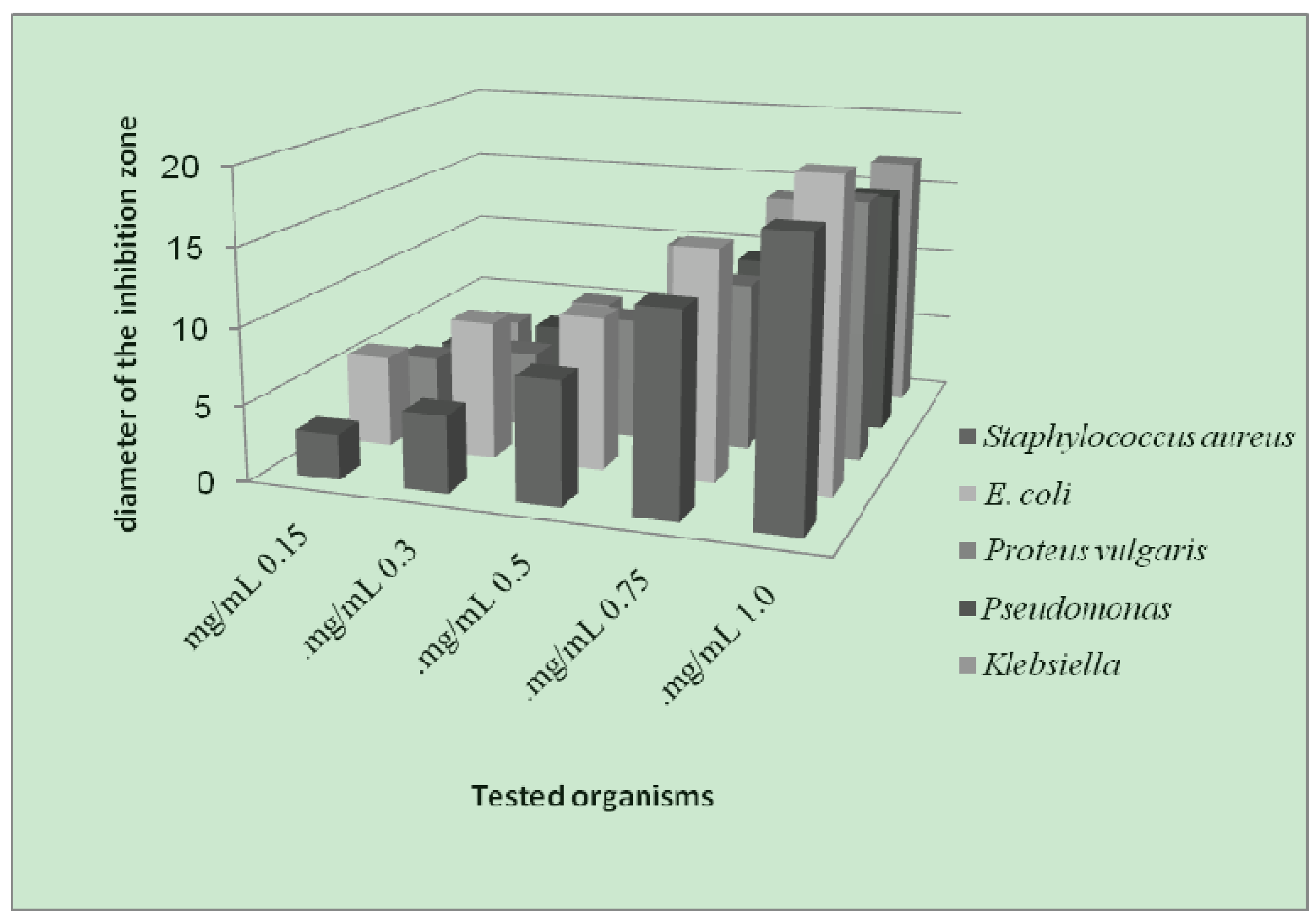
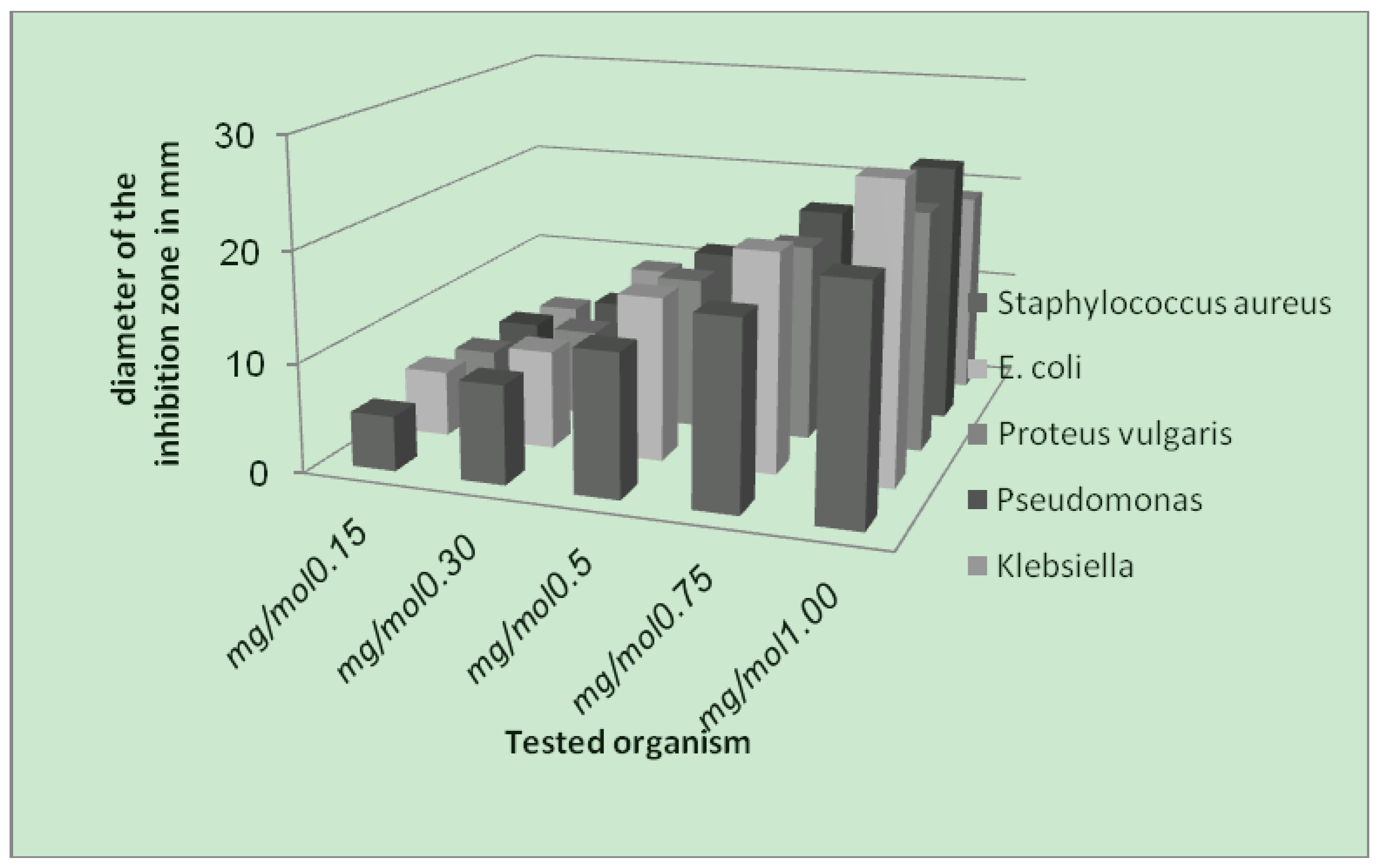
2.2.1. Antibacterial Activity

The antibacterial screening data show that the complexes exhibit antimicrobial properties, and we note that the metal chelates exhibit more inhibitory effects than the parent 3-aminocoumarin ligand. The increased activity of the metal chelates can be explained on the basis of chelation theory [39]. It is known that chelation tends to make the ligand act as powerful and potent bactericidal agents, thus killing more of the bacteria than the ligand alone. It is observed that, in a complex, the positive charge of the metal is partially shared with the donor atoms present in the ligands, and there may be π-electron delocalization over the whole chelating space [40]. This increases the lipophilic character of the metal chelate and favours its permeation through the lipoid layer of the bacterial membranes. The increased lipophilic character of these complexes seems to be responsible for their enhanced potent antibacterial activity. It may be suggested that these complexes deactivate various cellular enzymes, which play a vital role in various metabolic pathways of these microorganisms. It has also been proposed that the ultimate action of the toxicant is the denaturation of one or more proteins of the cell, which as a result, impairs normal cellular processes. There are other factors which also increase the activity, which are solubility, conductivity, and bond length between the metal and the ligand.
Figure 4. Effect of the Cr-complex toward test organisms.
Molecules 16 06969 g004
Figure 5. Effect of Ni-complex toward test organisms.
Molecules 16 06969 g005
Figure 6. Effect of Cu-complex toward test organisms.
Molecules 16 06969 g006
As a result from the study of antibacterial of the prepared metal complexes (Figure 4, Figure 5 and Figure 6), the following conclusions may be stated:
  • (1). Generally, the result of prepared complexes exhibited antibacterial activity toward E. coli bacteria was more than the inhibition on other types of bacteria.
  • (2). The copper complex has more activity toward all the kinds of tested bacteria, compared to other complexes. The stability of the copper complex and the coupling with the ligand may be the reasons for this activity against bacteria.

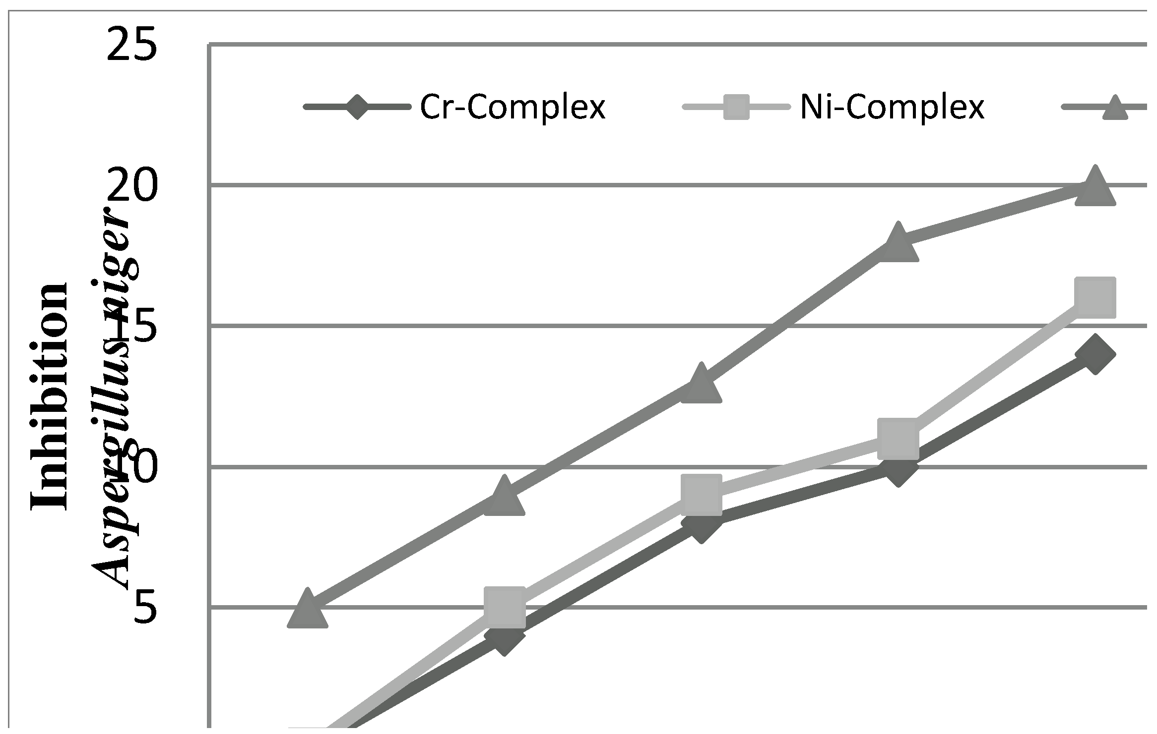
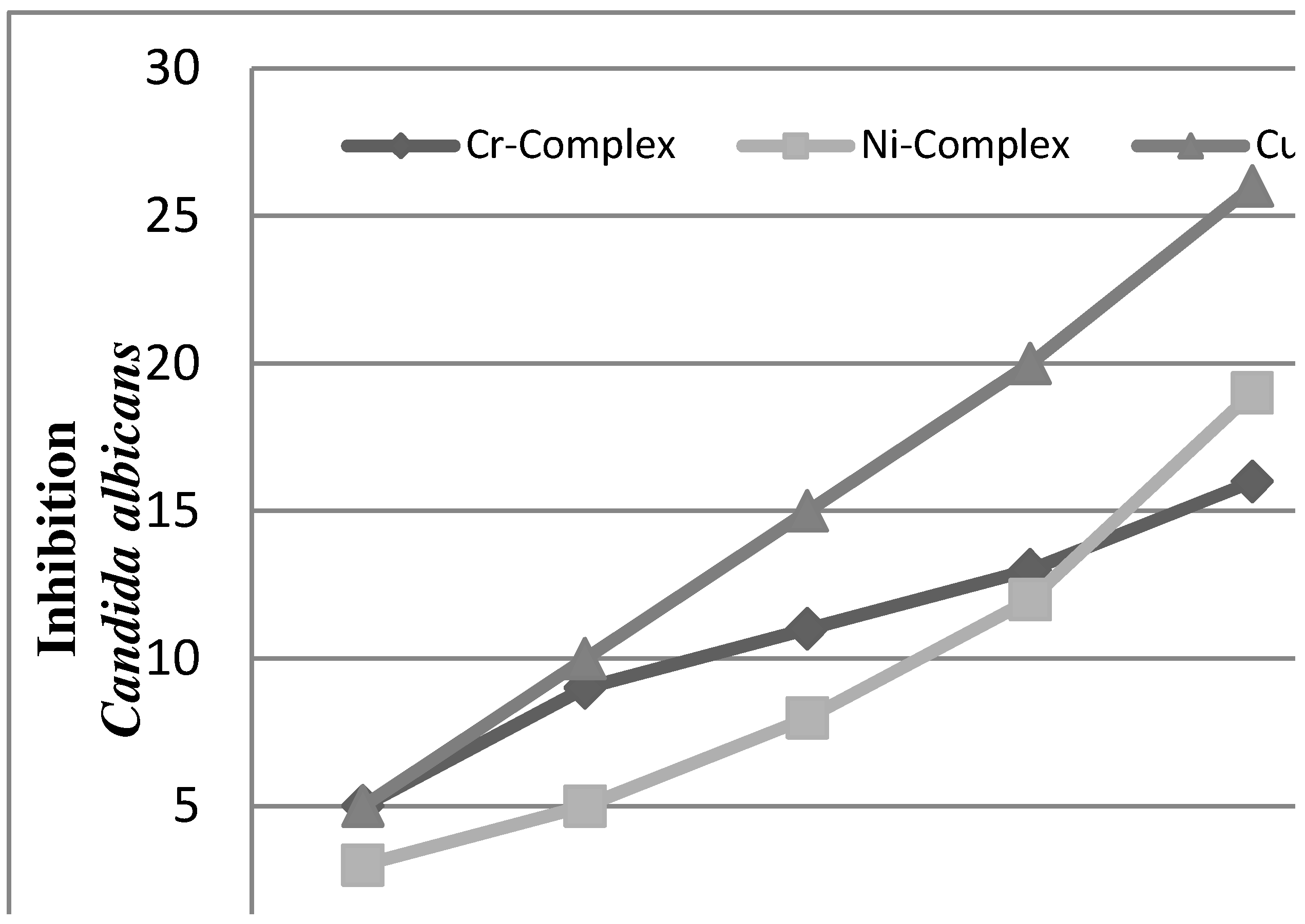
2.2.2. Antifungal Activities

Figure 7. The effect of complexes toward Aspergillus niger.
Molecules 16 06969 g007
In vitro antifungal screening effects of the investigated compounds were tested against some fungal species (Aspergillus niger and Candida albicans). The Cu(II) complex was found to exhibit antifungal activity against all the fungi in this study (Figure 7 and Figure 8).
Figure 8. The effect of complexes toward Candida albicans.
Molecules 16 06969 g008

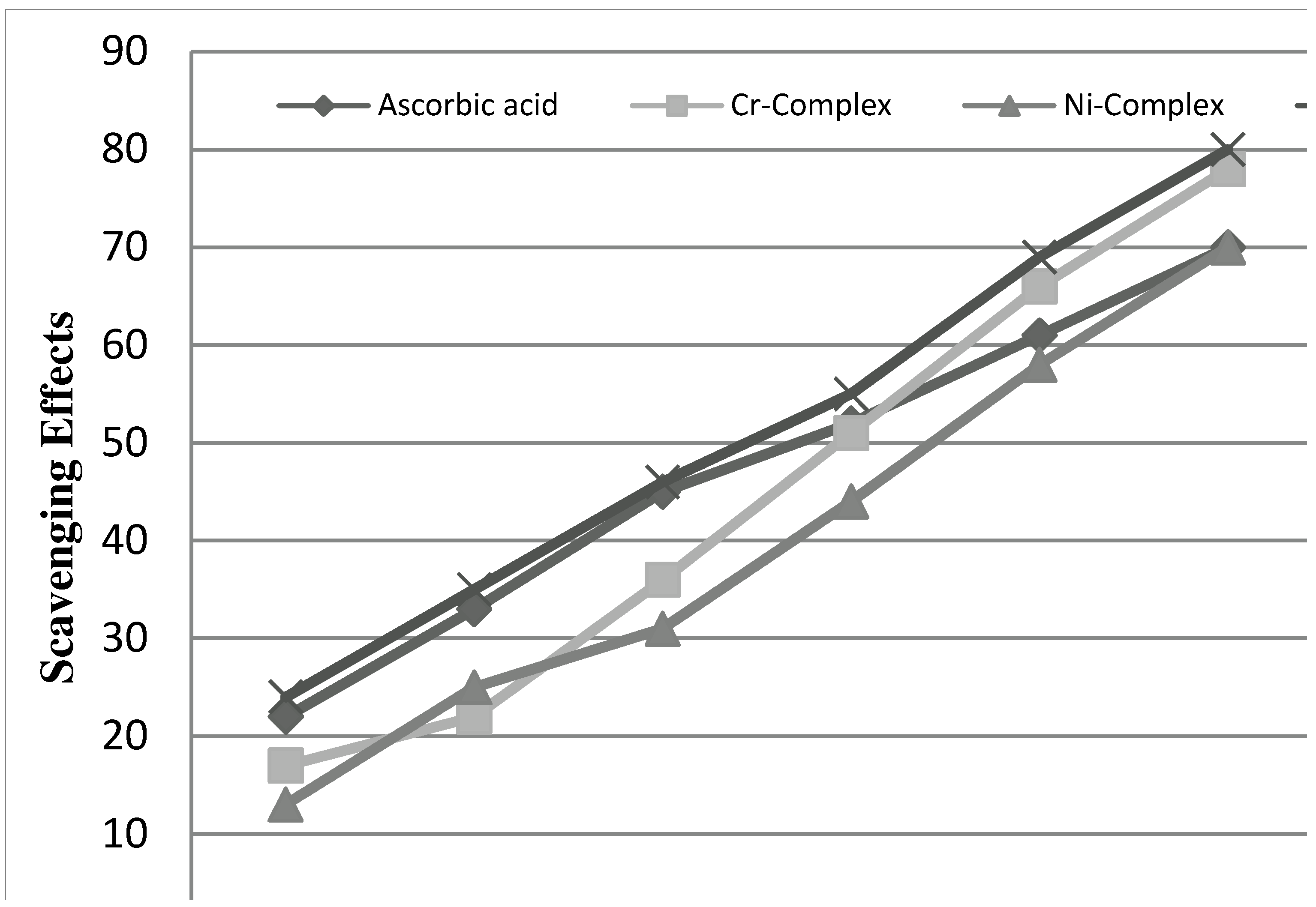
2.2.3. Radical Scavenging Activity

The 2,2′′-diphenyl-1-picrylhydrazyl (DPPH) radical assay provides an easy and rapid way to evaluate the antiradical activities of antioxidants. Determination of the reaction kinetic types DPPHH is a product of the reaction between DPPH• and an antioxidant:
Molecules 16 06969 i001
The reversibility of the reaction is evaluated by adding DPPHH at the end of the reaction. If there is an increase in the percentage of remaining DPPH• at the plateau, the reaction is reversible, otherwise it is a complete reaction.
DPPH was used as stable free radical electron accepts or hydrogen radical to become a stable diamagnetic molecule [41]. DPPH is a stable free radical containing an odd electron in its structure and usually used for detection of the radical scavenging activity in chemical analysis. [42]. The reduction capability of DPPH radicals was determined by decrease in its absorbance at 517 nm induced by antioxidants. [43]. The graph was plotted with percentage scavenging effects on the y-axis and concentration (µg/mL.) on the x-axis. The scavenging ability of the metal complexes was compared with ascorbic acid as a standard. The metal complexes showed good activities as a radical scavenger compared with ascorbic acid, Figure 9. These results were in agreement with previous metallic complexes studies where the ligand has the antioxidant activity and it is expected that the metal moiety will increase its activity [44,45,46].
Figure 9. Scavenging effect of metal complexes, and ascorbic acid at different concentrations (15, 30, 45, 60, 80 and 100 μg/mL).
Molecules 16 06969 g009

3. Experimental

3.1. General

All chemicals used in this work were of reagent grade (supplied by either Sigma-Aldrich or Fluka) and used without farther purifications. The FTIR spectra were recorded in the (4000–200) cm−1 range on cesium iodide disks using a Shimadzu FTIR 8300 Spectrophotometer. Proton NMR spectra were recorded on Bruker-DPX 300 MHz spectrometer with TMS as internal standard. The UV-Visible spectra were measured in ethanol using a Shimadzu UV-Vis. 160A spectrophotometer in the range (200–1000) nm. Magnetic susceptibility measurement for complexes was obtained at room temperature using a Magnetic Susceptibility Balance Model MSB-MKI. The flame atomic absorption of a Shimadzu AA-670 elemental analyzer was used for metal determination. Elemental micro analysis was carried out using a CHN elemental analyzer model 5500-Carlo Erba instrument. A Gallenkamp M.F.B.600.010 F melting point apparatus was used to measure the melting points of all the prepared compounds.

3.2. Chemistry

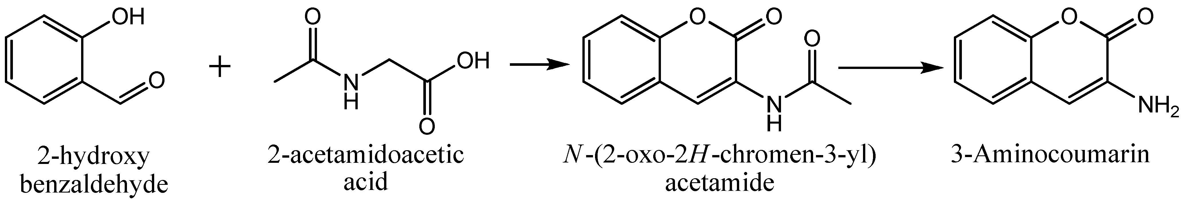
3.2.1. Synthesis of the Ligand

A mixture of 2-acetamidoacetic acid (58.5 g, 0.5 mol) and 2-hydroxybenzaldehyde (92.7 g, 0.76 mol) in acetic anhydride (0.5 mL, 0.053 mol) with few drops of piperidine was refluxed at 130 °C for 8 hours. After cooling to room temperature the product is separated out and washed with diethyl ether several times, dried and then recrystallized from ethanol to give N-(2-oxo-2H-chromen-3-yl)acetamide in 85% yield. The latter (5 g, 0.02 mol) was refluxed in ethanol (25 mL), containing conc. hydrochloric acid (2 mL) for 4 hours. The crystallized compound (impure 3-aminocoumarin) mixture was cooled, poured on to ice before neutralization with sodium bicarbonate. The solid was separated out, filtered and left to dry. 3-Aminocoumarin was recrystallized from ethanol, giving a pale yellow powder, yield 17%, m.p. 129 °C (lit. [47] 130 oC); 1H-NMR (CDCl3): δ 7.97 (s, NH2), δ 6.11 (s, 1H) for –C=C-H), δ 7.40-7.61 (m, 1H) for aromatic ring); IR: 3407 and 3299 cm−1 (NH2, amine), 1709 cm−1 (C=O, lactone); Anal. Calcd. for C9H7NO2: C 67.07%, H 4.38%, N 8.69%. Found: C 66.98%, H 4.27%, N 8.58%.

3.2.2. Synthesis of the Complexes

Metal salts (CrCl3·6H2O, NiCl2·6H2O and CuCl2·2H2O; 5 mmol) in hot ethanol (20 mL) were mixed with hot ethanolic solution of the 3-aminocoumarin (1.61 g, 10 mmol) and refluxed for 4 hours. On cooling the contents, the colored complexes were separated out. The products were filtered [48], washed with cold 50% ethanol and dried in vacuum over P4O10. Purity of the complexes was checked by Thin Layer Chromatography (TLC). The TLC plates (20 × 10 cm) were recoated silica gel on aluminum 60F – 254, with a stationary phase thickness of about 0.5 mm. Five μL each of test solution was applied on each plate. The TLC plate was placed in a saturated chromatographic tank containing an ethyl acetate-methanol-acetone (25:25:50) solvent system, Rf values for Cu(II), Ni(II) and Cr(III) were 0.31, 3.0 and 4.3, respectively.

3.2.3. Study of Complex Formation in Solution

The complexes of the 3-aminocoumarin with metal ions were studied in dimethylformamide (DMF), in order to determine the M:L ratio in the complex following the molar ratio method. Several series of solutions were prepared having constant concentration (10−3 M) of the metal ion and (L). The M:L ratios were determined from the relationship between the absorption of light [49] and the M:L mole ratio. The results of complexes formation in solution were listed in Table 2.

3.3. Pharmacology

3.3.1. Evaluation of Antibacterial Activities

The in vitro antibacterial effects of the complexes were evaluated against a sp. of Gram-positive bacteria (Staphylococcus aureus) and four Gram-negative bacteria (Escherichia coli, Pseudomonas aeruginosa, Klebsiella pneumonia and Proteus vulgaris) by the disc diffusion method [50] using nutrient agar medium. The bacteria were sub-cultured in the agar medium and were incubated for 24 h. at 37 °C. The discs having a diameter of 5 mm, then soaked in the test solutions (Sterile filter paper discs, Whatman No. 1.0) with the appropriate equivalent amount of the metal complexes dissolved in sterile dimethyl sulphoxide (DMSO) at concentrations of 1–10 mg/disc) and were placed in Petri dishes on an appropriate medium previously seeded with organisms and stored in an incubator for the above mentioned period of time. The inhibition zone around each disc was measured and the results recorded in the form of inhibition zones (diameter, mm). To clarify any effect of DMSO on the biological screening, separate studies were carried out using DMSO as control and it showed no activity against any bacterial strains.

3.3.2. Evaluation of Antifungal Assay

Antifungal activity [51,52], based on the determined growth inhibition rates of the mycelia of strain (Aspergillus niger and Candida albicans) in Potato Dextrose Broth medium (PDB). Under aseptic conditions, one mL of spore suspension (5 × 10⁶ cfu/mL) of tested fungi was added to 50 mL PDB medium in a 100 mL Erlenmeyer flask. Appropriate volumes of tested metal complexes were added to produce concentrations ranging from 10 to 100 μg mL−1. Flasks were incubated at 27 ± 1 °C in the dark for 5 days and then the mycelium was collected on filter papers. The filter papers were dried to constant weight and the level of inhibition, relative to the control flasks was calculated from the following formula:
Molecules 16 06969 i002
where T = weight of mycelium from test flasks and C = weight of mycelium from control flasks.

3.3.3. Evaluation of Antioxidant Activity

Stock solution (1 mg/mL) was diluted to final concentrations of 20–100 μg/mL. Ethanolic DPPH solution (1 mL, 0.3 mmol) was added to sample solutions in DMSO (3 mL) at different concentrations (50–300 μg/mL) [53]. The mixture was shaken vigorously and allowed to stand at room temperature for 30 min. The absorbance was then measured at 517 nm in a UV-Vis Spectrophotometer. The lower absorbance of the reaction mixture indicates higher free radical scavenging activity. Ethanol was used as the solvent and ascorbic acid as the standard. The DPPH radical scavenger was calculated using the following equation:
Molecules 16 06969 i003
where Ao is the absorbance of the control reaction and A1 is the absorbance in the presence of the samples or standards. A0-A1

4. Conclusions

In this study, Cr(III), Ni(II) and Cu(II) complexes of 3-aminocoumarin have been successfully synthesized and characterized by using various spectroscopic methods, elemental analysis, magnetic moment and molar conductance studies. The synthesized complexes were tested for antioxidant and antimicrobial activities. Out of these complexes, Cu(II) indicated significant antimicrobial activities as compared to either Cr(III) or Ni(II). In addition the Cu(II) complex is also found to be a superior antioxidant complex as compared to ascorbic acid.

References

  1. Raman, N.; Sakthivel, A.; Rajasekaran, K. Synthesis and spectral characterization of antifungal sensitive Schiff base transition metal complexes. Mycobiology 2007, 35, 150–153. [Google Scholar] [CrossRef]
  2. Rice, S.A.; Givskov, M.; Steinberg, P.; Kjelleberg, S. Bacterial signals and antagonists: The interaction between bacteria and higher organisms. J. Mol. Microbiol. Biotechnol. 1999, 1, 23–31. [Google Scholar]
  3. Ironmonger, A.; Whittaker, B.; Andrew, J.; Baron, B.; Chris, J.; Alison, E.; Ashcroft, G.; Nelson, A. Scanning conformational space with a library of stereo- and regiochemically diverse aminoglycoside derivatives: The discovery of new ligands for RNA hairpin sequences. Org. Biomol. Chem. 2007, 5, 1081–1086. [Google Scholar] [CrossRef]
  4. Kumar, G.; Kumar, D.; Devi, S.; Verma, R.; Johari, R. Synthesis, spectral characterization of biologically active compounds derived from oxalyldihydrazide and 5-tert-butyl-2-hydroxy-3-(3-phenylpent-3-yl)benzaldehyde and their Cu(II), Ni(II) and Co(II) Complexes. Int. J. Eng. Sci. Technol. 2011, 3, 1630–16354. [Google Scholar]
  5. Parmer, S.; Kumar, R. Substituted quinazolone hydrazides as possible antituberculous agents. J. Med. Chem. 1968, 11, 635–636. [Google Scholar]
  6. Reddy, Y.D.; Somayajulu, V.V. Synthesis, spectra and physiological activity of 7H-pyrano[3,2-c]benzoxazole-7-ones. J. Ind. Chem. Soc. 1981, 58, 599–601. [Google Scholar]
  7. Soine, T.O. Naturally occuring coumarins and related physiological activities. J. Pharm. Sci. 1964, 53, 231–264. [Google Scholar] [CrossRef]
  8. Jund, L.; Corse, J.; King, A.D.; Bayne, H.; Mihara, K. Antimicrobial properties of 6,7-dihydroxy-, 7,8-dihydroxy-, 6-hydroxy- and 8- hydroxycoumarins. Phytochemicals 1971, 10, 2971–2974. [Google Scholar] [CrossRef]
  9. Abd Allah, O.A. Synthesis and biological studies of some benzopyrano[2,3-c]pyrazole derivatives. II Farmacogn. 2000, 55, 641–649. [Google Scholar] [CrossRef]
  10. Abdel-Al, E.H.; Al-Ashamawi, M.I.; Abd El-Fattah, B. Synthesis and antimicrobial testing of certain oxadiazoline and triazole derivatives. Die Pharm. 1983, 38, 833–838. [Google Scholar]
  11. Bhamaria, R.P.; Bellare, R.A.; Deliwala, C.V. In intro effect of 1-acyl-4-alkyl-(or aryl)-thiosemicarbazides 1-(5-chlorosalicylidine)-4-alkyl-(or aryl)-thiosemicarbazones and some hydrazones of 5-chlorosalicylaldehyde against pathogenic bacteria including mycobacterium tuberculosis (H37Rv). Indian J. Exp. Biol. 1968, 6, 62–63. [Google Scholar]
  12. Dutta, M.M.; Goswani, B.N.; Kataky, J.C.S. Studies on biologically active heterocycles. Part I. Synthesis and antifungal activity of some new aroyl hydrazones and 2,5-disubstituted-1,3,4-oxadiazoles. J. Heterocycl. Chem. 1986, 23, 793–795. [Google Scholar] [CrossRef]
  13. Gupta, A.K.S.; Garg, M.; Chandra, U. Synthesis of some new Mannich bases derived from substituted benzimidazole, benzoxazol-2-one, benzoxazol-2-thione, oxadiazol-2-thiones and their biological activities. J. Indian Chem. Soc. 1979, 56, 1230–1232. [Google Scholar]
  14. Wenner, W. Antitubercular agents. Derivatives of pyridinecarboxylic acid hydrazides. J. Org. Chem. 1953, 18, 1333–1337. [Google Scholar] [CrossRef]
  15. Mansour, A.K.; Eid, M.M.; Khalil, N. Synthesis and reactions of some new heterocyclic carbohydrazides and related compounds as potential anticancer agents. Molecules 2003, 8, 744–755. [Google Scholar] [CrossRef]
  16. Hoult, J.R.S.; Paya, M. Pharmacological and biochemical actions of simple coumarins: Natural products with therapeutic potential. Gen. Pharmacol. 1996, 27, 713–722. [Google Scholar] [CrossRef]
  17. Matsumoto, P.; Hanawalt, C. Histone H3 and heat shock protein GRP78 are selectively cross-linked to DNA by photoactivated gilvocarcin V in human fibroblasts. Cancer Res. 2000, 60, 3921–3926. [Google Scholar]
  18. Kelly, V.P.; Ellis, E.M.; Manson, M.M. Chemoprevention of aflatoxin B1 hepatocarcinogenesis by coumarin, a natural benzopyrone that is a potent inducer of aflatoxin B1-aldehyde reductase, the glutathione S-transferase A5 and P1 subunits, and NAD(P)H: Quinone oxidoreductase in rat liver. Cancer Res. 2000, 60, 957–969. [Google Scholar]
  19. Kirkiacharian, S.; Thuy, D.T.; Sicsic, S.; Bakhchinian, R.; Kurkjian, R.; Tonnaire, T. Structure-activity relationships of some 3-substituted-4-hydroxycoumarins as HIV-1 protease inhibitors. II Farmacogn. 2002, 57, 703–708. [Google Scholar] [CrossRef]
  20. Suzuki, D.; Yu, M.; Xie, L.; Morris, S.L.; Lee, K.H. Recent progress in the development of coumarin derivatives as potent anti-HIV agents. Med. Res. Rev. 2003, 23, 322–345. [Google Scholar] [CrossRef]
  21. Marshall, E.M.; Ryles, M.; Butler, K.; Weiss, L. Treatment of advanced renal cell carcinoma (RCC) with coumarin and cimetidine: longterm follow-up of patients treated on a phase I trial. J. Cancer Res. Clin. Oncol. 1994, 120, 535–538. [Google Scholar]
  22. Fylaktakidou, K.C.; Hadjipavlou-Litina, D.J.; Litinas, K.E.; Nicolaides, D.N. Natural and synthetic coumarin derivatives with anti-inflammatory/antioxidant activities. Curr. Pharm. Des. 2004, 10, 3813–3833. [Google Scholar] [CrossRef]
  23. Tianzhi, Y.; Yuling, Z.; Duowang, F. Synthesis, crystal structure and photoluminescence of 3-(1-benzotriazole)-4-methyl-coumarin. J. Mol. Struct. 2006, 791, 18–22. [Google Scholar] [CrossRef]
  24. Yu, T.Z.; Zhao, Y.L.; Ding, X.S.; Fan, D.W.; Qian, L.; Dong, W.K. Synthesis, crystal structure and photoluminescent behaviors of 3-(1H-benzotriazol-1-yl)-4-methylbenzo[7,8]coumarin. J. Photochem. Photobiol. A 2007, 188, 245–251. [Google Scholar] [CrossRef]
  25. Ray, D.; Bharadwaj, P.K. A coumarin-derived fluorescence probe selective for magnesium. Inorg. Chem. 2008, 7, 2252–2254. [Google Scholar]
  26. Trenor, S.R.; Shultz, A.R.; Love, B.J.; Long, T.E. Coumarins in polymers: From light harvesting to photo-cross-linkable tissue scaffolds. Chem. Rev. 2004, 104, 3059–3077. [Google Scholar] [CrossRef]
  27. Tianzhi, Y.; Peng, Z.; Yuling, Z.; Hui, Z.; Jing, M.; Duowang, F. Synthesis and photoluminescent properties of two novel tripodal compounds containing coumarin moieties. Spectrochim. Acta Part A 2009, 73, 168–173. [Google Scholar] [CrossRef]
  28. Ruikui, C.; Xichuan, Y.; Haining, T.; Licheng, S. Tetrahydroquinoline dyes with different spacers for organic dye-sensitized solar cells. J. Photochem. Photobiol. A 2007, 189, 295–300. [Google Scholar] [CrossRef]
  29. Yu, T.Z.; Zhang, P.; Zhao, Y.L.; Zhang, H.; Meng, J.; Fan, D.W.; Dong, W.K. Photoluminescence and electroluminescence of a tripodal compound containing 7-diethylamino-coumarin moiety. J. Phys. D: Appl. Phys. 2008, 41, 235406–235413. [Google Scholar]
  30. Tang, C.W.; VanSlyke, S.A. Organic electroluminescent diodes. Appl. Phys. Lett. 1987, 51, 913–915. [Google Scholar] [CrossRef]
  31. Musa, A.Y.; Khadom, A.A.; Kadhum, A.A.H.; Mohamad, A.B.; Takriff, M.S. Kinetic behavior of mild steel corrosion inhibition by 4-amino-5-phenyl-4H-1,2,4-trizole-3-thiol. J. Taiwan Inst. Chem. Eng. 2010, 41, 126–128. [Google Scholar] [CrossRef]
  32. Tianzhi, Y.; Peng, Z.; Yuling, Z.; Hui, Z.; Jing, M.; Duowang, F. Synthesis, characterization and high-efficiency blue electroluminescence based on coumarin derivatives of 7-diethylaminocoumarin-3-carboxamide. Org. Electron. 2009, 10, 653–660. [Google Scholar] [CrossRef]
  33. Asish, R.; Arunima, M.; Raghunath, S. Synthesis of biologically potent new 3-(heteroaryl)aminocoumarin derivatives via Buchwald–Hartwig C–N coupling. Tetrahedron Lett. 2010, 51, 1099–1102. [Google Scholar] [CrossRef]
  34. Amit, A.; Jamie, K.; Chad, C.; Natasha, D.; Graham, J. Hydrolysis-free synthesis of 3-aminocoumarins. Tetrahedron Lett. 2007, 48, 5077–5080. [Google Scholar] [CrossRef]
  35. Yadav, L.D.S.; Singh, S.; Rai, V.K. A one-pot [Bmim]OH-mediated synthesis of 3-benzamidocoumarins. Tetrahedron Lett. 2009, 50, 2208–2212. [Google Scholar] [CrossRef]
  36. Guru, P. Microbial Studies on Some Coordination Compound of Metals with Tetracycline. J. Appl. Chem. Res. 2010, 12, 7–16. [Google Scholar]
  37. Mishra, A.P.; Mishra, R.K.; Shrivastava, S.P. Structural and antimicrobial studies of coordination compounds of VO(II), Co(II), Ni(II) and Cu(II) with some Schiff bases involving 2-amino-4-chlorophenol. J. Serbian Chem. Soc. 2009, 74, 523–535. [Google Scholar] [CrossRef]
  38. Al-Amiery, A.A.; Al-Majedy, K.; Abdulhadi, S.A. Design, synthesis and bioassay of novel metal complexes of 3-amino-2-methylquinazolin-4(3H)-one. Afr. J. Pure Appl. Chem. 2009, 3, 218–227. [Google Scholar]
  39. Sengupta, S.K.; Pandey, O.P.; Srivastava, B.K.; Sharma, V.K. Synthesis, structural and biochemical aspects of titanocene and zirconocene chelates of acetylferrocenyl thiosemicarbazones. Transit. Met. Chem. 1998, 23, 349–353. [Google Scholar] [CrossRef]
  40. Rohaya, A.; Abdul Manaf, A.; Daud, A.; Israf, T.; Nor, H.; Khozirah, S.; Nordin, H. Antioxidant, radical-scavenging, anti-inflammatory, cytotoxic and antibacterial activities of methanolic extracts of some Hedyotis species. Life Sci. 2005, 76, 1953–1964. [Google Scholar] [CrossRef]
  41. Soares, J.R.; Dinis, T.C.P.; Cunha, A.P.; Almeida, L.M. Antioxidant activities of some extracts of thymus zygis. Free Radic. Res. 1997, 26, 469–478. [Google Scholar] [CrossRef]
  42. Duh, P.D.; Tu, Y.Y.; Yen, G.C. Antioxidant activity of water extract of Harng Jyur (Chyrsanthemum morifolium Ramat). Lebensm. Wiss. Technol. 1999, 32, 269–277. [Google Scholar] [CrossRef]
  43. Matthaus, B. Antioxidant activity of extracts obtained from residues of different oilseeds. J. Agric. Food Chem. 2002, 50, 3444–3452. [Google Scholar]
  44. Bukhari, S.B.; Memon, S.; Mahroof-Tahir, M.; Bhanger, M.I. Synthesis, characterization and antioxidant activity copper-quercetin complex. Spectrochim. Acta A Mol. Biomol. Spectrosc. 2009, 71, 1901–1906. [Google Scholar] [CrossRef]
  45. Gabrielska, J.; Soczynska-Kordala, M.; Hladyszowski, J.; Zylka, R.; Miskiewicz, J.; Przestalski, S. Antioxidative effect of quercetin and its equimolar mixture with phenyltin compounds on liposome membranes. J. Agric. Food Chem. 2006, 54, 7735–7746. [Google Scholar] [CrossRef]
  46. Bukhari, S.B.; Memon, S.M.; Tahir, M.M.; Bhanger, I. Synthesis, characterization and investigation of antioxidant activity of cobalt-quercetin complex. J. Mol. Struct. 2008, 892, 39–46. [Google Scholar] [CrossRef]
  47. Tripathy, K.P.; Mukerjee, K.A. A Facile synthesis of 3-acylaminocoumarins. Indian J. Chem. 1987, 26, 61–62. [Google Scholar]
  48. Al-Amiery, A.A.; Al-Majedy, K.; Abdulreazak, H.; Abood, H. Synthesis, characterization, theoretical crystal structure and antibacterial activities of some transition metal complexes of the thiosemicarbazone (Z)-2-(pyrrolidin-2-ylidene)hydrazinecarbothioamide. Bioinorg. Chem. Appl. 2011, 2011. [Google Scholar] [CrossRef]
  49. Al-Majedy, K.; Al-Amiery, A.A.; Almoussaoy, H.H.; Khweter, R. Novel analytical method for the determination of theophylline in pharmaceutical preparations. J. Appl. Sci. Res. 2011, 7, 470–475. [Google Scholar]
  50. Al-Amiery, A.A.; Mohammed, A.; Ibrahim, H.; Abbas, A. Study the biological activities of tribulus terrestris extracts. World Acad. Sci. Eng. Technol. 2009, 57, 433–435. [Google Scholar]
  51. Daw, Z.Y.; EL-Baroty, G.S.; Mahmoud, A.E. Inhibition of Aspergillus parasiticus growth and aflatoxin production by some essential oils. Chem. Mikrobiol. Technol. Lebensm. 1994, 16, 129–135. [Google Scholar]
  52. Myiut, S.; Daud, W.R.W.; Mohamed, A.B.; Kadhum, A.A.H. Gas chromatographic determination of eugenol in ethanol extract of cloves. J. Chromatogr. B: Biomed. Appl. 1996, 676, 193–195. [Google Scholar]
  53. Chen, Y.; Wong, M.; Rosen, R.; Ho, C. 2,2-Diphenyl-1-picrylhydrazyl radical scavenging active components from Polygonum multiflorum Thunb. J. Agric. Food Chem. 1999, 47, 2226–2228. [Google Scholar] [CrossRef]
  • Sample Availability: Samples of 3-aminocoumarin and its complexes are available from the authors.

