Article

Unsymmetrical Mesoporphyrinic Complexes of Copper (II) and Zinc (II). Microwave-Assisted Synthesis, Spectral Characterization and Cytotoxicity Evaluation

Faculty of Pharmacy, “Carol Davila” University of Medicine and Pharmacy, 6 Traian Vuia St., 020956 Bucharest, Romania
Molecules 2011, 16(7), 5604-5617; https://doi.org/10.3390/molecules16075604
Submission received: 16 May 2011 / Revised: 27 June 2011 / Accepted: 29 June 2011 / Published: 30 June 2011

Abstract

:
New unsymmetrical mesoporphyrinic complexes, namely 5-(4-hydroxyphenyl)-10,15,20–tris-(4-carboxymethylphenyl)–21,23-Zn(II)-porphine and 5-(4-hydroxyphenyl)-10,15,20–tris-(4-carboxymethylphenyl)–21,23-Cu(II)-porphine, were synthesized using a microwave irradiation method. The structures of the porphyrinic complexes were confirmed using FT-IR, UV–Vis, EPR and NMR spectral data. The spectral absorption and emission properties of the porphyrinic complexes were studied in organic solvents of different polarities and the influence of solvent polarity on the wavelengths of the absorbance and fluorescence band maxima is described. The cytotoxicity evaluation of the porphyrinic complexes was performed on human colon adenocarcinoma cell line HT29 for different doses and incubation times. The obtained result indicates a lack of or low toxicity for both compounds, thus recommending them for further testing in light activation protocols.

1. Introduction

Metalloporphyrins have attracted attention for many decades because of their importance in the field of biomedicine, especially in diagnosis and treatment of cancer [1,2,3,4,5,6] as well as in photodynamic antimicrobial therapy [7,8,9,10].
Photodynamic therapy as a selective treatment method that involves the administration of a pharmaceutical formulation containing a tetrapyrrolic photosensitizer, its selective localization at the cellular level, followed by generation by irradiation with light in the red region of the visible spectrum of cytotoxic species such as singlet oxygen (1O2) that destroys the sick cells [11,12,13,14,15]. The development of new candidates for potential application as sensitizers in photodynamic therapy requires of them a high purity and simple production under laboratory conditions, a good localization at cellular and subcellular level, great selectivity for the malignant tissue, photoactivity at wavelengths higher than 600 nm, lack of toxicity in the absence of the exciting light, rapid elimination from the body after the treatment is performed and nontoxic metabolites [16,17,18,19,20,21].
Porphyrins and metalloporphyrins have also been investigated for use as fluorescent markers in cancer diagnosis because have appropriate fluorescence characteristics such as convenient emissions in the phototherapeutic window (~600 to 1,100 nm), high singlet oxygen quantum yield, large Stokes shifts, and reduced photolability [1,3,4]. For diagnostic and photodynamic therapeutic applications the biomedical efficiency of the porphyrinic structures is determined by its subcellular localization, which depends on the structural and physicochemical characteristics of the photosensitizer [22]. Therefore, depending on the nature of the peripheric substituents and the metallic ion, photosensitizers can be localized within the mitochondria, lysosomes, endoplasmic reticulum, Golgi apparatus and plasma membranes [23]. Thus, by modifying the charge density and its distribution at the periphery of the tetrapyrrolic macrocycle it is possible to control the route of these compounds to the target cells [24,25]. The purity of these compounds is also very important because the presence of trace amounts of impurities can introduce significant errors in the photodynamic activity.
This paper reports the microwave-assisted synthesis of some new unsymmetrical porphyrinic complexes, namely 5-(4-hydroxyphenyl)-10,15,20–tris-(4-carboxymethylphenyl)–21,23-Zn(II)-porphine (denoted as Zn(II)TCMPOHp - Figure 1) and 5-(4-hydroxyphenyl)-10,15,20–tris-(4-carboxy-methylphenyl)–21,23-Cu(II)-porphine (denoted as Cu(II)TCMPOHp - Figure 1), their spectral properties and cytotoxicity evaluation with a view to assessing their possible use in the photodiagnosis and photodynamic therapy of cancer.
Figure 1. General structures of 5-(4-hydroxyphenyl)-10,15,20–tris-(4-carboxymethyl-phenyl)–21, 23 M(II) porphines.
Molecules 16 05604 g001

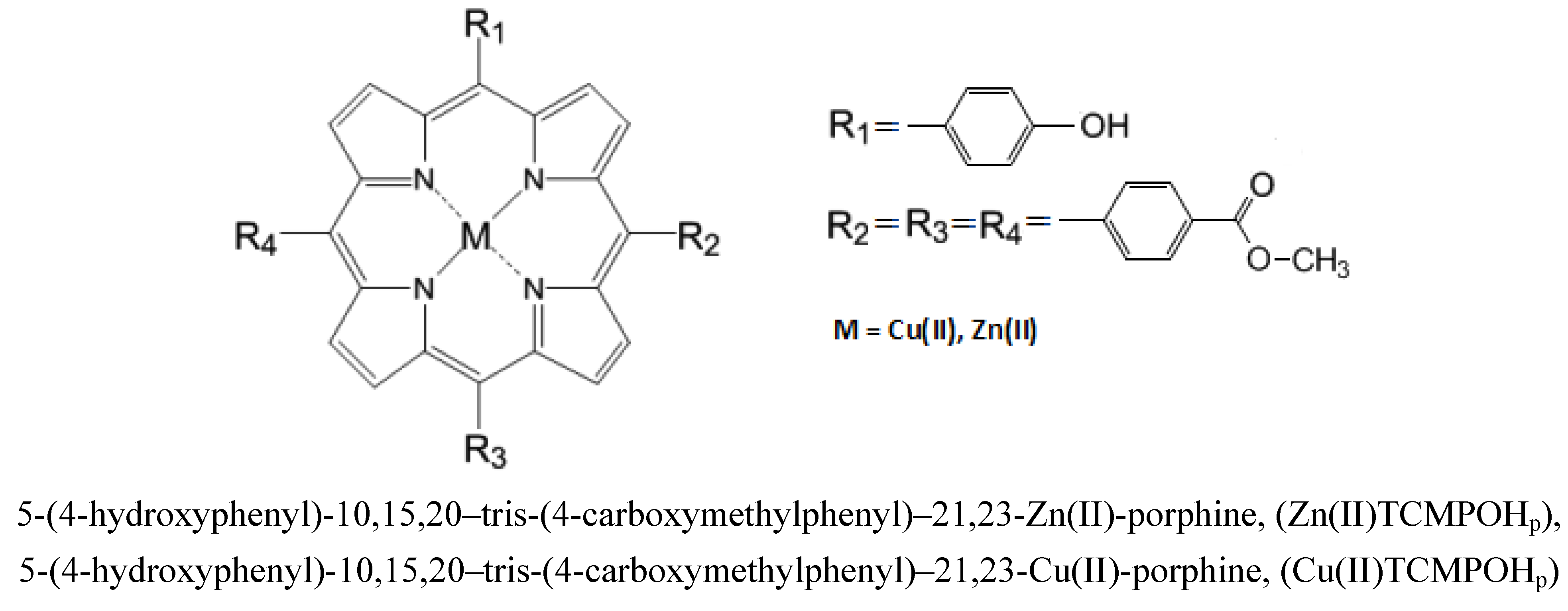
2. Results and Discussion

Microwave assisted synthesis has attracted considerable attention over the last few years and has been applied successfully in the preparation of tetrapyrrolic complexes. The main advantages of synthesis via microwave irradiation are significant reductions in reaction times and side reactions, increased yields, ease of purification and minimization of the amount of solvent used [26,27,28,29,30,31]. For thse these reasons in this paper the synthesis of the porphyrinic complexes was carried out using microwave irradiation assisted synthesis. The synthetic reactions have been successfully repeated several times with identical results and the porphyrinic complexes thus formed were then characterized by elemental analysis, IR, UV–Vis, NMR, EPR spectrometry and evaluated in terms of cytotoxicity using HT29 (human colon adenocarcinoma) cell line for different doses and incubation times.

2.1. Infrared Spectra

The most relevant results extracted from the IR spectra of the synthesized unsymmetrical porphyrinic complexes are presented in Table 1.
Table 1. Characteristic IR vibrations of the unsymmetrical porphyrinic complexes.
Characteristic vibrationWavenumber of the IR band (cm−1)
Zn(II)TCMPOHpCu(II)TCMPOHp
νO-H3490 m3489 m
νC-H2919 m2920 m
νC-H from -O-CH32850 m2850 m
νC=O1716 m1718 m
νC-N1595 s1596 s
νC=N1451 m1453 m
νC-H pyrrole1390 m1384 w
νC-O1158 m1157 m
δC-H1029 m1032 w
γC-C859 w857 w
γC-Npyrrole790 m789 m
The intensities of the signals are described as weak (w), medium (m), strong (s) and very strong (v.s.).
The IR spectra of the porphyrinic ligand [32] shows a medium band at ~3,310 cm−1 assigned to the N-H stretching vibration of the porphyrinic core, which not found in the spectrum of the mesoporphyrinic complexes. This observation supports the coordination of both nitrogen atoms of the porphyrinic core to the metal center.
The IR spectrum of the synthesized copper and zinc complexes includes typical vibration modes of both porphyrin macrocycle and phenyl substituents. Thus, the medium band at ~2,919 cm−1 is attributed to the C–H stretching of the phenyl groups. The bands at 3,490 cm−1 are assigned to the O-H stretching vibration of the –OH functional group in the Zn(II)TCMPOHP and Cu(II)TCMPOHP. The bands of the complexes in the range of 1,716–1,718 cm−1 are assigned to C=O stretching vibrations. In addition, in the IR spectra of the unsymmetric porphyrinic complexes the medium absorption band at about 1,158 cm−1 can be attributed to the C-O groups. The IR spectrum of the porphyrinic complexes indicates the presence of the -O-CH3 group at 2,850 cm−1. A band attributed to νC=N was present at 1,451–1,453 cm−1. In addition, the band at about 1,595 cm−1 can be assigned to C-N stretching vibrations and the band at ~1,390 cm−1 includes the C–H stretching of the pyrrole ring.

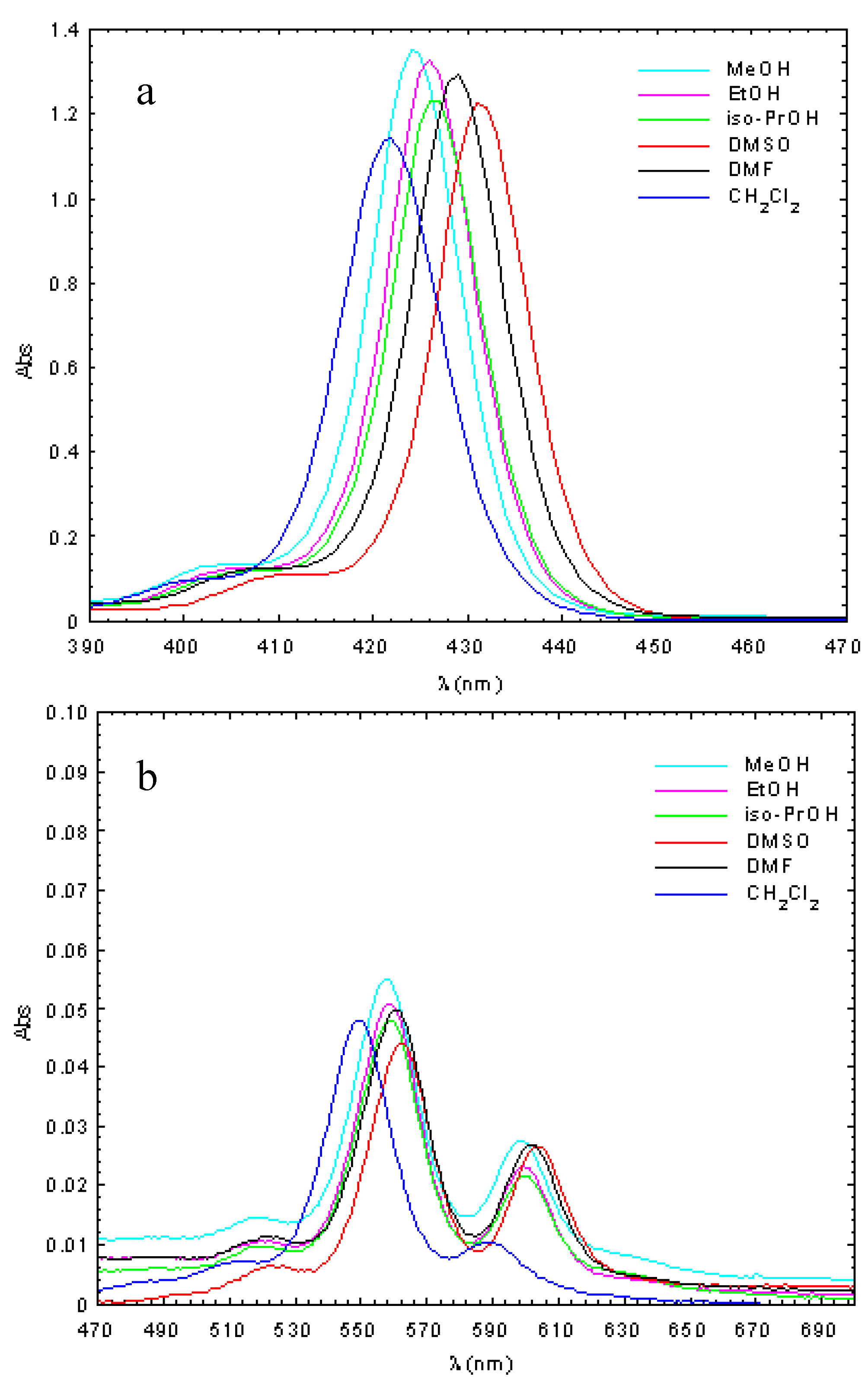
2.2. Absorption and Fluorescence Spectra

The absorption spectra of Zn(II)TCMPOHp and Cu(II)TCMPOHp were studied in solvents with different polarities (methanol, ethanol, isopropyl alcohol, dimethyl sulfoxide, dimethylformamide, dichloromethane) for solutions containing 2.5 × 10⁻⁶ M of the porphyrinic complex. The obtained results are presented in Figure 2 and Figure 3 and Table 2.
Figure 2. Absorption spectra of 5-(4-hydroxyphenyl)-10,15,20–tris-(4-carboxymethyl-phenyl) – 21, 23 Zn(II) porphine in different solvents (c = 2.5 × 10⁻⁶ M, a-Soret band; b-Q bands).
Molecules 16 05604 g002
Figure 3. Absorption spectra of 5-(4-hydroxyphenyl)-10,15,20–tris-(4-carboxymethyl-phenyl)– 21, 23 Cu(II) porphine in different solvents (c = 2.5 × 10⁻⁶ M, a-Soret band; b-Q bands).
Molecules 16 05604 g003
Table 2. Maximum wavelength and molar absorptivity of the Zn(II)TCMPOHp and Cu(II)TCMPOHp in different solvents (c = 2.5 × 10⁻⁶ M).
Solventλmax (nm) [lgε (L mol−1 cm−1)]
Soret bandQ bands
B(0,0)Qy(0,0)Qx(1,0)
5-(4-hydroxyphenyl)-10, 15, 20–tris-(4-carboxymethylphenyl)–21,23-Zn(II) porphine
MeOH424.5 [5.732]557.7 [4.342]598.8 [4.049]
EtOH426.0 [5.723]558.6 [4.310]599.7 [3.964]
iso-PrOH426.7 [5.693]558.6 [4.283]600.0 [4.033]
CH2Cl2421.9 [5.659]549.1 [4.282]588.6 [3.602]
DMF428.9 [5.713]560.7 [4.301]601.8 [4.033]
DMSO431.5 [4.954]562.8 [4.246]603.6 [4.028]
MeOH414.2 [5.532]538.6 [4.260]-
EtOH414.5 [5.571]538.9 [4.283]-
iso-PrOH414.8 [5.540]538.9 [4.259]-
CH2Cl2417.1 [5.649]540.1 [4.356]-
DMF418.8 [5.555]541.3 [4.298]578.1(sh)
DMSO422.9 [5.507]545.2 [4.203]587.4 [3.477]
sh – shoulder; MeOH = methanol, EtOH = ethanol, iso-PrOH = isopropyl alcohol, DMF = dimethylformamide, CH2Cl2= dichloromethane, DMSO = dimethyl sulfoxide.
The obtained molecular electronic spectra of the copper and zinc synthesized complexes are typical of metalloporphyrins [33,34,35,36,37]. These consist of the Soret band, as a result of the a1u (π) → eg (π∗) transition and two Q bands bands corresponding to the a2u (π) → eg (π∗) transition.
Thus, the UV–Vis spectra of the Zn(II)TCMPOHp complex exhibited one Soret band in the spectral range of 421–432 nm accompanied by two Q bands, in the 549–563 nm and 588–604 nm spectral range, respectively. The absorption spectra of Cu(II)TCMPOHp display a Soret band in the 414–423 nm wavelength region and either one or two Q bands between 538–590 nm, depending on solvent polarity.
The main differences observed in the absorption spectra of the two porphyrinic complexes are determined by the nature of the metallic center and solvent polarity. Thus, in the absorption spectra of metalloporphyrinic complexes in various solvents, a blue shift of the spectral bands was observed with increasing solvent polarity [38]. These spectral changes can be ascribed to the formation of proton bridges between the polar solvent molecules and the porphyrinic substitutents due to the polarity-induced and proton properties of the alcohol. Also, from the absorption spectra of copper porphyrin can be observed that the second Q band has much smaller intensity or disappeared with increasing solvent polarity.
Compared with Zn(II)TCMPOHp,the spectral bands of the Cu(II)TCMPOHpshows a blue shift around 10–12 nm for the Soret band and about 20 nm for Q bands. In agreement with Gouterman’s theory [39], these spectral differences can be explained by stronger conjugation effects for the copper 4pz orbital with the π electrons of the tetrapyrrolic ring, effects that cause a decrease the energy of the a1u (π) and a2u (π) orbitals relative to the eg (π∗) orbitals with increased energy available for electron transitions and the blue shift of the spectral bands.
Of the synthesized metalloporphyrins, fluorescence signals were only detected for Zn(II)TCMPOHp under the experimental conditions used. The emission spectra of 5-(4-hydroxyphenyl)-10,15,20–tris-(4-carboxymethylphenyl)–21,23-Zn(II) porphine in different solvents, at room temperature, were recorded at λex = 420 nm.
The fluorescence spectral data shows for Zn(II)TCMPOHp two bands located in the 605–660 nm spectral region (see Table 3) and reveal smaller shifts of the emmision maxima by changing the solvent polarity.
Table 3. The fluorescence data of Zn(II)TCMPOHp(c = 2.5 × 10⁶M, λex = 420 nm).
Solventλmax (nm) [If] (a.u.)
Qx(0,0)Qx(0,1)
MeOH605.2 [379.3]651.8 [63.2]
EtOH606.8 [740.2]651.6 [118.0]
iso-PrOH607.0 [776.8]653.1 [126.4]
CH2Cl2608.9 [585.5]653.5 [130.7]
DMF609.7 [490.7]654.9 [81.5]
DMSO610.9 [380.8]656.0 [49.9]
MeOH = methanol, EtOH = ethanol, iso-PrOH = isopropyl alcohol, DMF = dimethylformamide, CH2Cl2= dichloromethane, DMSO = dimethyl sulfoxide.

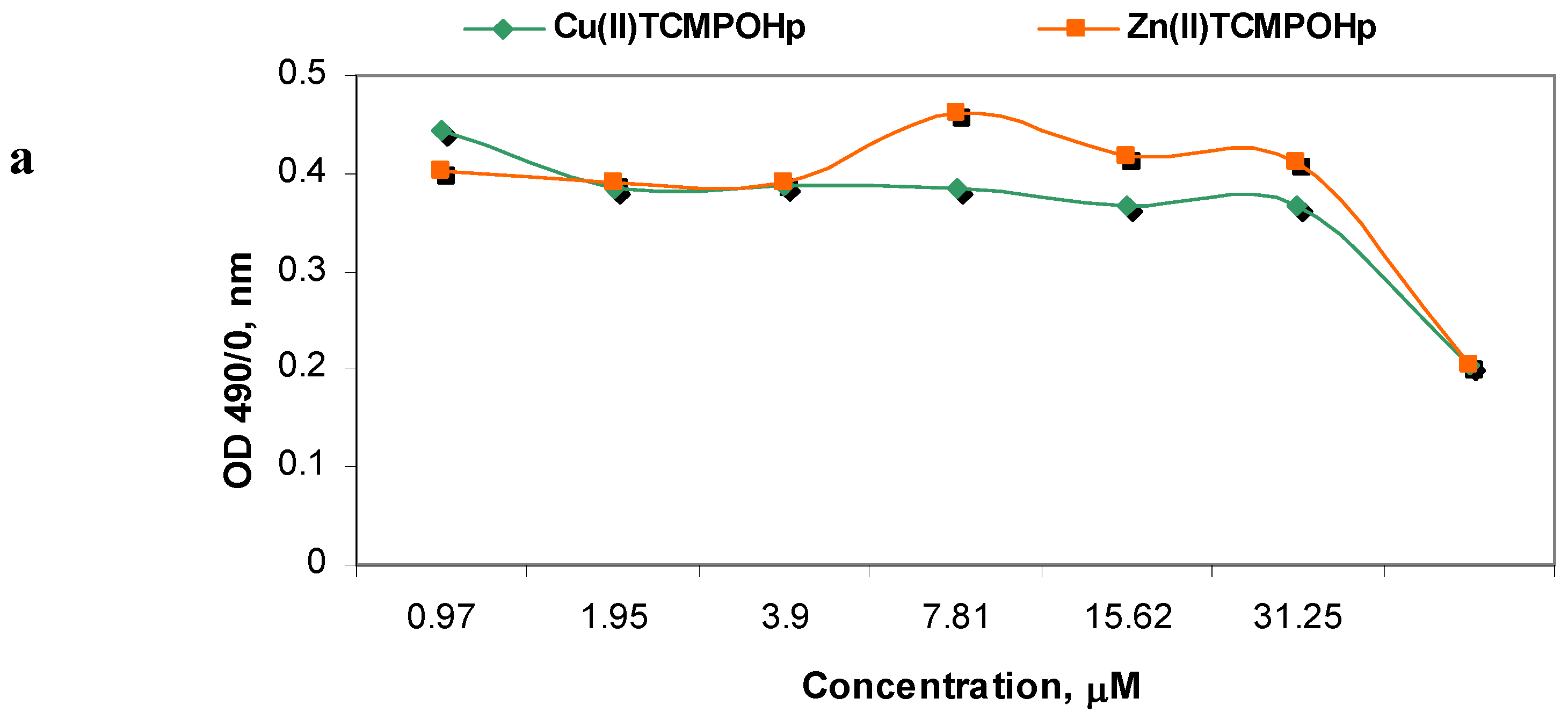
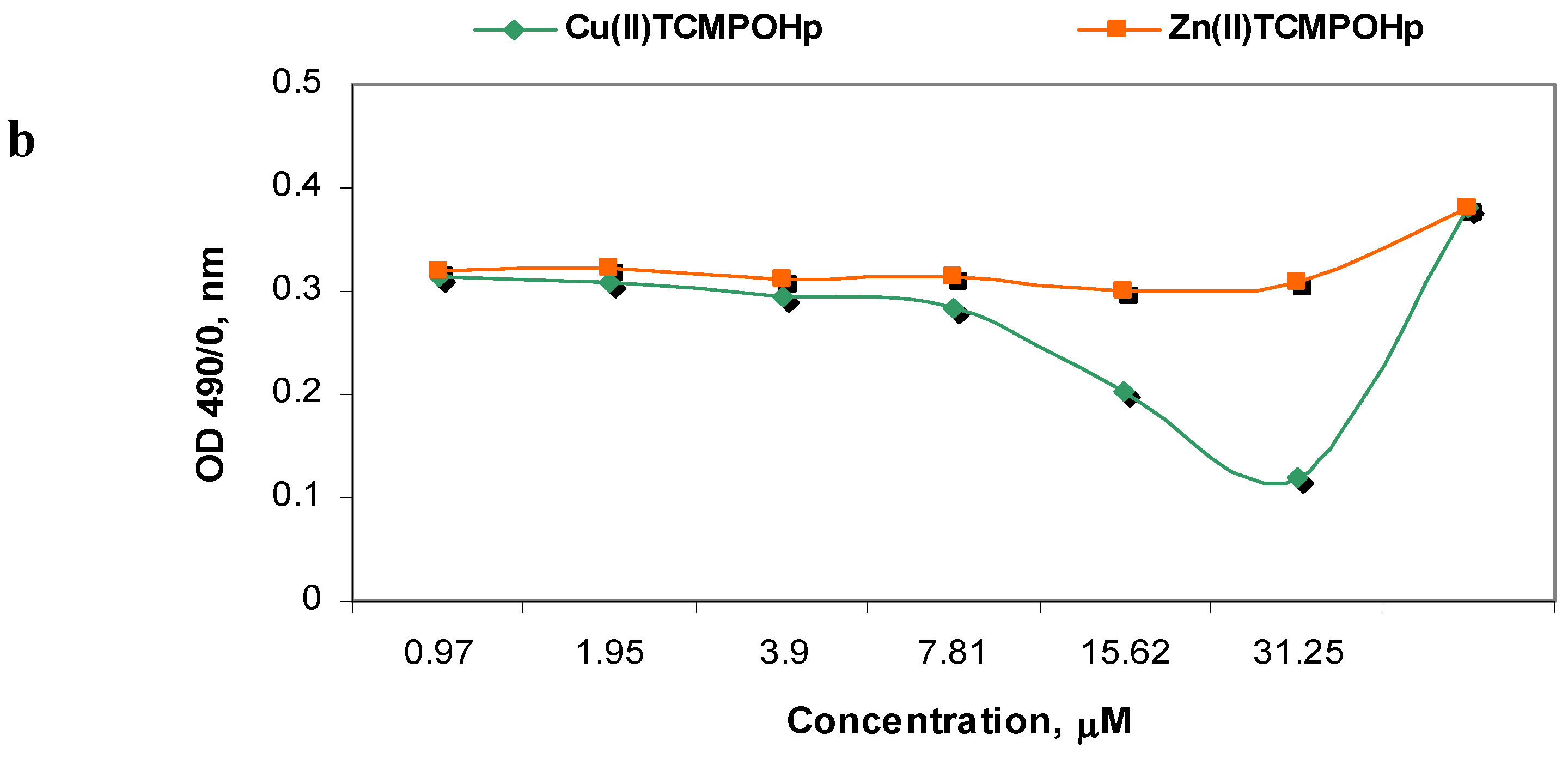
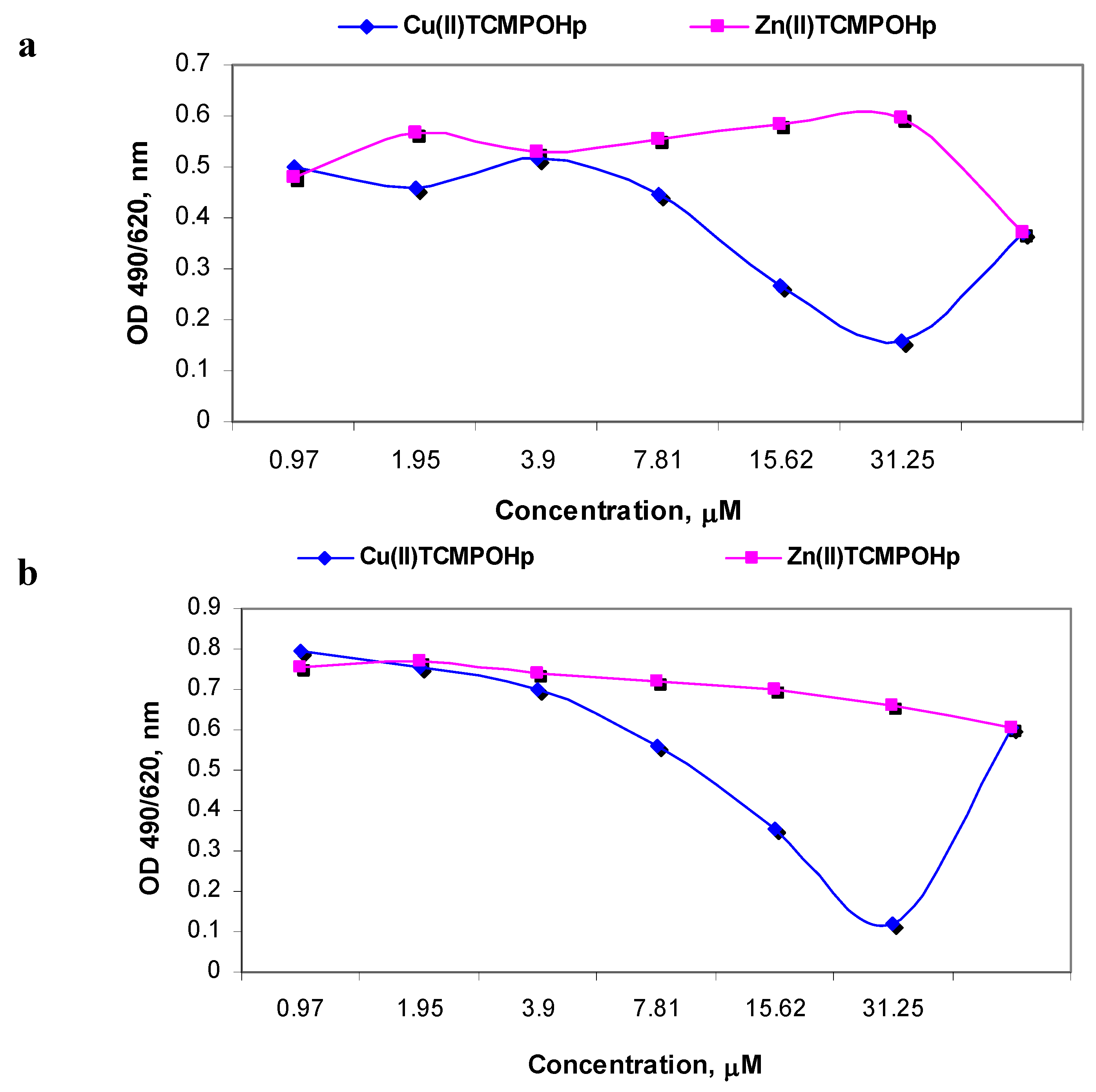
2.3. Dark Cytotoxicity Tests

The short-term cytotoxicity evaluation of a metalloporphyrinic compound designed to be used as a photosensitizer is mandatory for its efficiency validation in antitumor therapy when seeking a new therapeutic agent [40] and of its use in imagistics [41]. As well, a long incubation period with the porphyrinic complexes structures provides the answer to the question concerning the possibility of a delayed cytotoxicity installation in the cells when the phototherapeutic aim is the objective, or on their permanence in the cellular structures when imagistic detection is sought.
In order establish the dark cytotoxicity profile, the new mesoporphyrinic complexes were evaluated in vitro using HT29 (human colon adenocarcinoma) cell line for different doses and incubation times. For this purpose the cellular viability and proliferative capacity were assessed upon each round of cell incubation with the synthesized metalloporphyrins.
Viability assay at short-time incubation stressed the point that at low loading doses (1–5 µM) there are no differences between the two porphyrinic complexes behavior. For doses greater than 5 µM, only Zn(II)TCMPOHp induces an increase in the LDH release by HT29 cells (Figure 3a). The effect of the metallic ion of the porphyrinic complex on the cell viability is highlighted at 24 h of incubation and for concentrations higher than 5 µM. In these conditions, only Cu(II)TCMPOHp has a visible influence on the cell viability (Figure 3b).
Figure 3. The viability of HT29 cells incubated 2h (a) and 24h (b) with Zn(II)TCMPOHpand Cu(II)TCMPOHp; the LDH release registered as OD at 490 nm.
Molecules 16 05604 g004aMolecules 16 05604 g004b
Proliferative capacity assay performed following two hours incubation suggests slight differences between the compounds (Figure 4a). The 3.95 µM concentration results in a homogeneous cell response for both compounds. Cu(II)TCMPOHp inhibits the cellular proliferation only in concentrations higher than 10 µM. Following 24 hours incubation Zn(II)TCMPOHp maintains its nontoxic characteristics related to the proliferative function of the cells, while for Cu(II)TCMPOHp the effect is dose-dependent (Figure 4b).
Figure 4. The proliferation of HT29 cells incubated 2 h (a) and 24 h (b) with Zn(II)TCMPOHp and Cu(II)TCMPOHp; the MTS reduction reaction registered as OD at 490/620 nm.
Molecules 16 05604 g005

3. Experimental

3.1. Materials and Methods

Commercially available chemicals and solvents were used as received from Sigma-Aldrich and Merck. For the microwave assisted synthesis, a domestic microwave oven was used, with temperature and power control. The elemental analysis of C, H and N was performed with an automatic Carlo Erba L-1108 analyzer. IR spectra were recorded with a FT-IR 400D Nicolet Impact spectrophotometer. The substances under analysis, previously dried for 24 h at 150 °C, were processed as KBr (spectrally pure) pellets. The spectra were recorded in the 4,000–500 cm−1 spectral range. The molecular absorption spectra of the mesoporphyrinic complexes were recorded with the use of a Lambda 35 Perkin-Elmer spectrophotometer in different solvents (MeOH, EtOH, iso-PrOH, DMF, DMSO, CH2Cl2) using a 10 mm path length quartz cell, in single beam mode. Fluorescence spectra were recorded with a Jasco FP 6500 spectrofluorimeter.
The metalloporphyrin solutions were freshly prepared in the spectrally pure solvents at the concentration 2.5 × 10⁻⁶ M and kept in dark until the measurement to prevent photodegradation. The NMR spectra of the zinc porphyrinic complex were recorded with a 400 MHz Bruker NMR spectrometer. EPR spectra of the copper porphyrinic complex were recorded on powders at room temperature using a ART-6 spectrometer, operating in the X band (9.01 GHz), equipped with a field modulation unit of 100 KHz.
The preliminary toxicological studies in the presence of the unsymmetrical mesoporphyrinic complexes consisted in viability and proliferation studies performed on human colon adenocarcinoma cell line HT29. The HT29 cell line was purchased from the European Collection of Cell Cultures (ECACC, catalog no. 91072201) and cultured in RPMI 1640 medium supplemented with 100 U/mL penicillin, 0.1 mg/mL streptomycin, 0.25 μg/mL amphotericin, 2 mM glutamine and 10% fetal bovine serum. The cells were maintained at 37 °C in a 5% CO2 humid atmosphere for standard cultivation and during all the toxicological tests.
The porphyrinic complexes solutions were handled under sterile conditions. Ten mM stock solutions of the compounds to be tested were prepared in DMSO by sonication at 22,000 Hz for 30 seconds. For the toxicological tests the solutions were further diluted in RPMI 1640 culture medium for the toxicological tests in the range 0.97–31.25 µM.
For the viability and the proliferation test assay, the cells (cell density 5 × 10⁻⁴ cells/mL) were incubated with each of the tested concentrations for 2 and 24 h. A cellular control consisting in unloaded/untreated HT29 cells was used for every replicate of the tests, corresponding to each incubation time.
For cellular viability we have used lactate dehydrogenase (LDH) release test [42], using CytoTox 96® Non-Radioactive Cytotoxicity Test (Promega). For testing cell proliferation by means of the tetrazolium salt (MTS) reduction test [43], we have used CellTiter 96® AQeous One Solution Cell Proliferation Assay kit (Promega). Briefly, at end of each incubation time with the mesoporphyrinic complexes, the cell supernatants were collected for LDH test whilst the cell sediment was used for MTS assay. Results were expressed as triplicate’s mean of optical density (OD) ± SD, recorded on a Jasco V630 spectrophotometer, in a single beam mode, at 490 nm without any reference (for LDH release in the cell viability test) and at 490 nm with reference at 640 nm (for MTS reduction test).

3.2. Synthesis of 5-(4-hydroxyphenyl)-10, 15, 20–tris-(4-carboxymethylphenyl)–21, 23-Zn(II)-porphine (Zn(II)TCMPOHP)

A mixture of 4-hydroxybenzaldehyde (1.12 g, 0.01 mol), methyl 4-formyl benzoate (4.92 g, 0.03 mol), freshly distilled pyrrole (2.76 mL, 0.04 mol), anhydrous zinc chloride (1.36 g, 0.01 mol) and 2–3 g of silica gel 60 (200–500 μm, 35–70 mesh) in the presence of 2,6-dimethylpyridine (1 mL) was irradiated in a Pyrex bottle with a microwave oven set at 175 °C, 450 W for 10 min. Extraction of samples for monitoring the synthesis by UV–Vis spectroscopy was performed after every 2 min of irradiation.
The crude product was first dissolved in dichloromethane, then filtered and finally purified on a chromatography column by repeated elution, using silica gel (100–200 mesh size) as stationary phase and dichloromethane as eluent. The solution of the zinc porphyrinic complex was concentrated by simple distillation. The obtained violet crystals were dried at ≈ 100 °C for 12 h. The compound of interest was obtained with a yield of about 48%. Elemental analysis for C50H34N4O7Zn: calculated C 69.2, H 3.92, N 6.45; found C 69.04, H 3.80, N 6.28. 1H-NMR: δH(400 MHz, CDCl3), ppm: 4.13 (9H, s, O-CH3), 6.5 (1H, s, brs.,OH), 8.00 (2H, d, Hmetha-Ph-OH), 8.2 (2H, d, Hortho-Ph-OH), 8.3 (6H, d, H metha-Ph-COOCH3), 8.4 (6H, d, H ortho-Ph-COOCH3), 8.9 (6H, d, Hβpyrr2), 9.0 (2H, d, Hβpyrr3).
The comparative study of the 1H-NMR data of the unsymmetrical mesoporphyrinic ligand and its zinc complex reveals the absence of the signal at −2.78 (2H, s, N-H) [32], indicating the involvement of both nitrogen atoms of porphyrinic ring in the coordination after deprotonation. Other signals in the NMR spectrum of the complex are similar to those of the ligand.

3.3. Synthesis of 5-(4-hydroxyphenyl)-10, 15, 20–tris-(4-carboxymethylphenyl)–21, 23-Cu(II)-porphine (Cu(II)TCMPOHP)

A mixture of anhydrous CuCl2 (1.34 g, 0.01 mol), methyl 4-formyl benzoate (4.92 g, 0.03 mol), 4-hydroxybenzaldehyde (1.12 g, 0.01 mol), freshly distilled pyrrole (2.76 mL, 0.04 moli), 2–3 g of silica gel 60 (200–500 μm, 35–70 mesh) and 2,6-dimethylpyridine (1 mL) was irradiated in a microwave oven at 180 °C, 475 W, for 8 min. The extent of the complexation reaction was monitored by UV–Vis spectroscopy and for this purpose extraction of samples was performed after every 2 min of irradiation.
The product was dissolved in dichloromethane, filtered and then purified on a chromatography column. The solid product was chromatographed on silica gel (100–200 mesh size) with dichloromethane as eluent. The solution of the copper porphyrinic complex was concentrated by simple distillation. The obtained dark red crystals were dried at ≈ 100 °C for 12 h.
The copper unsymmetrical porphyrinic complexes were obtained with a yield of about 52%. Elemental analysis for C50H34N4O7Cu: calculated C 69.32, H 3.93, N 6.47; found C 69.19, H 3.82, N 6.36. The EPR parameters are: g||=2.172, g=2.050, A||= 202G, A=30 G. These values are in good agreement with literature data and confirm a square planar geometrical arrangement of nitrogen atoms around the copper ion [44,45]. Also, the g|| value (g|| < 2.3) indicates a covalent character of the Cu-N bonds in the copper-porphyrinic complex [46].

4. Conclusions

The paper describes the microwave irradiation assisted synthesis, spectral properties and preliminary cytotoxicity investigation of some new unsymmetrical porphyrinic complexes, namely 5-(4-hydroxyphenyl)-10,15,20–tris-(4-carboxymethylphenyl)–21,23-Zn(II)-porphine and 5-(4-hydroxy-phenyl)-10, 15, 20–tris-(4-carboxymethylphenyl)–21,23-Cu(II)-porphine. These complexes were synthesized for use as active substances in the photodiagnosis and photodynamic therapy of cancer.
The syntheses of the copper and zinc porphyrinic complexes were confirmed by FT-IR, UV–Vis, EPR and NMR analysis. The influence of environment polarity on the wavelengths of the absorbance and fluorescence band maxima is also described. The spectral changes that occur by increasing environmental polarity can be ascribed to the physical interaction between the porphyrinic substituents and the solvent molecules.
Both tetrapyrrolic complexes were evaluated in terms of cytotoxicity using HT29 (human colon adenocarcinoma) cell line for different doses and incubation times. The results showed no significant toxicity on the investigated cell line and suggest that these novel complexes are promising active substances for the photodiagnosis and photodynamic therapy use.

Acknowledgements

The research was supported by CNMP project No. 41-047/2007, granted from the Romanian Ministry of Education, Research, Youth and Sport. The author thanks Carolina Constantin from “Victor Babes” National Institute of Pathology, Bucharest, Romania, for experimental assistance in preliminary cytotoxicity investigation of the newly synthesized porphyrinic complexes.

References and Notes

  1. Detty, M.R.; Gibson, S.L.; Wagner, S.J. Current Clinical and Preclinical Photosensitizers for Use in Photodynamic Therapy. J. Med. Chem. 2004, 47, 3897–3915. [Google Scholar] [CrossRef]
  2. Kamuhabwa, A.; Agostinis, P.; Ahmed, B.; Landuyt, W.; Van Cleynenbreugel, B.; Van Poppel, H.; De Witte, P. Hypericin as a potential phototherapeutic agent in superficial transitional cell carcinoma of the bladder. Photochem. Photobiol. Sci. 2004, 3, 772–780. [Google Scholar] [CrossRef]
  3. Hilderbrand, S.; Weissleder, R. Near-infrared fluorescence: Application toin vivo molecular imaging. Curr. Opin. Chem. Biol. 2010, 14, 71–79. [Google Scholar] [CrossRef]
  4. Chatterjee, D.K.; Fong, L.S.; Zhang, Y. Nanoparticles in photodynamic therapy: An emerging paradigm. Adv. Drug Deliv. Rev. 2008, 60, 1627–1637. [Google Scholar] [CrossRef]
  5. Bonneau, S.; Bizet, C.V.; Mojzisova, H.; Brault, D. Tetrapyrrole-photosensitizers vectorization and plasma LDL: A physic-chemical approach. Int. J. Pharm. 2007, 344, 78–87. [Google Scholar] [CrossRef]
  6. Chin, W.W.L.; Lau, O.W.K.; Bhuvaneswari, R.; Heng, P.W.S.; Olivo, M. Chlorin e6-polyvinylpyrrolidone as a fluorescent marker for fluorescence diagnosis of humanbladder cancer implanted on the chick chorioallantoic membrane model. Cancer Lett. 2007, 245, 127–133. [Google Scholar] [CrossRef]
  7. Mora, J.S.; Cormick, P.M.; Milanesio, E.M. Durantini, E.N. The photodynamic activity of a novel porphyrin derivative bearing a fluconazole structure in different media and against Candida albicans. Dyes Pigments 2010, 87, 234–240. [Google Scholar] [CrossRef]
  8. Banfi, S.; Caruso, E.; Buccafurni, L; Battini, V.; Zazzaron, S.; Barbieri, P.; Orlandi, V. Antibacterial activity of tetraaryl-porphyrin photosensitizers: An in vitro study on Gram negative and Gram positive bacteria. J. Photochem. Photobiol. B-Biol. 2006, 85, 28–38. [Google Scholar] [CrossRef]
  9. Cormick, P.M.; Alvarez, G.; Rovera, M.; Durantini, E.N. Photodynamic inactivation of Candida albicans sensitized by tri- and tetra-cationic porphyrin derivatives. Eur. J. Med. Chem. 2009, 44, 1592–1599. [Google Scholar] [CrossRef]
  10. Munin, E.; Giroldo, L.M.; Alves, L.P.; Costa, M.S. Study of germ tube formation by Candida albicans after photodynamic antimicrobial chemotherapy (PACT). J. Photochem. Photobiol. B-Biol. 2007, 88, 16–20. [Google Scholar] [CrossRef]
  11. Stockert, J.C.; Cañete, M.; Juarranz, A.; Villanueva, A.; Horobin, R.W.; Borrell, J.I.; Teixidó, J.; Nonell, S. Porphycenes: Facts and Prospects in Photodynamic Therapy of Cancer. Curr. Med. Chem. 2007, 14, 997–1026. [Google Scholar] [CrossRef]
  12. Banfi, S.; Caruso, E.; Caprioli, S.; Mazzagatti, L.; Canti, G.; Ravizza, R.; Gariboldia, M.; Montia, E. Photodynamic effects of porphyrin and chlorin photosensitizers in human colon adenocarcinoma cells. Bioorg. Med. Chem. 2004, 12, 4853–4860. [Google Scholar]
  13. Chen, J.Y.; Mak, N.K.; Yow, C.M.N.; Fung, M.C.; Chiu, L.C.; Leung, W.N.; Cheung, N.H. The Binding Characteristics and Intracellular Localization of Temoporfin (mTHPC) in Myeloid Leukemia Cells: Phototoxicity and Mitochondrial Damage. Photochem. Photobiol. 2000, 72, 541–547. [Google Scholar] [CrossRef]
  14. Grosseweiner, L.I. The Science of Phototherapy; CRC Press: London, UK, 1994; pp. 139–155, Chapter 8. [Google Scholar]
  15. Schweiter, C.; Schmidt, R. Physical Mechanisms of Generation and Deactivation of Singlet Oxygen. Chem. Rev. 2003, 103, 1685–1758. [Google Scholar] [CrossRef]
  16. Bonnett, R. Chemical Aspects of Photodynamic Therapy. In Advanced Chemistry Texts; Gordon and Breach Science Publishers: Amsterdam, The Netherlands, 2000; Volume 1, pp. 57–112. [Google Scholar]
  17. Postino, F.; Mora, M.; DeMadariaga, M.A.; Nonell, S.; Sagrista, M.L. Incorporation of hydrophobic porphyrins into liposomes: Characterization and structural requirements. Int. J. Pharma. 2004, 278, 239–254. [Google Scholar] [CrossRef]
  18. Scalise, I.; Durantini, E.N. Photodynamic effect of metallo 5-(4-carboxyphenyl)-10,15,20-tris(4-methylphenyl) porphyrins in biomimetic AOT reverse micelles containing urease. J. Photochem. Photobiol. A 2004, 162, 105–113. [Google Scholar] [CrossRef]
  19. Boyle, R.B.; Dolphin, D. Structure and Biodistribution Relationships of Photodynamic Sensitizers. Photochem. Photobiol. 1996, 64, 469–485. [Google Scholar] [CrossRef]
  20. Mac Donald, I.J.; Dougherty, T.J. Basic principles of photodynamic therapy. J. Porphyrins Phthalocyanines 2001, 5, 105–129. [Google Scholar] [CrossRef]
  21. Nyman, E.S.; Hynninen, P.H. Research advances in the use of tetrapyrrolic photosensitizers for photodynamic therapy. J. Photochem. Photobiol. B-Biol. 2004, 73, 1–28. [Google Scholar] [CrossRef]
  22. Kessel, D. Correlation between subcellular localization and photodynamic efficacy. J. Porphyrins Phthalocyanines 2004, 8, 1009–1014. [Google Scholar] [CrossRef]
  23. Rosenkranz, A.A.; Jans, D.A.; Sobolev, A.S. Targeted intracellular delivery of photosensitizers to enhance photodynamic efficiency. Immunol. Cell Biol. 2002, 78, 452–464. [Google Scholar]
  24. Osterloh, J.; Vicente, M.G.H. Mechanisms of porphyrinoid localization in tumors. J. Porph. Phthal. 2002, 5, 305–325. [Google Scholar] [CrossRef]
  25. Milgrom, L.; MacRobert, S. Light years ahead. Chem. Brit. 1998, 34, 45–50. [Google Scholar]
  26. Adam, D. Microwave chemistry: Out of the kitchen. Nature 2003, 421, 571–572. [Google Scholar] [CrossRef]
  27. Wei, T.H. Microwave-assisted synthesis and reverse saturable absorption of phthalocyanines and porphyrins. J. Organomet. Chem. 2004, 689, 1078–1084. [Google Scholar] [CrossRef]
  28. Liu, M.O.; Hu, A.T. Microwave-assisted synthesis of phthalocyanine–porphyrin complex and its photoelectric conversion properties. J. Organomet. Chem. 2004, 689, 2450–2455. [Google Scholar] [CrossRef]
  29. Lindsey, J.S. Synthetic Routes to meso-Patterned Porphyrins. Acc. Chem. Res. 2010, 43, 300–311. [Google Scholar] [CrossRef]
  30. Senge, M.O. Nucleophilic Substitution as a Tool for the Synthesis of Unsymmetrical Porphyrins. Acc. Chem. Res. 2005, 38, 733–743. [Google Scholar] [CrossRef]
  31. Senge, M.O.; Shaker, Y.M.; Pintea, M.; Ryppa, C.; Hatscher, S.S.; Ryan, A.; Sergeeva, Y. Synthesis of meso-substituted ABCD-type porphyrins by functionalization reactions. Eur. J. Org. Chem. 2010, 2, 237–258. [Google Scholar]
  32. Socoteanu, R.; Boscencu, R.; Nacea, V.; Constantin, C.; Manda, G.; Neagu, M.; Ilie, M.; Oliveira, A.S.; Vieira Ferreira, L.F. Bio functionalized porphyrinic compound has tetrapyrrolic heterocycles class, synthesized and optimized for cellular load assays. Romanian Patent Application No 0200900452/17.06.2009, published in the Official Bulletin of Intelectual Property (BOPI) 11/2009 with No.125018AO.
  33. Gouterman, M. Optical Spectra and Electronic Structure of Porphyrins and Related Rings. In The Porphyrins; Dolphin, D., Ed.; Academic Press: New York, NY, USA, 1978; Volume 3, pp. 11–87. [Google Scholar]
  34. Boscencu, R.; Socoteanu, R.; Oliveira, A.S.; Vieira Ferreira, L.F.; Nacea, V.; Patrinoiu, G. Synthesis and Characterization of Some Unsymmetrically-substituted Mesoporphyrinic Mono-Hydroxyphenyl Complexes of Copper(II). Pol. J. Chem. 2008, 82, 509–522. [Google Scholar]
  35. Boscencu, R.; Socoteanu, R.; Oliveira, A.S.; Ferreira, L.F.V. Studies on Zn(II) monohydroxyphenyl mesoporphyrinic complexes. Synthesis and characterization. J. Serb. Chem. Soc. 2008, 73, 713–726. [Google Scholar] [CrossRef]
  36. Boscencu, R.; Ilie, M.; Socoteanu, R.; Oliveira, A.S.; Constantin, C.; Neagu, M.; Manda, G.; Vieira Ferreira, L.F. Microwave Synthesis, Basic Spectral and Biological Evaluation of Some Copper (II) Mesoporphyrinic Complexes. Molecules 2010, 15, 3731–3743. [Google Scholar] [CrossRef]
  37. Boscencu, R.; Socoteanu, R.; Ilie, M.; Oliveira, A.S.; Constantin, C.; Vieira Ferreira, L.F. Synthesis, Spectral and Biological Evaluation of Some Mesoporphyrinic Zn(II) Complexes. Rev. Chim. 2009, 10, 1006–1011. [Google Scholar]
  38. Reichardt, C.H. Solvent effects on the absorption spectra of organic compounds. In Solvents and Solvent Effects in Organic Chemistry; VCH: New York, NY, USA, 1988; pp. 329–352. [Google Scholar]
  39. Gouterman, M.; Wagniere, G.H.; Snyder, L.C. Spectra of porphyrins: Part II. Four orbital model. J. Mol. Spectrosc. 1963, 11, 108–127. [Google Scholar] [CrossRef]
  40. Allison, R.R.; Sibata, C.H. Oncologic photodynamic therapy photosensitizers: A clinical review. Photodiagnosis Photodyn. Ther. 2010, 7, 61–75. [Google Scholar] [CrossRef]
  41. Pandey, R.K.; Goswami, L.N.; Chen, Y.; Gryshuk, A.; Missert, J.R.; Oseroff, A.; Dougherty, T.J. Nature: A rich source for developing multifunctional agents. Tumor - imaging and photodynamic therapy. Lasers Surg. Med. 2006, 38, 445–467. [Google Scholar] [CrossRef]
  42. Korzeniewski, C.; Callewaert, D.M. An enzyme-release assay for natural cytotoxicity. J. Immunol. Meth. 1983, 64, 313–320. [Google Scholar] [CrossRef]
  43. Barltrop, J.A.; Owen, T.C.; Cory, A.H.; Cory, J.G. 5-(3-carboxymethoxyphenyl)-2-(4,5-dimethylthiazolyl)-3-(4-sulfophenyl)tetrazolium, inner salt (MTS) and related analogs of 3-(4,5-dimethylthiazolyl)-2,5-diphenyltetrazolium bromide (MTT) reducing to purple water-soluble formazans as cell-viability indicators. Bioorg. Med. Chem. Lett. 1991, 1, 611–614. [Google Scholar] [CrossRef]
  44. Lin, W.C. Electron Spin Resonance and Electronic Structure of Metalloporphyrins. In The Porphyrins; Dolphin, D., Ed.; Academic Press: New York, NY, USA, 1978; Volume 4, pp. 358–364. [Google Scholar]
  45. Manoharan, P.T.; Roger, M.T. ESR study of copper (II) and silver (II) tetraphenylporphyrin. In Electron Spin Resonance of Metal Complexes; Yen, T.F., Ed.; Plenum Press: New York, NY, USA, 1969; pp. 143–173. [Google Scholar]
  46. Kivelson, D.; Neiman, R.R. ESR Studies on the Bonding in Copper Complexes. J. Chem. Phys. 1961, 35, 149–155. [Google Scholar] [CrossRef]
  • Sample Availability: Samples of the compounds are available from the authors.

