Article

Crystal Structure and Hydrogen Bonding Study of (10E)-2,2-Dimethyl-3,4-dihydro-2H-benzo[g]chromene-5,10-dione 10-Oxime Derived From a-Lapachone

by
Andrea R. Da Silva
1,
Marcelo H. Herbst
1,*,
Aurelio B. B. Ferreira
1,
Ari M. Da Silva
1 and
Lorenzo C. Visentin
2,*
1
Departamento de Química, Universidade Federal Rural do Rio de Janeiro, BR 465, km 47, Seropédica, RJ, 23890-000, Brazil
2
Instituto de Química, Universidade Federal do Rio de Janeiro, Avenida Athos da Silveira Ramos, 149, Bloco A, 7° andar, Cidade Universitária, Rio de Janeiro, RJ, 21941-909, Brazil
*
Authors to whom correspondence should be addressed.
Molecules 2011, 16(2), 1192-1200; https://doi.org/10.3390/molecules16021192
Submission received: 15 October 2010 / Revised: 25 January 2011 / Accepted: 26 January 2011 / Published: 27 January 2011

Abstract

:
The compound (10E)-2,2-dimethyl-3,4-dihydro-2H-benzo[g]chromene-5,10-dione-10-oxime (1) was synthesized from α-lapachone and hydroxylamine chloride in alkaline medium. Single-crystals suitable for X-ray diffraction measurements were grown from an ethanol solution, and the crystal structure of the title molecule is reported for the first time. The title molecule was also characterized by 1H- and 13C-NMR in CDCl3 solution, FTIR and MS. The crystal structure of 1 shows an E stereochemistry and dimers formed through classical hydrogen bonds.

Graphical Abstract

1. Introduction

The chemistry of oximes derived from lapachol and lapachones has been studied by several groups because these substances show important biologic and organic chemical applications [1,2,3,4]. α-Lapachone (2,2-dimethyl-3,4-dihydro-2H-benzo[g]chromene-5,10-dione) is a natural naphtha-quinone found in the wood of trees of the genus Tabebuia (family Bignoniaceae), which occur in most of Central and South Americas and are known as ipê or pau d’arco in Brazil (e.g., ipê-roxo, Tabebuia serratifolia) and lapacho in Argentina and other Spanish-speaking countries. α-Lapachone, (2,2-dimethyl-3,4-dihydro-2H-benzo[g]chromene-5,10-dione) and β-lapachone, (2,2-dimethyl-3,4-dihydro-2H-benzo[h]chromene-5,6-dione) occur in small quantities, while the isomer lapachol (2-hydroxy-3-(3-methyl-2-butenyl)-1,4-naphtoquinone) is the most abundant naphtoquinone found in the ipê-roxo wood [5]; these substances have been under medical study for their varied biological activities and ipê-roxo has been traditionally used in folk medicine [6,7,8]. The lapachones may be easily synthesized, from lapachol (2-hydroxy-3-(3-methyl-2-butenyl)-1,4-naphtoquinone) through cyclization in acidic media [7]. Lemos et al., studying Brazilian natural quinones and their derivatives by 1H- and 13C-NMR spectroscopy in CDCl3 solutions (including 2D experiments 1H-1H-COSY, HMQC 1JCH and HMBC nJCH (n = 2 and 3)), have assigned Z stereochemistry for the 10-oxime derived from α-lapachone [4]. Herein we report a single crystal X-ray diffraction study that presents the first report of the crystal structure of the (10E)-2,2-dimethyl-3,4-dihydro-2H-benzo[g]chromene-5,10-dione10-oxime (1).

2. Results and Discussion

The oxime 1, one of two possible regioisomers obtainable from α-lapachone, adopts an E stereochemistry in the crystalline state. The conformation is governed by intra- and intermolecular interactions of classical and non classical hydrogen bond types. The structural formula of the title molecule is depicted in Figure 1. Although the numbering scheme recommended by IUPAC has been used for the name of the title molecule, in the NMR assignments a different numbering scheme was chosen, as shown in the figure. This numbering scheme was further used for refining the X-ray structure.
Figure 1. Structural scheme and numbering label for 1.
Molecules 16 01192 g001

2.1. Infrared, 1H- and 13C-Nuclear Magnetic Resonance Spectroscopy

The infrared spectra obtained in the solid state (KBr) for 1 in the range from 4,000 to 400 cm−1 shows characteristic oxime group absorption bands. The principal absorption frequencies in this molecule are attributed to (O-H), (C=O), (C=N), (C-H) and (C=C) bonds, which show absorption bands in the range of the 3,160 cm−1 (νOH), 2,975–2,937 cm−1 (CH2 and CH3), 1,639 cm−1 (C=N), 1,621 cm−1 (C=O) and 1,272 cm−1 (C-O). These absorption frequencies supply important information on the molecular structure of 1. The IR spectrum of 1 shows broad hydroxyl absorption due to hydrogen bonding around this group centered in 3,160 cm−1. These data agree with literature reports [1]. The X-ray diffraction reveals the same hydrogen bonds around the hydroxyl group.
The 1H-NMR spectrum of the 10-oxime from α-lapachone (of which 1 is the E stereoisomer) in CDCl3 solution shows two triplet signals at δ 2.63 and δ 1.88 ppm assigned to the two methylene groups at the C11 and C12 labeled atoms, which are part of the 2-oxene ring moiety. Also observed is a cumulative singlet signal at δ 1.48 ppm, assigned to the methyl groups (C14 and C15) in this same ring. The singlet signal attributed to the OH group was observed at δ 12.25 ppm in CDCl3, in accordance with literature [4]. In DMSO-d6 this signal shifted to 13.65 and in pyridine-d5 to 15.85 ppm, but there were no significant alterations in the other peaks. A NOESY experiment in CDCl3 solution (not shown) was inconclusive and did not elucidate the chemical neighborhood of the OH hydrogen atom. The 13C-NMR spectrum indicated the presence of fourteen carbon atom signals. A cumulative signal at δ 26.8 ppm is assigned to the carbon atoms labeled C14 and C15, and there is only one signal indicating the presence of a carbonyl group in δ 184.0 ppm. The signal in 139.7 ppm was assigned to the quaternary carbon of the oxime group. Table 1 lists all the 1H- and 13C-NMR data.
Table 1. 1H- and 13C-NMR data for 1.
Carbon Atomδ 13C δ 1H ( J in Hz )
1184.0-
2113.6-
3156.6-
4139.7-
5126.9-
6129.89.06 (d, 7.8)
7132.67.65 (td, 1.4, 7.6)
8130.77,58 (td, 1.2, 7.7)
9126.88.27 (dd, 1.2, 7.6)
10130.7-
1116.92.63 (t, 6.6)
1231.81.88 (t, 6.6)
1378.4-
1426.81.48 (s)
1526.81.48 (s)
16-12.25 (s)

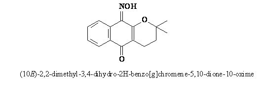
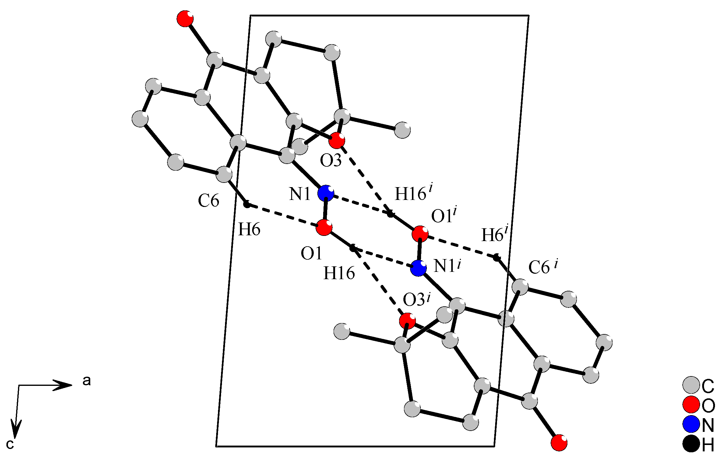
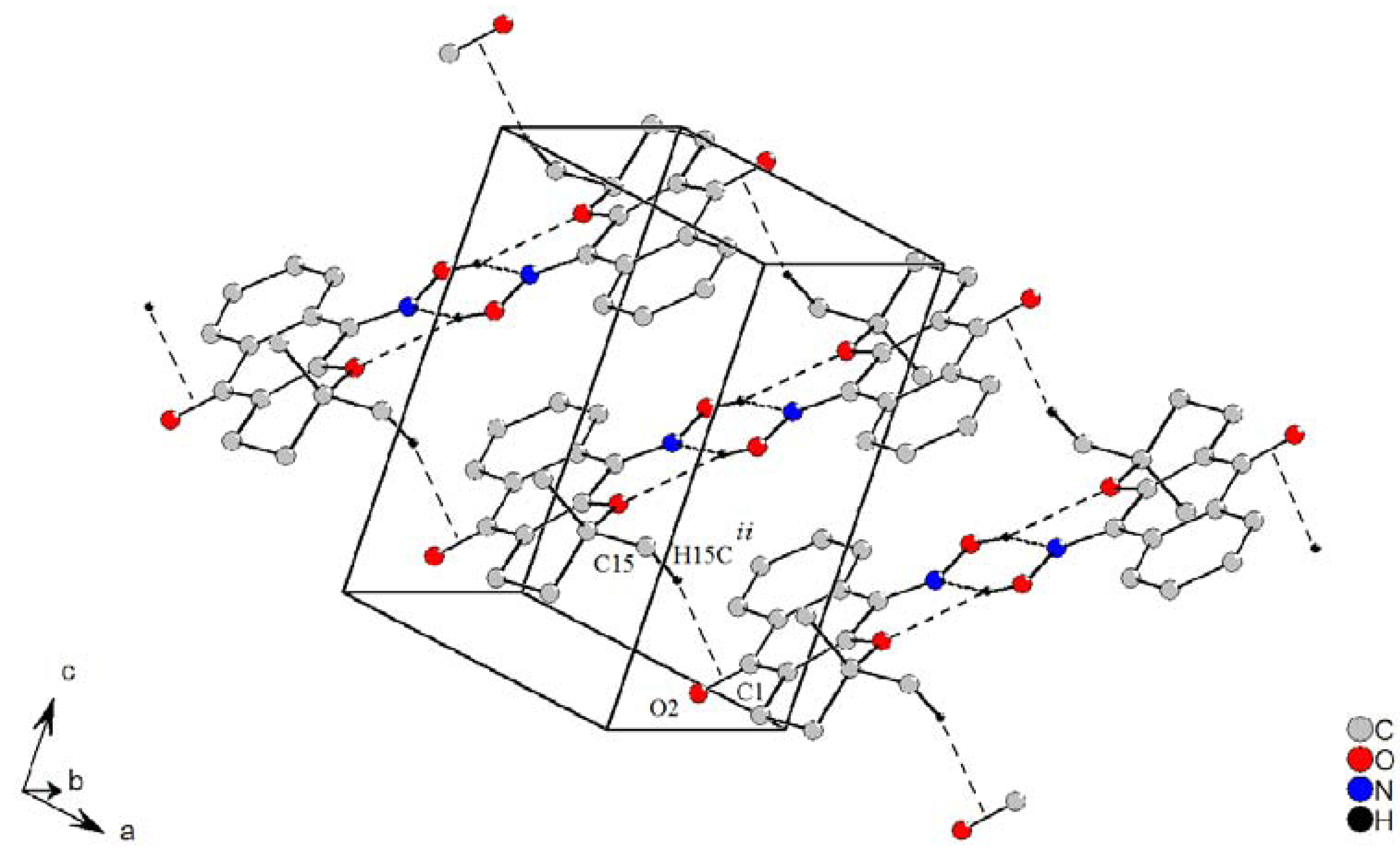
2.2. Crystal Structure

The crystal structure of 1 is reported here for the first time. The atomic arrangement and numbering scheme for 1 are shown in Figure 2. Selected bond lengths and bond angles are listed in Table 2. These parameters are in the expected ranges reported in the literature [1,9,10,11] for compounds of this class. For instance, the oxime 6-hydroxy-3-(hydroxyimino)indolin-2-one [9], shows bond lengths around the N-OH group of 1.361(3)Å for N2-O3 and 1.286(4)Å for N2=C8. For 4-(1-methylvinyl)cyclohexene-1-carbaldehyde oxime [10] these distances are of 1.407(3)Å for O1-N1 and 1.266(4)Å for N1=C1, and the (E)-4-nitrobenzaldehyde oxime [11] shows similar distances around the oxime group, which are 1.401(3)Å for O3-N2 and 1.264(3)Å for N2-C7.
Figure 2. View of the ORTEP projection for (1) with respective atom-numbering scheme [19]. Displacement ellipsoids are draw at the 50% probability level.
Molecules 16 01192 g002
Table 2. Selected geometric parameters in (1), (Å/°).
Bonds
N1-O11.382(1)
N1-C41.290(2)
C1-O21.234(2)
C3-O3 1.353(2)
C13-O31.473(2)
Angles
O1-N1-C4117.2(1)
C3-O3-C13118.1(1)
O2-C1-C10120.8(1)
O2-C1-C2120.6(1)
C12-C13-C15111.8(1)
O3-C13-C14107.8(1)
The average bond lengths in 1 also agree with the typical interatomic distance for C=N-OH oxime groups (R = aryl) in oxime molecules. The International Tables for X-ray Crystallography [12] list these parameters for typical bond distances, which are 1.281 Å for Csp2=N [σ = 0.013, ql = 1.273 Å and qu = 1.288 Å] and 1.416 Å for N-OH [σ = 0.006, ql = 1.416 Å and qu = 1.420 Å].
The phenyl ring and the ring fused to it in 1 form an almost planar system: the dihedral angle between C1-C10 [r.m.s. 0.0306 Å] and C10-C9 [r.m.s. 0.0073Å] rings is 4.7(1)°. The six-membered (oxene) ether ring is distorted and the torsion angle in O3-C3-C2-C11 is −3.6(2)°, whereas in O3-C13-C12-C11 it is −59.1 (2)°. In 1, the C6-H6…O1 intramolecular hydrogen bond gives rigidity to the oxime group by the formation of one six-membered ring (Figure 3).
Figure 3. View of the centrosymmetric R22(6) dimers in (101) plane by intra- and intermolecular interactions. [Symmetry code (i) = 1-x, 2-y, 1-z]
Molecules 16 01192 g003
In addition, a dimer arrangement is created in 1 by O1-H16…O3i and O1-H16℘?℘N1i bifurcate hydrogen bonds, which link the molecules into centrosymmetric R22(6) dimers on the (101) plane, symmetry code (i) = 1-x, 2-y, 1-z. These bifurcate interactions cement the crystal structure by formation of one six- and two five-membered rings, which are formed by 1-x, 2-y, 1-z equivalent symmetry. All noncovalent bonds, that is, classical and non-classical hydrogen bonds, enforce the E stereochemistry around oxime group in this crystal structure. These hydrogen bonds were calculated by PLATON program [13] and their parameters are in accordance with the literature [14].
Besides that, the centrosymmetric dimers are self-arranged in a 1-D fashion through four C-Hπ intermolecular interactions along the [100] crystallography direction (Figure 4). The π electrons from the carbonyl group are responsible for these interactions and the molecular self-organization.
Figure 4. Self assembly by tectons linked through C-Hπ noncovalent interactions. The intramolecular interaction is omitted. [Symmetry code (ii) = 1+x, y, z]
Molecules 16 01192 g004
The self assembly of predictable supramolecular aggregates of this self-organization can be induced by selective, directional attractive noncovalent interactions [15]. The molecules which play the role of building blocks in a self-assembled, ordered supramolecular structure are called tectons [16,17,18]. The crystal packing is accomplished by these weak interactions, and its unusual noncovalent bond grows a staircase on (101) plane, where the steps are formed with tectons linked by four C15-H15cCgii from the center double bond in the C1=O2 group [symmetry code (ii) = 1 + x, y, z] formed by side-on noncovalent interactions (Figure 4). The bond length C15-H15c is slightly longer than a normal C-H bond in methyl groups. These noncovalent bonds are equivalent by symmetry, with the following symmetry codes, 1-x, 2-y, 1-z; -x, 2-y, 1-z; and-1+x, y, z. Geometric parameters for H-bonds in (1) are show in Table 3.
Table 3. Geometric parameters for H-bonds in (1), (Å/°).
D-H···AD-HH··· AD···A Molecules 16 01192 i001D-H···A
O1-H16...O3 i0.86(2)2.35(2)3.070(2)142(2)
O1-H16...N1 i0.86(2)2.01(2)2.771(2)149(2)
C6-H6O10.932.162.785(2)123
D-H···CgD-HH··· CgD···Cg Molecules 16 01192 i001D-H···Cg
C15-H15cCgii1.02(2)2.7953.797160.28
[Symmetry code (i) = 1-x, 2-y, 1-z and(ii) = 1+x, y, z; and Cg = center CO double bond]

3. Experimental

3.1. General

The 1H- and 13C-NMR spectra were obtained in a Bruker Avance 400 (400 MHz 1H and 100 MHz 13C), in CDCl3, DMSO-d6 and pyridine-d5, using TMS as internal standard. The IR spectra were measured in a Perkin-Elmer 1605 spectrophotometer, using KBr pellets. Mass spectra were measured in a Varian Saturn 2000 spectrometer. The melting point was determined with a Büchi 510 apparatus.

3.2. Synthesis of 1

The title molecule was synthesized as follows [4] (all the manipulations were carried out at room temperature): α-lapachone (2,2-dimethyl-3,4-dihydro-2H-benzo[g]chromene-5,10-dione) (0.242 g, 1 mmol) was added to a methanol solution (10 mL) of hydroxylamine chloride (NH2OHHCl, 0.075 g, 1 mmol) in the presence of sodium hydroxide (5%, w/v). After 2 h of magnetic stirring, the solution was neutralized with acetic acid and filtered. Anhydrous sodium sulphate was added to the solution, which was filtered once more. The solvent was removed under vacuum, and the crude yellow product was isolated. Yellow block-shaped crystals suitable for X-ray diffraction were obtained by recrystallization from ethanol. Yield: 0.962 g, 37%; Melting point = 172.4 °C; MS (EI): m/z (%) 257 (90), 241 (45), 240 (50), 226 (100), 212 (10), 201 (25), 186 (10), 158 (10), 143 (5), 130 (50), 115 (10), 102 (40), 89 (5), 76 (25), 63 (10), 50 (20). NMR data: see Table 1.

3.3. X-ray diffraction Experiment

The X-ray data for the title compound were collected from a Bruker KAPPA CCD diffractometer [20], at 295K and MoKα monochromatic-graphite radiation. The crystal data are listed in Table 4. The cell parameters for the oxime molecule were obtained using the PHICHI and DIRAX programs [21,22]. The average data were reduced using the EvalCCD program and the absorption correction was performed with the SADABS programs [23,24]. The structure was solved by direct methods via SHELXS97 and refined via SHELXL97 by a full-matrix least-squares treatment with anisotropic temperature parameters for all non H atoms [25]. H atoms of the unsaturated carbon were positioned geometrically (C–H = 0.93 Å for Csp2 atoms) and treated as riding on their respective C atoms, with Uiso(H) values set at 1.2UeqCsp2. The hydrogen atoms of the oxime group, methyl, and methylene groups were located in Fourier map and free refined to position.
Table 4. Crystal data and structure refinement parameters for (1).
Empirical formulaC15H15NO3
Formula weight257.28
Temperature295(2) K
Wavelength0.71073 A
Crystal system, space grouptriclinic, P-1
Unit cell dimensionsa = 6.6069(13) Å α = 91.47(3)°
b = 9.6001(19) Å β = 94.47(3)°
c = 10.176(2) Å γ = 94.27(3)°
Volume641.3(2) Å3
Z, Calculated density2, 1.332 mg/m3
Absorption coefficient0.093 mm-1
F(000)272
Crystal size0.47 × 0.40 × 0.20 mm
Theta range for data collection2.97 to 25.00°
Limiting indices−7<=h<=7, −11<=k<=11, −12<=l<=12
Reflections collected / unique10469 / 2246 [R(int) = 0.0339]
Completeness to theta = 25.0099.5 %
Max. and min. transmission0.9816 and 0.9574
Refinement methodFull-matrix least-squares on F2
Data / restraints / parameters2246 / 0 / 216
Goodness-of-fit on F21.032
Final R indices [I>2sigma(I)]R1 = 0.0390, wR2 = 0.1010
R indices (all data)R1 = 0.0550, wR2 = 0.1103
Largest diff. peak and hole0.137 and −0.190 e.Å-3

4. Conclusions

The single-crystal X-ray diffraction studies reported in this work have established an E stereochemistry for compound 1, whereas the literature reports a Z stereochemistry for 1 in solution, as derived from NMR spectroscopy data. In this way, either 1 presents different stereochemistries in the solid state and in solution, or the assignment of the stereochemistry of 1 in solution is actually wrong. However, to our knowledge, there are no conclusive studies in the literature on the stereochemistry of complex oxime molecules in solution, including the 10-oxime of α-lapachone. In this work we have shown that in the solid state H-bonds link two oxime molecules, which form centrosymmetric dimers with an R22(6) graph-set motif on the (101) plane. Furthermore, C-Hπ noncovalent bonds grow in a 1D self-arrangement along the [100] crystallography direction. In the case of an eventual change in the stereochemistry of 1 upon crystallization, this should be related to the intra- and intermolecular hydrogen bonding. Further studies are in progress, as well as calculations, in an attempt to establish conclusively the stereochemistry of 1 in solution.

Acknowledgements

The authors are grateful to their sponsors, CNPq, FAPERJ and to Laboratório de Difração de Raios X, (LDRX), Universidade Federal Fluminense, Brazil, for the diffractometer facility.

Supplementary Material

CCDC 690106 contains the supplementary crystallographic data for this paper. These data can be obtained free of charge from The Cambridge Crystallographic Data Centre via www.ccdc.cam.ac.uk/data_request/cif.

References and Notes

  1. Alcalde, E.; Mesquida, N.; Alvarez-Rúa, C.; Cuberes, R.; Frigola, J.; García-Granda, S. 1,2-Diaryl(3-pyridyl)ethanone Oximes. Intermolecular Hydrogen Bonding Networks Revealed by X-ray Diffraction. Molecules 2008, 13, 301–318. [Google Scholar] [CrossRef]
  2. Pérez-Sacau, E.; Estévez-Braum, A.; Ravelo, A.G.; Ferro, E.A.; Tokuda, H.; Mukainaka, T.; Nishino, H. Inhibitory effects of lapachol derivatives on Epstein-Barr virus activation. Bioorg. Med. Chem. 2003, 11, 483–488. [Google Scholar] [CrossRef]
  3. Pérez-Sacau, E.; Diaz-Peñate, R.G.; Estévez-Braun, A.; Ravelo, A.G.; García-Castellano, J.M.; Pardo, L.; Campillo, M. Synthesis and Pharmacophore Modeling of Naphthoquinone Derivatives with Cytotoxic Activity in Human Promyelocytic Leukemia HL-60 Cell Line. J. Med. Chem. 2007, 50, 696–706. [Google Scholar]
  4. Lemos, T.L.G.; Monte, F.J.Q.; Santos, A.K.L.; Fonseca, A.M.; Santos, H.S.; Oliveira, M.F.; Costa, S.M.O.; Pessoa, O.D.L.; Braz-Filho, R. Quinones from plants of Northeastern Brazil: Structural diversity, chemical transformations, NMR data and biological activities. Nat. Prod. Res. 2007, 21, 529–550. [Google Scholar] [CrossRef]
  5. Burnett, A.R.; Thomson, R.H. Naturally occurring quinones. Part X. The quinonoid constituents of Tabebuia avellanedae (Bignoniaceae). J. Chem. Soc. C 1967, 2100–2104. [Google Scholar]
  6. Hussain, H.; Krohn, K.; Ahmad, V.U.; Miana, G.A.; Green, I.R. Lapachol: an overview. ARKIVOC 2007, ii, 145–171. [Google Scholar]
  7. Hooker, S.C. LVII.—The constitution of “lapachic acid” (lapachol) and its derivatives. J. Chem. Soc. Trans. 1892, 61, 611–650. [Google Scholar] [CrossRef]
  8. Pinto, A.V.; Pinto, M.C.F.R.; Oliveira, C.G.T. Synthesis of the α- and nor-β-lapachones. Properties in acid and reactions with N-bromosuccinimide. An. Acad. Bras. Cienc. 1982, 54, 107–114. [Google Scholar]
  9. Yu, H. l. 6-Hydroxy-3-(hydroxyimino)indolin-2-one. Acta Crystallogr. 2009, E65, o2328. [Google Scholar]
  10. Yuan, X.-Y.; Zhang, M.; Ng, S.W. Perillartine. Acta Crystallogr. 2009, E65, o2149. [Google Scholar]
  11. Abbas, A.; Hussain, S.; Hafeez, N.; Badshah, A.; Hasan, A.; Lo, K.M. (E)-4-Nitrobenzaldehyde oxime. Acta Crystallogr. 2010, E66, o1130. [Google Scholar]
  12. Allen, F.H.; Watson, D.G.; Brammer, L.; Orpen, A.G.; Taylor, R. Typical Interatomic Distances: Organic Compounds. In International Tables for Crystallography; Prince, E., Ed.; Wiley: New York, NY, USA, 2006; Volume C, pp. 790–811. Chapter 9.5. [Google Scholar]
  13. Spek, A.L. Single-crystal structure validation with the program PLATON. J. Appl. Crystallogr. 2003, 36, 7–13. [Google Scholar] [CrossRef]
  14. Jeffrey, G.A.; Saenger, W. Hydrogen Bonding in Biological Structures; Springer-Verlag: Berlin, Germany, 1994; p. 569. [Google Scholar]
  15. Hörner, M.; Oliveira, G.M.; Visentin, L.C.; Cezar, R. Dimerization and polymeric self arrangement of [HgII(Py)2(PhN2C6H4NNNC6H4R)2] (Py = pyridine; R = NO2) through reciprocal metal-η2-arene π-interactions and non-classical C–H…O bonding: Synthesis and X-ray characterization of a polyaryl asymmetric-substituted triazenide complex polymer of Hg(II). Inorg. Chim. Acta 2006, 359, 4667–4671. [Google Scholar] [CrossRef]
  16. Haiduc, I.; Edelmann, F.T. Supramolecular Organometallic Chemistry; Wiley–VCH: Weinheim, Germany, 1999; p. 486. [Google Scholar]
  17. Simard, S.; Su, D.; Wuest, J.D. Use of hydrogen bonds to control molecular aggregation. Self-assembly of three-dimensional networks with large chambers. J. Am. Chem. Soc. 1991, 113, 4696–4698. [Google Scholar] [CrossRef]
  18. Fyfe, M.C.T.; Stoddart, J.F. Synthetic Supramolecular Chemistry. Acc. Chem. Res. 1997, 30, 393–401. [Google Scholar] [CrossRef]
  19. Farrugia, L.J. ORTEP-3 for Windows - a version of ORTEP-III with a Graphical User Interface (GUI). J. Appl. Crystallogr. 1997, 30, 565–566. [Google Scholar] [CrossRef]
  20. Hooft, R.W.W. Collect; Nonius BV: Delft, The Netherlands, 1998. [Google Scholar]
  21. Duisenberg, A.J.M.; Hooft, R.W.W.; Schreurs, A.M.M.; Kroon, J. Accurate cells from area-detector images. J. Appl. Crystallogr. 2000, 33, 893–898. [Google Scholar] [CrossRef]
  22. Duisenberg, A.J.M. Indexing in single-crystal diffractometry with an obstinate list of reflections. J. Appl. Crystallogr. 1992, 25, 92–96. [Google Scholar] [CrossRef]
  23. Duisenberg, A.J.M.; Kroon-Batenburg, L.M.J.; Schreurs, A.M.M. An intensity evaluation method: EVAL-14. J. Appl. Crystallogr. 2003, 36, 220–229. [Google Scholar] [CrossRef]
  24. Sheldrick, G.M. SADABS, Program for Empirical Absorption Correction of Area Detector Data; University of Göttingen: Germany, 1996. [Google Scholar]
  25. Sheldrick, G.M. A short history of SHELX. Acta Crystallogr. 2008, A64, 112–122. [Google Scholar]
  • Sample Availability: Samples of compound 1 are available from the authors.

