Communication

Chemical Constituents from Clematis delavayi var. spinescens

1
Laboratory of Ethnobotany, Kunming Institute of Botany, Chinese Academy of Sciences, Kunming 650204, China
2
Institute of Tibetan Plateau Research at Kunming, Kunming Institute of Botany, Chinese Academy of Sciences, Kunming 650204, Yunnan, China
3
Biology Institute of Shandong Academy of Sciences, Jinan 250014, Shandong, China
*
Authors to whom correspondence should be addressed.
Molecules 2009, 14(11), 4433-4439; https://doi.org/10.3390/molecules14114433
Submission received: 20 October 2009 / Revised: 29 October 2009 / Accepted: 30 October 2009 / Published: 5 November 2009

Abstract

:
A new coumarin, 7-hydroxy-4,6-dimethoxy-5-methylcoumarin (1), was isolated from the aerial parts of Clematis delavayi var. spinescens together with 17 known compounds. Their structures were identified by extensive spectral analysis, especially 2D NMR techniques. Antiangiogenic effects of all compounds were evaluated using a zebrafish model.

1. Introduction

The genus Clematis, belonging to the family Ranunculaceae, is a large genus with about 300 species. The roots and rhizomes of Clematis are traditionally used as an analgesic, abirritative, antibacterial, antiphlogistic, anticancer and diuretic agent. Crude extracts from plants of this genus showed diuretic [1], antimicrobial [2], anti-inflammatory [3] biological activities. Some triterpenoid saponins and alkaloids isolated from this genus showed cytotoxic [4], antibacterial [5], and antifungal [6] activities.
Clematis delavayi var. spinescens is an apically spinescent shrub, which is widely distributed in the dry valleys of the upper reaches of the Yangtze River in Southwest China [7]. Up to now, no phytochemical study on C. delavayi var. spinescens was reported. Our investigation on the aerial parts of this plant lead to the identification of a new coumarin, 7-hydroxy-4,6-dimethoxy-5-methylcoumarin (1), and 17 known compounds, including (E)-para-coumatic acid (2) [8], coniferaldehyde (3) [9], caffeic acid (4) [10], caffeic acid methyl eater (5) [11], ethyl caffeate (6) [12], ferulic acid (7) [17], isoferulic acid (8) [10], 4,7-dimethoxy-5-methylcoumarin (9) [14], 4,6,7-trimethoxy-5-methylcoumarin (10) [14], (–)-secoisolariciresinol (11) [15], (+)-dihydrodehydrodiconiferyl alcohol (12) [16], dehydrodiconiferyl alcohol (13) [17], (+)-syringaresinal-4′-O-β-D-glucopyranoside (14) [18], 3, 3′,7-trihydroxy-4′,5-di-methoxyflavone (15) [19], 5-hydroxy-4-oxopentanoic acid (16) [20], 4-carbonyl-5-hydroxy methyl valerate (17) [21] and daucosterol (18) (Figure 1). In addition, the antiangiogenic effects of all compounds were evaluated using a zebrafish model and none of them were bioactive in this assay.
Figure 1. The structures of compounds 1-17.
Molecules 14 04433 g001

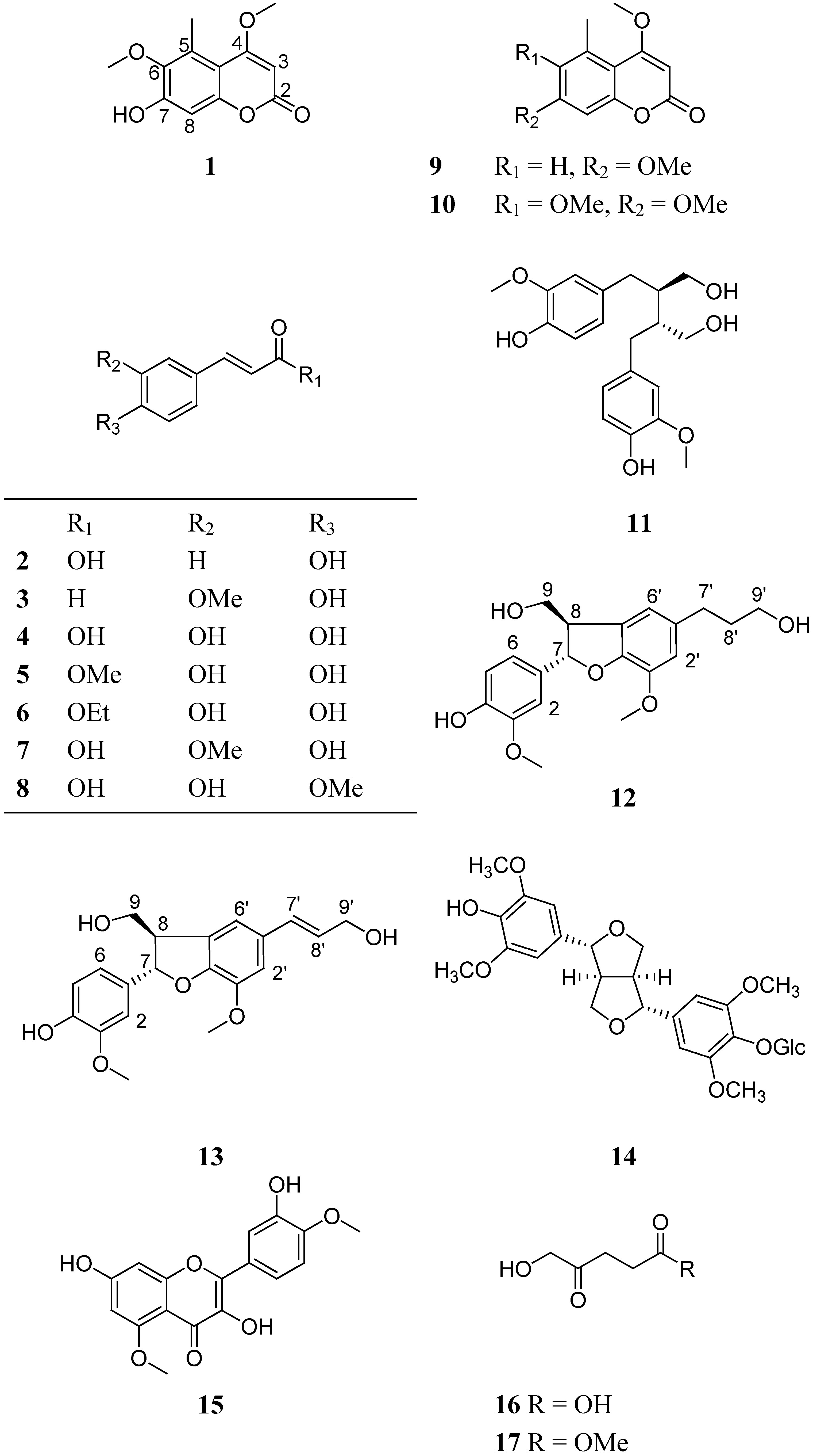
2. Results and Discussion
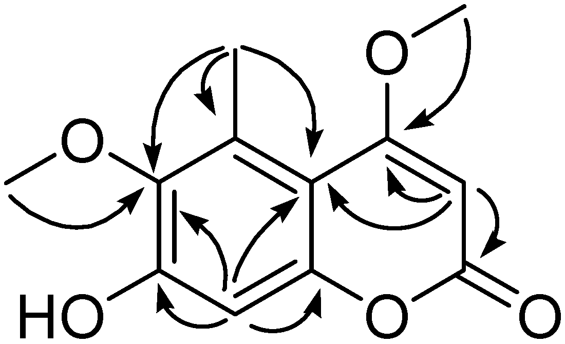
Compound 1 was isolated as white powder. Its molecular formula was established as C12H12O5 on the basis of HRESIMS ([M + H]+, found 237.0762, calcd 237.0762). The 1H- and 13C-NMR data (Table 1), coupled with UV absorptions at 208, 291, 325 nm and IR bands at 1,704 and 1,618 cm-1, revealed that 1 has a coumarin skeleton.
Table 1. The NMR data of 1 (in DMSO-d6). a
PositionδCδHPositionδCδH
2161.9 (s)8101.6 (d)6.64 (s)
387.2 (d)5.59 (s)9151.6 (s)
4169.5 (s)10106.1 (s)
5129.5 (s)OMe-(4)56.6 (q)3.90 (3H, s)
6143.5 (s)OMe-(6)60.0 (q)3.62 (3H, s)
7154.4 (s)Me-(5)14.0 (q)2.50 (3H, s)
a 1H- and 13C-NMR spectra were obtained at 500 and 125 MHz, respectively.
Only two downfield signals (δH 6.64 s, 5.59 s) were observed in the 1H-NMR spectrum of 1, suggesting that 1 was a tetrasubstituted coumarin. The 13C-NMR spectrum showed the presence of one carbonyl carbon, two methines, six quaternary carbons, one methyl group, and two methoxyl groups. Initial comparison of 1H-NMR and 13C-NMR data of 1 with those of known compound 4,6,7-trimethoxy-5-methylcoumarin (10) [8] showed compound 1 is very similar to 10. The big difference is the presence of only two methoxyl groups in 1, which is one less than those of 10. The methyl group was assigned to be located at C-5 by the HMBC correlations of proton of the methyl group (δH 2.48, s, 3H) to C-5 (δC 129.5), C-6 (δC 143.5) and C-10 (δC 106.1) (Figure 2).
Figure 2. Key HMBC (H → C) correlations of 1.
Molecules 14 04433 g002
A methoxyl group was established to be located at C-4 by the HMBC correlation of OMe (δH 3.90, s, 3H) to C-4 (δC 169.5), which was also supported by the ROESY correlation between the proton signal of this methoxyl group and H-3. The HMBC correlation of OMe (δH 3.62, s, 3H) to C-6 (δC 143.5) showed that another methoxyl group was located at C-6. According to its molecular formula, the remaining substitute group was assigned to be a hydroxyl group, which was determined to be located at C-7 by the downfield chemical shift of C-7 (δC 154.4), and the side-by-side NMR data comparison of 1 with 10. Therefore, the structure of 1 was elucidated as 7-hydroxy-4, 6-dimethoxy-5-methylcoumarin.
The antiangiogenic activities of all compounds were evaluated using a zebrafish model, in terms of the inhibition on the growth of intersegmental vessels, with PTK787 as a positive control (IC50 0.15 μg/mL) [22]. All compounds in the assay showed bioactivity with IC50 values above 5 µg/mL.

3. Conclusions

A new coumarin, 7-hydroxy-4, 6-dimethoxy-5-methylcoumarin (1) and 17 known compounds were isolated from the EtOAc extract of C. delavayi var. spinescens aerial parts. Their structures were identified by extensive spectral analysis, especially 2D NMR techniques. Most of these compounds are belonging are phenolics, including coumarins, lignans and flavonoids. The antiangiogenic activities of all compounds were evaluated using a zebrafish model, and did not show obvious bioactivity, with IC50 values more than 5 µg/mL.

4. Experimental Section

4.1. General

1D and 2D NMR spectra were recorded on Brucker AM-400 and DRX-500 spectrometers. Unless otherwise specified, chemical shift (δ) were expressed in ppm with reference to the solvent signals. MS were performed on a VG Autospec-3000 spectrometer under 70 eV. Optical rotation was measured with a Horiba SEPA-300 polarimeter. A Bio-Rad FTS-135 spectrophotometer was used for scanning IR spectroscopy of compounds with KBr pellets. Column chromatography was performed on silica gel (200–300 mesh, Qingdao Marine Chemical Inc., Qing-dao, Peoples Republic of China) and silica gel H (10–40 μm, Qingdao Marine Chemical Inc.). Fractions were monitored by TLC and spots were visualized by heating plates spraying with 10% H2SO4 in EtOH.

4.2. Plant Materials

The aerial parts of C. delavayi var. spinescens were collected from Derong County of Sichuan Province. The plant material was identified by Prof. Yongping Yang. A voucher specimen (No. LY 200709002) was deposited at Kunming Institute of Botany, Chinese Academy of Sciences.

4.3. Extraction and Isolation

The air-dried aerial parts of C. delavayi var. spinescens (6.0 kg) were powdered and extracted with 70% aqueous acetone (3 × 15 L) for 24 h at room temperature and concentrated in vacuo to give a crude extract (100 g), which was suspended in H2O and partitioned with EtOAc. The EtOAc extract was evaporated and the residue (90 g) was subjected to open column chromatography over MCI-gel CHP-20P eluting with 95% ethanol. The eluent from 95% ethanol (68 g) was concentrated in vacuo and subjected to column chromatography over silica gel (200–300 mesh) eluting with petroleum ether and acetone (1:0, 4:1, 2:1, 1:1, 1:2 and 0:1) to afford fractions A–E. Fraction B was subjected to Sephadex LH-20 (CHCl3/MeOH 1:1) and column chromatography over silica gel (petroleum ether/acetone) to yield 10 (10.2 mg), 16 (135.0 mg). Fraction C was chromatographed on Sephadex LH-20 (CHCl3/MeOH 1:1) and silica gel (CHCl3/MeOH) columns to yield 1 (46.1 mg), 2 (9.0 mg), 5 (1.0 mg), 7 (16.2 mg), 8 (6.3 mg), 11 (5.1 mg), 12 (2.0 mg), 13 (2.1 mg). Fraction D was also subjected to sephadex LH-20 (CHCl3/MeOH 1:1) and column chromatography over silica gel (CHCl3/MeOH) to yield 3 (3.2 mg), 4 (374.0 mg), 6 (6.1 mg), 9 (3.1 mg), 14 (25.2mg), 15 (1.0 mg), 17 (2.3 mg), 18 (47.5 mg).
Compound 1: white powder; UV max (MeOH): 208, 291, 325 nm; IR (KBr); υmax 3424, 1704, 1691, 1618, 1563 cm−1; For 1H- and 13C-NMR see Table 1. HRESIMS ([M + H]+, found 237.0762, calcd 237.0762).

4.4. Antiangiogenesis Assay [22]

Stock solutions (10 mg/mL) of all samples were prepared by dissolving the test compounds in 100% DMSO. These solutions were diluted in sterile salt water (5 mM NaCl, 0.17 mM KCl, 0.4 mM CaCl2, 0.16 mM MgSO4) to obtain solutions with the test compounds dissolved in 0.1% DMSO. These solutions were aliquoted into 96-well plates, and embryos at 24 hpf (hours post-fertilization) were also transferred randomly into the above wells. After 24-h treatment, the intersegmental vessels of embryos were visualized with methods of green fluorescent protein labeling and endogenous alkaline phosphatase staining. The antiangiogenic activities of compounds were calculated from the inhibition ratio of angiogenesis. PTK787 was used as the positive control.

Acknowledgements

The authors are grateful to the Department of Instrumental Analysis of Kunming Institute of Botany for measuring UV, IR, NMR, and mass spectra. The work was supported by MOST grant (2008BAD98B06), MAPs Traditional Knowledge Network in Southwest China, the Ford Foundation (1075-0729 and 1090-0448) and the West Light Foundation of the Chinese Academy of Sciences (awarded to Xiaoli Li).

References and Notes

  1. Alvarez, M.E.; Maria, A.O.; Villegas, O.; Saad, J.R. Evaluation of diuretic activity of the constituents of Clematis montevidensis Spreng. (Ranunculaceae) in rats. Phytother. Res. 2003, 17, 958–960. [Google Scholar] [CrossRef]
  2. Buzzini, P.; Pieroni, A. Antimicrobial activity of extracts of Clematis vitalba towards pathogenic yeast and yeast-like microorganisms. Fitoterapia 2003, 74, 397–400. [Google Scholar] [CrossRef]
  3. Park, E.K.; Ryu, M.H.; Kim, Y.H.; Lee, Y.A.; Lee, S.H.; Woo, D.H.; Hong, S.J.; Han, J.S.; Yoo, M.C.; Yang, H.I.; Kim, K.S. Anti-inflammatory effects of an ethanolic extract from Clematis mandshurica Rupr. J. Ethnopharmacol. 2006, 108, 142–147. [Google Scholar] [CrossRef]
  4. Yan, L.H.; Xu, L.Z.; Lin, J.; Yang, S.L.; Feng, Y.L. Triterpenoid saponins from the stems of Clematis parviloba. J. Asian. Nat. Prod. Res. 2009, 11, 332–338. [Google Scholar] [CrossRef]
  5. Ding, Q.; Yang, L.X.; Yang, H.W.; Jiang, C.; Wang, Y.F.; Wang, S. Cytotoxic and Antibacterial Triterpenoids Derivatives from Clematis ganpiniana. J. Ethnopharmacol. 2009. [Google Scholar] [CrossRef]
  6. Chen, J. H.; Du, Z. Z.; Shen, Y. M.; Yang, Y. P. Aporphine alkaloids from Clematis parviloba and their antifungal activity. Arch. Pharm. Res. 2009, 32, 3–5. [Google Scholar] [CrossRef]
  7. Wang, W.C.; Liu, L. Flora Reipublicae Popularis Sinicae [Zhongguo zhiwu Zhi]; Science Press: Beijing, China, 1988; Volume 28, pp. 154–156. [Google Scholar]
  8. Yang, L.J.; Yang, X.D.; Li, L. Study on chemical constituents of Lagotis yunnanensis. Zhong Yao Cai 2005, 28, 767–768. [Google Scholar]
  9. Shen, C.C.; Ni, C.L.; Shen, Y.C.; Huang, Y.L.; Kuo, C.H.; Wu, T.S.; Chen, C.C. Phenolic constituents from the stem bark of Magnolia officinalis. J. Nat. Prod. 2009, 72, 168–171. [Google Scholar] [CrossRef]
  10. Tao, T.T.; Sei, Y.; Wang, T.Z.; Bai, C.; Chang, Y.B. Chemical constituents of Sibiraea angustata. Chin. J. Nat. Med. 2006, 4, 257–259. [Google Scholar]
  11. Guan, Y.X.; Yang, X.S.; Tong, L.H.; Yang, B.; Hao, X.J. Chemical constituents in Ficus tikoua of Miao nationality. Chin. Tradition. Herbal Drugs 2007, 38, 342–344. [Google Scholar]
  12. Cheng, J.; Bai, Y.J.; Zhao, Y.Y.; Wang, B.; Cheng, T.M. Studies on the Phenylpropanoids from Eucommia ulmoides. Chin. J. Nat. Med. 2002, 27, 38–40. [Google Scholar]
  13. He, C.N.; Wang, C.L.; Guo, S.X.; Yang, J.S.; Xiao, P.G. Study on chemical constituents in herbs of Anoectochilus roxburghii II. China J. Chin. Mat. Med. 2005, 30, 761–763. [Google Scholar]
  14. Qi, S.H.; Wu, D.G.; Ma, Y.B.; Luo, X.D. Chemical constituents of Ailanthus triphysa. Chin. Tradition. Herbal Drugs 2003, 34, 590–592. [Google Scholar]
  15. Zhang, X.Y.; Li, B.G.; Zhou, M.; Yuan, X.H.; Zhang, G.L. Chemical constituents from Gymnosporia varialilis Loes. Chin. App. Environ. Bio. 2006, 12, 163–169. [Google Scholar]
  16. Fu, Z.H.; Zhang, Y.M.; Tan, N.H.; Chu, H.B.; Ji, C.J. Chemical constituents of Keteleeria evelyniana. Nat. Prod. Res. Develop. 2008, 20, 257–261. [Google Scholar]
  17. Zhang, M.; Dong, X.P.; Deng, Y.; Wang, H.; Li, X.N.; Song, Q. A new sesqui-norlignan from Herpetospermum pedunculosum. Acta Pharma. Sin. 2006, 41, 659–661. [Google Scholar]
  18. Yan, L.H.; Xu, L.Z.; Lin, J.; Zou, Z.M.; Zhao, B.H.; Yang, S.L. Studies on lignan constituents of Clematis parviloba. Chin. J. Nat. Med. 2008, 33, 1839–1843. [Google Scholar]
  19. Ho, L.K.; Lin, W.N. Quercetin 5,4′-dimethyl Ether from Rhododendron ellipticum. Phytochemistry 1995, 39, 463–464. [Google Scholar]
  20. Ju, Y.; Jia, Z.J.; Zhu, Z.Q. Chemical constituents of Anemone altaica. Chin. Tradition. Herbal Drugs 1986, 17, 388–391. [Google Scholar]
  21. Lüönd, R.M.; Walker, J.; Neier, R.W. Assessment of the Active-site Requirements of 5-aminolaevulinic Acid Dehydratase: Evaluation of substrate and product analogues as competitive inhibitors. J.Org. Chem. 1992, 57, 5005–5013. [Google Scholar] [CrossRef]
  22. Murphey, R.D.; Zon, L.I. Small molecule screening in the zebrafish. Methods 2006, 39, 255–261. [Google Scholar] [CrossRef]
  • Sample Availability: Samples of the compounds 1-17 are available from the authors.

