Article

Different Supramolecular Coordination Polymers of [N,N'-di(pyrazin-2-yl)-pyridine-2,6-diamine]Ni(II) with Anions and Solvent Molecules as a Result of Hydrogen Bonding

by
Gene-Hsiang Lee
1 and
Hsin-Ta Wang
1,*
1
Institute of Organic and Polymeric Materials, National Taipei University of Technology, Taipei 106, Taiwan
2
Instrumentation Center, National Taiwan University, Taipei 106, Taiwan
*
Author to whom correspondence should be addressed.
Molecules 2007, 12(4), 821-831; https://doi.org/10.3390/12040821
Submission received: 15 March 2007 / Revised: 18 April 2007 / Accepted: 19 April 2007 / Published: 23 April 2007

Abstract

:
Ni(II) complexes of N,N'–di(pyrazin–2–yl)pyridine–2,6–diamine (H2dpzpda) with different anions were synthesized and their structures were determined by X-ray diffraction. Hydrogen bonds between the amino groups and anions assembled the mononuclear molecules into different architectures. The perchlorate complex had a 1-D chain structure, whereas switching the anion from perchlorate to nitrate resulted in a corresponding change of the supramolecular structure from 1-D to 3-D. When the nitrate complex packed with the co-crystallized water, a double chain structure was formed through hydrogen bonding. The magnetic studies revealed values of g = 2.14 and D = 3.11 cm-1 for [Ni(H2dpzpda)2](ClO4)2 (1) and g = 2.18 and D = 2.19 cm-1 for [Ni(H2dpzpda)2](NO3)2 (2), respectively.

Introduction

Hydrogen bonding has attracted much attention since it plays a crucial role in biological systems, supramolecular chemistry, molecular recognition and crystal engineering [1,2,3]. As one of the most important noncovalent interactions, it participates in the assembly of new functional materials into 2- or 3-D networks with channels or huge pores [4,5,6]. Although the factors affecting molecular crystallization are intricate, self-assembly combined with hydrogen bonding is often a necessary ingredient for obtaining novel architectures. It was also found that solvent is an important factor in the packing of molecules into crystal frameworks [7].
Oligo–α–pyridyl(pyrazyl)amine ligands are very effective in producing linear metal string complexes and are also capable of forming coordination polymers [8,9]. In the latter case, the amino groups are not deprotonated but usually remain uncoordinated, with the free –NH groups acting as excellent donors for hydrogen bonding. In this paper we describe three Ni(II) complexes of N,N'–di(pyrazin–2–yl)pyridine–2,6–diamine (H2dpzpda) built with different anions and solvent molecules displaying 1-D, 3-D and double chain structures as a result of hydrogen bonds.

Results and Discussion

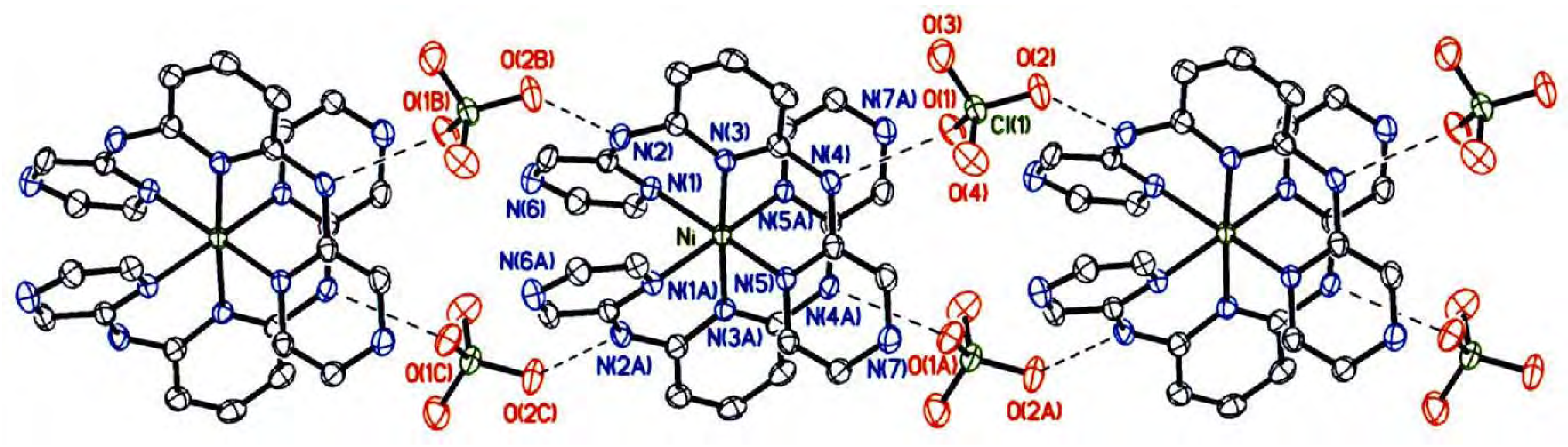
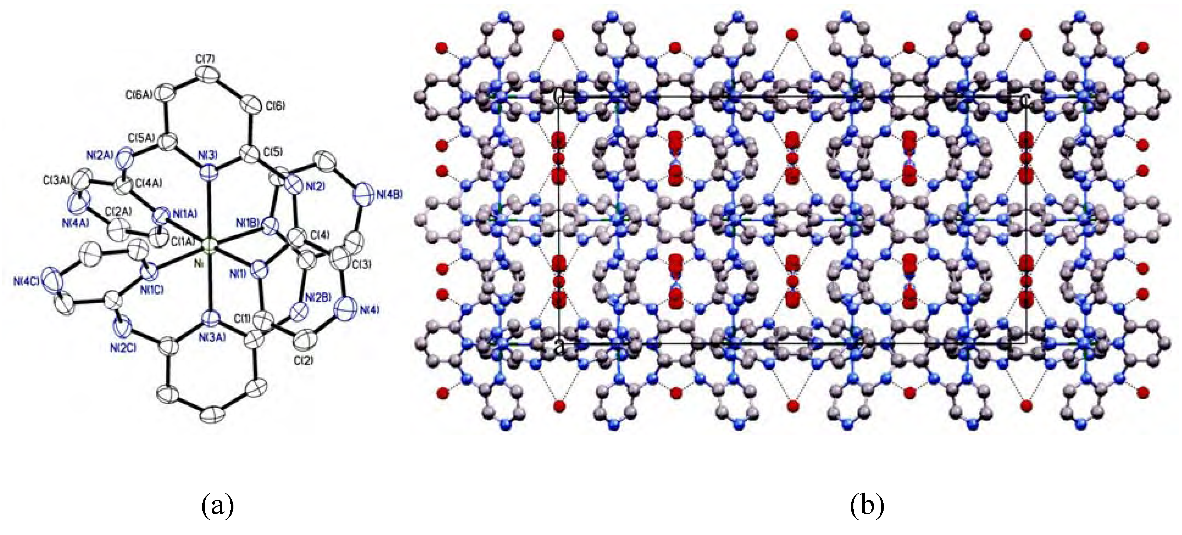
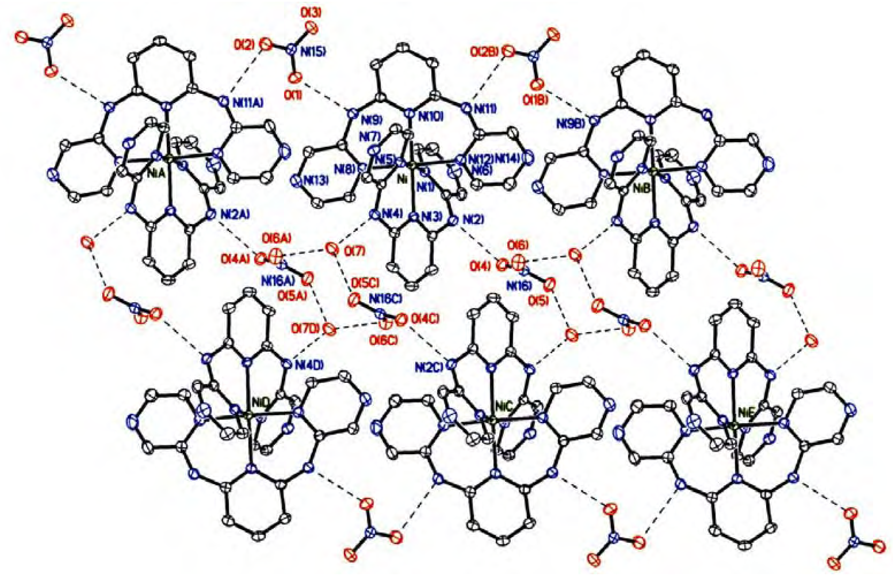
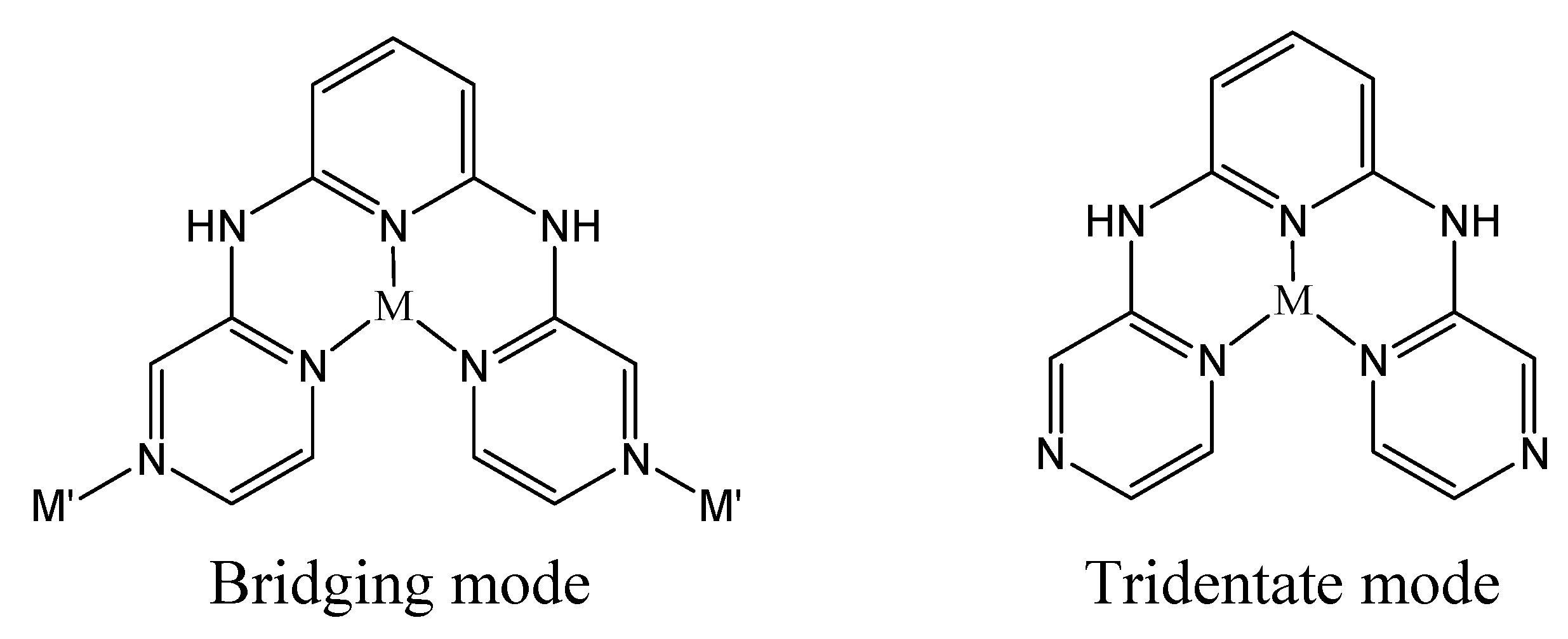
Crystal structure description

Complexes 1, 2 and 2·H2O were synthesized by reacting nickel(II) salts with the (H2dpzpda) ligand in methanol. The structures of these complexes were then established by X-ray crystallography. Selected bond lengths and angles are listed in Table 1 and the corresponding hydrogen bond geometries are listed in Table 2. The three complexes are mononuclear species with a hexacoordinated, octahedral geometry. In complex 1, the nickel atom sits in the C2 axial position. The crystal structure of 2 without a crystallized water molecule has D2 symmetry, whereas the 2·H2O species has rather low symmetry. The H2dpzpda ligand is coordinated with the Ni(II) atom as a tridentate species and the amino group nitrogen atoms are free. One of nitrogen atoms in the pyrazine ring (4-N) is uncoordinated, which is unexpected and very different from the previously reported H2dpzpda complex of copper(II), [Cu2(μ3-H2dpzpda)2(H2O)2]n(ClO4)4n [8], in which H2dpzpda coordinates to three metal atoms acting as a bridging ligand and leads to the formation of a 2-D complex with a [2×2] grid (Scheme 1). The Ni-N bond distances of high spin Ni(II) compounds are in the 2.062 to 2.103 Å range.
Scheme 1. Coordination modes of H2dpzpda.
Molecules 12 00821 sch001
The nitrogen atoms of free amino groups provided excellent donors for hydrogen bonds, and actually all the NH groups formed N−H···O type hydrogen bonding with counteranions or water molecules. For complex 1, hydrogen bonds between −NH and ClO4‾ gave rise to the 1-D chain along the b axis through N(4)−H···O(1) (2.895 Å) and N(2)−H···O(2B) (2.933 Å, where the symmetry operation: B = x, y-1, z.) linkages (Figure 1). A short 3.069 Å distance between the N(4) and N(5A) atoms was also observed, although intramolecular hydrogen bonding could be excluded because the H(4) atom of N(4) points away from the N(5A) atom.
Figure 1. 1-D chain structure of [Ni(H2dpapda)2](ClO4)2 (1). Atomic displacement ellipsoids were drawn at the 50% probability level. Dashed lines represent the hydrogen bonding.
Molecules 12 00821 g001
In nitrate complex 2 hydrogen bonds were observed between the amino group and nitrate (N(2)−H···O(1), 2.998 Å & N(2)−H···O(2), 3.108 Å). As the molecule was highly symmetrical (I41/acd in tetragonal crystal system), the hydrogen bonds (N−H···O) extended the complex in four directions, linking the original molecule with four different molecules and forming a three dimensional network (Figure 2). The change of anion from perchlorate to nitrate thus resulted in a change in the supramolecular structure from 1-D to 3-D.
Figure 2. (a) The molecular structure of [Ni(H2dpzpda)2]2+; (b) The hydrogen bond linkage in [Ni(H2dpzpda)2](NO3)2 (2); dashed lines represent the hydrogen bonding.
Molecules 12 00821 g002
When complex 2 included a water molecule into the crystal packing as 2•H2O, the symmetry decreased to a triclinic crystal system with P ī space group. The intermolecular hydrogen bonds through amino group and nitrate, [N(9)−H···O(1), 2.805 and N(11)−H···O(2B), 2.892 Å], still existed, and 1-D chains were formed along the a axis. It is very interesting that a 6-member oxygen ring, [O(6A)···O(7)···O(5C)···O(6C)···O(7D)···O(5A)···O(6A)], formed through the hydrogen bonding between nitrate anion and the cocrystallized water molecule [O(6A)···O(7), 2.781 Å; O(5C)···O(7), 2.830 Å; O(5C)···O(7)···O(6A), 122.0°; symmetry operation code: A = x+1, y, z, B = x-1, y, z, C = -x+1, -y, -z.] was found and further linked into the 1-D chain structure through hydrogen bonds [N(4)−H···O(7) (2.820 Å) and N(2)−H···O(4) (2.896 Å)], and the paired 1-D chains became a double chain (Figure 3). The 6-member ring adopted a chair configuration and stabilized the double chain structure.
Figure 3. The double chain structure of complex 2·H2O; dashed lines represent the hydrogen bonding.
Molecules 12 00821 g003
Table 1. Selected bond distances (Å) and angles (°) of complexes 1, 2 and 2•H2O.
1
Ni-N(1)2.1003(17)Ni-N(3)2.0797(16)
Ni-N(5)2.0654(17)
N(1)-Ni-N(3)87.48(7)N(1)-Ni-N(5)172.91(6)
N(1)-Ni-N(1A)85.75(9)N(1)-Ni-N(3A)95.42(7)
N(1)-Ni-N(5A)89.65(7)N(3)-Ni-N(5)87.59(7)
N(3)-Ni-N(3A)176.05(10)N(3)-Ni-N(5A)89.76(6)
N(5)-Ni-N(5A)95.42(9)
2
Ni-N(1)2.0708(17)Ni-N(3)2.076(2)
N(1)-Ni-N(1A)90.91(8)N(1)-Ni-N(1B)175.31(7)
N(1)-Ni-N(1C)89.29(8)N(1)-Ni-N(3)87.65(4)
N(1)-Ni-N(3A)92.35(4)N(3)-Ni-N(3A)180.0
2·H2O
Ni-N(1)2.0626(17)Ni-N(3)2.0884(16)
Ni-N(5)2.0637(17)Ni-N(8)2.0911(17)
Ni-N(10)2.1030(16)Ni-N(12)2.0781(17)
N(1)-Ni-N(3)89.11(7)N(1)-Ni-N(5)176.11(6)
N(1)-Ni-N(8)91.45(6)N(1)-Ni-N(10)91.90(6)
N(1)-Ni-N(12)90.04(6)N(3)-Ni-N(5)87.18(6)
N(3)-Ni-N(8)89.66(6)N(3)-Ni-N(10)178.34(6)
N(3)-Ni-N(12)92.63(6)N(5)-Ni-N(8)89.71(6)
N(5)-Ni-N(10)91.84(6)N(5)-Ni-N(12)88.95(6)
N(8)-Ni-N(10)89.00(6)N(8)-Ni-N(12)177.28(6)
N(10)-Ni-N(12)88.68(6)
Symmetry operation for 1: A = -x, y, -z+1/2; for 2: A = y-1/4, x+1/4, -z+1/4, B = -x, -y+1/2, z, C = -y+1/4, -x+1/4, -z+1/4.
Table 2. Hydrogen-bond geometry of complexes 1, 2 and 2·H2O. (Å, °).
D−H···Ad(D−H)d(H···A)d(D···A)< (DHA)
1
N(2)−H(2)···O(2B)0.882.0812.933(2)162.6
N(4)−H(4)···O(1)0.882.0532.895(2)159.9
2
N(2)−H(2)···O(1)0.882.1272.998(2)170.6
2•H2O
N(2)−H(2)···O(4)0.882.1502.896(2)142.2
N(4)−H(4)···O(7)0.882.0702.820(2)142.6
N(9)−H(9)···O(1)0.882.1572.805(2)123.0
N(11)−H(11)···O(2B)0.882.2342.892(2)131.4
O(7)−H(7)···O(6A)0.801.9862.781(2)170.0
O(7)−H(7’)···O(5C)0.802.0362.830(2)168.0
Symmetry operation for 1: B = x, y-1, z; for 2·H2O: A = x+1, y, z, B = x-1, y, z, C = -x+1, -y, -z.

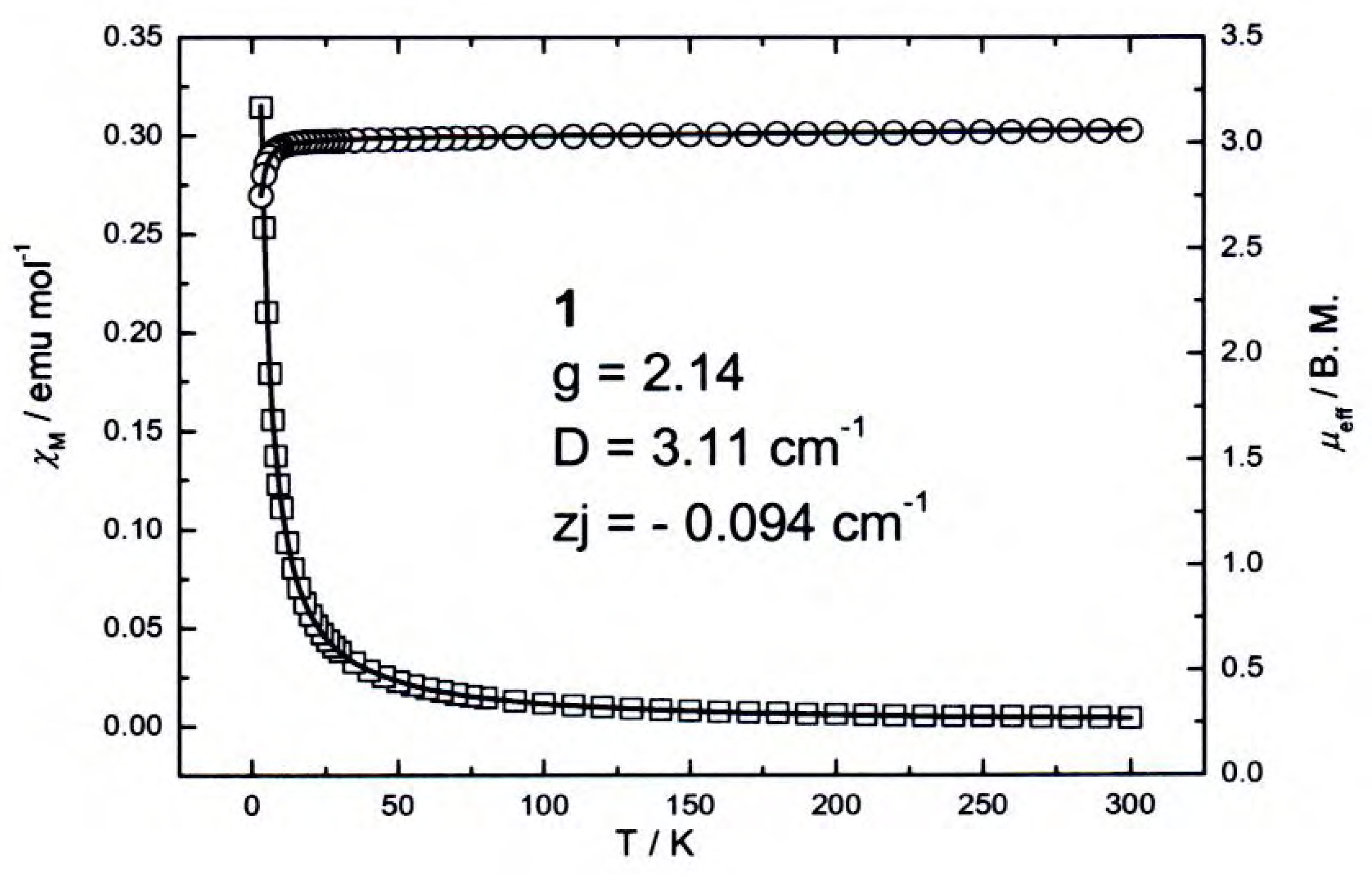
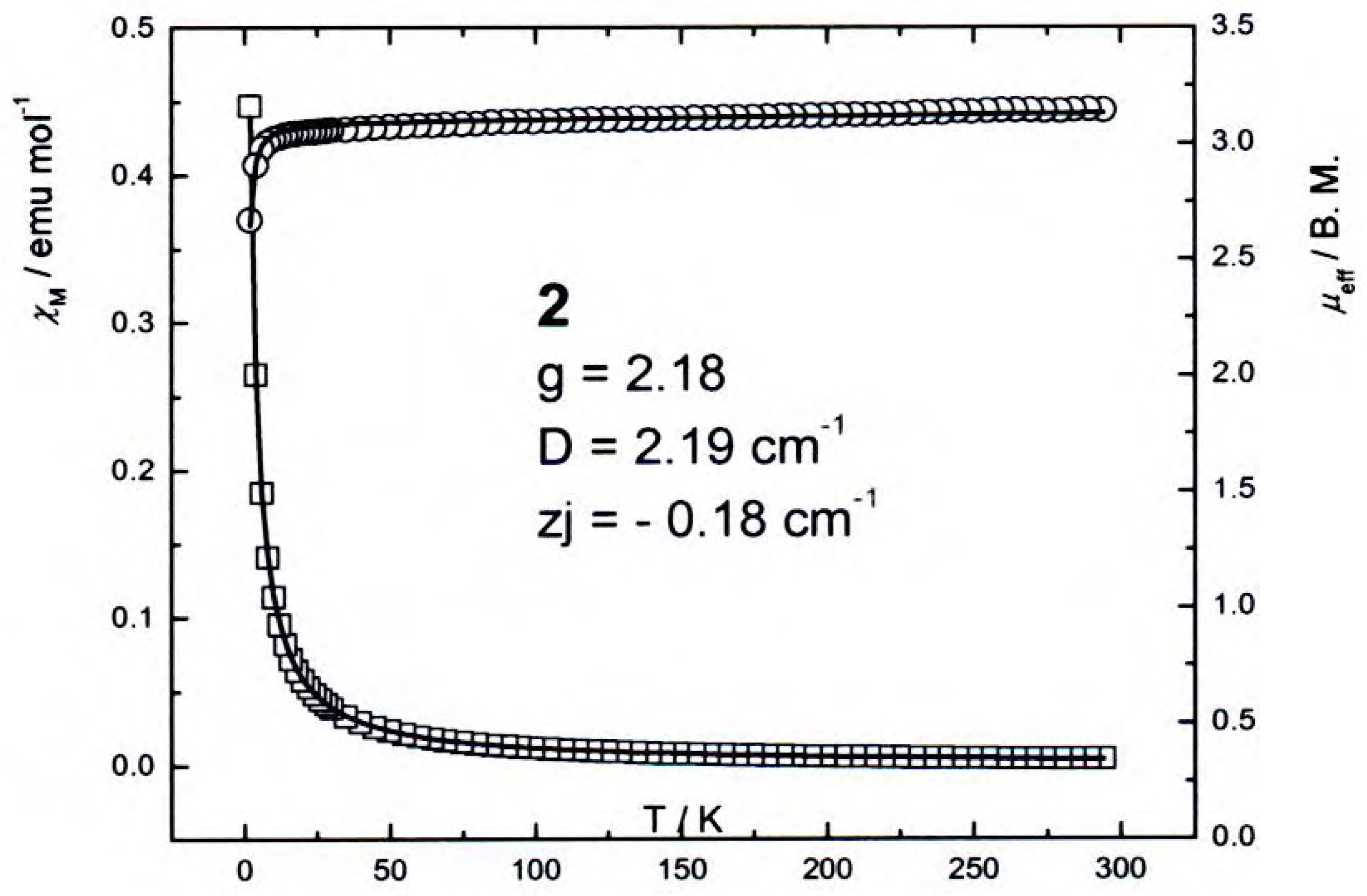
Magnetic and spectroscopic properties

The temperature-dependent magnetic susceptibilities of 1 and 2 are displayed in Figure 4 and Figure 5, respectively. Both of them exhibited a monotonic decrease of magnetic susceptibility with increasing temperature from 2K to 300K. The effective magnetic moment at room temperature was 3.06 B. M. for 1 and 3.15 B. M. for 2, which corresponds to expected spin-only value of two unpaired electrons with considerable orbital contribution. A substantial drop of magnetic moment was observed below 16 K due to spin-orbital coupling.
Figure 4. Temperature dependence of the magnetic moment μeff (O) and the molar susceptibility χM (□) for 1. Solid lines result from least-squares fits using equation (1) and (2).
Molecules 12 00821 g004
Figure 5. Temperature dependence of the magnetic moment μeff (O) and the molar susceptibility χM (□) for 2. Solid lines result from least-squares fits using equation (1) and (2).
Molecules 12 00821 g005
The experimental data were fitted with the axial anisotropic model for an S = 1 system:
χ// = (2Ng//2β 2/kT)exp(-D/kT) × [1+2exp(-D/kT)]-1
χ = (2Ng2β 2/D)[1-exp(-D/kT)] × [1+2exp(-D/kT)]-1
where D is the zero-field splitting parameter and χ = (χ// +2χ)/3. To simplify the calculation only one g value was considered (g// = g = g). A least squares fitting method taking into account MFA (Mean-Field Approximation) led to g = 2.14, D = 3.11 cm-1, zj = -0.09 cm-1 and F = 6.89×10⁻⁷ for 1 and g = 2.18, D = 2.19 cm-1, zj = -0.18 cm-1 and F = 2.70×10⁻⁶ for 2, in which zj corresponds to intermolecular interaction and F is defined as Σ((χi exp)-1i expi theo)2). The results are consistent with literature values [10,11]. The positive D value indicates the doubly degenerate levels (MS = ±1) lie above the singly degenerate (MS = 0) one, and also implies all spin entropy can be removed on cooling without intervention of long-range magnetic ordering processes.
In the infrared spectra, vibrations for the free amino group –NH moieties were observed at 3314 cm-1 for 1, and 3328 cm-1 for 2. Strong absorptions in the 1420-1600 cm-1 range can be assigned to the υ(C=C) and υ(C=N) vibrations of pyridine and pyrazine rings, respetively. The strong bands observed in the IR spectra of 1 and 2 in the 1077-1122 cm-1 region are assigned to the ClO4 or NO3 anions.
The electronic spectra of 1 and 2 in DMF solution were measured in the 11100-50000 cm-1 region. Two very strong absorptions at 37040 and 28570 cm-1 for 1 and 2, along with another strong band at 31740 cm-1 for 2 are attributed to the π-π* transitions of the ligand. Three weak absorptions were observed for 1 at 22730 cm-1 (shoulder, ε = 388 L mol-1 cm-1), 17240 cm-1 (ε = 56 L mol-1 cm-1) and 15270 cm-1 (ε = 90 L mol-1 cm-1), which can be assigned to spin-allowed d-d transitions, according to the literature [12,13]. For the nearly octahedral Ni(II) (d8) complex, they are attributed to the transitions of 3A2g3T1g(P), 3A2g3T1g(F) and 3A2g3T2g(F), respectively. The shoulder absorption corresponded to 3A2g3T1g(P) was not observed in spectrum of 2, perhaps because it was overlapped by the strong π-π* absorption of the complex.

Conclusions

In this paper we describe three Ni(II) complexes of N,N'–Di(pyrazin–2–yl)pyridine–2,6–diamine (H2dpzpda) built with different anions. Hydrogen bonds between amino groups and anions assembled the mononuclear molecules into different architectures. The perchlorate complex had a 1-D chain structure, whereas the nitrate complex displayed a 3-D network. When the nitrate complex packed with cocrystallized water, a double chain structure was formed through hydrogen bonds. The magnetic studies revealed g = 2.14 and D = 3.11 cm-1 for [Ni(H2dpzpda)2](ClO4)2 (1) and g = 2.18 and D = 2.19 cm-1 for for [Ni(H2dpzpda)2](NO3)2 (2), respectively.

Experimental

General

All reagents and solvents were obtained from commercial sources and were used without further purification. The ligand H2dpzpda was synthesized using the previously reported method [8]. Absorption spectra were recorded on a Hewlett Packard Model 8453 spectrophotometer. IR spectra were measured on KBr discs in the 400-4000 cm–1 range with a Perkin Elmer FT-IR spectrometer (PARAGON1000).

Preparation of compounds

[Ni(H2dpzpda)2](ClO4)2 (1)
A mixture of H2pzppda (50 mg, 0.19 mmol) and Ni(ClO4)2•6H2O (37 mg, 0.10 mmol) in methanol (50 mL) was stirred overnight. The solution was then filtered to remove insoluble impurities and concentrated under vacuum. After slow evaporation of the solvent orange-yellow crystals suitable for X-ray diffraction were produced (62 mg, yield: 83%). IR (KBr) υ/cm-1 = 3314s, 3237m, 3098m, 1636s, 1586m, 1555m, 1540m, 1499s, 1474m, 1449s, 1397w, 1343w, 1222m, 1180m, 1153s, 1122s, 1102s, 1077s, 1026m, 836m, 805m, 626m, 442m; UV/Vis (DMF) λmax/nm (ε/dm3mol-1cm-1) = 270 (5.63 × 10⁴), 350 (4.95 × 10⁴), 440 (sh) (3.88 × 10²), 580 (56), 655 (90); Analysis (%) C26H22Cl2NiN14O8: calcd.: C, 39.62; H, 2.81; N, 24.88; found: C, 39.40; H, 2.46; N, 24.53.
[Ni(H2dpzpda)2](NO3)2 (2)
A mixture of H2pzppda (50 mg, 0.19 mmol) and Ni(NO3)2•6H2O (29 mg, 0.10 mmol) in methanol (50 mL) was stirred overnight. Then solution was filtered and concentrated to 1/3 volume under vacuum. After slow evaporation of the green solution, light-brown crystals of 2·H2O suitable for X-ray diffraction were obtained (20 mg, Yield: 62%). Crystals of 2 without cocrystallized water were prepared by the same procedure as above, but using less methanol (6 mL) and by treating under solvothermal conditions at 150°C for 2 days and cooling down to room temperature at 5°C/h. The resulting orange crystals were obtained in 50% yield. IR (KBr) υ/cm-1 = 3328m, 3058m, 1633s, 1568m, 1511m, 1472s, 1421m, 1397m, 1190m, 1151s, 1122s, 1090s, 1109s, 943m, 820m, 626m, 466m; UV/Vis (DMF) λmax/nm (ε/dm3mol-1cm-1) = 270 (4.58 × 10⁴), 315 (3.16 × 10⁴), 350 (3.95 × 10⁴), 583 (1.08 × 10²), 657 (141); Analysis (%) C26H22NiN16O6·H2O: calcd.: C, 43.78; H, 3.11; N, 31.43; found: C, 43.50; H, 3.14; N, 30.84.

Crystal Structure Determinations

The chosen crystals were mounted on a glass fiber. Data collection was carried out using a Mo-Kα radiation (λ = 0.71073 Å) and a liquid nitrogen low-temperature controller at 150(1) K on a NONIUS KappaCCD diffractometer for 1 and 2, and BRUKER SMART ApexCCD for 2·H2O. Data reduction was performed on the DENZO-SMN [14] software for 1 and 2, and Bruker SAINT [15] for 2·H2O. The absorption correction was applied with SORTAV [16] program for 1 and 2, and SADABS [17] for 2•H2O. All structures were solved by the SHELXS-97 [18] and refined with SHELXL-97 [19] using the full-matrix least squares on F2 values. Molecular graphics were plotted using SHELXTL [20]. The detailed crystal data are listed in Table 3. CCDC-640746~8 contains the supplementary crystallographic data for this paper. These data can be obtained free of charge from the Cambridge Crystallographic Data Centre via www.ccdc.cam.ac.uk/data_rquest/cif
Table 3. Crystallographic and experimental data for complexes 1, 2 and 2•H2O.
Compound[Ni(H2dpzpda)2](ClO4)2 (1)[Ni(H2dpzpda)2](NO3)2 (2)[Ni(H2dpzpda)2](NO3)2•H2O (2•H2O)
FormulaC26H22N14O8C2NiC26H22N16O6Ni C26H24N16O7Ni
Formula weight788.19713.31731.32
Crystal systemMonoclinicTetragonalTriclinic
Space groupC2/cI41/acdP ī
Formula per unit cell, Z482
Unit-cell dimensionsa = 18.0619(5) Åa = 14.6836(3) Åa = 10.3287(6) Å
b = 11.0820(3) Åb = 14.6836(3) Åb = 10.3728(6) Å
c = 16.9851(4) Åc = 28.3900(5) Åc = 16.6563(9) Å
α = 90°α = 90°α = 73.506(1)°
β = 118.082(2)°β = 90°β = 82.367(1)°
γ = 90°γ = 90°γ = 61.950(1)°
Unit-cell volume, V (Å 3)2999.5(1)6121.1(2)1510.1(2)
Dcalcd. (g/cm3)1.7451.5481.608
Absorption coefficient (mm-1)0.9030.7050.719
F(000)16082928752
Crystal size (mm)0.40 x 0.20 x 0.150.18 x 0.15 x 0.150.28 x 0.25 x 0.25
θ ranges for data collection (o)2.24 ~ 27.502.43 ~ 27.501.27 ~ 27.50
Reflections collected151891360319579
Independent reflections3444 (Rint = 0.0476)1765 (Rint = 0.0475)6901 (Rint = 0.0273)
Completeness to θ=27.50 (%)99.810099.7
Absorption correctionMulti-scanMulti-scanMulti-scan
Max. and min. transmission0.876 and 0.7140.907 and 0.8620.841 and 0.824
Refinement methodFull-matrix L. S. on F2Full-matrix L. S. on F2Full-matrix L. S. on F2
Data / restraints / parameters3444 / 0 / 2311765 / 2 / 1236901 / 0 / 453
Goodness-of-fir on F21.0431.0991.082
R1, wR2 [I > 2σ(I)]0.0341, 0.08660.0336, 0.09040.0405, 0.0987
R1, wR2 (all data)0.0487, 0.09360.0633, 0.09990.0456, 0.1017
Largest diff. peak and hole 0.401 and -0.493 e/ Å 30.275 and -0.331 e/ Å 30.526 and -0.595 e/ Å 3

Acknowledgments

The authors thank the National Science Council of the Republic of China for financial support.

References

  1. Ferlay, S.; Hosseini, W. Crystalline molecular alloys. Chem. Commun. 2004, 788–789. [Google Scholar] [CrossRef] [PubMed]
  2. Dechambenoit, P.; Ferlay, S.; Hosseini, M. W. From tectons to composite crystals. Cryst. Growth Des. 2005, 5, 2310–2312. [Google Scholar] [CrossRef]
  3. Nguyen, T. L.; Fowler, F. W.; Lauher, J. W. Commensurate and Incommensurate Hydrogen Bonds. An Exercise in Crystal Engineering. J. Am. Chem. Soc. 2001, 123, 11057–11064. [Google Scholar] [CrossRef] [PubMed]
  4. Prior, T. J.; Bradshaw, D.; Teat, S. J.; Rosseinsky, M. J. Designed layer assembly: a three-dimensional framework with 74% extra-framework volume by connection of infinite two-dimensional sheets. Chem. Commun. 2003, 500–501. [Google Scholar] [CrossRef]
  5. Lee, G. H.; Wang, H. T. Synthesis and Molecular Structures of Two [1,4-bis(3-pyridyl)-2,3-diazo-1,3-butadiene]-dichloro-Zn(II) Coordination Polymers. Molecules 2006, 11, 589–596. [Google Scholar] [CrossRef] [PubMed]
  6. Lee, G. H.; Wang, H. T. Synthesis and Crystal Structure of a Three Dimensional Nickel(II) Coordination Polymer with 1,4-Bis(3-pyridyl)-2,3-diazo-1,3-butadiene as Ligand. Anal. Sci. 2007, 23, x1–x2. [Google Scholar] [CrossRef]
  7. Lu, Y. L.; Wu, J. Y.; Chan, M. C.; Huang, S. M.; Lin, C. S.; Chiu, T. W.; Liu, Y. H.; Wen, Y. S.; Ueng, C. H.; Chin, T. M.; Hung, C. H.; Lu, K. L. Influence of Water Content on the Self-Assembly of Metal-Organic Frameworks Based on Pyridine-3,5-dicarboxylate. Inorg. Chem. 2006, 45, 2430–2437. [Google Scholar] [CrossRef] [PubMed]
  8. Ismayilov, R. H.; Wang, W. Z.; Lee, G. H.; Peng, S. M. One-, two- and three-dimensional Cu(II) complexes built via new oligopyrazinediamine ligands: from antiferromagnetic to ferromagnetic coupling. Dalton Trans. 2006, 478–491. [Google Scholar] [CrossRef] [PubMed]
  9. Yeh, C. Y.; Chou, C. H.; Pan, K. C.; Wang, C. C.; Lee, G. H.; Su, Y. O.; Peng, S. M. Linear Pentacobalt Complexes: Synthesis, Structures, and Physical Properties of Both Neutral and One-electron Oxidation Compounds. J. Chem. Soc. Dalton Trans. 2002, 2670–2677. [Google Scholar] [CrossRef]
  10. Carlin, R. L.; O’Connor, C. J.; Bhatia, S. N. Large Zero Field Splitting and Subcritical Antiferro-magnetic Interactions in [Ni(C5H5N0)6](Cl04)2. J. Am. Chem. Soc. 1976, 98, 3523–3526. [Google Scholar] [CrossRef]
  11. Boča, R. Zero-field splitting in metal complexes. Coord. Chem. Rev. 2004, 248, 757–815. [Google Scholar] [CrossRef]
  12. Xu, J. Y.; Bian, H. D.; Gu, W.; Yan, S. P.; Cheng, P.; Zheng, D. Z.; Jiang, Z. H.; Shen, P. W. X-ray structural and spectroscopic studies of a mononuclear nickel(II) complex with tris(2-pyridyl-methyl)amine and thiocyanate. J. Mol. Struct. 2003, 646, 237–242. [Google Scholar] [CrossRef]
  13. Mašlejová, A.; Boca, R.; Dlhán, L.; Papánková, B.; Svoboda, I.; Fuess, H. Structure and zero-field splitting in bis(1,2-dimethylimidazole)bis(acetato)nickel(II) molecular complex. Chem. Phys. Lett. 2001, 347, 397–402. [Google Scholar] [CrossRef]
  14. Otwinowski, Z.; Minor, W. Meth. Enzymol. 1997, 276, 307–326.
  15. SAINT. Program for cell refinement and data reduction; Bruker AXS Inc.: Madison, Wisconsin, USA, 2005.
  16. Blessing, R. H. An empirical correction for absorption anisotropy. Acta. Cryst. Sect. A. 1995, 51, 33–38. [Google Scholar] [CrossRef]
  17. Sheldrick, G. M. Program for absorption correction; University of Göttingen: Göttingen, Germany, 2003. [Google Scholar]
  18. Sheldrick, G. M. Phase annealing in SHELX-90: direct methods for larger structures. Acta Cryst. 1990, A46, 467. [Google Scholar] [CrossRef]
  19. Sheldrick, G. M. Program for the refinement of crystal structure; University of Göttingen,: Göttingen, Germany, 1997. [Google Scholar]
  20. SHELXTL. Software used to prepare material for publication; Bruker AXS Inc.: Madison, Wisconsin, USA, 2000.
  • Sample Availability: Available from the authors.

