Article

Synthesis, Molecular Structure and Characterization of Allylic Derivatives of 6-Amino-3-methyl-1,2,4-triazolo[3,4-f][1,2,4]-triazin-8(7H)-one

1
Faculty of Medicinal and Applied Chemistry, Kaohsiung Medical University, Kaohsiung City 80708, Taiwan, ROC
2
Department of Psychiatry, Longcyuan Veterans Hospital, Pingtung 912, Taiwan
3
Instrumentation Center, College of Science, National Taiwan University, Taipei City 106, Taiwan
*
Author to whom correspondence should be addressed.
Molecules 2006, 11(6), 444-452; https://doi.org/10.3390/11060444
Submission received: 31 May 2006 / Revised: 17 June 2006 / Accepted: 19 June 2006 / Published: 22 June 2006

Introduction

Many aza/deaza analogues of purine have been found to display interesting biological activities. In the last decade numerous fused 1,2,4-triazines and fused 1,2,4-triazoloes have been synthesized and screened in vitro/vivo, thus revealing their varied biochemical, biological, pharmacological or cellular activities [1,2,3,4,5,6,7,8,9,10,11,12,13,14,15,16,17,18,19]. Recently, we have explored a ring cyclization mechanism [20,21] in connection with the preparation of the tautomeric heterobicyclic 6-amino-1,2,4-triazolo[3,4-f][1,2,4]triazin-8(7H)-one (4,8-diaza-9-deazaguanine), the isosteric isomer of guanine and additionally we have proven by X-ray crystallographic analysis [22,23] that the predominant tautomeric structure of the former is the 7H-tautomer. To our knowledge, no report describing N-substituted derivatives of the tautomeric structure of 6-amino-1,2,4-triazolo[3,4-f][1,2,4]triazin-8(7H)-one has appeared to date. In continuation of our studies on the synthesis of derivatives and the tautomeric structures of the 6-amino-1,2,4-triazolo[3,4-f][1,2,4]triazin-8(7H)-one moiety, we report in this paper the synthesis and characterization of some N-allylated derivatives of 6-amino-3-methyl-1,2,4-triazolo[3,4-f][1,2,4]triazin-8(7H)-one (1), viz., 1-allyl-6-amino-3-methyl-1,2,4-triazolo[3,4-f][1,2,4]triazin-8(7H)-one (2) and 7-allyl-6-amino-3-methyl-1,2,4-triazolo[3,4-f][1,2,4]triazin-8(7H)-one (3) as potential bioactive molecules (Scheme 1). The molecular structures of 2 and 3 have been confirmed by spectral analysis, mainly their 2D-NMR. In addition, the single X-ray crystal structural analysis of molecule 3 is also reported, which supports the conclusions reached from the interpretation of the 2D-NMR spectral data.
Scheme 1.
Scheme 1.
Molecules 11 00444 g004

Results and Discussion

Compound 1 was reacted with potassium carbonate and allyl bromide in dry acetone at room temperature for 16 h, under 18-crown-6-ether catalysis, to afford a 1:3 ratio of a minor product 2 (Rf = 0.59) and a major product 3 (Rf = 0.41) in 36% total yield. The structures of 1, 2 and 3 were completely assigned by 1H- and 13C-NMR spectroscopy. The presence of an allyl group and eight carbon signals were observed in both compounds 2 and 3. A detailed 2D-NMR study, including gHSQC and gHMBC measurements was necessary to confirm the allylation sites of 2 and 3. The gHSQC 1H-13C experiments allowed segregation of the chemical shifts of proton or carbon for the 3-methyl, C-3 or allyl groups in 1, 2 and 3. The long-range gHMBC 1H-13C correlations showed the attachment of the allyl group on N-1 for structure 2, since the methylene protons (δH 5.18 ppm) of this group show a 3J (H,C) coupling to the C-8a carbon atom (δC 133.40 ppm). On the other hand, the fact that the methylene protons (δH 4.52 ppm) of the allyl group have a 3J (H,C) coupling to both the C-6 (δC 151.88 ppm) and C-8 (δC 153.22 ppm) carbon atoms proved the attachment of the allyl group on N-7 for structure 3.
Scheme 2 presents the numbering system of the 6-amino-1,2,4-triazolo[3,4-f][1,2,4]triazin-8(7H)-one moiety and complete assignment of 1H- and 13C-NMR signals for 1, 2 and 3, together with the corresponding long-range 1H-13C correlations. The δ values of 1H- and 13C-NMR of this moiety were similar in 1 and 3. On the other hand, there were differences in the δ values of the 1H- and 13C-NMR spectra between the N-1-allyl 2 and N-7-allyl 3; namely, the upfield shifts for 6-NH2 group and C-8a between 2 and 3 were +0.72 and +6.54 ppm, respectively. Meanwhile, the δ values of C-6 and C-8 of 2 showed significant downfield shifts of -5.8 and -10.13 ppm, respectively, as compared to 3. Similarly, the δ values of the methylene on the allyl group of 2 showed significant downfield shifts of -0.64 and -9.67 ppm, respectively, as compared to 3. Taken together, molecule 2 revealed obvious differences in the NMR chemical shifts of C-6, C-8, C-8a, and the methylene on the allyl group, as compared to molecules 1 and 3. This evidence suggests that 2 may be an aromatic molecule.
Scheme 2.
Scheme 2.
Molecules 11 00444 g005
The structure of 3 was unambiguously confirmed by X-ray crystallography of its adduct with dimethyl sulfoxide (DMSO), revealing the structural framework to be the 7-allyl-6-amino-3-methyl-1,2,4-triazolo[3,4-f][1,2,4]triazin-8(7H)-one connected with the oxygen of DMSO via N-H···O intermolecular hydrogen bonding (Figure 1). Obviously, the allyl group is located at N(5) (i.e., N-7), which is compatible with the tautomeric 7-H proton located at N-7 in the compounds 6-amino-3-ethyl-1,2,4-triazolo[3,4-f][1,2,4]triazin-8(7H)-one and 6-amino-3-benzylmercapto-1,2,4-triazolo[3,4-f]-[1,2,4]-triazin-8(7H)-one, as previously reported by us [22,23]. The mean plane of the 1,2,4-triazolo[3,4-f][1,2,4]triazine ring forms a dihedral angle of 89.14° with the N(5)-allyl group (N(5)-C(5)-C(6)-C(7)).
The molecular packing is shown in Figure 2. In the molecular structure of 3 the short bonds 1.306(3) Å (N(4)-C(4)), 1.316(3) Å (N(1)-C(2)) and 1.319(3) Å (N(2)-C(1)) have an appreciable double-bond character. Notably, the bond length 1.328(3) Å between C(4)-N(6) is shorter than 1.355 Å of Car-NH2 (Nsp2: planar) [24].
Figure 1. ORTEP drawing and atom labelling scheme of the compound 3 (attached to DMSO) with thermal ellipsoids drawn at the 30% probability level.
Molecules 11 00444 g001
Figure 2. A perspective drawing of the packing arrangement of molecule 3, showing (a) the molecules’ direction along the a-view and (b) c-view with π-π stacking interactions. Dashed lines are intermolecular N−H···O hydrogen bonds.
Molecules 11 00444 g002
Meanwhile, the 117.78°(19) value of the N(4)-C(4)-N(6) angle, close to 120°, confirms the sp2 hybridization of the nitrogen atom, which implies that the 6-amino group strongly donates the unpaired electrons and resonates with the [1,2,4]triazolo[3,4-f][1,2,4]triazine ring. The C(3)-O(1) bond length (1.214(3) Å) is shorter than the 1.240 Å bond length of Csp2=O(1) in δ-lactams [24]. This may be attributed to the electron abstraction by the π-deficient heterobicyclic ring. In the structure of the DMSO moiety the bond lengths of 1.771 (3) (S(1)-C(9)), 1.759(3) Å (S(1)-C(10)) and 1.482(2) Å (S(1)=O(2)) are all shorter than the 1.809 Å (S-C) and 1.497 Å (S=O) bond lengths in pure crystallized DMSO [24]. This is a result from the attraction of the intermolecular N-H···O hydrogen bonding in the crystal structure of 3.
Analysis of the molecular packing in unit cell reveals that each molecular structure of 3 is linked by intermolecular hydrogen bonds and π-π stacking interactions (Figure 3 and Table 3). Each molecule is linked into Molecules 11 00444 i001
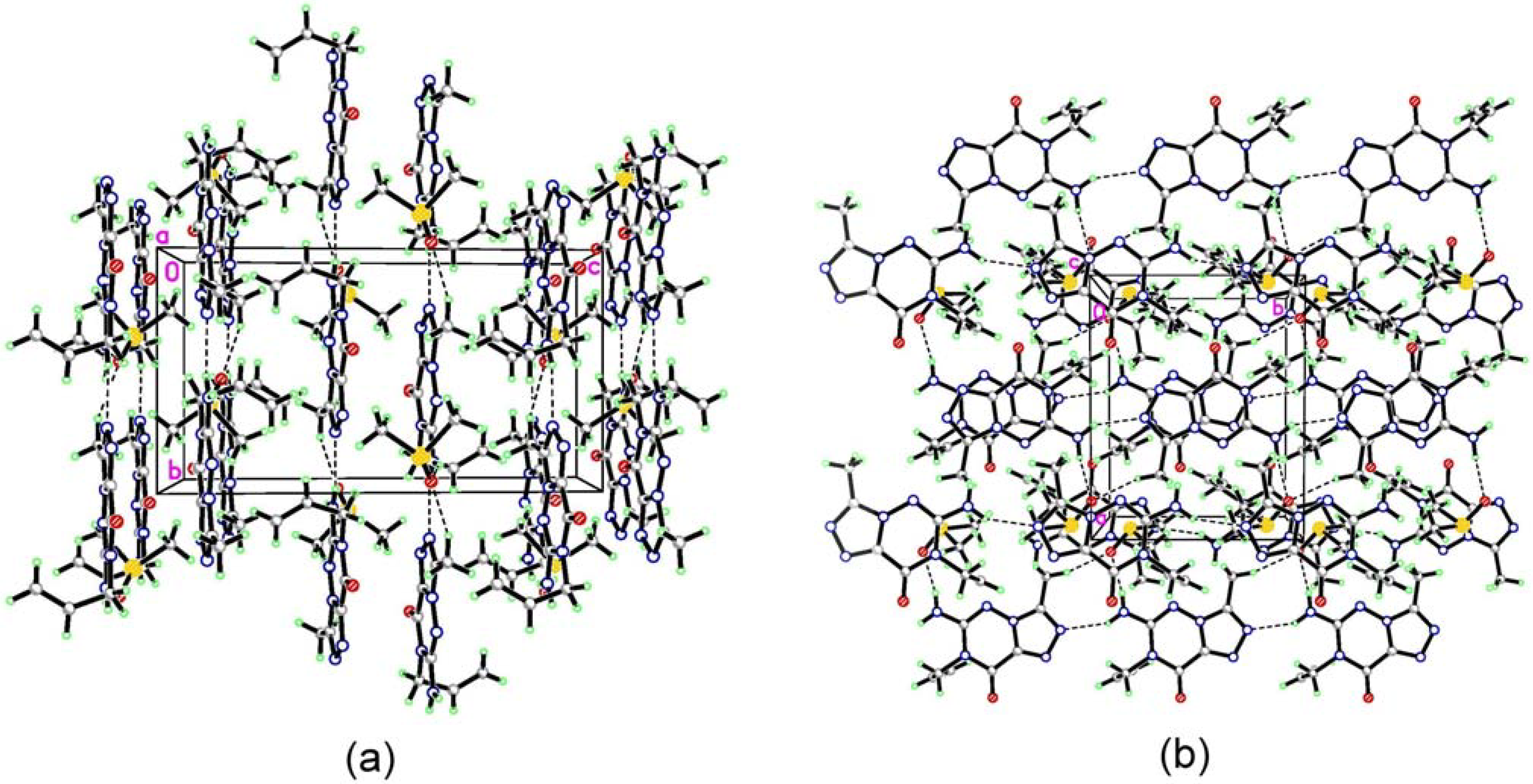
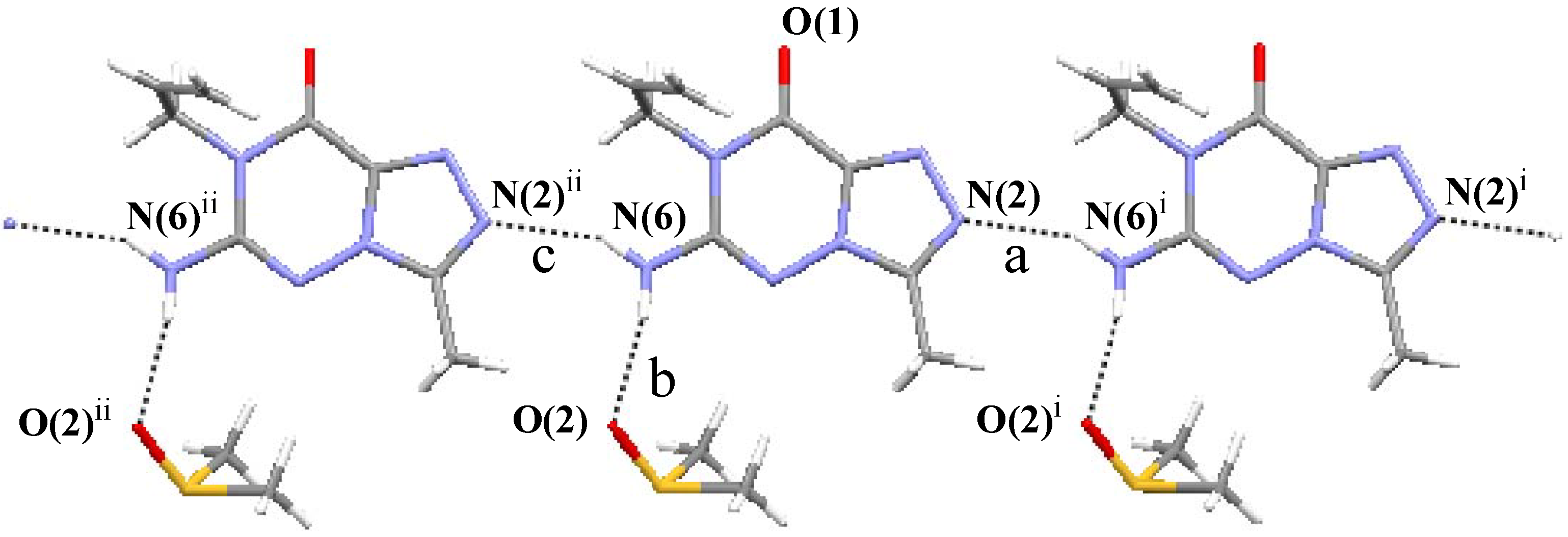
(7) graph set association via N-H···N hydrogen bond interactions (Figure 3 notation [a] and [c]). Assignment of the H-bond descriptors is based on the graph-set theory [25]. The molecular graphic was obtained using the Mercury program (version 1.4, CCDC, Cambridge, UK). Meanwhile, each molecule links with the oxygen of DMSO via N-H···O hydrogen bonding ([b]). The structure is further stabilized by π-π stacking interactions (Figure 2: (b)), which results in the centroid···centroid distance (3.351 Å) being that between the layer of [1,2,4]triazolo[3,4-f][1,2,4]triazine ring.
Figure 3. A part of the crystal structure for molecule 3, showing the molecules’ direction along the a-view with x+90°. Broken lines indicate the intermolecular hydrogen bonding patterns. For notation and symmetry codes see Table 1.
Molecules 11 00444 g003
Table 1. Hydrogen bond geometry in compound 3.
NotationD-H···AD-H (Å)H···A (Å)D···A (Å)Length-VdWD-H···A(°)
a
b
c
N(2)···H(6C)−N(6)i
N(6)−H(6B)···O(2)
N(6)−H(6C)···N(2)ii
0.860
0.860
0.860
2.190
2.033
2.190
2.978
2.848
2.978
–0.122
–0.222
–0.122
152.27
157.57
152.27
* Note. Symmetry codes: (i) x, 1+y, z; (ii) x, –1+y, z.

Conclusions

In summary, we have reported the synthesis and molecular structure characterization of N-1-allyl 2 and N-7-allyl 3. The 2D-NMR spectral analysis allowed complete assignments of various carbons of these molecules. Obviously different δ values in 1H- and 13C-NMR spectra between 2 and 3 were found. A single X-ray crystal structural analysis of 3 supported the evidence gathered from the NMR spectral interpretation.

Experimental Section

General

Melting points were measured on a YANACO micromelting point apparatus and were uncorrected. The Infrared spectra were recorded as KBr discs on a Perkin-Elmer FTIR 1650 instrument. The UV-VIS spectra were measured on a GBC Cintra 6 UV-Visible spectrometer. The 1H NMR and 13C NMR spectra were obtained in dimethyl sulfoxide-d6 (DMSO-d6) on a Varian Mercury-plus 400 (400 MHz) spectrometer. Chemical shifts are expressed in ppm (δ) with tetramethylsilane (TMS) as an internal standard. For the assignments of signals, standard and long-range 1H-13C heteronuclear chemical shift correlation 2D-NMR experiments (gHSQC and gHMBC) were used. Thin layer chromatography (tlc) analyses were performed on silica gel plates (Merck 60 F254, 0.2 mm thickness), and the components were detected by UV light (254 nm). Mass spectra were obtained on a Quattro VG-5022 spectrometer with an ionization potential of 70 eV. Elemental analyses were performed on a Heraeus CHN-O-Rapid elemental analyzer. All the solvents used were dried and distilled under argon prior to use.

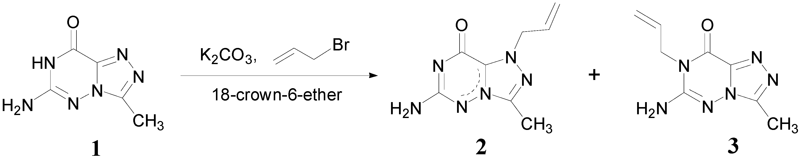
Syntheses: 6-Amino-3-methyl-1,2,4-triazolo[3,4-f][1,2,4]triazin-8(7H)-one (1)

Compound 1 was prepared as described by Lovelette [26], m.p. >300°C (from water); IR (cm–1), νmax: 3340, 3185, 1729 (C=O), 1625(C=N), 1517, 1408, 1283, 1131; UV, λmax nm (log ε): (0.1 N HCl) 213 (4.35); (H2O) 208 (4.56), 220 (4.56); (0.1 N NaOH) 208 (4.25), 220 (4.39); 1H-NMR, δ: 2.44 (s, 3H, CH3); 6.39 (s, 2H, NH2); 11.48 (br s, 1H, NH); 13C-NMR, δ: 9.40 (CH3), 139.55 (C-8a), 146.93 (C-3), 150.46 (C-6), 151.97 (C-8); MS, m/z (%): 166 (M+, 35), 138 (3), 123 (5), 111 (8), 97 (6), 91 (4), 77 (5), 69 (24), 54 (32), 43 (100). Anal. calcd for C5H6N6O: C, 36.15%; H, 3.64%; N, 50.58%. Found: C, 36.10%; H, 3.70%; N, 50.74%.

1-Allyl-6-amino-3-methyl-1,2,4-triazolo[3,4-f][1,2,4]triazin-8(7H)-one (2) and 7-Allyl-6-amino-3-methyl-1,2,4-triazolo[3,4-f][1,2,4]triazin-8(7H)-one (3)

A solution of 1 (0.83 g, 5 mmol) in dry acetone (30 mL) was mixed with anhydrous potassium carbonate (0.69 g, 5 mmol) and a catalytic amount of 18-crown-6-ether (0.13 g, 0.5 mmol). Then allyl bromide (0.60 g, 5 mmol) was added and the mixture stirred at room temperature for 16 h. The solvent was evaporated to afford a crude product which was then applied to a silica gel (230-400 mesh) column. The column was eluted with a mixture of chloroform and methanol (50:1) and the appropriate fractions were combined and evaporated.
The Rf value of the minor adduct was 0.59 (chloroform- methanol = 5:1). The residue thus obtained was recrystallized from ethanol to give 2 (0.09 g, 9%) as bright white crystals, m.p. 278-279°C; IR (cm–1), νmax: 3384, 3321, 3141, 1706 (C=O), 1657 (C=N), 1591, 1493, 1425, 1315, 1267, 1098, 969, 919; UV, λmax nm (log ε): (0.1 N HCl) 217 (4.41); (H2O) 224 (4.66); (0.1 N NaOH) 218 (4.07); 1H-NMR, δ: 2.44 (3H, CH3), 5.18 (2H, CH2), 5.19 (2H, CH=CH2), 5.93 (1H, CH=CH2), 5.88 (2H, 3-NH2); 13C-NMR, δ: 10.08 (CH3), 54.24 (CH2), 120.81 (CH=CH2), 132.21 (CH=CH2), 133.40 (C-8a), 146.54 (C-3), 157.68 (C-6), 163.35 (C-8); MS, m/z (%): 206 (M+, 20), 165 (21), 164 (100), 111 (3), 84 (2), 69 (6), 54 (4), 42 (10); Anal. calcd for C10H10N4O: C, 46.60%; H, 4.89%; N, 40.76%. Found: C, 46.72%; H, 4.86%; N, 40.65%.
The Rf value for the second (major) product was 0.41 (chloroform-methanol = 5:1). The residue thus obtained was recrystallized from ethanol to give 3 (0.28 g, 27%) as bright white crystals, m.p. 260-261°C; IR (cm–1), νmax: 3383, 3321, 3141, 1706 (C=O), 1658 (C=N), 1591, 1493, 1426, 1315, 1267, 1104, 967, 920; UV, λmax nm (log ε): (0.1 N HCl) 208 (4.66), 213 (4.66), 218 (4.66); (H2O) 220 (4.41); (0.1 N NaOH) 218 (4.10); 1H-NMR, δ: 2.43 (3H, CH3), 4.52 (2H, CH2), 5.11 (2H, CH=CH2), 5.82 (1H, CH=CH2), 6.60 (2H, 3-NH2); 13C-NMR, δ: 9.92 (CH3), 44.57 (CH2), 117.96 (CH=CH2), 131.50 (CH=CH2), 139.94 (C-8a), 148.86 (C-3), 151.88 (C-6), 153.22 (C-8); MS, m/z (%): 206 (M+, 100), 191 (71), 177 (8), 165 (17), 164 (35), 151 (10), 135 (13), 123 (6), 111 (6), 96 (5), 84 (7), 69 (10), 54 (15); Anal. calcd for C10H10N4O: C, 46.60%; H, 4.89%; N%, 40.76. Found: C, 46.60%; H, 4.93%; N, 40.70%.
Table 2. Crystal and experimental data for compound 3.
FormulaC10H16N6O2S
Formula weight284.35
Crystal systemMonoclinic
Space groupP21/n
Unit-cell dimensions (Å)a = 10.4850(6)
b = 8.4409(5)
c = 15.5152(9)
β = 95.963(1)°
Unit-cell volume, V3)1365.71(14)
Formula per unit cell, Z4
Dcalcd (g/cm3)1.383
Absorption coefficient, μ (mm-1)0.246
F(000)600
Crystal size (mm)0.36 × 0.25 × 0.08
Index ranges– 13 ≤ h ≤ 13
– 10 ≤ k ≤ 10
– 20 ≤ l ≤ 20
Max. and min. transmission0.9806 and 0.9167
Independent reflections3133 (Rint = 0.0480)
Reflections/restraints/parameters3133 /0 /175
Final R indices [I > 2σ(I)]R1 = 0.0590, wR2 = 0.1371
R indices (all data)R1 = 0.0736, wR2 = 0.1454
Goodness-of-fit on F21.078
Max. shift/error0.000

X-ray techniques

X-ray quality crystals of compound 3 were obtained by crystallization from dimethyl sulfoxide at room temperature. A summary of the crystallographic data and details of the structure refinements is listed in Table 2. The data was collected on a BRUKER SMART ApexCCD diffractometer with a graphite-monochromated Mo radiation (λ = 0.71073 Å) at 295(2) K. The crystal structure has been deposited at the Cambridge Crystallographic Data Centre [27]. A total of 13191 reflections and 3133 independent reflections (Rint = 0.0480) were collected within the range of 2.24 <θ< 27.50° by using the ω scan technique, of which 2535 observed reflections with I > 2σ(I) were used in the structural analysis. The crystal structure was solved by direct methods using SHELXS-97 [28] and refined by full-matrix least-squares methods on F2 using SHELXL-97 [29]. All non-H atoms were refined anisotropically. The hydrogen atoms were placed geometrically and refined as riding. The final cycle of full-matrix least-squares refinement gave R1 = 0.0416, wR2 = 0.1142 (w = 1/[σ2( Molecules 11 00444 i002) + (0.0584P)2 + 0.7097P], where P = ( Molecules 11 00444 i002 + 2 Molecules 11 00444 i003)/3). S = 1.078 and (Δ/σ)max= 0.000. The maximum peak on the final difference Fourier map is 0.321 and the minimum peak −0.319 eÅ–3.

Acknowledgements

The authors would like to thank the National Science Council, Republic of China, for support under grant NSC 90-2113-M-037-015.

References and Notes

  1. Pomarnacka, E. Acta Pol. Pharm. 1998, 55, 481.
  2. Abdel-Rahman, R. M.; Morsy, J. M.; Hanafy, F.; Amene, H. A. Pharmazie 1999, 54, 347.
  3. Abdel-Rahman, R. M.; Morsy, J. M.; El-Edfawy, S.; Amene, H. A. Pharmazie 1999, 54, 667.
  4. Abdel-Rahman, R. M. Pharmazie 2001, 56, 18.
  5. Abdel-Rahman, R. M. Pharmazie 2001, 56, 275.
  6. El-Gendy, Z.; Morsy, J. M.; Allimony, H. A.; Ali, W. R.; Abdel-Rahman, R. M. Pharmazie 2001, 56, 376.
  7. Holla, B. S.; Rao, B. S.; Gonsalves, R.; Sarojini, B. K.; Shridhara, K. Farmaco 2002, 57, 693.
  8. Habib, N. S.; Soliman, R.; Ismail, K.; Hassan, A. M.; Sarg, M. T. Boll. Chim. Farm. 2003, 142, 396.
  9. Iwashita, A.; Maemoto, T.; Nakada, H.; Shima, I.; Matsuoka, N.; Hisajima, H. J. Pharmacol. Exp. Ther. 2003, 307, 961. [CrossRef]
  10. Guertin, K. R.; Setti, L. Qi. L.; Dunsdon, R. M.; Dymock, B. W.; Jones, P. S.; Overton, H.; Taylor, M.; Williams, G.; Sergi, J. A.; Wang, K.; Peng, Y.; Renzetti, M.; Boyce, R.; Falcioni, F.; Garippa, R.; Olivier, A. R. Bioorg. Med. Chem. Lett. 2003, 13, 2895.
  11. Hunt, J. T.; Mitt, T.; Borzilleri, R.; Gullo-Brown, J.; Fargnoli, J.; Fink, B.; Han, W. C.; Mortillo, S.; Vite, G.; Wautlet, B.; Wong, T.; Yu, C.; Zheng, X.; Bhide, R. J. Med. Chem. 2004, 47, 4054.
  12. Chambers, M. S.; Atack, J. R.; Carling, R. W; Collinson, N.; Cook, S. M.; Dawson, G. R.; Ferris, P.; Hobbs, S. C.; O'Connor, D.; Marshall, G.; Rycroft, W.; Macleod, A. M. J. Med. Chem. 2004, 47, 5829. [CrossRef]
  13. Borzilleri, R. M.; Cai, Z. W.; Ellis, C.; Fargnoli, J.; Fura, A.; Gerhardt, T.; Goyal, B.; Hunt, J. T.; Mortillo, S.; Qian, L.; Tokarski, J.; Vyas, V.; Wautlet, B.; Zheng, X.; Bhide, R. S. Bioorg. Med. Chem. Lett. 2005, 15, 1429. [CrossRef]
  14. Borzilleri, R. M.; Zheng, X.; Qian, L.; Ellis, C.; Cai, Z. W.; Wautlet, B. S.; Mortillo, S.; Jeyaseelan, R., Sr.; Kukral, D. W.; Fura, A.; Kamath, A.; Vyas, V.; Tokarski, J. S.; Barrish, J. C.; Hunt, J. T.; Lombardo, L. J.; Fargnoli, J.; Bhide, R. S. J. Med. Chem. 2005, 48, 3991.
  15. Fink, B. E.; Vite, G. D.; Mastalerz, H.; Kadow, J. F.; Kim, S. H.; Leavitt, K. J.; Du, K.; Crews, D.; Mitt, T.; Wong, T. W.; Hunt, J. T.; Vyas, D. M.; Tokarski, J. S. Bioorg. Med. Chem. Lett. 2005, 15, 4774. [CrossRef]
  16. Sztanke, K. Acta Pol. Pharm. 2005, 62, 221.
  17. Jones, P.; Atack, J. R.; Braun, M. P.; Cato, B. P.; Chambers, M. S.; O'Connor, D.; Cook, S. M.; Hobbs, S. C.; Maxey, R.; Szekeres, H. J.; Szeto, N.; Wafford, K. A.; MacLeod, A. M. Bioorg. Med. Chem. Lett. 2006, 16, 872. [CrossRef]
  18. Gupta, R.; Gupta, A. K.; Paul, S.; Kachroo, P. L. Indian J. Chem. 1998, 37B, 1211.
  19. Hiremath, S. P.; Ullagaddi, A.; Shivaramayya, K.; Purohit, M.G. Indian J. Heterocycl. Chem. 1999, 3, 145.
  20. Hwang, L. C.; Wu, R. R.; Tu, C. H. J. Heterocycl. Chem. 2005, 42, 851.
  21. Hwang, L. C.; Tu, C. H.; Wang, J. H. J. Heterocycl. Chem. 2006, in press.
  22. Hwang, L. C.; Tu, C. H.; Wang, J. H.; Lee, G. H.; Wang, Y. Anal. Sci. 2002, 18, 853.
  23. Hwang, L. C.; Tu, C. H.; Wang, J. H.; Lee, G. H. Molecules 2006, 11, 169.
  24. Lide, D. R. CRC Handbook of Chemistry and Physics, 80th ed.; CRC Press LLC: Boca Raton, FL, 1999-2000; Section 9; pp. 1–14. [Google Scholar]
  25. Bernstein, J.; Davis, R. E.; Shimoni, L.; Chang, N. L. Angew. Chem. Int. Ed. Engl. 1995, 34, 1555. [CrossRef]
  26. Lovelette, C. A. J. Heterocycl. Chem. 1979, 16, 555.
  27. CCDC 608157 contains the supplementary crystallographic data for this paper. These data can be obtained free of charge via www.ccdc.cam.ac.uk/conts/retrieving.html (or from the CCDC, 12 Union Road, Cambridge CB2 1EZ, UK; fax: +44 1223 336033; e-mail: [email protected]).
  28. Sheldrick, G. M. SHELXS-97, Program for the Solution of Crystal Structure; Univ. of Göttingen: Göttingen, Germany, 1990. [Google Scholar]
  29. Sheldrick, G. M. SHELXL-97, Program for the Refinement of Crystal Structure; Univ. of Göttingen: Göttingen, Germany, 1997. [Google Scholar]
  • Sample Availability: Samples of the compounds mentioned are available from the authors.

